REVIEW Biomedical nanoparticle carriers with combined ... · 54 T.-Y. Liu et al. Table 1 Thermal...
14
Nano Today (2009) 4, 52—65 available at www.sciencedirect.com journal homepage: www.elsevier.com/locate/nanotoday REVIEW Biomedical nanoparticle carriers with combined thermal and magnetic responses Ting-Yu Liu a,b , Shang-Hsiu Hu b , Dean-Mo Liu b , San-Yuan Chen b , I-Wei Chen a,∗ a Department of Materials Sciences and Engineering, University of Pennsylvania, Philadelphia, PA 19104-6272, USA b Department of Materials Science and Engineering, National Chiao Tung University, Hsinchu, Taiwan, ROC Received 18 September 2008; received in revised form 13 October 2008; accepted 13 October 2008 KEYWORDS Nanoparticles; Biomedical; Thermal response; Magnetic response; Drug delivery Summary Several biocompatible polymers are capable of large responses to small temper- ature changes around 37 ◦ C. In water, their responses include shrinkage and swelling as well as transitions in wettability. These properties have been harnessed for biomedical applications such as tissue engineering scaffolds and drug delivery carriers. A soft material/hard material hybrid in which a magnetic metal or oxide is embedded in a temperature-responsive polymer matrix can combine the thermal sensitivity with magnetic signatures. Importantly, nanosizing such construct brings about new desirable features of extremely fast thermal response time, small magnetic hysteresis and enhanced magnetic susceptibility. Remote magnetic maneuver- ing and heating of the hybrid nanocolloids makes possible such applications as high-throughput enzyme separation and cell screening. Robust drug release on demand may also be obtained using these colloids and nanoparticle-derived thin film devices of combined thermal magnetic sensitivity. © 2008 Elsevier Ltd. All rights reserved. Introduction Smart materials responsive to multiple environmental stim- uli are of interest to biotechnology because of possible applications such as delivery carriers, separation plat- forms and environment sensors. Since body temperature is nearly constant, a small temperature excursion about it provides an environmental stimulus to be exploited. Temperature-responsive soft materials used in conjunction ∗ Corresponding author. E-mail address: [email protected] (I.-W. Chen). with localized heating (e.g., via hyperthermia) are there- fore prime candidates for biomedical applications [1]. Other stimuli such as pH, glucose, stress or strain, and electro- magnetic fields can be combined with thermal stimulus to create a multi-stimuli-responsive system. Here we focus on magnetic stimulus which can be applied remotely. One pos- sible application of magnetically and thermally responsive smart nanomaterials is illustrated in Fig. 1 that pertains to remotely controlled drug delivery. Since none of the soft materials suitable for biomedi- cal applications is magnetic, a soft—hard hybrid construct is required to combine magnetic and thermal sensitivities. The soft temperature-responsive materials of choice are those 1748-0132/$ — see front matter © 2008 Elsevier Ltd. All rights reserved. doi:10.1016/j.nantod.2008.10.011
Transcript of REVIEW Biomedical nanoparticle carriers with combined ... · 54 T.-Y. Liu et al. Table 1 Thermal...

N
R
Bt
TS
a
b
R
I
SuafiiT
1d
ano Today (2009) 4, 52—65
avai lab le at www.sc iencedi rec t .com
journa l homepage: www.e lsev ier .com/ locate /nanotoday
EVIEW
iomedical nanoparticle carriers with combinedhermal and magnetic responses
ing-Yu Liua,b, Shang-Hsiu Hub, Dean-Mo Liub,an-Yuan Chenb, I-Wei Chena,∗
Department of Materials Sciences and Engineering, University of Pennsylvania, Philadelphia, PA 19104-6272, USADepartment of Materials Science and Engineering, National Chiao Tung University, Hsinchu, Taiwan, ROC
eceived 18 September 2008; received in revised form 13 October 2008; accepted 13 October 2008
KEYWORDSNanoparticles;Biomedical;Thermal response;Magnetic response;Drug delivery
Summary Several biocompatible polymers are capable of large responses to small temper-ature changes around 37 ◦C. In water, their responses include shrinkage and swelling as wellas transitions in wettability. These properties have been harnessed for biomedical applicationssuch as tissue engineering scaffolds and drug delivery carriers. A soft material/hard materialhybrid in which a magnetic metal or oxide is embedded in a temperature-responsive polymermatrix can combine the thermal sensitivity with magnetic signatures. Importantly, nanosizingsuch construct brings about new desirable features of extremely fast thermal response time,
small magnetic hysteresis and enhanced magnetic susceptibility. Remote magnetic maneuver-ing and heating of the hybrid nanocolloids makes possible such applications as high-throughputenzyme separation and cell screening. Robust drug release on demand may also be obtainedusing these colloids and nanoparticle-derived thin film devices of combined thermal magneticsensitivity.ts re
wfsmc
© 2008 Elsevier Ltd. All righ
ntroduction
mart materials responsive to multiple environmental stim-li are of interest to biotechnology because of possiblepplications such as delivery carriers, separation plat-
orms and environment sensors. Since body temperatures nearly constant, a small temperature excursion aboutt provides an environmental stimulus to be exploited.emperature-responsive soft materials used in conjunction∗ Corresponding author.E-mail address: [email protected] (I.-W. Chen).
mssr
crs
748-0132/$ — see front matter © 2008 Elsevier Ltd. All rights reserved.oi:10.1016/j.nantod.2008.10.011
served.
ith localized heating (e.g., via hyperthermia) are there-ore prime candidates for biomedical applications [1]. Othertimuli such as pH, glucose, stress or strain, and electro-agnetic fields can be combined with thermal stimulus to
reate a multi-stimuli-responsive system. Here we focus onagnetic stimulus which can be applied remotely. One pos-
ible application of magnetically and thermally responsivemart nanomaterials is illustrated in Fig. 1 that pertains to
emotely controlled drug delivery.Since none of the soft materials suitable for biomedi-al applications is magnetic, a soft—hard hybrid construct isequired to combine magnetic and thermal sensitivities. Theoft temperature-responsive materials of choice are those

Biomedical nanoparticle carriers 53
Figure 1 Two drug release mechanisms under magnetic heating. Gentle magnetic heating causes temperature-responsive polymeragne
tsdf
T
Ltrgmhsiplsracec
sPspwacfb
to shrink, squeezing drug out from the nanoparticle. Intense mburst-like drug release.
that form hydrogel [2], which is a three-dimensional net-work of polymer that retains its structure while being waterabsorbent; i.e., it swells, but does not dissolve, in water.Common biomedical uses of hydrogels include soft contactlenses made of silicone or polyacrylamide and medical elec-trodes made of polyethylene oxide. In some hydrogels, it ispossible to couple water absorption and network deforma-tion to a temperature-stimulated phase transition, so thetemperature response may be manifested as a large changein the shape, rigidity, water content or hydrophobicity ofthe gel. The hard magnetic material of choice is iron oxide,which is relatively safe for biomedical applications and canbe readily synthesized in a form of small particles to beembedded into the soft material. Iron oxide can be attractedto a magnet. Moreover, using a high-frequency field remotemagnetic heating of iron oxide becomes possible therebyconverting a magnetic stimulus to a thermal stimulus.
Nanotechnology offers several advantages to these mate-rials. Nanoparticles of iron oxide do not have multipledomains found in larger magnets; the unit-cell spinsof the entire nanoparticle line up and act as a sin-gle ‘‘super’’ spin that aligns more perfectly with theapplied field giving rise to a higher magnetic susceptibility.This ‘‘superparamagnetism’’ unique to nanoparticles pro-vides a stronger magnetic response than bulk magnetism.Meanwhile, breathing water in a temperature-responsivehydrogel is easier for nanoparticles because of shorter trans-port distance, so their response to a temperature stimulus ismuch faster than that of a bulk hydrogel. In addition, smallerhybrid particles form more stable colloids and they circulatebetter in the body; at the same time they can more easilypenetrate and accumulate in the leaky, defective archi-tecture of growing, vascularizing tumors [3,4]. Nanosizediron oxide and polymer particles can also be more readily
digested in the body through biodegradation and clearance[5]. On the other hand, the stability of the nanoparticleconstruct and its cargo against chemical dissolution anddegradation may be questionable. Moreover, the magneticforce on nanoparticle is very small because of small mass. InNsns[
tic heating additionally ruptures the nanoparticle, triggering a
he following we will discuss the current status and under-tanding of the nanoscale hybrid systems which have beeneveloped to exploit these thermal and magnetic responsesor biomedical applications.
emperature-responsive polymers
ike all materials polymers manifest thermodynamic struc-ural transitions along with associated physical or chemicalesponses. These changes are categorized by the phase dia-rams. Polymers, however, are unique in that their solutionsay thermodynamically separate into two distinct phases at
igh temperatures, whereas in other materials such phaseeparations usually occur at low temperatures. Of specialnterest for biomedical applications is the behavior of aolymer—water solution which is stable below a so-calledower critical solution temperature (LCST), above which theolution partitions into two phases: water and a polymer-ich phase. This is in contrast to the phase separation belown upper critical solution temperature (UCST) that is moreommonly encountered in non-polymer systems. Such LCSTxists for both homopolymers and block copolymers. Someommon ones are listed in Table 1.
Among the homopolymers that exhibit LCST, the mosttudied is poly(N-isopropylacrylamide) (poly(NIPPAm) orNIPPAm) [6] (Fig. 2a) in which the LCST behavior repre-ents a coil-to-globule transition in the shape of a hydratedolymer chain [7]. At low temperature, the chain solubilizesater which keeps the chain extended. At higher temper-ture, the lost entropy of the ordered water around thehain becomes energetically costly, so the water leavesor the bulk and the coil collapses under the hydropho-ic force between polymer segments. Slightly crosslinked
IPPAm is therefore a thermally responsive hydrogel thathrinks above the LCST by rejecting water from the polymeretwork. Poly(N-vinylcaprolactam) (PVCL) is another exten-ively studied homopolymer with a similar LCST behavior8].
54T.-Y.
Liuet
al.
Table 1 Thermal transitions of selected homopolymers, their modified copolymers, Pluronics®, synthetic elastin-like polypeptides and natural polymers.
Homopolymers Modified copolymers Pluronic® series and similar triblockcopolymers
Natural polymersa
Materials LCST (◦C) Materials LCST (◦C) Materials CMT (◦C) Materials Tgel—sol (◦C)a
Poly(N-isopropylacrylamide),PNIPAAm [71]
30—34 Poly(NIPAAm-co-AAm)[1,21]
35—55 L64 [12] 24—45 Gelatin/collagen[48,49]
∼40
Poly(N-vinylcaprolactam),PVCL [2,71,74]
25—50 Poly(NIPAAm-co-N-tBAAm)[1]
<30 P65 [12] 26—49 Polysaccharides[2,86]
30—50
Poly(vinyl methyl ether), PVME[71]
37 PNIPAAm—PEG [77,78] 30—39 F68 [12] 27—53 Natural polymersb
Poly(N,N-diethylacrylamide),PDEAAm [56,71]
25—34 PNIPAAm—CA—PCL [67] 37—38 P84/P85[12]
19—47 Materials Tsol—gelb (◦C)
Poly(methacrylic acid), PMAA[2]
∼75 PNIPAAm-b-PMMA/PBMA[79,30]
32—35 F88 [12] 22—53 Methylcellulose,MC [2]
∼80
Poly(vinyl methyl oxazolidone),PVMO [2]
∼65 P(NIPAAm-co-SMA) [80] ∼40 P103/P104/P105[12]
18—32 Hydroxypropylcellulose,HPC [2]
∼55
poly(dimethylaminoethylmethacrylate), PDMAEMA[75]
∼50 Poly(NIPAAm-co-DMAAm)[81]
32—44 F108 [12] 21—41 Polyphosphazenederivatives [2]
33—100
poly(N-(L)-(1-hydroxymethyl)propylmethacrylamide) [76]
∼30 Poly(NIPAAm)-PL(G)A[68,69]
34—50 P123 [12] 13—26 Elastin-like polypeptides (ELPs)
Poly(silamine) [2] ∼37 poly(NIPAAm-co-HPMAm)series [82]
10—50 F127 [12] 20—36 Materials LCST (◦C)
Poly(siloxyethylene glycol) [2] 10—60 PUA-b-PNIPAAm [83] ∼31 PEO—PLA—PEO[60]
19—32 Poly(GVGVP)[71,74]
28—30
Poly(vinyl alcohol), PVA [2] ∼125 Peptide-modifiedP(NIPAAm-co-AAc) [84]
∼34 PEO—PHA—PEO[85]
22—45 Poly(GVG(50%Val-30% Gly-20%Ala)P) [21,74]
40—42
Poly(vinyl pyrrolidone), PVP [2] ∼160 PVCL-g-PTHF [2] 35—50 PEO—PEA—PEO[85]
14—44 Poly(GVG(6%Val-50% Gly-44%Ala)P) [21]
67
a Most natural polymers form a gel phase below Tgel—sol. At high temperatures, they have a random coil configuration forming a sol. At low temperature, renaturation to the triple helicalconformation in gelatin and the double helical conformation in polysaccharides drives the formation of physical junctions, causing gelation.
b Some natural biopolymers (e.g., cellulose) undergo reverse thermogelation (gelation at elevated temperature from a sol state at low temperature) at Tsol—gel.

Biomedical nanoparticle carriers
oebShtaobtuttettchhnetspa
stIawaaatpbo4fa[
ttsbidceepmvsrMoreover, the chemistry and physical properties of the
Figure 2 Chemical formula of two polymers that exhibitLCST. (a) PNIPAAm homopolymer and (b) PEO—PPO—PEO triblockcopolymer.
Among block copolymers, the most studied are thepoly(ethylene oxide)—poly(propylene oxide)—poly(ethyleneoxide) (PEO—PPO—PEO) triblock copolymers [9] (Fig. 2b).PEO, also known as PEG, is frequently present as a biocom-patible hydrophilic coating on nanoparticles to improve theirin vivo circulation [10]; PPO, on the other hand, is morehydrophobic. Commercially known as Pluronics® (BASF) orpoloxamers® (ICI) this amphiphilic polymer is a non-ionicsurfactant because within each chain the PEO blocks andthe PPO blocks can self-segregate into hydrophilic andhydrophobic domains, respectively. Above the LCST, inter-chain aggregation also occurs, forming alternating PEO andPPO layers arranged into micelles (with a hydrophobic PPOcore and a hydrophilic PEO shell), cylinders, lamellas orother supramolecular structures [11]. In this sense, theLCST also represents the critical micellization tempera-ture (CMT) [12—13]. Stabilized supramolecular structures ofPEO—PPO—PEO (via chemical crosslinking, physical entan-glement with another interpenetrating polymer network, oradsorption to a water/oil interface) undergo a volumetrictransition at the LCST due to water solubilization/rejectionin the PPO layer. Moreover, at higher concentrations swollenmicelles may gel reflecting an ordering tendency akinto colloidal crystallization which maximizes the free vol-ume, hence entropy, around individual micelles. SomePEO—PPO—PEO polymers listed in Table 1 have an LCST closeto the physiological temperature (37 ◦C).
Natural biopolymers generally exhibit multiple structuraltransitions at increasing temperatures, some causing largeshape changes. For example, a single strand polypeptide canreversibly transform from a helix to a coil above a character-istic temperature, and two helical strands of complementaryDNA reversibly dissociate when heated above the ‘‘melting’’temperature. Such changes of secondary and tertiary struc-tures of natural biopolymers have a profound effect ontheir biological functionalities. The helix-to-coil transitionis not the LCST type, however, unlike the coil-to-globule
transition in PNIPPAm. This is because the conformationchange from helix to coil [14] is mainly controlled byhydrogen bonding between amino acids (base pairs) andis relatively immune to the entropy-dominated influencesmisr
55
f solubilization and hydrophobicity. So the UCST here isssentially the ‘‘melting’’ temperature of the hydrogenond (between a carbonyl oxygen and an amine hydrogen).ynthetic block copolypeptides containing hydrophobic andydrophilic blocks have also been synthesized to exploitheir thermal responses. Hydrophobic blocks in these diblocknd triblock copolypeptides typically appear as �-helicesr �-sheets, whereas random coils serve as the hydrophiliclocks. However, unlike PEO—PPO—PEO block copolymershat form micelles, lamellas or other ordered supramolec-lar structures, the aggregation of hydrophobic blocks inhese copolypeptides commonly leads to long range gela-ion forming an ‘‘amorphous’’ hydrogel instead [15,16]. Forxample, between two helices of the ‘‘leucine zipper’’ typehe aggregation takes the form of side-wise lineup of thewo helices, providing physical (as opposed to chemical)rosslinks for the gel [17]. The thermal behavior of theseydrogels is again the non-LCST type since they ‘‘melt’’ atigh temperatures by breaking loose the crosslinks. Similaron-LCST behavior is found in natural hydrogels and somexamples are listed in Table 1. When gelatin is cooled belowhe gelation temperature, random coils of polypeptideself-assemble into triple-helices of the collagen structure,roviding crosslinks [18]. In this case, both hydrogen bondingnd hydrophobic aggregation contribute to gelation.
Since any protein solution eventually precipitates atufficiently high temperatures, hydrophobic collapse ofhe polypeptide backbone must be ultimately inevitable.ndeed, linear polypeptides made of monomers of a singlemino acid species have a well defined collapse temperaturehich rises with the hydrophilicity of the respective aminocid: 24 ◦C for valine, 40 ◦C for proline, 45 ◦C for alaninend 55 ◦C for glycine [19]. Therefore, by combining differentmino acids, it is possible to design linear homopolypeptideshat hydrophobically collapse near the physiological tem-erature. These so-called ‘‘elastin-like polypeptides’’ (ELP)ehave like PNIPPAm. For example, the LCST of an ELP madef Val-Pro-Gly-Val-Gly repeats is 26 ◦C [19], which is raised to2 ◦C by randomly substituting 50% Val, 30% Gly and 20% Alaor the second valine in the repeats. Such ELP may be suit-ble for temperature-responsive drug delivery applications20,21].
It is clear from the above discussion that the phaseransitions and the associated property changes of theemperature-responsive polymers are fundamentally sen-itive to the chemical and structural features of theiruilding blocks as well as their surrounding [1]. Thiss unavoidable because the LCST transition reflects aelicate balance between solubilization and hydrophobicollapse, which involve electrochemical equilibrium andlectrostatic/electrodynamic interactions. These influence-xerting features start with the primary structure of theolymer, including the hydrophilicity/hydrophobicity of theonomers and their arrangement (e.g., random copolymer
ersus block copolymer). They also extend to the secondarytructure; for example, whether the hydrophobic block is aandom coil, �-helix or �-sheet makes a difference [15—16].
odifications to the polymer and its environment, includ-ng crosslinking agents, intentionally incorporated additivesuch drugs and imaging agents or unintentionally incorpo-ated additives such as absorbed serum proteins, and the

56
Frc
adtasp
T
Acfwttptcpbi
icnvafa
2tmists
ivppror[mhwaicaPtTalatc
nmids
igure 3 Size range of particles made of temperature-esponsive polymers, as well as that of the iron oxide particlesontained therein.
queous environment it is in (pH, salt concentration andielectric constant), can all have a profound effect. Lastly,he molecular weight and polydispersity of the polymerre obviously important parameters as well. These factorshould be taken into account in the design of any materialsackage involving temperature-responsive polymers.
emperature-responsive nanocolloids
lthough temperature-responsive polymers may be directlyonjugated with drugs and used as such, a preferred formor controlled drug delivery entails the colloidal state inhich the therapeutic substance is encapsulated inside
he suspended nanoparticles [4]. Nanocolloids based onemperature-responsive polymers must remain stable inhysiological electrolytes such as phosphate buffered solu-ion (PBS) and serum. The typical size range of stableolloids prepared from common temperature-responsiveolymers is shown in Fig. 3. Some examples of polymer-ased temperature-responsive colloidal particles are givenn Table 2.
Being an amphiphilic surfactant, PEO—PPO—PEO read-ly forms oil-in-water micelles with a PPO core and a PEOorona. Using double emulsion (water-in-oil-in-water) tech-
iques (e.g., Fig. 4), one can also form PEO—PPO—PEOesicles (liposomes or nanocapsules) with a shell made ofbilayer membrane that has hydrophilic, PEO-rich outeraces [22]. These colloids dilate below the LCST and shrinkbove the LCST, with a radius ratio typically ranging from
Ofapn
Table 2 Volume changes and transition temperatures of colloidalchange is generally larger for the Pluronic® series than for the PNmicrospheres/beads and nanocapsules.
Materials
PNIPAAm/iron oxide Beads [87]a
PNIPAAm microsphere [88]Au/Boltorn H40-NIPAAm nanoparticle [89]Pluronic® F127/iron oxide nanoparticles [90]Pluronic® F127 nanocapsules [91]Pluronic® F127/heparin nanocapsules [22]Pluronic® F127/poly(ethylenimine) nanocapsules [92]Au/Pluronic® F127 core—shell nanocapsules [93]Pluronic® F127/PEG nanocapsules [94]Pluronic® F68 nanocapsules [91]Pluronic® F68/iron oxide nanocapsules [91]
a mm sized.
T.-Y. Liu et al.
to 5 (Fig. 5). Post-formation crosslinking adds stabilityo the colloids without substantially affecting their ther-al responses. The core of the PEO—PPO—PEO micelle can
ncorporate hydrophobic substance such as drug, as can thehell of the bilayer nanocapsule; meanwhile the core ofhe bilayer nanocapsule can be loaded with hydrophilic sub-tance as illustrated in Fig. 4.
PNIPPAm is a homopolymer and does not self-assemblento micelles. However, latex-like colloids which exhibitolumetric responses to temperature changes can be pre-ared starting with NIPPAm monomers and proceeding witholymerization under emulsifying conditions that limit theeactions within emulsion micro-reactors. The product isften referred to as microgel [23,24] which may actuallyeach the nanosize (less than, say, 300 nm) for PNIPPAm25] and PVCL [26]. More generally, PNIPPAm may beodified in two ways to become sufficiently amphiphilic,
ence capable of self-assembly into nanocolloids [1]. First,hen the NIPPAm blocks copolymerize with blocks thatre more hydrophobic, the block copolymer self-assemblesnto micelles with a hydrophobic core and a PNIPPAm-richorona. Conversely, when more hydrophilic pendants aredded to NIPPAm, micelles form above the LCST with aNIPPAm core and a hydrophilic corona; the micelles canhen be crosslinked to maintain stability below the LCST.riblock copolymer with both a hydrophobic end block andhydrophilic end block can also be prepared [27]. A simi-
ar approach may be applied to form ELP colloids [20]. Thebove colloids also undergo volumetric transitions with aypical radius ratio ranging from 2 to 4, while their coresan again incorporate hydrophobic drugs.
The volume reduction of the colloid is obviously accompa-ied by water rejection. Accordingly, bulk or shell diffusivityay change significantly. In the case of hydrogel, there
s evidence of a ‘‘dry skin’’ forming above the LCST thatecreases the diffusivity [28,29]. Pronounced changes inurface properties are also experienced by some colloids.
n colloids that have a PNIPPAm or ELP corona the sur-ace switches from being hydrophilic to being hydrophobics the LCST is exceeded, causing colloid to aggregate or evenrecipitate from the water solution [30]. The hydrophobicanoparticles in the aggregate actually experience an addi-
particles made of temperature-responsive polymers. VolumeIPAAm series. It also increases in the order of nanoparticles,
Volume changes (%) Transition temperature (◦C)
∼85 ∼35∼83 ∼35∼64 ∼32∼78 20—25∼97 ∼26∼99 ∼25
92—97 ∼21∼96 ∼18∼89 ∼23∼98 ∼40∼94 ∼40
Biomedical nanoparticle carriers 57
Figure 4 A self-assembly strategy of aqueous nanocapsules using two water phases and one oil phase for drug delivery undercombined magnetic and thermal stimuli. ‘‘W1’’: a water phase made of PBS into which a hydrophilic drug and Fe salts are dissolved.‘‘W2’’: a water phase made of PBS. ‘‘Oil’’: an oil phase made of methylene chloride solution containing PEO—PPO—PEO triblockcopolymer (e.g., Pluronic® 68). The triblock copolymer is modified by reacting 4-nitrophenyl chloroformate (NPC) with PEO formingPluronic®—NPC which can later react with gelatin for crosslinking. (a) Adding W1 to oil forms an inverse micelle emulsion; (b)adding this emulsion to W2 forms a liposome suspension containing nanocapsules with a bilayer PEO—PPO—PEO shell. (c) The PEOshell can be crosslinked by adding gelatin and held at 4 ◦C, and gelatin itself can be crosslinked by reacting with 1-ethyl-3-(3-dimethylaminopropyl)carbodiimide (EDC) at 4 ◦C; meanwhile, theevaporation. (d) Iron oxide nanoparticles can be precipitated by addThe final F68 nanocapsule has a diameter of 108 nm at 25 ◦C and 43 n
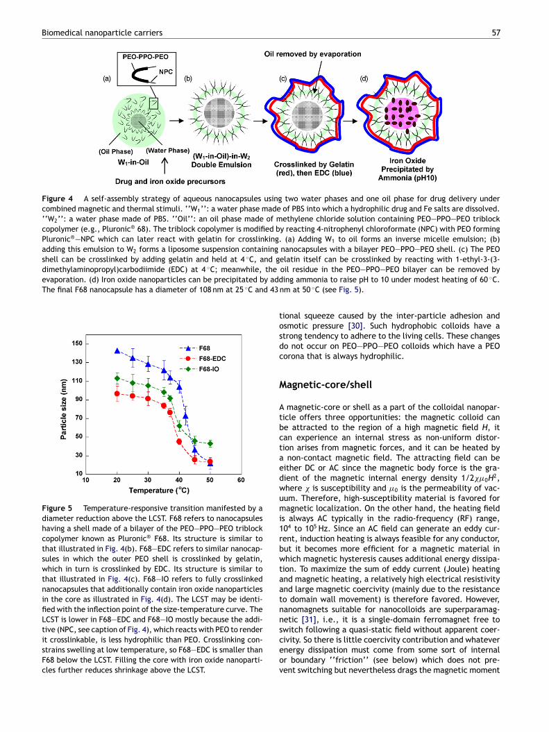
Figure 5 Temperature-responsive transition manifested by adiameter reduction above the LCST. F68 refers to nanocapsuleshaving a shell made of a bilayer of the PEO—PPO—PEO triblockcopolymer known as Pluronic® F68. Its structure is similar tothat illustrated in Fig. 4(b). F68—EDC refers to similar nanocap-sules in which the outer PEO shell is crosslinked by gelatin,which in turn is crosslinked by EDC. Its structure is similar tothat illustrated in Fig. 4(c). F68—IO refers to fully crosslinkednanocapsules that additionally contain iron oxide nanoparticlesin the core as illustrated in Fig. 4(d). The LCST may be identi-fied with the inflection point of the size-temperature curve. TheLCST is lower in F68—EDC and F68—IO mostly because the addi-tive (NPC, see caption of Fig. 4), which reacts with PEO to renderit crosslinkable, is less hydrophilic than PEO. Crosslinking con-strains swelling at low temperature, so F68—EDC is smaller thanF68 below the LCST. Filling the core with iron oxide nanoparti-cles further reduces shrinkage above the LCST.
tosdc
M
Atbctaedwumi1rbwtaatnnsceov
oil residue in the PEO—PPO—PEO bilayer can be removed bying ammonia to raise pH to 10 under modest heating of 60 ◦C.m at 50 ◦C (see Fig. 5).
ional squeeze caused by the inter-particle adhesion andsmotic pressure [30]. Such hydrophobic colloids have atrong tendency to adhere to the living cells. These changeso not occur on PEO—PPO—PEO colloids which have a PEOorona that is always hydrophilic.
agnetic-core/shell
magnetic-core or shell as a part of the colloidal nanopar-icle offers three opportunities: the magnetic colloid cane attracted to the region of a high magnetic field H, itan experience an internal stress as non-uniform distor-ion arises from magnetic forces, and it can be heated by
non-contact magnetic field. The attracting field can beither DC or AC since the magnetic body force is the gra-ient of the magnetic internal energy density 1/2��0H2,here � is susceptibility and �0 is the permeability of vac-um. Therefore, high-susceptibility material is favored foragnetic localization. On the other hand, the heating field
s always AC typically in the radio-frequency (RF) range,04 to 105 Hz. Since an AC field can generate an eddy cur-ent, induction heating is always feasible for any conductor,ut it becomes more efficient for a magnetic material inhich magnetic hysteresis causes additional energy dissipa-
ion. To maximize the sum of eddy current (Joule) heatingnd magnetic heating, a relatively high electrical resistivitynd large magnetic coercivity (mainly due to the resistanceo domain wall movement) is therefore favored. However,anomagnets suitable for nanocolloids are superparamag-etic [31], i.e., it is a single-domain ferromagnet free to
witch following a quasi-static field without apparent coer-ivity. So there is little coercivity contribution and whatevernergy dissipation must come from some sort of internalr boundary ‘‘friction’’ (see below) which does not pre-ent switching but nevertheless drags the magnetic moment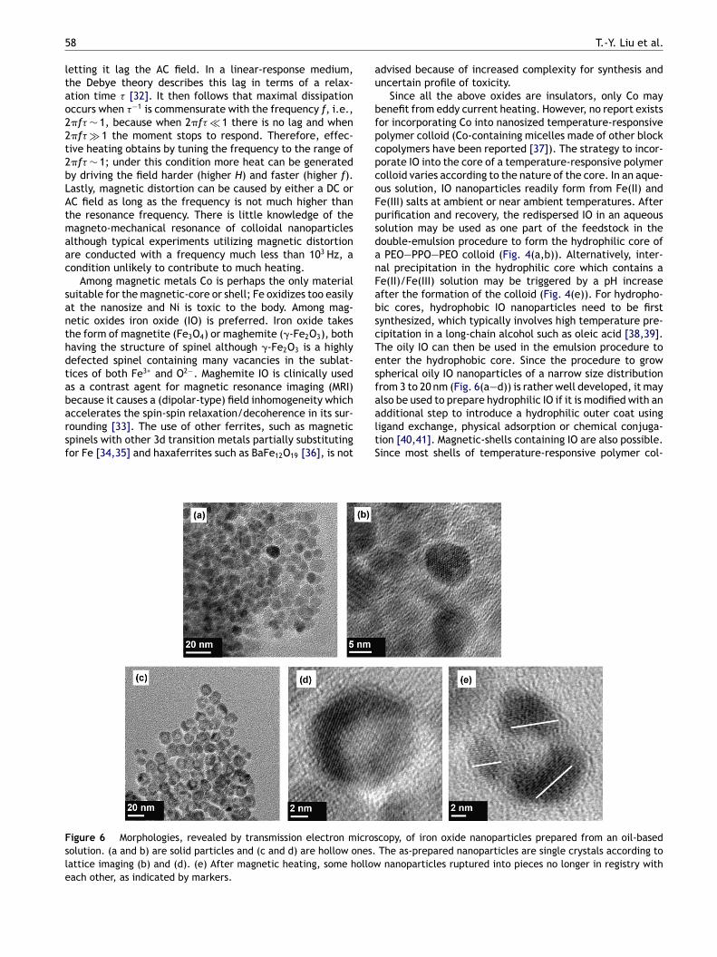
5
ltao22t2bLAtmaac
santhdtabarsf
au
bfpcpcoFpsdanFabscTesf
Fsle
8
etting it lag the AC field. In a linear-response medium,he Debye theory describes this lag in terms of a relax-tion time � [32]. It then follows that maximal dissipationccurs when �−1 is commensurate with the frequency f, i.e.,�f� ∼ 1, because when 2�f� � 1 there is no lag and when�f� � 1 the moment stops to respond. Therefore, effec-ive heating obtains by tuning the frequency to the range of�f� ∼ 1; under this condition more heat can be generatedy driving the field harder (higher H) and faster (higher f).astly, magnetic distortion can be caused by either a DC orC field as long as the frequency is not much higher thanhe resonance frequency. There is little knowledge of theagneto-mechanical resonance of colloidal nanoparticles
lthough typical experiments utilizing magnetic distortionre conducted with a frequency much less than 103 Hz, aondition unlikely to contribute to much heating.
Among magnetic metals Co is perhaps the only materialuitable for the magnetic-core or shell; Fe oxidizes too easilyt the nanosize and Ni is toxic to the body. Among mag-etic oxides iron oxide (IO) is preferred. Iron oxide takeshe form of magnetite (Fe3O4) or maghemite (�-Fe2O3), bothaving the structure of spinel although �-Fe2O3 is a highlyefected spinel containing many vacancies in the sublat-ices of both Fe3+ and O2−. Maghemite IO is clinically useds a contrast agent for magnetic resonance imaging (MRI)
ecause it causes a (dipolar-type) field inhomogeneity whichccelerates the spin-spin relaxation/decoherence in its sur-ounding [33]. The use of other ferrites, such as magneticpinels with other 3d transition metals partially substitutingor Fe [34,35] and haxaferrites such as BaFe12O19 [36], is notaaltS
igure 6 Morphologies, revealed by transmission electron microsolution. (a and b) are solid particles and (c and d) are hollow ones.attice imaging (b) and (d). (e) After magnetic heating, some hollowach other, as indicated by markers.
T.-Y. Liu et al.
dvised because of increased complexity for synthesis andncertain profile of toxicity.
Since all the above oxides are insulators, only Co mayenefit from eddy current heating. However, no report existsor incorporating Co into nanosized temperature-responsiveolymer colloid (Co-containing micelles made of other blockopolymers have been reported [37]). The strategy to incor-orate IO into the core of a temperature-responsive polymerolloid varies according to the nature of the core. In an aque-us solution, IO nanoparticles readily form from Fe(II) ande(III) salts at ambient or near ambient temperatures. Afterurification and recovery, the redispersed IO in an aqueousolution may be used as one part of the feedstock in theouble-emulsion procedure to form the hydrophilic core ofPEO—PPO—PEO colloid (Fig. 4(a,b)). Alternatively, inter-
al precipitation in the hydrophilic core which contains ae(II)/Fe(III) solution may be triggered by a pH increasefter the formation of the colloid (Fig. 4(e)). For hydropho-ic cores, hydrophobic IO nanoparticles need to be firstynthesized, which typically involves high temperature pre-ipitation in a long-chain alcohol such as oleic acid [38,39].he oily IO can then be used in the emulsion procedure tonter the hydrophobic core. Since the procedure to growpherical oily IO nanoparticles of a narrow size distributionrom 3 to 20 nm (Fig. 6(a—d)) is rather well developed, it may
lso be used to prepare hydrophilic IO if it is modified with andditional step to introduce a hydrophilic outer coat usingigand exchange, physical adsorption or chemical conjuga-ion [40,41]. Magnetic-shells containing IO are also possible.ince most shells of temperature-responsive polymer col-copy, of iron oxide nanoparticles prepared from an oil-basedThe as-prepared nanoparticles are single crystals according tonanoparticles ruptured into pieces no longer in registry with

Biomedical nanoparticle carriers 59
apsule na
dwvmFbsFlriqcoit�
�
wfctcspbpli
B
Madbdac
Figure 7 Transmission electron micrographs of F68—IO nanocexposure to 45 ◦C, above the LCST. After magnetic heating, somones (c).
loids are hydrophilic, magnetic-shells are synthesized usinghydrophilic IO. This is typically achieved by either adsorp-tion of IO nanoparticles or precipitation from aqueousFe precursors [26,42]. Using IO nanoparticles as seeds toinitiate polymerization, other magnetic-core/polymer-shellnanocolloids can also be synthesized as reviewed by Schmidt[31].
Under magnetic heating the temperature of the mag-netic nanocolloid solution gradually rises reaching a steadystate of several to several tens of degrees of centigradehigher. At this temperature, the heat input from the mag-netic nanoparticles equals the heat loss at the externalboundary (the container, fixtures, surfaces). What is infor-mative of magnetic dissipation is the initial heating rate,typically of the order of 0.1—1 ◦C/s for colloids containingIO nanoparticles. Since the energy input of the solution isentirely from the energy input of the magnetic nanopar-ticles, the initial heating rate of the nanoparticle shouldbe precisely CW/CMVM times that of the (water) solution.Here CW and CM are the volumetric specific heat of waterand the magnetic material, respectively, and VM is the vol-ume fraction of the magnetic material in the solution. SinceCW/CM ∼ 1 for IO and VM is of the order of 10−3, the initialheating rate experienced by the IO nanoparticle must be ofthe order of 102 to 103 ◦C/s. The steady state temperatureof the IO nanoparticle depends on the heat exchange mech-anisms between IO and the surrounding, which are currentlyunknown. However, microscopy evidence presented in Fig. 7for IO nanoparticles in the core of a PEO—PPO—PEO colloidafter RF heating suggests a rather high temperature of pos-sibly several hundred degrees of centigrade. Clearly, veryefficient ‘‘frictional’’ heating has been achieved. Magneti-cally caused fracture of hollow IO nanoparticles is also seenin Fig. 6(e), and similar transmission electron microscopyobservations of magnetic-heat-rupture have been reportedfor silica nanoparticles coated with an (single crystalline) IOshell [43].
Assuming magnetic heating involves isolated, indepen-dent nanoparticles only, in an RF field friction arises inand around a magnetic particle from two sources [44].
First, particle may tumble causing frictional heating at theparticle—water interface. The relaxation time �B for thismode can be estimated as the time required for Brownianmotion over a characteristic distance of the order of one par-ticle diameter. From Stoke—Einstein equation and viscousihatl
es (see caption of Fig. 5) that show (a) uneven shrinkage afternocapsules ruptured (b), other coarsened into irregular shaped
rag on a spherical particle, one can estimate �B = 3�V/kT,here � is the viscosity at the interface, V is the particleolume and kT has its usual meaning. Brownian relaxationay not be responsible for the frictional heating of IO seen in
ig. 7, though, because the heat from this mechanism shoulde about equally shared between the nanoparticle and watero it is unlikely for IO alone to reach a very high temperature.riction may also arise from spin rotation without crystal-attice rotation. The relaxation time �N for this mode (Neelelaxation) is the reciprocal of the spin flipping rate whichs of the order of �D exp(−KV/kT). Here �D is the Debye fre-uency of the order of 1012/s and KV is the energy barrier foroherent spin flipping which may be of a magnetocrystalliner shape origin. Most IO nanoparticles of several nanometersn size are superparamagnetic with a blocking temperatureypically around 50 K or lower. At the blocking temperature,N should be of the order of 10−2 to 102 s, so we estimateN to be of the order of 10−10 s at room temperature. Thisould make Neel relaxation too fast to add to any significant
riction in a RF field. However, the IO nanoparticles in Fig. 7ame from internal precipitation at the ambient tempera-ure, so they are not perfect and most likely contain a highoncentration of crystalline defects. Such defects may notignificantly affect the blocking temperature and the super-aramagnetic characteristics measured at low frequency,ut they can greatly increase the friction against spin flip-ing thus causing lattice heating. This seems to be the mostikely magnetic heating mechanism for the IO nanoparticlesn Fig. 7.
iomedical applications
agnetically and thermally responsive nanocolloids may findpplications in medicine and biotechnology such as drugelivery and enzyme immobilization/separation. Magneticody force can align or relocate the colloid and magneticissipation provides a means of remote heating. Temper-ture excursions can trigger a change in the size, waterontent, diffusivity, surface properties and hydrogen bond-
ng of the colloid. Although all of these individual effectsave been separately illustrated in numerous studies, therere very few biomedically relevant reports that demonstratehe combined magnetic and thermal actions in the nanocol-oid setting—–bulk hydrogels and large (�m to mm) latex
6 T.-Y. Liu et al.
ps
M
Cdaobptiettsspahtd[p
M
MianpoaimFalv(aeoapri
M
Mtsbmspm
Figure 8 Cumulative release of a model drug (vitamin B12)from F68—IO nanocapsules (see caption of Fig. 5) at varioustemperatures. The rapid increase from 37 to 45 ◦C is mostlydue to nanocapsule shrinkage from 90 to 45 nm (see Fig. 5). Themt
thItdualc[ls
M
IacamwLadstifor LCST colloids since above the LCST the colloid will pre-
0
articles are excluded. In the following, we summarize thesetudies and comment on the pertinent mechanisms.
agnetic heating of UCST colloids
onventional synthetic polymers may experience increasediffusivity and water content when magnetically heatedbove the UCST, which may accelerate the releasef trapped drug or a model dye. This was reportedy Schmidt and coworkers for an IO-core-containingoly(�-caprolactone) (PCL) nanocolloid loaded with a solva-ochromic dye; PCL exhibits an UCST of 35 ◦C when dispersedn dimethyl sulfoxide as in this study [45,46]. A more inter-sting study concerns a biopolymer with hydrogen bondinghat melts above the UCST; magnetic heating then causeshe release of hydrogen-bonded drug. This was demon-trated by Derfus et al. using IO nanoparticles to which singletrand DNA was grafted: the DNA binds a dye-labeled com-lement below the UCST, then releases it above the UCSTt the implanted site in a mouse tumor model [47]. Meltingydrogen-bonding in a bulk gel magnetically heated abovehe UCST has been used to increase the diffusivity, hencerug release from IO-containing collagen [48] and gelatin49]. Extension to microgels of submicrometer sizes is inrinciple feasible but not yet reported.
agnetic heating of LCST colloids
agnetic heating of the NIPPAm colloid above the LCSTnduces aggregation and size shrinkage. Wakamatsu etl. [50] applied the first effect to IO-core/PNIPPAm-shellanoparticles to trigger their entrapment in a columnacked with hydrophobic beads. We have applied the sec-nd effect to PEO—PPO—PEO nanoparticles to squeeze outhydrophilic drug from the core (the preparation method
s shown in Fig. 4, size isotherm in Fig. 5, reconstructedagnetic-cores in Fig. 7, and the release mechanism in
ig. 1). This is the first example of utilizing magnetic heatingnd size shrinkage to control drug release from a nanocol-oid. The profile of drug release rates shown in Fig. 8 isery favorable: very slow at 4 ◦C and 25 ◦C, modest at 37 ◦Cbelow the LCST), much faster at 45 ◦C (above the LCST)nd bursting upon magnetic heating. Compared to an earlierxample of �m-sized colloid (NIPPAm with IO) [51], the ratiof release under magnetic heating to that of 25 ◦C is at leastfactor of 100 higher in this nanocolloid. A further com-
arison to other examples of magnetically triggered drugelease (with or without a temperature-responsive polymer)s shown in Table 3.
agnetic separation of LCST/UCST colloids
agnetic support particles have been investigated for a longime as a separation platform in biotechnology. Nanometerized colloids can reduce fouling, but magnetic separation
ecomes much more difficult because of the smalleragnetic force in comparison to colloidal forces that favoruspension and Brownian motion. Colloids made of LCSTolymers aggregate above the LCST, so they experience auch larger magnetic force and smaller colloidal forces,
chd(n
uch faster burst-like release during magnetic heating is dueo rupture of the nanocapsule (see Fig. 7).
hus allowing easy separation by a relatively low field. Thisas been demonstrated by Kondo and Fukuda [52] by heatingO-containing PNIPPAm colloids (150—250 nm) above 32 ◦Co separate immobilized enzymes on the nanoparticles. Theispersion-to-flocculation transition at the LCST was alsotilized by the same group [53] to achieve magneticallyided affinity selection of target cells from phage displayibraries. Similarly, magnetic separation of UCST colloidsan be practiced below the UCST as illustrated by Kaiser54] for IO-containing polystyrene nanoparticles. In theatter case, a hydrophobic solution (cyclohexane in thistudy) must be used.
agnetic directing of LCST colloids
n vivo localization of nanomagnetic particles is feasibleccording to the study of Deng et al. [55] who localized IO-ontaining PNIPPAm nanoparticles (300—500 nm) to liver inrabbit using a DC magnetic field; without a field accu-ulation in other organs (lung, spleen, kidney and heart)as observed. The colloid was initially placed below theCST to access the swollen state to soak up doxorubicin,hydrophilic drug for cancer treatment, although in vivo
emonstration of drug release was not performed in thistudy apparently because the LCST (32—37 ◦C) is no higherhan the body temperature. In principle, AC magnetic heat-ng (hyperthermia) can also provide a localization effect
ipitate with a tendency to adhere to the cells. Localizedyperthermia was proposed as a targeting tool to directrug-loaded LCST colloids to tumors which are warmer∼42 ◦C) than the rest of the body [30], but this idea hasot been demonstrated for magnetic colloids.

Biomedical nanoparticle carriers 61
Table 3 Half-life (t50) for drug release (typically a small molecule) from particles with and without magnetic heating. t50 isthe time to reach Mt/M∞ = 0.5, where Mt/M∞ is the amount released at time t normalized by the total amount of drug contained(see Fig. 8). Much faster release with magnetic heating.
Materials Half-time (t50) Released molecules
Without magnetic heating With magnetic heating
Pluronic® F68/iron oxidenanocapsules [91]
42 h (37 ◦C)/5 h (45 ◦C) 5 min Vitamin B12
Pluronic® F127/iron oxidenanoparticles [90]
18 h (15 ◦C)/3 h (45 ◦C) 5 min Doxorubicin
Fe3O4/PAH capsules [95]a 15 h (25 ◦C) 30 min FITC—dextranSilica/iron oxide nanospheres [43]a >20 days (25 ◦C) 3 min Fluorescent dyeSilica/iron oxide nanospheres [96]a >10 days (25 ◦C) 15 min IbuprofenEthylene—vinyl acetate with
embedded magnetic sphere [97]bNot measured (>40 days) 10 times shorterb Bovine serum albumin
r, no
vdhtc5wlfaIm
dabtttIthiciftratt(omaM
a Without a temperature-responsive polymer.b 10 mm × 10 mm × 2 mm, not a temperature-responsive polyme
Membranes of magnetically and thermallyresponsive colloids
Various magnetic hydrogels not unlike those previouslymentioned [48—49] have been studied but one serious short-coming of the macroscopic gels is their slow response time,which scales with the size to the second power reflect-ing the diffusion limit of water transport [56]. This canbe overcome if the macroscopic construct is itself madeof nanoparticles of temperature-responsive hydrogel. Sincethe diffusion time of nanoparticle is very short, the responseof the construct is also very fast despite its macroscopicdimension. Indeed, the nanoparticles can even be embed-ded in another gel without affecting the response time aslong as water exchange in and out of the nanoparticles canproceed locally. One such construct with a magnetic sig-nature is a membrane made by gelling nanoparticles or bydepositing nanoparticle colloids. For example, Csetneki etal. reported a membrane made of nanoparticles with an IO-containing polystyrene core which is coated with PNIPPAm[57]. The membrane was endowed with a special microstruc-ture by applying a magnetic field during gelation (belowthe LCST) with poly(vinyl alcohol) crosslinking: the mag-netic nanoparticles are lined up into necklace strings due todipole—dipole interactions. Above the LCST, shrunk nanopar-ticles disrupt the microstructure causing a rapid increase inpermeability as demonstrated by bovine serum albumin pen-etration. Using spin coating, we have fabricated a 50-�mfilm of IO-containing PEO—PPO—PEO nanoparticles on a sil-icon substrate to demonstrate magnetically actuated rapiddye release from this device (Fig. 9). Micro-implant devicesconstructed in a similar way may be used for magneticallycontrolled drug delivery.
In vivo delivery
Hyperthermia via magnetically heating of IO has been stud-ied in mice and human cadavers to treat breast tumors,showing tumor shrinkage and nuclear degenerations inheated malignant cells [58]. A maximum temperature ele-
d(smc
magnetic heating, faster response due to magnetic distortion.
ation �T up to 88 ◦C was reported. A recent studyemonstrated deep cranial thermotherapy using magneticeating of aminosilane-coated IO applied to human glioblas-oma multiforme patients who also received MRI andomputed tomography (CT) for evaluation [59]. At a �T of—12 ◦C, patients reported no discomfort. For drug release,e already mentioned (see Magnetic heating of UCST col-
oids section) the study of fluorophore (a model drug) releaserom magnetically heated IO that was pre-implanted intomouse tumor model [47]. Clinical use of dextran-coated
O as a MRI contrast agent has also been a well-establishedodality for liver imaging [33].In the above applications IO colloids were delivered by
irect injection to the target sites. In recent years, in vivonimal studies have been used to demonstrate the possi-ility of targeted delivery and imaging of IO with tetheredargeting moieties; for example, folate ligand has beenethered to the dextran coating of IO via a linker to targetumor xenografts that overexpress folate receptors [60].n theory, if the self-directed IO colloids are well localizedo the targeted tumor site, they can also be magneticallyeated to treat tumor, but this has not been demonstratedn vivo. Indeed, although multifunctional nanoparticlesapable of targeted delivery of imaging agents and drugss a much discussed concept, its in vivo demonstrationor magnetic colloids is so far rare; we know of none foremperature-responsive magnetic colloids. In a recenteview of application of nanotechnology in cancer therapynd imaging [61], only one was cited for simultaneousargeted delivery of drug and imaging agent: it deliverso targeted tumor cells small interfering ribonucleic acidssiRNA) that are covalently tethered to the dextran coatingf IO [62]. This study did not utilize magnetic heating,agnetic directing or thermal sensitivity. Recently, Yang et
l. used core-shell magnetic nanoparticles (core containingnFe2O4, a spinel ferrite and doxorubicin, an anticancer
rug) tethered with a breast-cancer-targeting antibodyhuman epidermal growth factor receptor 2 (HER2)) toimultaneously detect and treat cancer xenografts in mouseodels [63]. Although this study used an amphiphilic blockopolymer of poly(D,L-lactide-co-glycolide) (PLGA) and PEG
62 T.-Y. Liu et al.
Figure 9 A device prototype made of iron-oxide-containing nanocapsules. Drug-containing F68—IO nanocapsules (see caption ofFig. 5) were spin coated onto a Si substrate to form a 60-�m thick film. Another PEO—PPO—PEO triblock polymer (Pluronic® F127)solution with a LCST of 22 ◦C was used in the spin-coating solution as a ‘‘binder’’ gel. After spin coating, a gelatin coating wasintroduced to crosslink the PEO shell of the nanocapsule. Magnetic heating triggers drug release from F68—IO. A similar implantd
fbPmifd
D
WrAtrmpfwHnf
Lssptnto(icptau
Lm
evice may be used for controlled drug release.
or the shell, which is not temperature-responsive, it shoulde possible to replaced PLGA—PEG by PEO—PPO—PEO or aNIPPA copolymer. Using such a construct, functionalities ofagnetic heating, magnetic directing and thermal sensitiv-
ty can in principle be incorporated into nanocolloid systemsor self-directed simultaneous detection and treatment ofiseases.
esigning nanoscale systems
e begin this section with a few comments on the drugelease mechanisms in magnetically heated LCST colloids.lthough a generic increase in diffusivity at higher tempera-ure may play a minor role, the dominant mechanisms are allelated to structural changes due to the LCST transition andagnetic field/heating. Clearly, the volumetric shrinkagerovides a potentially powerful driving force for drug release
rom the core. Effective actuation requires core shrinkage,hich is easier for a soft core than for a hard core [64].owever, volumetric shrinkage cannot account for the mag-etically triggered burst-like release in Fig. 8, which is muchaster than that achieved by heating to 45 ◦C (above thesdopt
CST) alone. The burst-like release is most likely due to theevere disruption of the IO core by magnetic heating. Othertructural changes in the pore structure of the shell may alsolay a role. The changes may be caused by a thermal distor-ion akin to the one associated with a heated heterogeneousetwork structure: some regions expand while others con-ract. Magnetic forces may also cause a structural disruptionf the shell when 2�f�B � 1, as shown in a low frequency300 Hz) study on magnetically triggered on—off permeabil-ty switch across a polyelectrolyte shell surrounding a Co/Auore of 5 �m [65]. Force-directed structural movement isrobably not important in the RF frequency range because,o effect shell distortion, 2�f�B � 1 must be satisfied forparticle of the size of the colloidal particle—–a condition
nlikely to be met.We have already emphasized the importance of the
CST/UCST temperature, the structural transitions and theagnetic constituent of the nanocolloid that is respon-
ive to both magnetic and temperature stimuli. For in vivorug delivery, these temperatures should be a few degreesf centigrade above the physiological temperature, andreferably there is a large change in size and surface func-ionality. Tuning the transition temperature must be tackled

iuatptempsstc
A
Tt2U2
R
[[[[[
[[[[
[[[[[[[[[[
[[[[
Biomedical nanoparticle carriers
at the system level, since as mentioned before the transi-tion temperature is sensitive to all chemical and physicalaspects of the constituents of the polymer and its surround-ing. A soft core is preferred to effect core shrinkage [64].Actuation will be more effective if the transition temper-ature and the magnetic response are sharp. This requiresa precise control of the composition and microstructureincluding a narrow distribution of the molecular weightof the polymer and of the size of the IO nanoparticles.Efficiency of magnetic heating is probably sensitive tothe defect chemistry of the IO, its control and charac-terization at the nanoscale presenting a challenge. Cost,synthetic ease and scalability for mass production are impor-tant and mostly dependent on the chemistry and processesselected.
A successful system design should also address otherissues of material chemistry and physics. First, safety andbiocompatibility demand rigorous screening to eliminateany toxic chemical in the composition of the polymerand the process residue. A particularly complicated issueis colloidal and drug stability. Structural integrity of thenanocolloid obviously calls for substantial stability of theconstituent polymer during storage and circulation, whichmay be improved by crosslinking. Nanocolloids tend to havelonger circulation half-lives, but to help escape the fateof rapid clearance by macrophages or the reticuloendothe-lial system surface hydrophilic tethers of PEG or dextran (apolysaccharide) are beneficial [66]. Tethers may also reducethe absorption of serum proteins, thus avoiding enzymaticattack at the same time. Meanwhile, biodegradability of thetemperature-responsive polymer would be desirable whichmay be introduced by incorporating biodegradable blocksor oligomers such as PCL [67], polylactic acid (PLA) [68] andPLGA [69], including their copolymers (in the PEO—PPO—PEOtriblock copolymers, they should substitute for the PPOblock) [60]. Concerning drug targeting, hydrophilic tethersmentioned above will mask the transition to hydrophobicityabove the LCST of PNIPPAm and PVCL, so temperature-triggered aggregation and cell adhesion is no longer possible.In this regard, moieties for receptor or ligand bonding toenable targeted delivery is a desirable functionality thatcan be attached to the nanoparticles via suitable surfacetethers [61]. Another important issue is the trigger for drugrelease. Although a long residence time after localizationat the target site may sometimes be enough for deliveringdrug, a more efficient scheme is to utilize a device thatallows for nanoparticle internalization (e.g., via receptor-mediated endocytosis) [70] and drug release (e.g., via anacid-labile linkage that is broken in the low-pH environmentof endosomes) [71,72]. Lastly, drug loading is dictated bythe physical chemistry of the polymer and the drug dur-ing fabrication, so a condition which simultaneously allowsfor polymer reaction (including self-assembly) and drugincorporation need to be found [64]. Since these aspectswill again impact the transition temperature and transi-tion characteristics, a system engineering approach must beadopted to find a satisfactory solution for this nanotechnol-
ogy.Finally, injection of particulate substance (liposomes,micelles and other natural or synthetic particles) in thesubmicron size range may elicit allergic reactions such ascardiovascular, respiratory and cutaneous symptoms, includ-
[[[
[
63
ng death [73]. Typically, such reactions are most severepon initial exposure, and the frequency of particulatellergy in the 5—45% range seems to be much higher thanhat of classical anaphylactic reactions to drugs (for exam-le, penicillin allergy occurs in <2%). Interestingly, therigger dose of hypersensitivity reactions in mouse mod-ls is two orders of magnitude higher than that in reactivean, so many animal studies may not foretell the threat ofossible allergic reactions (Pig models appear to exhibit aimilar trigger dose as reactive man). Therefore, designingafe nanoparticle delivery systems for in vivo applica-ions may pose the most serious though least consideredhallenge.
cknowledgements
his work was supported by the National Science Council ofhe Republic of China, Taiwan under contract No. NSC96-627-B-009-006 and NSC96-2113-M009-027-MY2, and by theS National Science Foundation under grant No. DMR-05-0020 (MRSEC).
eferences
[1] E.S. Gil, S.M. Hudson, Prog. Polym. Sci. 29 (2004) 1173.[2] B. Jeong, et al., Adv. Drug Deliv. Rev. 54 (2002) 37.[3] G.J. Kim, S. Nie, Nanotoday August (2005) 28.[4] I. Brigger, et al., Adv. Drug Deliv. Rev. 54 (2002) 631.[5] E. Okon, et al., Lab. Invest. 91 (1994) 895.[6] H.G. Schild, Prog. Polym. Sci. 17 (1992) 163.[7] Y. Maeda, et al., Langmuir 16 (2000) 7503.[8] V. Boyko, et al., Polymer 19 (2003) 8675.[9] P. Alexandridis, T.A. Hatton, Colloids Surf. A 96 (1995) 1.10] S.M. Daly, et al., Langmuir 21 (2005) 1328.11] K. Mortensen, J. Phys.: Condens. Matter 8 (1996) A13.12] P. Alexandridis, et al., Macromolecules 27 (1994) 2414.13] P. Alexandridis, et al., Langmuir 11 (1995) 1468.14] A. Chakrabartty, R.L. Baldwin, Adv. Protein Chem. 46 (1995)
141.15] A.P. Nowak, et al., Nature 417 (2002) 424.16] J. Kopecek, Nature 417 (2002) 388.17] W.A. Petka, et al., Science 281 (1998) 389.18] A.G. Ward, A. Courts, The Science and Technology of Gelatin,
Academic Press, New York, 1977.19] D.W. Urry, J. Phys. Chem. B 101 (1997) 11007.20] D.E. Meyer, et al., J. Control. Release 74 (2001) 213.21] A. Chilkoti, et al., Adv. Drug Deliv. Rev. 54 (2002) 613.22] S.H. Choi, et al., Langmuir 22 (2006) 1758.23] T.G. Park, A.S. Hoffman, Biotechnol. Prog. 10 (1994) 82.24] R. Pelton, Adv. Colloid Interface Sci. 85 (2000) 1.25] J. Rubio-Retama, et al., Langmuir 23 (2007) 10280.26] S. Bhattacharya, et al., Small 3 (4) (2007) 650.27] K.S. Soppimath, et al., Adv. Funct. Mater. 17 (2007) 355.28] T. Okano (Ed.), Biorelated Polymers and Gels, Academic Press,
San Diego, CA, 1998.29] N.S. Satarkar, J.Z. Hilt, Acta Biomater. 4 (2008) 11.30] J.E. Chung, et al., J. Control. Release 62 (1999) 115.31] A.M. Schmidt, Colloid Polym. Sci. 285 (2007) 953.32] P. Debye, Polar Molecules, Dover, 1929.
33] R. Weissleder, Radiology 193 (1994) 593.34] D.-H. Kim, et al., J. Magn. Magn. Mater. 293 (2005) 320.35] D.-H. Kim, et al., J. Magn. Magn. Mater. 320 (2008)2390.36] P. Xu, et al., J. Phys. Chem. 111 (2007) 5866.

6
[[[[[[[[[[[[[[
[
[
[
[[[[[[[[[[[[[[[[[[[[[
[
[[[[[[[[[[[[[[[[[[[[[[
ccg
thbt
afRca
4
37] J. Connolly, et al., J. Phys. D: Appl. Phys. 37 (2004) 2475.38] S. Sun, H.J. Zeng, J. Am. Chem. Soc. 124 (2002) 8204.39] S. Sun, et al., J. Am. Chem. Soc. 126 (2003) 273.40] J.-H. Lee, et al., Nat. Med. 13 (1) (2007) 95.41] J. Qin, et al., Adv. Mater. 19 (2007) 1874.42] S. Bi, et al., Mater. Lett. 62 (2008) 2963.43] S.-H. Hu, et al., Adv. Mater. 20 (2008) 2690.44] R.E. Rosensweig, J. Magn. Magn. Mater. 252 (2002) 370.45] A.M. Schmidt, J. Magn. Magn. Mater. 289 (2005) 5.46] A. Kaiser, et al., J. Phys.: Condens. Matter 18 (2006) S2563.47] A.M. Derfus, et al., Adv. Mater. 19 (2007) 3932.48] V.M. De Paoli, et al., Langmuir 22 (2006) 5894.49] S.-H. Hu, et al., Macromolecules 40 (2007) 6786.50] H. Wakamatsu, et al., J. Magn. Magn. Mater. 302 (2006)
327.51] D. Muller-Schulte, T. Schmitz-Rode, J. Magn. Magn. Mater. 302
(2006) 267.52] A. Kondo, H. Fukuda, J. Ferment. Bioeng. 84 (4) (1997)
337.53] H. Furukawa, et al., Appl. Microbiol. Biotechnol. 62 (2003)
478.54] Kaiser, J. Phys.: Condens. Matter (2006).55] Y. Deng, et al., Chem. Eur. J. 11 (2005) 6006.56] Y. Qiu, K. Park, Adv. Drug Deliv. Rev. 53 (2001) 321.57] H. Csetneki, et al., Macromolecules 39 (2006) 1939.58] I. Hilger, et al., Radiology 218 (2001) 570.59] K. Maier-Hauff, et al., J. Neuro-Oncol. 81 (2007) 53.60] S.W. Choi, et al., J. Polym. Sci. A 37 (1999) 2207.61] X. Wang, et al., CA Cancer J. Clin. 58 (2008) 97.62] Z. Medarova, et al., Nat. Med. 13 (2007) 372.63] J. Yang, et al., Angew. Chem. Int. Ed. 46 (2007) 8836.64] J.E. Chung, et al., J. Control. Release 65 (2000) 93.65] Z. Lu, et al., Langmuir 21 (2005) 2042.66] A.K. Gupta, M. Gupta, Biomaterials 26 (2005) 3995.67] W.-Q. Chen, et al., Polymer 49 (18) (2008) 3965.68] F. Kohori, et al., J. Control. Release 55 (1998) 87.69] S.Q. Liu, et al., Mol. Biosyst. 1 (2005) 158.70] S. Wang, P.S. Low, J. Control. Release 53 (1998) 39.71] D. Schmaljohann, Adv. Drug Deliv. Rev. 58 (2006) 1655.72] Z. Zhang, R.D.K. Misra, Acta Biomater. 3 (2007) 838.73] J. Szebeni, et al., J. Liposome Res. 17 (2007) 107.74] C. de las Heras Alaró, et al., Chem. Soc. Rev. 34 (2005)
276.75] H.S. Cho, et al., J. Polym. Sci. B: Polym. Phys. 35 (1997)
595.76] A.T. Muramaysu, et al., Macromolecules 34 (2001) 3118.77] Z. Yang, et al., Polymer 48 (2007) 931.78] M.D.C. Topp, et al., Macromolecules 30 (1997) 8518.79] H. Wei, et al., Biomaterials 30 (2007) 99.80] Y.A. Han, et al., Polym. Test. 21 (2002) 913.81] J.E. Chung, et al., J. Control. Release 53 (1998) 119.82] D. Neradovic, et al., Macromolecules 34 (2001) 7589.83] H. Wei, et al., J. Control. Release 116 (2006) 266.84] R.A. Stile, K.E. Healy, Biomacromolecules 2 (2001) 185.85] M.J. Song, et al., J. Polym. Sci. A 427 (2004) 772.86] E. Miyoshi, et al., Polym. Gels Netw. 6 (1998) 273.87] M. Zrínyi, Colloid. Polym. Sci. 278 (2000) 98.88] C.-J. Cheng, et al., Colloid. Polym. Sci. 286 (2008) 571.89] H. Xu, et al., Chem. Mater. 19 (2007) 2489.90] T.Y. Liu et al., Langmuir, in press.91] T.Y. Liu et al., Adv. Func. Mater., in press.92] S.H. Choi, et al., Biomacromolecules 7 (2006) 1864.93] K.H. Bae, et al., Langmuir 22 (2006) 6380.
94] K.H. Bae, et al., Biomacromolecules 8 (2007) 650.95] S.-H. Hu, et al., Langmuir 24 (2008) 11811.96] S.-H. Hu, et al., Langmuir 24 (2008) 239.97] E.R. Edelman, et al., J. Biomed. Mater. Res. 19 (1985)67.
T.-Y. Liu et al.
Ting-Yu Liu (BS — Chemical Engineering,Yuan-Ze University, 2001; MS — PolymerEngineering, National Taiwan University ofScience and Technology, 2003; PhD — Mate-rials Science and Engineering, National ChiaoTung University, 2008) spent a year as Visit-ing Scholar in the Department of MaterialsScience and Engineering at University of Penn-sylvania (2007—2008). He has authored over20 papers on nanotechnology, polymer hydro-gels and biomedical devices (dialysis and drug
arriers). His current research interest includes ferrogels for drugontrolled release and core—shell magnetic nanoparticles for tar-eted drug delivery and magnetic resonance imaging.
Shang-Hsiu Hu (BSc — Chemical Engineer-ing, National Chung-Hsin University, Taiwan,2004; MSc — Material Science and Engineer-ing, National Chiao Tung University, Taiwan,2006) is currently pursuing a PhD under theguidance of Prof. San-Yuan Chen. His researchfocus is on novel process development andcontrolled drug release in nano-biomaterialcomposites. He received National InnovationAward for Biotechnology and Medicine Indus-try (2007, 2008).
Dean-Mo Liu (MEng — Chemical Engineer-ing, Chuan Yuan Christian University, Taiwan86’; MSc — Materials Science, VPI&SU, USA,1991; PhD Materials Science and Engineer-ing, University of British Columbia, Canada2004) joined National Chiao-Tung University,Taiwan as a professor of Materials Scienceand Engineering in 2007. Prior to that hespent 6 years in biomedical industry in Canada(2001—2007). His current research focuses ondeveloping new (bio)materials and devices
hrough colloidal-based assembly and biomimetic technologies. Heas authored more than 150 technical papers and several scientificooks. He has also served as a scientific advisory member for severalechnical journals and international conferences since 1998.
San-Yuan Chen (PhD, University of Michigan,Ann Arbor, USA, 1994) has been Profes-sor of Material Science and Engineering atNational Chiao Tung University of Taiwan since1996. His current research is focused onfunctional photoelectronic inorganic nanoma-terials, novel process development and con-trolled drug release in nano-biomaterial com-posites, and high-dielectric and ferroelectricmemory thin-film processing. Honors include:National Innovation Award for Biotechnology
nd Medicine Industry (2007, 2008); Distinguished Engineering Pro-essor Award from Chinese Engineering Institute (2005); Excellentesearch Award from National Chiao Tung University (2003); Second-lass Research Award from National Science Council (2005, 2006)nd Marquis Who’s Who in the World (since 2006).
I-Wei Chen (BS — Physics — 1972, Tsinghua;MS — Physics — 1975, Penn; PhD — Met-allurgy — 1980, MIT) has been SkirkanichProfessor of Materials Innovation at Univer-sity of Pennsylvania since 1997. He taught
at the University of Michigan (Materials)during 1986—1997 and MIT (Nuclear Engineer-ing; Materials) during 1980—1986. He beganceramic research studying martensitic trans-formations in zirconia nanocrystals, which led
Edward C. Henry Award (1999) and Sosman Award (2006), he
Biomedical nanoparticle carriers
to studies on transformation plasticity, superplasticity, fatigue,
grain growth and sintering in various oxides and nitrides. He iscurrently interested in nanograin ferroelectrics, nanoparticles forbiomedical applications, nanostructured resistance memory devicesand electromigration in fuel cells. A Fellow of American CeramicSociety (1991) and recipient of its Ross Coffin Purdy Award (1994),aSSo
65
uthored over 90 papers in the Journal of the American Ceramicociety (1986—2006). He received Humboldt Research Award forenior U.S. Scientists (1997) and is a Chong-Kong Chair Professorf Tsinghua University in Beijing (2006—2009).


![Static and Dynamic Density Functional Theory and ...called copolymers. Here we consider the class of copolymers called \block copolymers" [7] while there are many kinds of copolymers.](https://static.fdocuments.in/doc/165x107/5eccfbf97d791301bb64d299/static-and-dynamic-density-functional-theory-and-called-copolymers-here-we.jpg)