CD163 versus CD68 in tumor associated macrophages of classical

Review ArticleTumor-Associated Macrophages and Neutrophils inTumor Microenvironment
Jaehong Kim1 and Jong-Sup Bae2
1Department of Biochemistry, School of Medicine, Gachon University, Incheon 406-799, Republic of Korea2College of Pharmacy, CMRI, Research Institute of Pharmaceutical Sciences, BK21 Plus KNUMulti-Omics basedCreative Drug Research Team, Kyungpook National University, Daegu 702-701, Republic of Korea
Correspondence should be addressed to Jaehong Kim; [email protected] and Jong-Sup Bae; [email protected]
Received 25 November 2015; Accepted 18 January 2016
Academic Editor: Amedeo Amedei
Copyright © 2016 J. Kim and J.-S. Bae.This is an open access article distributed under the Creative Commons Attribution License,which permits unrestricted use, distribution, and reproduction in any medium, provided the original work is properly cited.
Distinct tumor microenvironment forms in each progression step of cancer and has diverse capacities to induce both adverseand beneficial consequences for tumorigenesis. It is now known that immune cells can be activated to favor tumor growth andprogression, most probably influenced by the tumor microenvironment. Tumor-associated macrophages and tumor-associatedneutrophils can exert protumoral functions, enhancing tumor cell invasion and metastasis, angiogenesis, and extracellular matrixremodeling, while inhibiting the antitumoral immune surveillance. Considering that neutrophils in inflammatory environmentsrecruit macrophages and that recruited macrophages affect neutrophil functions, there may be various degrees of interactionbetween tumor-associated macrophages and tumor-associated neutrophils. Platelets also play an important role in the recruitmentand regulation of monocytic and granulocytic cells in the tumor tissues, suggesting that platelet function may be essential forgeneration of tumor-associatedmacrophages and tumor-associated neutrophils. In this review,wewill explore the biology of tumor-associated macrophages and tumor-associated neutrophils and their possible interactions in the tumor microenvironment. Specialattention will be given to the recruitment and activation of these tumor-associated cells and to the roles they play in maintenanceof the tumor microenvironment and progression of tumors.
1. Introduction
Cancer-related nonresolving inflammation in the tumormicroenvironment (TME) is a hallmark of cancer, andcancer cells are confronted with various types of stromaland immune cells across all stages of the disease, from earlycarcinogenesis to tumor progression and metastasis [1, 2].The progression of cancer has traditionally been regardedas a multistep process with genetic and epigenetic changestargeting only cancer cells. However, studies over the pasttwo decades have revealed that the TME is an equallyimportant determinant of tumor behavior. The componentsof the TME include local stromal cells, such as residentfibroblasts and macrophages, and distant recruited cells suchas endothelial cells, immune cells including myeloid andlymphoid cells, bone marrow-derived precursor cells, andcirculating platelets. To note, tumor-associated myeloid cells(TAMCs) comprise five distinct myeloid populations: tumor-associated macrophages (TAMs), monocytes expressing the
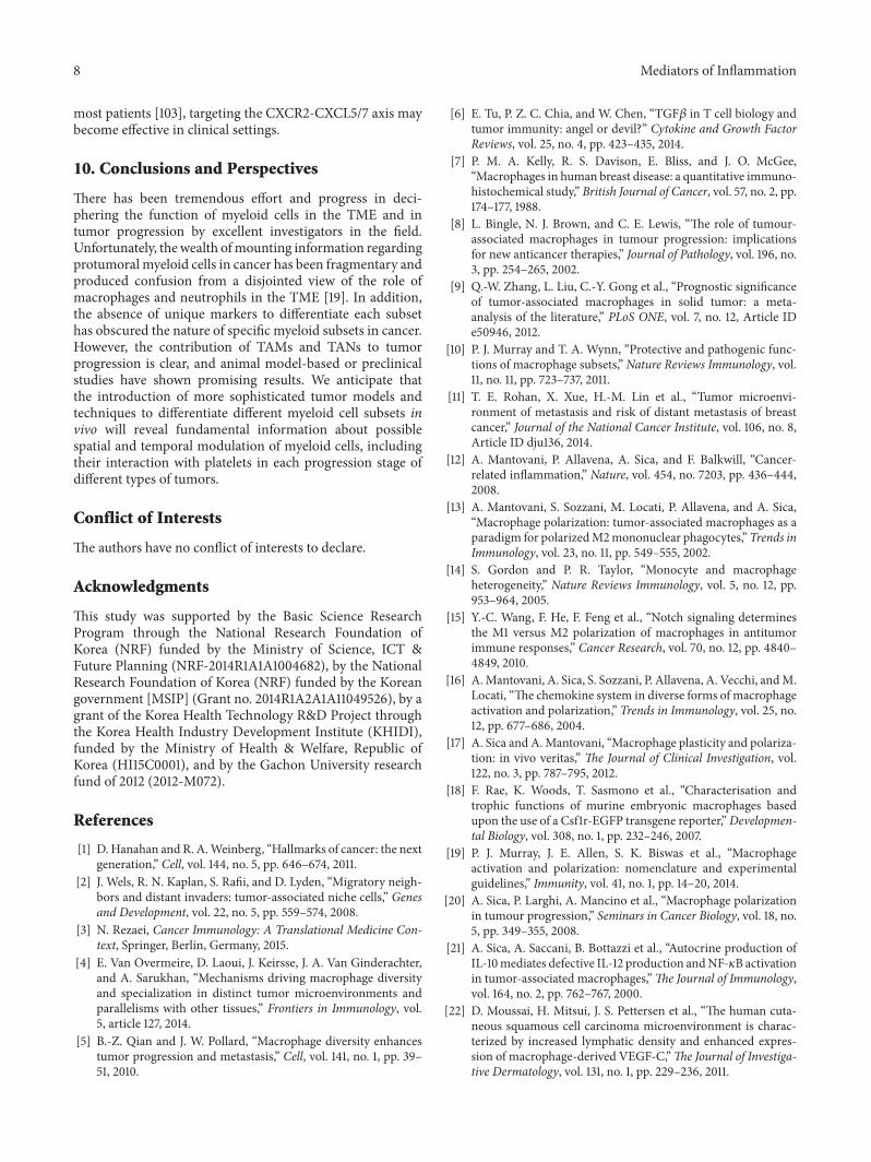
angiopoietin-2 receptor Tie2 (Tie2-expressing monocytes orTEMs), myeloid-derived suppressor cells (MDSCs), tumor-associated neutrophils (TANs), and tumor-associated den-dritic cells (Figure 1) [3]. Of these, TAMCs result in TAMsand TANs to be discussed in this review.
2. General Characteristics of TAMs
Macrophages are the most well-characterized type of tumor-infiltrating immune cell, and it is not surprising that they playa prominent active role from early carcinogenesis to tumorprogression including metastasis [4]. While macrophagesinvolved in cancer-initiating conditions are immune acti-vated (e.g., antitumoral), once tumors are established, themacrophages are educated to become protumoral [5]. Cur-rently, the majority of evidence supports a tumor-promotingrole of a specific subpopulation of macrophages, TAMswithin the primary TME. Surprisingly, macrophages canconstitute up to 50% of a tumor mass, forming a major
Hindawi Publishing CorporationMediators of InflammationVolume 2016, Article ID 6058147, 11 pageshttp://dx.doi.org/10.1155/2016/6058147

2 Mediators of Inflammation
?
HSC CMP IMC
TEM TEM
Monocyte
MDSCM-MDSC
G-MDSC G-MDSC
M-MDSC
TAM
M1-like TAM
M2-like TAM
Neutrophil TAN
iDC TADC
N1-like TAN
N2-like TAN
Bone marrow Blood/spleen Tumor
Figure 1: Differentiation of tumor-associated myeloid cells beginsfrom hematopoietic stem cells (HSC) in the bone marrow. CMP:common myeloid progenitors, IMC: immature myeloid cells, TEM:Tie2-expressingmonocyte,MDSC:myeloid-derived suppressor cell,M-MDSC: myeloid MDSC, G-MDSC: granulocytic MDSC, iDC:immature dendritic cells, TADC: tumor-associated dendritic cells,TAM: tumor-associated macrophage, and TAN: tumor-associatedneutrophil [63].
component of immune cell infiltrate in the TME [4, 6, 7].This was long considered to be an indication of antitumorimmunity, considering the inherent phagocytic and cytotoxicproperties of macrophages. However, high frequencies ofTAMs are generally associated with poor prognosis in mosthuman cancers [8, 9], and this is in stark contrast with thetraditional notion that macrophages play host-protectingroles in inflammatory microenvironments. When exposed tosignals from the TME, macrophages show a surprisingdegree of plasticity in functional reprogramming and adopteither pro- or anti-inflammatory phenotypes in responseto environmental stimuli [10]. Importantly, another tumor-promoting structure—the TME for metastasis, consisting ofmacrophages, endothelial cells, and tumor cells—is recogniz-able inmetastatic sites and has been shown to be predictive ofmetastatic potential in human breast cancers [11].This obser-vation is explained by the role of TAMs in cancer cell sur-vival through immunosuppression, invasion, metastasis, andangiogenesis. In the transition from benign to malignant in-vasive cancer, the TME is flooded with cytokines and growthfactors. TAMs display delayed and defectiveNF-𝜅B activationin response to signals such as LPS andTNF-𝛼 and this enablesTAMs to sustain “smouldering inflammation” in the TME,which is responsible for the protumor phenotypes [12].
Available information suggests that TAMs infiltratingestablished tumors acquire the properties of M2-like phago-cytic population and phenotypes such as promotion oftumor growth and angiogenesis, remodeling of tissues, andsuppression of antitumor immunity [12]. Analogously tothe T helper (Th1) and Th2 dichotomy, macrophages havebeen classified into specific M1-like (activated) or M2-like(alternatively activated) functional status based on functionalpolarization by the microenvironment [13, 14]. It has been
widely accepted that IFN-𝛾 alone or with microbial LPSor cytokines such as TNF and GM-CSF induces classicallyactivated M1 macrophages and immune complexes, IL-4, IL-6, IL-10, IL-13, IL-21, IL-33, and Notch can elicit the M2 formof macrophage activation [15, 16]. However, M1- and M2-polarizedmacrophages are extremes in a continuum in awiderange of functional states and truly polarized macrophagesare rare [17, 18]. Instead, TAM can be described as M(IL-4),M(Ig),M(IL-10),M(GC: glucocorticoid),M(IFN-𝛾),M(LPS),and so forth, according to recently attempted nomenclaturelinked to the activation standard [19]. In turn, TAMs con-tribute to high IL-10 and TGF-𝛽 levels in the TME [20]and they express inflammatory cytokines (e.g., IL-1𝛽, IL-6, IL-12, and TNF-𝛼), albeit at low levels [21]. In responseto stimuli from TEMs, TAMs can promote tumor growththrough the production of activation factors for stromal andcancer cells (EGF, bFGF, VEGF, PDGF, and TGF-𝛽) [22–25].These findings indicate mutual interactions between TAMsand the TME for tumor progression.
Recently emerging efforts to establish a common lan-guage for describing the properties of the macrophagesunder investigation prefer the term “activation” rather than“polarization” for the classification of functional status ofTAMs [19]. Because TAMs are not truly polarized populationof macrophages, we will use the term “activation” instead of“polarization” in this review to avoid further confusions.
As macrophages in human cancer can neither be uni-formly classified into classically activated M1-like or alter-natively activated M2-like macrophages, they are collectivelytermed TAMs and the former view of TAMs as a skewedM2-like single macrophage population is an oversimplification[26]. Rather, M1- and M2-polarized macrophages are twoextremes in a continuum in a wide range of functionalstates [17, 18, 27] and recent study with highly standardizedstimulation of human macrophages showed that current M1versusM2 polarizationmodel can be extended to a “spectrummodel” with at least nine distinct macrophage activation pro-grams [27]. It has become clear that dynamic alterations in thephenotypes of macrophages occur during tumor initiation,progression, andmetastasis and that subpopulations of TAMsare responsible for distinct tumor-promoting activities [5, 28,29]. Notably, tumors have a diverse spectrumof disorders andthe distribution and function of TAMs differ considerably indifferent microregions of the neoplastic tissue; recent large-scale transcriptome analyses revealed that macrophages havea mixed phenotype expressing both M1-like and M2-likemarkers [5, 13]. Different signals from particular locationsin the TME seem to influence activation of TAMs andoverall tumor prognosis [30]. For example, within canceroustissue, TAMs can bemicroanatomically diverse, including theaccumulation of cells with protumor properties in hypoxicareas [31] and differences in inflammatory components andpathways between tumors originating in distinct anatom-ical sites [31, 32]. TAMs have proangiogenic activity, andmacrophage infiltration in tumors is generally associatedwithhigh vascular density [33]. M2-like TAMs, highly localized inhypoxic tumor areas, have displayed superior proangiogenicactivity in vivo, and the numbers increased as the tumorsprogressed [31]. TAMs express variousmoleculesmodulating

Mediators of Inflammation 3
angiogenesis, such as VEGF, bFGF, TNF-𝛼, IL-1𝛽, CXCL8,cyclooxygenase 2, plasminogen activator (uPA), PDGF-𝛽,MMP7, MMP9, and MMP12 [34]. Of note, the compo-sition of the immune microenvironment and the overallactivation state of TAMs become more favorable for tumorgrowth during tumor progression, and the functional rolesof macrophages during tumor initiation become changedduring tumor progression.
Reversion of M2-like macrophages to M1-like cells andreduction of immunosuppressive effects from the M2 pop-ulation have been reported when TAMs recovered an M1phenotype following IFN-𝛾 treatment [35, 36]. These resultsindicate that activation of TAMs can be reversible and suggestnew possible therapeutic strategies targeting reeducation ofTAMs. The identification of genetic and epigenetic mech-anisms [37–39] underlying macrophage diversity in tissuesand their different forms of activation may pave the way toreeducation strategies.
3. Origin and Recruitment of TAMs inTumor Sites
It is now known that chemokines (e.g., CCL2: monocytechemotactic protein 1), cytokines (e.g., colony-stimulatingfactor-1 (CSF-1)), and products of the complement cascadeare major determinants of macrophage recruitment andpositioning in tumors (Figure 2) [40–43]. Simply stated,peripheral blood monocytes are recruited locally and differ-entiate into macrophages in response to a wide spectrumof chemokines and growth factors produced by stromaland tumor cells in the TME [41]. Do TAMs differentiateonly frommonocytes recruited from peripheral blood? Lungalveolar and peritoneal macrophages, Kupffer cells, epider-mal Langerhans cells, and brain microglia are derived fromprimitive yolk sac precursors and can be self-maintainedlocally. These are referred to as tissue-resident macrophagesand the evidence that local proliferation of macrophages cancontribute to the TAM pool was suggested from a Her2/Neudriven mammary carcinoma animal study [44, 45]. Thoughwe have evidence that both tissue-resident and recruitedmacrophages may coexist in tumors, that TAMs in a murinemammary tumor model are phenotypically and functionallydistinct from mammary tissue-resident macrophages, andalso that recruited macrophages may differentiate and formthe majority of TAMs, we cannot currently quantify theirrespective contribution to various stages of progression inmany different murine and human tumors [4, 41, 46, 47].Recently, CSF-1 whose expression was controlled by STAT1was reported to play an important role at several levels of themonocyte-to-macrophage differentiation pathway in tumors,implying M-CSFR and GM-CSFR signaling in governing thephenotype of macrophage subsets in tumors [45, 48]. Cur-rently, the precise origin of TAMs is thought to be either bonemarrow [47] or extramedullary hematopoiesis-like spleen[49] in several studies, indicating that the dominant originof TAMs appears to be tumor type- or stage-dependent.Overall, the understanding of both of the origin of TAMsandmechanism of their recruitment and differentiation is notcompletely clear.
4. General Characteristics of TANs
In inflamed tissues, neutrophils engage in sophisticatedbidirectional interactions with macrophages, dendritic cells,natural killer cells, lymphocytes, andmesenchymal stem cells[50]. However, the interactions have not been significantlyunderstood in the TME. Traditionally, the mechanism ofrecruitment and function of neutrophils and platelets havebeen studied mostly in inflammation or bleeding. Neu-trophils account for about 60% of all leukocytes in thecirculation and are usually the first line of defense at the site ofinfection or inflammation. Contrary to the well-known abil-ity of inflammatory neutrophils to engulf bacteria, activatethe immune system, and induce tissue damage in infections,it appears that TANs can function as immunosuppressivecells in the context of tumors [51]. Neutrophils may influencethe phenomenon of macrophage differentiation into pro-or anti-inflammatory subtypes indicated from many studiesshowing that activated neutrophils, by releasing variouschemokines, activate and recruit monocytes/macrophagesat the site of inflammation [52]. Besides cytokines, neu-trophils also secrete myeloperoxidase (MPO), also importantfor recruitment of monocytes/macrophages and activationof platelets [53]. These findings and some epidemiologi-cal studies indicate that the recruitment and function ofneutrophils and platelets may be linked, either directly orindirectly, with those of TAMs and that they are important incancer progression and also possibly in maintenance of theTME.
Recently, the neutrophil-to-lymphocyte ratio used incombination with elevated platelet count was found to bepredictive of the future clinical course of colorectal cancer[54], and, as mentioned, products of the complement cascadeare major determinants of macrophage recruitment andpositioning in tumors [40–42]. Indeed, TANs have beensuggested as key players in malignant transformation, tumorprogression, antitumoral immunity, and angiogenesis [50].It has been suggested that TANs from early tumors aremore cytotoxic toward tumor cells and produce higherlevels of TNF-𝛼, NO, and H
2O2and, in established tumors,
these functions are downregulated and TAN acquire a moreprotumorigenic phenotype [55]. Neutrophil depletion intwo murine models of melanoma and fibrosarcoma revertsthe increased tumor growth, angiogenesis, and metastasisobserved in IFN-𝛽-deficient mice with skewed N2 pheno-types [56], and recent reviewof the relationship betweenTANinfiltration and prognosis in human cancer demonstrates thefunction of TANs in murine and human tumor progression[57]. It is increasingly becoming clear and important thatTANs and their myeloid precursors (peripheral neutrophilsand granulocytic MDSCs [G-MDSCs]) in the spleen, bonemarrow, and blood have important roles in cancer biology[58]. Neutrophils also make up a significant portion of theinflammatory cell infiltrate inmanymodels of cancer, thoughthey release far less cytokine when compared with othermyeloid cells in the TME [59]. It was reported that, atearly stages of tumor development, neutrophils are almostexclusively at the periphery of the tumor [55]. At later stages,neutrophils are also found scattered among the tumor cells.

4 Mediators of Inflammation
Monocyte
G-MDSC
M-MDSC
Neutrophil/TAN
TAM
Platelets
(a) Tumor initiation
(b) Proliferation and inflammation
(c) Invasion
(d) Intravasation
(e) Circulation(f) Extravasation
(g) Colonization at distant organ
CAF
Normal cell
Tumor cell
ECM
Blood vessel
Basement membrane
IL-1, IL-8, and HMGB1:angiogenesis
GM-CSF, CSF-1 CCL2, CCL5,CCL20, CXCL5, CXCL12, TNF-𝛼, TGF-𝛽, IL-1𝛽, IL-6,IL-8, IL-10, IL-23, SDF-1, andC5a
TNF-𝛼, TGF-𝛽, NK-𝜅B,Notch, Wnt, IL-6, IL-23, and Hedgehog: EMT inducer
MMP-2,MMP-3,MMP-9,CTSB,and tryptase
TF, CXCR4, CCR4, CCR7, and CCR9
S100A8, S100A9
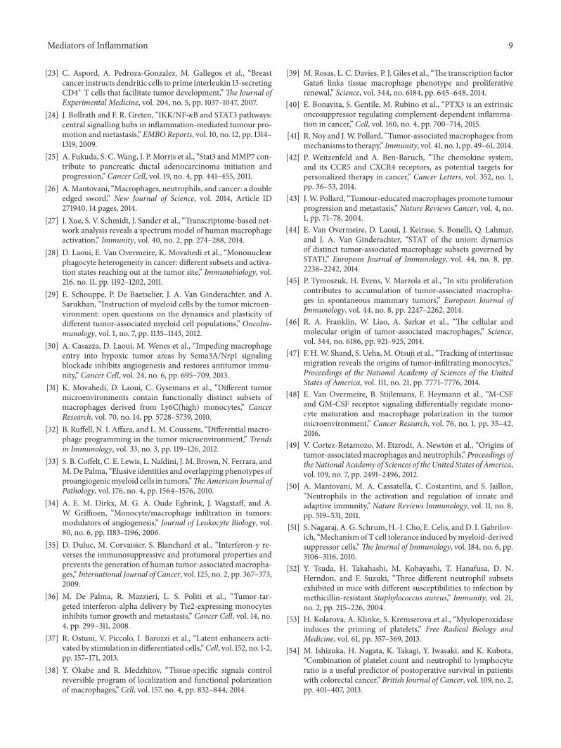
Figure 2: Recruitment pattern of myeloid cells in tumor progression and metastasis. Stages in tumor progression and metastasis includinginitiation, proliferation and tumor site inflammation, invasion, intravasation, circulation in blood stream, extravasation, and colonization areshown with associated myeloid cells, platelets, and cytokines. The contribution of TAM and TAN at early stage of distant colonization sitesis not clear. Green: cytokines/chemokines in recruitment or suppression of immune cells, black: metastasis associated proteins, red arrow:movement of myeloid cells, EMT: epithelial mesenchymal transition, and CTSB: cysteine protease cathepsin B based on [105].
Studies have shown that, analogously to the M1 and M2dichotomy, TANs develop a protumorigenic (N2) phenotypein untreated tumors, largely driven by the presence of TGF-𝛽[58], and that blocking the effects of TGF-𝛽 or augmentingIFN-𝛽 can also alter the phenotype of TANs to a moreantitumor (N1) phenotype [56]. Antitumor “N1-like” cells
generated in the absence of TGF-𝛽 produced higher levels ofTNF-𝛼, MIP-1𝛼, H
2O2, and NO and were cytotoxic to tumor
cells both in vitro and in vivo [59].Respiratory burst and granule proteins are two main
mechanisms of cell killing by neutrophils. Transcriptomeanalysis of naive bone marrow neutrophils (NN) from

Mediators of Inflammation 5
nontumor bearing mice and G-MDSC and TAN from micein which AB12 mesothelioma tumors were growing showedthat expression levels of both proteins involved in respi-ratory burst and granule proteins were downregulated andthat those of chemokine, cytokine, and APC genes wereupregulated in TANs [58]. N2-like neutrophils may also syn-ergistically interact with tumor-resident mesenchymal stemcells (MSCs) to prompt cancer progression [60]. TANs fromestablished tumors produce CCL17 or CCL22, recruitingimmunosuppressive regulatory T cells (Tregs) with defectivecytotoxic functions into the tumor and leading to suppressionof antitumoral immunity [61]. Of note, similarly to TAMs,TANs from early tumors were more cytotoxic toward tumorcells, while in established tumors TANs acquire a moreprotumoral phenotype, showing how the evolvement of theTME influences TAN phenotype [55]. Unlike TAMs, it isnot certain whether activation of TANs is reversible, and ithas been suggested that N1-like and N2-like phenotypes ofneutrophilsmay be fromdifferent degrees of activation ratherthan polarization [62]. The important question whetherTANs can be manipulated to undergo frank irreversibleactivation or possibly reversible activation states remainsunresolved and should be a matter of further research.
5. Recruitment of TANs
Do we know the origin of TANs? It is known that the spleenis the site of localization of TAM and TAN precursors, fromwhere they physically relocate to the tumor stroma, andthat CXCL8 (IL-8), a chemoattractant for neutrophils, is alsochiefly responsible for the recruitment of TANs (Figure 2)[49]. A recent transcriptome study showed that TANs are not“tissue-based G-MDSCs” modulated by the TME but are adifferent population of neutrophils from both bone marrow-derived neutrophils and G-MDSCs [58]. However, we are notsure whether the majority of TANs are actually differentiatedfrom G-MDSCs that have been recruited to the tumor orwhether they are bone marrow-/blood-derived neutrophils,converted to N2 TANs in the TME specifically by the highlocal concentrations of TGF-𝛽 [63]. Though the study doesnot clarify whether the cells were recruited from the bonemarrow/blood pool of neutrophils or the splenic G-MDSCpopulation, the two studies support the idea that TGF-𝛽 andother factors in the TME may affect the local “education” ofrecruited neutrophils.
6. Possible Interaction of TANs with TAMs
Do TANs then recruit TAM precursors to the tumor siteor are they responsible for the M2-like activation of macro-phages in the TME? It is known that activated neutrophilsreleasing IL-8 and TNF-𝛼 activate and recruit macrophagesat the site of inflammation [64]. Neutrophils secrete MPO,and MPO binding to the MMR induces secretion of reactiveoxygen intermediates, IL-8, TNF-𝛼, and GM-CSF in chronicinflammatory environments such as rheumatoid joints [65].M2-like macrophages express high levels of macrophagemannose receptor (MMR) and IL-10 and low levels of HLA-DR and IL-1𝛽 [66]. Though we still lack direct evidence
that supports TAN and TAM interaction through MPO andthe MMR, massive MPO-positive neutrophil infiltration hasbeen found in established colorectal cancer [67] and lungcancer [68]. Also, similar influence of TGF-𝛽 on activationof macrophages and neutrophils (M2-like and N2-like, resp.)indicates a close link between TAMs and TANs in the sameTME and the possibility that recruitment of macrophagesby neutrophils may precede their N2-like polarization. Itwould be necessary to confirm whether the interactionbetween TANs and TAMs in the TME is similar to well-known interactions between neutrophils andmacrophages ina nontumoral chronic inflammatory environment.
7. Nuclear Extracellular Trap (NET) Formationin the TME
NETs are neutrophil-derived structures composed of decom-pacted chromatin (DNA and citrullinated associating his-tones) and antimicrobial peptides, and NET-producing“NETosis” is a form of neutrophil death, distinct fromapoptosis or necrosis [69]. NETs are introduced to trap andkill microorganisms and facilitate a final form of neutrophil-mediated host defense against microorganisms. They havealso been found in non-microorganism-induced inflamma-tory environments in autoimmune diseases such as systemiclupus erythematous (SLE) and rheumatoid arthritis (RA)[70–73] and tumors [74, 75]. In autoimmune diseases suchas RA, neutrophils are mostly responsible for the cyto-toxic effects of immune cells and NETs appear to provideautoantigens and mediate organ damage [70, 76]. However,the function of NETs in tumor progression is still not clear,although they have been suggested to contribute tometastasisfrom trapping of circulating tumor cells at distant metastaticsites [74, 77] and to tumor progression at primary sites byproviding a high local concentration of biologically activeproteins [75, 77].The available data indicate a lack of evidenceto conclusively demonstrate whether TANs actually produceNETs and to indicate which signaling is involved in NETosisin the TME. Though we know the relationship betweendeposition of NETs and recruitment ofMPO-rich populationof neutrophils in tumors, it seems that there is not enoughevidence to indicate the existence of TAN specific NETosis[74, 75, 78].The animal studies were performed with infusionof bone marrow- or spleen-derived naive neutrophils and thelocalization of general neutrophils, not specifically TANs, wascharacterized fromMPO staining in tumor. The recent iden-tification of TAN specific signatures such as CD62LloCD54hiphenotype with a distinct repertoire of chemokine receptorsincluding CCR5, CCR7, CXCR3, and CXCR4 in human lungcancer indicates that further study to validate TAN specificNETosis may be possible in animal studies [79]. Anotherfunction of NETs is to provide autoantigens. In SLE andRA patients, specific autoantigens, such as anti-dsDNA andanti-citrullinated protein antibodies and rheumatoid factor,respectively, have been detected. However, there seems to bea relative paucity of tumor-derived autoantigens identifiedthus far, and this suggests that a major function of tumoralNETs is more likely to trap migrating tumor cells and to pro-vide protumoral substances rather than immunomodulating

6 Mediators of Inflammation
autoantigens. Still, it is becoming clear that NETs are a veryrecently introduced component of theTMEand that they playanother protumoral role in tumor progression. Future studieswill probably investigate (i) identification of the N1-like, N2-like, or general neutrophils that actually form NETs and thespecific tumor progression stage to which NETs primarilycontribute; (ii) whether retention of TAMsorTANs in tumorsalso requires formation of NETs; (iii) whetherM2-like or N2-like activation requires NETs; (iv) which signals are involvedin the formation of NETs.
8. Platelets as a Potential Hubfor the Recruitment ofMacrophages and Neutrophils
Platelets also contribute to tumor progression [80, 81]. Highplatelet count in blood (thrombocytosis) is associated withdecreased survival in awide range of cancers including breast,colorectal, and lung cancer [82, 83]. An increased plateletcount in blood in malignancy is associated with poor patientprognosis [84, 85]. It has been suggested that platelets mayprotect tumor cells from immune attack in the circulation,may provide adhesive sites for tumor dissemination, mayprovide chemokine signals for macrophage recruitment intumors, and may even shuttle growth factors and cytokinesfrom one site to another [2]. By forming microthrombi,platelets may function as a “shield” to protect disseminatingcancer cells in microcirculation from immune cell attack.Platelets store various chemokines and the majority (∼80%)of VEGF detectable in blood and platelets induces angiogen-esis in vivo [85].
Platelets play key roles in directing homing and reten-tion signals for bone marrow-derived cells (BMDCs) andcancer cells and also secrete SDF-1, critical for macrophagerecruitment and positioning in tumors [2]. Also, platelet-derived SDF-1 is critical for migration of CXCR4+ tumorcells, hematopoietic progenitor cells (HPCs), and endothelialprogenitor cells (EPCs) [86]. This is meaningful in thatBMDCs homing to the primary tumor niche may remain inan undifferentiated state in the formofHPCs, EPCs,MSCs, orGR-1+CD11b+MDSCs ormay differentiate intomore special-ized cell types including TAMs [2]. It is known that plateletssupport the recruitment of leukocytes in inflammation andvice versa and that the interaction between platelets andneutrophils can happen not only at the inflammatory site,but also in the circulation, indicating the role of plateletsin metastasis [87]. Platelets can recruit themselves andneutrophils via various mechanisms, such as the formationof platelet/leukocyte complexes, secretion of serotonin, andinduction of P-selectin on platelets and ICAM-1 and 𝛼v𝛽3on endothelial cells [87]. All of these findings indicate thatplatelets may play a central role in recruiting neutrophils ina chronically “persistent” inflammatory environment, that is,the TME. Tumor cells express tissue factor (TF), which isa receptor for coagulation factors VIIa and X [88, 89]. Clotformation by TF expressed by tumor cells enhances recruit-ment of macrophages in a lung metastasis model throughvarious mechanisms including protease-activated receptor
[90], and recruitment of granulocytic cells by the platelet-secreted CXCR2 ligands, CXCL5 and CXCL7 chemokines,upon platelet contact with tumor cells is essential mechanismfor the guidance of granulocytes to form “early metastaticniches” [81, 91, 92]. Importantly, recent results indicate thatcomplement components and platelets are key players incancer-related inflammation and mediate recruitment ofmacrophages at least partially via CCL2 [40]. Summary ofrepresentative interactions between TAM, TAN, and plateletsdescribed in this review can be found in Figure 3.
All of this evidence emphasizes the role of platelets inrecruitment of macrophages and neutrophils in tumor sites.Though we still lack evidence to support the role of plateletsin activation of macrophages and neutrophils—and it is gen-erally accepted that their tumor-protective role in the bloodstream may be the most profound influence of platelets ontumor progression—thrombocytopenicmice show increasedblood TNF-𝛼 and IL-6 and decreased TGF-𝛽 [87], possiblyfavoring antitumoral polarization; as stated, platelets areinvolved in recruitment of bothmacrophages andneutrophilsin both primary and metastatic tumor sites. At present, thereare important questions to be solved: which stages in tumorprogression, including metastasis, are primarily affected byplatelet functions, which of the adhesive or paracrine func-tions of platelets are more important for tumor progression,and which platelet factor or traditionally emphasized tissuefactor is more important for the protumoral activity of thecoagulation system? Further research will likely demonstratethe functional contribution of platelets in tumor progression,including the development of protumoral TAMs and TANs.
9. Clinical Implications
All the summarized data describing the protumoral role ofthe myeloid infiltrate of tumors in this review emphasize thatTAMCs are reasonable targets for new anticancer therapeuticapproaches. It is now becoming clear that host-protectiveproperties of macrophages are suppressed in the TME andthat therapeutic intervention can reverse this suppression.Recently explored strategies have focused on ablation ofmacrophages or reduction of recruitment of myeloid cellsand repolarization of M2-like protumoral macrophages toantitumoral M1-like cells. CD40 agonist antibody [93] andTLR9 agonist (CpG-oligodeoxynucleotide) [94] have beenshown to be effective in repolarizing M2-like protumoralmacrophages. CCL2/CCR2 antagonist [95, 96] and CSF-1inhibitory antibodies or Yondelis (trabectedin) [97] wereeffective in blocking recruitment of macrophages in tumorsites. Bisphosphonate zoledronic acid [36, 98] and clodronate[99] have been used to inhibit TAM effectors and to depleteTAMs, respectively.
Rather than depleting the entire population of neu-trophils, the usual strategy is to deplete TANs or disrupt theirhoming ability, migration. For this purpose, the deploymentof anti-CXCR2 antibodies to deplete TANs or the targetingof specific neutrophil-derived or recruiting chemokines, suchas CXCL-5, Gro-𝛼, or IL-8, was performed and reportedto be successful [59]. Furthermore, targeting TGF-𝛽 oraugmenting the activity of IFN-𝛽 to block its skewing of

Mediators of Inflammation 7
Platelet Activated platelet
P-selectinP2Y
ADP
TF
Thrombin
PARs
GPIIbIIIa(𝛼IIb𝛽3)
EGF,cathepsin, CCL18, and osteonectin: migration of tumor cells
CXCL12, HRG𝛽1
IL-4, IL-10prostaglandinHypoxia
TAN
Neutrophil
Macrophage
TAM
CAF
Tumor cellBlood vessel
CCL2, CXCL12
CSF-1, and SDF-1
complements
SEMA3APl
atelet
-tum
or ce
ll
aggr
egati
onvW
F, fib
rone
ctin,
and
fibrin
ogen
Macrophage recruitment
CXCL8
CXCL15 and HMGB1
MPO
NETosis
MMP9-ECM interaction,
cathepsin-mediated
aggregate, and E-cadherin
disruption via NE,
VEGF release from ECM
Platelet-neutrophil
aggregation
Neutrophil activation
(N2-like activation?)
Angiog
enic
or pr
oteo
lytic
prot
ein re
lease
(𝛼-g
ranu
le)
VEG
F, WN
T7B:
angiogenesis
IL-8, TNF-𝛼, MPO
TGF-𝛽
Figure 3: Summary of representative interactions between TAM, TAN, platelet, and tumor cells. The interactions between neutrophil andmacrophages have not been significantly understood in the TME and the contribution of platelet in differentiation of TAMandTAN suggestedin this review awaits further studies. Tumor cells, blood vessels, and CAF comprise TME. CCL2, CXCL12, CSF-1, SDF-1, complements, andSEMA3A for macrophage recruitment [30, 106]. CSF-1 prompts TAMs to produce EGF.The EGF-CSF-1 loop can be initiated by CAF derivedfactors, such as CXCL12 and HRG𝛽1 [106]. IL-4 fromCD4+ T cells or tumor cells can activate macrophages to TAMs. CCL18 and osteonectincan increase migration and intravasation of tumor cells in metastasis. CXCL-8, CXCL15, and HMGB1 secreted from tumor cells can recruitTANs in metastatic sites. MPO and cytokines from neutrophil recruit platelet and macrophages. PAR and P2Y receptor are involved inthrombin and ADP mediated platelet activation, respectively. P-selectin is involved in platelet leukocyte tethering and leukocyte activation.𝛼-granule is a storage of proteins that enhance adhesive process, angiogenesis, and extracellular matrix (ECM) degradation [81]. GPIIbIIIamediates tumor cell and platelet interaction via vWF, fibronectin, and fibrinogen [80]. Red arrow: neutrophil-mediated recruitment ofmacrophages in tumor.Thick arrow: conversion of platelets, neutrophils, andmacrophages to activated platelets, TAN, and TAM, respectively.GPIIbIIIa, glycoprotein IIbIIIa; vWF, Von Willebrand factor; ADP, adenosine diphosphate; PARs, proteinase-activated receptors; P2Y, P2Yreceptors; TF, tissue factor; NE, neutrophil elastase; HMGB1, high mobility group protein B1; HRG𝛽1, heregulin 𝛽1.
TANs toward an N2 phenotype may have potential as anew therapeutic approach [56, 63]. As neutrophil-derivedmolecules play critical roles in a wide range of stages of tumorprogression [59], targeting neutrophil-secreted enzymes orcytokines could be another effective approach [100]. Target-ing TANs may indirectly affect TAM populations, consid-ering the interaction between neutrophils and macrophagesmentioned above.
Because aggressive anticoagulant therapy in cancerpatients carries the risk of bleeding complications, selectiveinhibition of TF signaling or platelet functions should be con-sidered for clinical settings. Currently, the benefit that directplatelet receptor antagonists may have on cancer prognosishas not been demonstrated, and the evidence to support acombined use of antiplatelet agents with current chemother-apeutic reagents is lacking [101]. The concept that tumor cells
alter their gene expression profiles to acquire a genopheno-type closely resembling that of platelets and express severalmegakaryocytic genes (adhesion receptors 𝛼IIb𝛽3, thrombinreceptor, and PECAM/CD31 and/or platelet-type 12-LOX) toactivate platelets or the coagulation cascade is referred to as“platelet mimicry” of tumor cells [102]. This well-describedepiphenomenon facilitates hematogenous dissemination oftumor cells in metastasis; thus, identification of moleculartargets to regulate platelet mimicry is also likely to providenew therapeutic modalities. Recently, the CXCR2 receptorfor the granulocyte- and platelet-derived ligand CXCL5/7was shown to be important for recruitment of neutrophilsto early metastatic niches [92], and CXCR2 inhibitors reducethe recruitment of granulocytes in primary tumor sites as well[103, 104]. Considering that anti-CXCR2 inhibitors evaluatedin the clinic for inflammatory disease are well tolerated by
8 Mediators of Inflammation
most patients [103], targeting the CXCR2-CXCL5/7 axis maybecome effective in clinical settings.
10. Conclusions and Perspectives
There has been tremendous effort and progress in deci-phering the function of myeloid cells in the TME and intumor progression by excellent investigators in the field.Unfortunately, thewealth ofmounting information regardingprotumoral myeloid cells in cancer has been fragmentary andproduced confusion from a disjointed view of the role ofmacrophages and neutrophils in the TME [19]. In addition,the absence of unique markers to differentiate each subsethas obscured the nature of specific myeloid subsets in cancer.However, the contribution of TAMs and TANs to tumorprogression is clear, and animal model-based or preclinicalstudies have shown promising results. We anticipate thatthe introduction of more sophisticated tumor models andtechniques to differentiate different myeloid cell subsets invivo will reveal fundamental information about possiblespatial and temporal modulation of myeloid cells, includingtheir interaction with platelets in each progression stage ofdifferent types of tumors.
Conflict of Interests
The authors have no conflict of interests to declare.
Acknowledgments
This study was supported by the Basic Science ResearchProgram through the National Research Foundation ofKorea (NRF) funded by the Ministry of Science, ICT &Future Planning (NRF-2014R1A1A1004682), by the NationalResearch Foundation of Korea (NRF) funded by the Koreangovernment [MSIP] (Grant no. 2014R1A2A1A11049526), by agrant of the Korea Health Technology R&D Project throughthe Korea Health Industry Development Institute (KHIDI),funded by the Ministry of Health & Welfare, Republic ofKorea (HI15C0001), and by the Gachon University researchfund of 2012 (2012-M072).
References
[1] D. Hanahan and R. A.Weinberg, “Hallmarks of cancer: the nextgeneration,” Cell, vol. 144, no. 5, pp. 646–674, 2011.
[2] J. Wels, R. N. Kaplan, S. Rafii, and D. Lyden, “Migratory neigh-bors and distant invaders: tumor-associated niche cells,” Genesand Development, vol. 22, no. 5, pp. 559–574, 2008.
[3] N. Rezaei, Cancer Immunology: A Translational Medicine Con-text, Springer, Berlin, Germany, 2015.
[4] E. Van Overmeire, D. Laoui, J. Keirsse, J. A. Van Ginderachter,and A. Sarukhan, “Mechanisms driving macrophage diversityand specialization in distinct tumor microenvironments andparallelisms with other tissues,” Frontiers in Immunology, vol.5, article 127, 2014.
[5] B.-Z. Qian and J. W. Pollard, “Macrophage diversity enhancestumor progression and metastasis,” Cell, vol. 141, no. 1, pp. 39–51, 2010.
[6] E. Tu, P. Z. C. Chia, and W. Chen, “TGF𝛽 in T cell biology andtumor immunity: angel or devil?” Cytokine and Growth FactorReviews, vol. 25, no. 4, pp. 423–435, 2014.
[7] P. M. A. Kelly, R. S. Davison, E. Bliss, and J. O. McGee,“Macrophages in human breast disease: a quantitative immuno-histochemical study,” British Journal of Cancer, vol. 57, no. 2, pp.174–177, 1988.
[8] L. Bingle, N. J. Brown, and C. E. Lewis, “The role of tumour-associated macrophages in tumour progression: implicationsfor new anticancer therapies,” Journal of Pathology, vol. 196, no.3, pp. 254–265, 2002.
[9] Q.-W. Zhang, L. Liu, C.-Y. Gong et al., “Prognostic significanceof tumor-associated macrophages in solid tumor: a meta-analysis of the literature,” PLoS ONE, vol. 7, no. 12, Article IDe50946, 2012.
[10] P. J. Murray and T. A. Wynn, “Protective and pathogenic func-tions of macrophage subsets,”Nature Reviews Immunology, vol.11, no. 11, pp. 723–737, 2011.
[11] T. E. Rohan, X. Xue, H.-M. Lin et al., “Tumor microenvi-ronment of metastasis and risk of distant metastasis of breastcancer,” Journal of the National Cancer Institute, vol. 106, no. 8,Article ID dju136, 2014.
[12] A. Mantovani, P. Allavena, A. Sica, and F. Balkwill, “Cancer-related inflammation,” Nature, vol. 454, no. 7203, pp. 436–444,2008.
[13] A. Mantovani, S. Sozzani, M. Locati, P. Allavena, and A. Sica,“Macrophage polarization: tumor-associated macrophages as aparadigm for polarizedM2mononuclear phagocytes,”Trends inImmunology, vol. 23, no. 11, pp. 549–555, 2002.
[14] S. Gordon and P. R. Taylor, “Monocyte and macrophageheterogeneity,” Nature Reviews Immunology, vol. 5, no. 12, pp.953–964, 2005.
[15] Y.-C. Wang, F. He, F. Feng et al., “Notch signaling determinesthe M1 versus M2 polarization of macrophages in antitumorimmune responses,” Cancer Research, vol. 70, no. 12, pp. 4840–4849, 2010.
[16] A.Mantovani, A. Sica, S. Sozzani, P. Allavena, A. Vecchi, andM.Locati, “The chemokine system in diverse forms of macrophageactivation and polarization,” Trends in Immunology, vol. 25, no.12, pp. 677–686, 2004.
[17] A. Sica and A.Mantovani, “Macrophage plasticity and polariza-tion: in vivo veritas,” The Journal of Clinical Investigation, vol.122, no. 3, pp. 787–795, 2012.
[18] F. Rae, K. Woods, T. Sasmono et al., “Characterisation andtrophic functions of murine embryonic macrophages basedupon the use of a Csf1r-EGFP transgene reporter,”Developmen-tal Biology, vol. 308, no. 1, pp. 232–246, 2007.
[19] P. J. Murray, J. E. Allen, S. K. Biswas et al., “Macrophageactivation and polarization: nomenclature and experimentalguidelines,” Immunity, vol. 41, no. 1, pp. 14–20, 2014.
[20] A. Sica, P. Larghi, A. Mancino et al., “Macrophage polarizationin tumour progression,” Seminars in Cancer Biology, vol. 18, no.5, pp. 349–355, 2008.
[21] A. Sica, A. Saccani, B. Bottazzi et al., “Autocrine production ofIL-10mediates defective IL-12 production andNF-𝜅B activationin tumor-associated macrophages,”The Journal of Immunology,vol. 164, no. 2, pp. 762–767, 2000.
[22] D. Moussai, H. Mitsui, J. S. Pettersen et al., “The human cuta-neous squamous cell carcinoma microenvironment is charac-terized by increased lymphatic density and enhanced expres-sion of macrophage-derived VEGF-C,”The Journal of Investiga-tive Dermatology, vol. 131, no. 1, pp. 229–236, 2011.
Mediators of Inflammation 9
[23] C. Aspord, A. Pedroza-Gonzalez, M. Gallegos et al., “Breastcancer instructs dendritic cells to prime interleukin 13-secretingCD4+ T cells that facilitate tumor development,”The Journal ofExperimental Medicine, vol. 204, no. 5, pp. 1037–1047, 2007.
[24] J. Bollrath and F. R. Greten, “IKK/NF-𝜅B and STAT3 pathways:central signalling hubs in inflammation-mediated tumour pro-motion andmetastasis,” EMBO Reports, vol. 10, no. 12, pp. 1314–1319, 2009.
[25] A. Fukuda, S. C.Wang, J. P. Morris et al., “Stat3 andMMP7 con-tribute to pancreatic ductal adenocarcinoma initiation andprogression,” Cancer Cell, vol. 19, no. 4, pp. 441–455, 2011.
[26] A.Mantovani, “Macrophages, neutrophils, and cancer: a doubleedged sword,” New Journal of Science, vol. 2014, Article ID271940, 14 pages, 2014.
[27] J. Xue, S. V. Schmidt, J. Sander et al., “Transcriptome-based net-work analysis reveals a spectrum model of human macrophageactivation,” Immunity, vol. 40, no. 2, pp. 274–288, 2014.
[28] D. Laoui, E. Van Overmeire, K. Movahedi et al., “Mononuclearphagocyte heterogeneity in cancer: different subsets and activa-tion states reaching out at the tumor site,” Immunobiology, vol.216, no. 11, pp. 1192–1202, 2011.
[29] E. Schouppe, P. De Baetselier, J. A. Van Ginderachter, and A.Sarukhan, “Instruction of myeloid cells by the tumor microen-vironment: open questions on the dynamics and plasticity ofdifferent tumor-associated myeloid cell populations,” OncoIm-munology, vol. 1, no. 7, pp. 1135–1145, 2012.
[30] A. Casazza, D. Laoui, M. Wenes et al., “Impeding macrophageentry into hypoxic tumor areas by Sema3A/Nrp1 signalingblockade inhibits angiogenesis and restores antitumor immu-nity,” Cancer Cell, vol. 24, no. 6, pp. 695–709, 2013.
[31] K. Movahedi, D. Laoui, C. Gysemans et al., “Different tumormicroenvironments contain functionally distinct subsets ofmacrophages derived from Ly6C(high) monocytes,” CancerResearch, vol. 70, no. 14, pp. 5728–5739, 2010.
[32] B. Ruffell, N. I. Affara, and L.M. Coussens, “Differential macro-phage programming in the tumor microenvironment,” Trendsin Immunology, vol. 33, no. 3, pp. 119–126, 2012.
[33] S. B. Coffelt, C. E. Lewis, L. Naldini, J.M. Brown,N. Ferrara, andM. De Palma, “Elusive identities and overlapping phenotypes ofproangiogenicmyeloid cells in tumors,”TheAmerican Journal ofPathology, vol. 176, no. 4, pp. 1564–1576, 2010.
[34] A. E. M. Dirkx, M. G. A. Oude Egbrink, J. Wagstaff, and A.W. Griffioen, “Monocyte/macrophage infiltration in tumors:modulators of angiogenesis,” Journal of Leukocyte Biology, vol.80, no. 6, pp. 1183–1196, 2006.
[35] D. Duluc, M. Corvaisier, S. Blanchard et al., “Interferon-𝛾 re-verses the immunosuppressive and protumoral properties andprevents the generation of human tumor-associated macropha-ges,” International Journal of Cancer, vol. 125, no. 2, pp. 367–373,2009.
[36] M. De Palma, R. Mazzieri, L. S. Politi et al., “Tumor-tar-geted interferon-alpha delivery by Tie2-expressing monocytesinhibits tumor growth and metastasis,” Cancer Cell, vol. 14, no.4, pp. 299–311, 2008.
[37] R. Ostuni, V. Piccolo, I. Barozzi et al., “Latent enhancers acti-vated by stimulation in differentiated cells,”Cell, vol. 152, no. 1-2,pp. 157–171, 2013.
[38] Y. Okabe and R. Medzhitov, “Tissue-specific signals controlreversible program of localization and functional polarizationof macrophages,” Cell, vol. 157, no. 4, pp. 832–844, 2014.
[39] M. Rosas, L. C. Davies, P. J. Giles et al., “The transcription factorGata6 links tissue macrophage phenotype and proliferativerenewal,” Science, vol. 344, no. 6184, pp. 645–648, 2014.
[40] E. Bonavita, S. Gentile, M. Rubino et al., “PTX3 is an extrinsiconcosuppressor regulating complement-dependent inflamma-tion in cancer,” Cell, vol. 160, no. 4, pp. 700–714, 2015.
[41] R.Noy and J.W. Pollard, “Tumor-associatedmacrophages: frommechanisms to therapy,” Immunity, vol. 41, no. 1, pp. 49–61, 2014.
[42] P. Weitzenfeld and A. Ben-Baruch, “The chemokine system,and its CCR5 and CXCR4 receptors, as potential targets forpersonalized therapy in cancer,” Cancer Letters, vol. 352, no. 1,pp. 36–53, 2014.
[43] J.W. Pollard, “Tumour-educatedmacrophages promote tumourprogression and metastasis,” Nature Reviews Cancer, vol. 4, no.1, pp. 71–78, 2004.
[44] E. Van Overmeire, D. Laoui, J. Keirsse, S. Bonelli, Q. Lahmar,and J. A. Van Ginderachter, “STAT of the union: dynamicsof distinct tumor-associated macrophage subsets governed bySTAT1,” European Journal of Immunology, vol. 44, no. 8, pp.2238–2242, 2014.
[45] P. Tymoszuk, H. Evens, V. Marzola et al., “In situ proliferationcontributes to accumulation of tumor-associated macropha-ges in spontaneous mammary tumors,” European Journal ofImmunology, vol. 44, no. 8, pp. 2247–2262, 2014.
[46] R. A. Franklin, W. Liao, A. Sarkar et al., “The cellular andmolecular origin of tumor-associated macrophages,” Science,vol. 344, no. 6186, pp. 921–925, 2014.
[47] F. H.W. Shand, S. Ueha,M. Otsuji et al., “Tracking of intertissuemigration reveals the origins of tumor-infiltrating monocytes,”Proceedings of the National Academy of Sciences of the UnitedStates of America, vol. 111, no. 21, pp. 7771–7776, 2014.
[48] E. Van Overmeire, B. Stijlemans, F. Heymann et al., “M-CSFand GM-CSF receptor signaling differentially regulate mono-cyte maturation and macrophage polarization in the tumormicroenvironment,” Cancer Research, vol. 76, no. 1, pp. 35–42,2016.
[49] V. Cortez-Retamozo, M. Etzrodt, A. Newton et al., “Origins oftumor-associatedmacrophages and neutrophils,” Proceedings ofthe National Academy of Sciences of the United States of America,vol. 109, no. 7, pp. 2491–2496, 2012.
[50] A. Mantovani, M. A. Cassatella, C. Costantini, and S. Jaillon,“Neutrophils in the activation and regulation of innate andadaptive immunity,” Nature Reviews Immunology, vol. 11, no. 8,pp. 519–531, 2011.
[51] S. Nagaraj, A. G. Schrum,H.-I. Cho, E. Celis, andD. I. Gabrilov-ich, “Mechanismof T cell tolerance induced bymyeloid-derivedsuppressor cells,”The Journal of Immunology, vol. 184, no. 6, pp.3106–3116, 2010.
[52] Y. Tsuda, H. Takahashi, M. Kobayashi, T. Hanafusa, D. N.Herndon, and F. Suzuki, “Three different neutrophil subsetsexhibited in mice with different susceptibilities to infection bymethicillin-resistant Staphylococcus aureus,” Immunity, vol. 21,no. 2, pp. 215–226, 2004.
[53] H. Kolarova, A. Klinke, S. Kremserova et al., “Myeloperoxidaseinduces the priming of platelets,” Free Radical Biology andMedicine, vol. 61, pp. 357–369, 2013.
[54] M. Ishizuka, H. Nagata, K. Takagi, Y. Iwasaki, and K. Kubota,“Combination of platelet count and neutrophil to lymphocyteratio is a useful predictor of postoperative survival in patientswith colorectal cancer,” British Journal of Cancer, vol. 109, no. 2,pp. 401–407, 2013.

10 Mediators of Inflammation
[55] I. Mishalian, R. Bayuh, L. Levy, L. Zolotarov, J. Michaeli, and Z.G. Fridlender, “Tumor-associated neutrophils (TAN) developpro-tumorigenic properties during tumor progression,” CancerImmunology, Immunotherapy, vol. 62, no. 11, pp. 1745–1756, 2013.
[56] J. Jablonska, S. Leschner, K. Westphal, S. Lienenklaus, and S.Weiss, “Neutrophils responsive to endogenous IFN-beta regu-late tumor angiogenesis and growth in a mouse tumor model,”The Journal of Clinical Investigation, vol. 120, no. 4, pp. 1151–1164,2010.
[57] F. Donskov, “Immunomonitoring and prognostic relevance ofneutrophils in clinical trials,” Seminars in Cancer Biology, vol.23, no. 3, pp. 200–207, 2013.
[58] Z.G. Fridlender, J. Sun, I.Mishalian et al., “Transcriptomic anal-ysis comparing tumor-associated neutrophils with granulocyticmyeloid-derived suppressor cells and normal neutrophils,”PLoS ONE, vol. 7, no. 2, Article ID e31524, 2012.
[59] A. D. Gregory and A. M. Houghton, “Tumor-associated neu-trophils: new targets for cancer therapy,” Cancer Research, vol.71, no. 7, pp. 2411–2416, 2011.
[60] X. Zhang, Q. Zhu, X. Yuan, H. Qian, andW. Xu, “Mesenchymalstem cells in cancer: a new link to neutrophils,” Cancer Cell &Microenvironment, vol. 1, no. 3, 2014.
[61] I. Mishalian, R. Bayuh, E. Eruslanov et al., “Neutrophils recruitregulatory T-cells into tumors via secretion of CCL17—a newmechanism of impaired antitumor immunity,” InternationalJournal of Cancer, vol. 135, no. 5, pp. 1178–1186, 2014.
[62] H. Piccard, R. J. Muschel, and G. Opdenakker, “On thedual roles and polarized phenotypes of neutrophils in tumordevelopment and progression,” Critical Reviews in Oncol-ogy/Hematology, vol. 82, no. 3, pp. 296–309, 2012.
[63] Z. G. Fridlender, J. Sun, S. Kim et al., “Polarization of tumor-associated neutrophil phenotype by TGF-beta: ‘N1’ versus ‘N2’TAN,” Cancer Cell, vol. 16, no. 3, pp. 183–194, 2009.
[64] V. Kumar and A. Sharma, “Neutrophils: Cinderella of innateimmune system,” International Immunopharmacology, vol. 10,no. 11, pp. 1325–1334, 2010.
[65] D. L. Lefkowitz and S. S. Lefkowitz, “Macrophage-neutrophilinteraction: a paradigm for chronic inflammation revisited,”Immunology and Cell Biology, vol. 79, no. 5, pp. 502–506, 2001.
[66] J. Sun, Y. Mao, Y.-Q. Zhang et al., “Clinical significance of theinduction of macrophage differentiation by the costimulatorymolecule B7-H3 in humannon-small cell lung cancer,”OncologyLetters, vol. 6, no. 5, pp. 1253–1260, 2013.
[67] R. A. Droeser, C. Hirt, S. Eppenberger-Castori et al., “Highmyeloperoxidase positive cell infiltration in colorectal cancer isan independent favorable prognostic factor,” PLoS ONE, vol. 8,no. 5, Article ID e64814, 2013.
[68] A. L. Rymaszewski, E. Tate, J. P. Yimbesalu et al., “The role ofneutrophil myeloperoxidase in models of lung tumor develop-ment,” Cancers, vol. 6, no. 2, pp. 1111–1127, 2014.
[69] V. Brinkmann, U. Reichard, C. Goosmann et al., “Neutrophilextracellular traps kill bacteria,” Science, vol. 303, no. 5663, pp.1532–1535, 2004.
[70] H. L.Wright, R. J.Moots, and S.W. Edwards, “Themultifactorialrole of neutrophils in rheumatoid arthritis,” Nature ReviewsRheumatology, vol. 10, no. 10, pp. 593–601, 2014.
[71] G. S. Garcia-Romo, S. Caielli, B. Vega et al., “Netting neutrophilsare major inducers of type I IFN production in pediatricsystemic lupus erythematosus,” Science Translational Medicine,vol. 3, no. 73, Article ID 73ra20, 2011.
[72] R. Lande, D. Ganguly, V. Facchinetti et al., “Neutrophils activateplasmacytoid dendritic cells by releasing self-DNA-peptidecomplexes in systemic lupus erythematosus,” Science Transla-tional Medicine, vol. 3, no. 73, Article ID 73ra19, 2011.
[73] R. Khandpur, C. Carmona-Rivera, A. Vivekanandan-Giri et al.,“NETs are a source of citrullinated autoantigens and stimulateinflammatory responses in rheumatoid arthritis,” Science Trans-lational Medicine, vol. 5, no. 178, Article ID 178ra40, 2013.
[74] J. Cools-Lartigue, J. Spicer, B. McDonald et al., “Neutrophilextracellular traps sequester circulating tumor cells and pro-mote metastasis,” The Journal of Clinical Investigation, vol. 123,no. 8, pp. 3446–3458, 2013.
[75] S. Sangaletti, C. Tripodo, C. Vitali et al., “Defective stromalremodeling and neutrophil extracellular traps in lymphoidtissues favor the transition from autoimmunity to lymphomas,”Cancer Discovery, vol. 4, no. 1, pp. 110–129, 2014.
[76] J. S. Knight, C. Carmona-Rivera, and M. J. Kaplan, “Proteinsderived from neutrophil extracellular traps may serve as self-antigens and mediate organ damage in autoimmune diseases,”Frontiers in Immunology, vol. 3, article 380, 2012.
[77] J. Cools-Lartigue, J. Spicer, S. Najmeh, and L. Ferri, “Neutrophilextracellular traps in cancer progression,” Cellular and Molecu-lar Life Sciences, vol. 71, no. 21, pp. 4179–4194, 2014.
[78] S. Berger-Achituv, V. Brinkmann, U. A. Abed et al., “A proposedrole for neutrophil extracellular traps in cancer immunoedit-ing,” Frontiers in Immunology, vol. 4, article 48, 2013.
[79] E. B. Eruslanov, P. S. Bhojnagarwala, J. G. Quatromoni et al.,“Tumor-associated neutrophils stimulate T cell responses inearly-stage human lung cancer,” The Journal of Clinical Inves-tigation, vol. 124, no. 12, pp. 5466–5480, 2014.
[80] N. M. Bambace and C. E. Holmes, “The platelet contributionto cancer progression,” Journal of Thrombosis and Haemostasis,vol. 9, no. 2, pp. 237–249, 2011.
[81] L. J. Gay and B. Felding-Habermann, “Contribution of plateletsto tumour metastasis,” Nature Reviews Cancer, vol. 11, no. 2, pp.123–134, 2011.
[82] P. Jurasz, D. Alonso-Escolano, and M. W. Radomski, “Platelet–cancer interactions: mechanisms and pharmacology of tumourcell-induced platelet aggregation,” British Journal of Pharmacol-ogy, vol. 143, no. 7, pp. 819–826, 2004.
[83] J. A. Joyce and J.W. Pollard, “Microenvironmental regulation ofmetastasis,” Nature Reviews Cancer, vol. 9, no. 4, pp. 239–252,2009.
[84] S. Taucher, A. Salat, M. Gnant et al., “Impact of pretreatmentthrombocytosis on survival in primary breast cancer,” Throm-bosis and Haemostasis, vol. 89, no. 6, pp. 1098–1106, 2003.
[85] E. Sierko and M. Z. Wojtukiewicz, “Platelets and angiogenesisinmalignancy,” Seminars inThrombosis andHemostasis, vol. 30,no. 1, pp. 95–108, 2004.
[86] H.-G. Kopp and S. Rafii, “Thrombopoietic cells and the bonemarrow vascular niche,” Annals of the New York Academy ofSciences, vol. 1106, pp. 175–179, 2007.
[87] A. Gros, V. Ollivier, and B. Ho-Tin-Noe, “Platelets in inflam-mation: regulation of leukocyte activities and vascular repair,”Frontiers in Immunology, vol. 5, article 678, 2014.
[88] J. H. Im, W. Fu, H. Wang et al., “Coagulation facilitates tumorcell spreading in the pulmonary vasculature during early me-tastatic colony formation,” Cancer Research, vol. 64, no. 23, pp.8613–8619, 2004.
[89] J. S. Palumbo, “Mechanisms linking tumor cell-associatedprocoagulant function to tumor dissemination,” Seminars inThrombosis and Hemostasis, vol. 34, no. 2, pp. 154–160, 2008.
Mediators of Inflammation 11
[90] I. Lal, K. Dittus, and C. E. Holmes, “Platelets, coagulationand fibrinolysis in breast cancer progression,” Breast CancerResearch, vol. 15, no. 4, article 207, 2013.
[91] A.M.Gil-Bernabe, S. Ferjancic,M.Tlalka et al., “Recruitment ofmonocytes/macrophages by tissue factor-mediated coagulationis essential for metastatic cell survival and premetastatic nicheestablishment in mice,” Blood, vol. 119, no. 13, pp. 3164–3175,2012.
[92] M. Labelle, S. Begum, and R. O. Hynes, “Platelets guidethe formation of early metastatic niches,” Proceedings of theNational Academy of Sciences of the United States of America,vol. 111, no. 30, pp. E3053–E3061, 2014.
[93] G. L. Beatty, E. G. Chiorean, M. P. Fishman et al., “CD40 ago-nists alter tumor stroma and show efficacy against pancreaticcarcinoma in mice and humans,” Science, vol. 331, no. 6024, pp.1612–1616, 2011.
[94] I. N. Buhtoiarov, P. M. Sondel, J. M. Wigginton et al., “Anti-tumour synergy of cytotoxic chemotherapy and anti-CD40 plusCpG-ODN immunotherapy through repolarization of tumour-associated macrophages,” Immunology, vol. 132, no. 2, pp. 226–239, 2011.
[95] B.-Z. Qian, J. Li, H. Zhang et al., “CCL2 recruits inflammatorymonocytes to facilitate breast-tumour metastasis,” Nature, vol.475, no. 7355, pp. 222–225, 2011.
[96] K. Garber, “First results for agents targeting cancer-relatedinflammation,” Journal of the National Cancer Institute, vol. 101,no. 16, pp. 1110–1112, 2009.
[97] G. Germano, R. Frapolli, C. Belgiovine et al., “Role of macro-phage targeting in the antitumor activity of trabectedin,”CancerCell, vol. 23, no. 2, pp. 249–262, 2013.
[98] E. Giraudo, M. Inoue, and D. Hanahan, “An amino-bisphos-phonate targets MMP-9-expressing macrophages and angio-genesis to impair cervical carcinogenesis,”The Journal of Clini-cal Investigation, vol. 114, no. 5, pp. 623–633, 2004.
[99] S. M. Zeisberger, B. Odermatt, C. Marty, A. H. M. Zehnder-Fjallman, K. Ballmer-Hofer, and R. A. Schwendener, “Clodron-ate-liposome-mediated depletion of tumour-associated macro-phages: a new and highly effective antiangiogenic therapyapproach,” British Journal of Cancer, vol. 95, no. 3, pp. 272–281,2006.
[100] A. M. Houghton, D. M. Rzymkiewicz, H. Ji et al., “Neutrophilelastase-mediated degradation of IRS-1 accelerates lung tumorgrowth,” Nature Medicine, vol. 16, no. 2, pp. 219–223, 2010.
[101] D. Sharma, K. E. Brummel-Ziedins, B. A. Bouchard, and C. E.Holmes, “Platelets in tumor progression: a host factor that offersmultiple potential targets in the treatment of cancer,” Journal ofCellular Physiology, vol. 229, no. 8, pp. 1005–1015, 2014.
[102] J. Tımar, J. Tovari, E. Raso, L. Meszaros, B. Bereczky, and K.Lapis, “Platelet-mimicry of cancer cells: epiphenomenon withclinical significance,”Oncology, vol. 69, no. 3, pp. 185–201, 2005.
[103] A. Stadtmann and A. Zarbock, “CXCR2: from bench to bed-side,” Frontiers in Immunology, vol. 3, article 263, 2012.
[104] H. Ijichi, A. Chytil, A. E. Gorska et al., “Inhibiting Cxcr2disrupts tumor-stromal interactions and improves survival ina mouse model of pancreatic ductal adenocarcinoma,” Journalof Clinical Investigation, vol. 121, no. 10, pp. 4106–4117, 2011.
[105] H. A. Smith and Y. Kang, “The metastasis-promoting roles oftumor-associated immune cells,” Journal of MolecularMedicine,vol. 91, no. 4, pp. 411–429, 2013.
[106] T. Kitamura, B.-Z. Qian, and J. W. Pollard, “Immune cell pro-motion of metastasis,” Nature Reviews Immunology, vol. 15, no.2, pp. 73–86, 2015.

Submit your manuscripts athttp://www.hindawi.com
Stem CellsInternational
Hindawi Publishing Corporationhttp://www.hindawi.com Volume 2014
Hindawi Publishing Corporationhttp://www.hindawi.com Volume 2014
MEDIATORSINFLAMMATION
of
Hindawi Publishing Corporationhttp://www.hindawi.com Volume 2014
Behavioural Neurology
EndocrinologyInternational Journal of
Hindawi Publishing Corporationhttp://www.hindawi.com Volume 2014
Hindawi Publishing Corporationhttp://www.hindawi.com Volume 2014
Disease Markers
Hindawi Publishing Corporationhttp://www.hindawi.com Volume 2014
BioMed Research International
OncologyJournal of
Hindawi Publishing Corporationhttp://www.hindawi.com Volume 2014
Hindawi Publishing Corporationhttp://www.hindawi.com Volume 2014
Oxidative Medicine and Cellular Longevity
Hindawi Publishing Corporationhttp://www.hindawi.com Volume 2014
PPAR Research
The Scientific World JournalHindawi Publishing Corporation http://www.hindawi.com Volume 2014
Immunology ResearchHindawi Publishing Corporationhttp://www.hindawi.com Volume 2014
Journal of
ObesityJournal of
Hindawi Publishing Corporationhttp://www.hindawi.com Volume 2014
Hindawi Publishing Corporationhttp://www.hindawi.com Volume 2014
Computational and Mathematical Methods in Medicine
OphthalmologyJournal of
Hindawi Publishing Corporationhttp://www.hindawi.com Volume 2014
Diabetes ResearchJournal of
Hindawi Publishing Corporationhttp://www.hindawi.com Volume 2014
Hindawi Publishing Corporationhttp://www.hindawi.com Volume 2014
Research and TreatmentAIDS
Hindawi Publishing Corporationhttp://www.hindawi.com Volume 2014
Gastroenterology Research and Practice
Hindawi Publishing Corporationhttp://www.hindawi.com Volume 2014
Parkinson’s Disease
Evidence-Based Complementary and Alternative Medicine
Volume 2014Hindawi Publishing Corporationhttp://www.hindawi.com