Review Article PRP Augmentation for ACL … Article PRP Augmentation for ACL Reconstruction...
16
Review Article PRP Augmentation for ACL Reconstruction Luca Andriolo, 1 Berardo Di Matteo, 1 Elizaveta Kon, 2 Giuseppe Filardo, 1 Giulia Venieri, 1 and Maurilio Marcacci 1 1 II Orthopaedic and Traumatology Clinic, Biomechanics and Technology Innovation Laboratory, Rizzoli Orthopaedic Institute, Via di Barbiano No. 1/10, 40136 Bologna, Italy 2 Nano-Biotechnology Laboratory, Rizzoli Orthopaedic Institute, Via di Barbiano No. 1/10, 40136 Bologna, Italy Correspondence should be addressed to Luca Andriolo; [email protected] Received 29 May 2014; Accepted 15 August 2014 Academic Editor: Tomokazu Yoshioka Copyright © 2015 Luca Andriolo et al. is is an open access article distributed under the Creative Commons Attribution License, which permits unrestricted use, distribution, and reproduction in any medium, provided the original work is properly cited. Current research is investigating new methods to enhance tissue healing to speed up recovery time and decrease the risk of failure in Anterior Cruciate Ligament (ACL) reconstructive surgery. Biological augmentation is one of the most exploited strategies, in particular the application of Platelet Rich Plasma (PRP). Aim of the present paper is to systematically review all the preclinical and clinical papers dealing with the application of PRP as a biological enhancer during ACL reconstructive surgery. irty-two studies were included in the present review. e analysis of the preclinical evidence revealed that PRP was able to improve the healing potential of the tendinous graſt both in terms of histological and biomechanical performance. Looking at the available clinical evidence, results were not univocal. PRP administration proved to be a safe procedure and there were some evidences that it could favor the donor site healing in case of ACL reconstruction with patellar tendon graſt and positively contribute to graſt maturation over time, whereas the majority of the papers did not show beneficial effects in terms of bony tunnels/graſt area integration. Furthermore, PRP augmentation did not provide superior functional results at short term evaluation. 1. Introduction Anterior cruciate ligament (ACL) tears are among the most common sport-related injuries and therefore ACL recon- structive surgery is one the most frequently performed procedures in the field of sport medicine [1]. Epidemiological data reveal that the majority of patients are young and sport- active, with high expectations in terms of functional recovery and return to sport [2]. ere is a flourishing literature concerning ACL reconstructive procedures: several different techniques have been documented over the years and, despite overall good clinical outcomes reported at mid/long-term followup, ACL is still on the edge of current clinical and preclinical research [3]. ACL surgery (and the related research) is a classic example of integration between biomechanics and biology: the progress made in recent decades can be attributed to the big steps forward in the knowledge of both the mechanical and biological properties of ACL and its healing process [4– 8]. Although almost all available techniques can provide sat- isfactory results, ACL reconstruction is not a “100%-success” procedure: not all patients are able to regain their previous sport activity level and many factors could influence the clinical outcome, such as the type of graſt used and the pre- op. knee laxity [2, 9]. is is a major concern since ACL injured patients can be a very demanding category, especially professional sport players who also need to return to the playing field as soon as possible. Current research is therefore investigating novel strategies to enhance ACL healing, to reduce the failure rate and accelerate recovery time. Among the different available options, biological augmentation is the most sought aſter approach and recently platelet rich plasma (PRP), which is an autologous blood derivative, largely applied in orthopaedic practice especially in the treatment of degenerative cartilage and tendon lesions, is gaining increasing interest [10–12]. PRP is a source of several growth factors (GFs) and other bioactive molecules that might promote tissue healing and regulate joint homeostasis [13, 14]. It is an easily available product obtained directly from the venous blood of the patient and its use is allowed also in athletes, since antidoping regulations do not consider this blood derivative as a banned substance [15]. Furthermore, it is Hindawi Publishing Corporation BioMed Research International Volume 2015, Article ID 371746, 15 pages http://dx.doi.org/10.1155/2015/371746
Transcript of Review Article PRP Augmentation for ACL … Article PRP Augmentation for ACL Reconstruction...

Review ArticlePRP Augmentation for ACL Reconstruction
Luca Andriolo,1 Berardo Di Matteo,1 Elizaveta Kon,2 Giuseppe Filardo,1
Giulia Venieri,1 and Maurilio Marcacci1
1II Orthopaedic and Traumatology Clinic, Biomechanics and Technology Innovation Laboratory,Rizzoli Orthopaedic Institute, Via di Barbiano No. 1/10, 40136 Bologna, Italy2Nano-Biotechnology Laboratory, Rizzoli Orthopaedic Institute, Via di Barbiano No. 1/10, 40136 Bologna, Italy
Correspondence should be addressed to Luca Andriolo; [email protected]
Received 29May 2014; Accepted 15 August 2014
Academic Editor: Tomokazu Yoshioka
Copyright © 2015 Luca Andriolo et al.This is an open access article distributed under the Creative Commons Attribution License,which permits unrestricted use, distribution, and reproduction in any medium, provided the original work is properly cited.
Current research is investigating new methods to enhance tissue healing to speed up recovery time and decrease the risk of failurein Anterior Cruciate Ligament (ACL) reconstructive surgery. Biological augmentation is one of the most exploited strategies, inparticular the application of Platelet Rich Plasma (PRP). Aim of the present paper is to systematically review all the preclinicaland clinical papers dealing with the application of PRP as a biological enhancer during ACL reconstructive surgery. Thirty-twostudies were included in the present review. The analysis of the preclinical evidence revealed that PRP was able to improve thehealing potential of the tendinous graft both in terms of histological and biomechanical performance. Looking at the availableclinical evidence, results were not univocal. PRP administration proved to be a safe procedure and there were some evidencesthat it could favor the donor site healing in case of ACL reconstruction with patellar tendon graft and positively contribute tograft maturation over time, whereas the majority of the papers did not show beneficial effects in terms of bony tunnels/graft areaintegration. Furthermore, PRP augmentation did not provide superior functional results at short term evaluation.
1. Introduction
Anterior cruciate ligament (ACL) tears are among the mostcommon sport-related injuries and therefore ACL recon-structive surgery is one the most frequently performedprocedures in the field of sport medicine [1]. Epidemiologicaldata reveal that the majority of patients are young and sport-active, with high expectations in terms of functional recoveryand return to sport [2]. There is a flourishing literatureconcerning ACL reconstructive procedures: several differenttechniques have been documented over the years and, despiteoverall good clinical outcomes reported at mid/long-termfollowup, ACL is still on the edge of current clinical andpreclinical research [3].
ACL surgery (and the related research) is a classicexample of integration between biomechanics and biology:the progress made in recent decades can be attributed to thebig steps forward in the knowledge of both the mechanicaland biological properties of ACL and its healing process [4–8]. Although almost all available techniques can provide sat-isfactory results, ACL reconstruction is not a “100%-success”
procedure: not all patients are able to regain their previoussport activity level and many factors could influence theclinical outcome, such as the type of graft used and the pre-op. knee laxity [2, 9]. This is a major concern since ACLinjured patients can be a very demanding category, especiallyprofessional sport players who also need to return to theplaying field as soon as possible. Current research is thereforeinvestigating novel strategies to enhance ACL healing, toreduce the failure rate and accelerate recovery time. Amongthe different available options, biological augmentation isthe most sought after approach and recently platelet richplasma (PRP), which is an autologous blood derivative,largely applied in orthopaedic practice especially in thetreatment of degenerative cartilage and tendon lesions, isgaining increasing interest [10–12]. PRP is a source of severalgrowth factors (GFs) and other bioactive molecules thatmight promote tissue healing and regulate joint homeostasis[13, 14]. It is an easily available product obtained directly fromthe venous blood of the patient and its use is allowed alsoin athletes, since antidoping regulations do not consider thisblood derivative as a banned substance [15]. Furthermore, it is
Hindawi Publishing Corporation
BioMed Research International
Volume 2015, Article ID 371746, 15 pages
http://dx.doi.org/10.1155/2015/371746

2 BioMed Research International
a versatile product since it can be prepared and used directlyin the operating theatre, through intra-articular injectionsor in the shape of a membrane that can be placed directlyonto the target site. The healing potential of PRP has beenshown in several preclinical and clinical studies [13, 16, 17],revealing that its action is directed toward all the articulartissues, ranging from meniscus to cartilage and even softtissues like synovium, tendons, and ligaments: the effects ofPRP are several, including an anabolic stimulus toward cells,an increase in extracellular matrix deposition, reduction ofproapoptotic signals, and even an anti-inflammatory effect inthe joint environment [13].
In light of such potential, the possibility of applying thisbiological product to enhance ACL reconstructive surgeryappears attractive: PRP not only might promote a better andfaster ligamentization of the graft used for ACL reconstruc-tion and reduce the proinflammatory factors released imme-diately after surgery, but might also contribute to a betterintegration of the graftwithin the bone tunnels, thus avoidingtheir enlargement and failure over time. Furthermore, PRPcould be used to accelerate healing and reduce donor-sitemorbidity at the harvest site of the tendon graft.
The aim of the present paper is to review systematicallythe current preclinical and clinical evidence concerning theapplication of PRP as a biological augmentation, to determinesafety and efficacy of this biological approach to improveACLreconstruction surgery.
2. Materials and Methods
A systematic review of the literature was performed on theuse of PRP in ACL reconstruction.The search was conductedon the PubMed database on February 20th, 2014 using thefollowing parameters: ((ACL) OR (ACL reconstruction) OR(ACL lesion)) AND ((PRP) OR (platelet rich plasma) OR(platelet gel) OR (platelet derived) OR (platelet concentrate)).The guidelines for Preferred Reporting Items for SystematicReviews and Meta-Analysis (PRISMA) were used. Screeningprocess and analysis were conducted separately by 2 indepen-dent observers (BDM and LA).
First, the articles were screened by title and abstract.Thefollowing inclusion criteria for relevant articles were usedduring the initial screening of titles and abstracts: clinicaland preclinical reports of any level of evidence, written inEnglish language, with no time limitation, on the use of PRPin ACL reconstruction, and reporting results on PRP effects.Exclusion criteria were articles written in other languages,reviews, or studies analyzing other applications of PRP inknee surgery not related to ACL procedures. In the secondstep, the full texts of the selected articles were screened,with further exclusions according to the previously describedcriteria. Moreover, the articles not reporting clinical, MRI,or histologic results were excluded. Reference lists fromthe selected papers were also screened. A flowchart of thesystematic review is provided in Figure 1. Relevant data werethen extracted and collected in a unique database withthe consensus of the two observers to be analyzed for thepurposes of the present paper.
3. Results
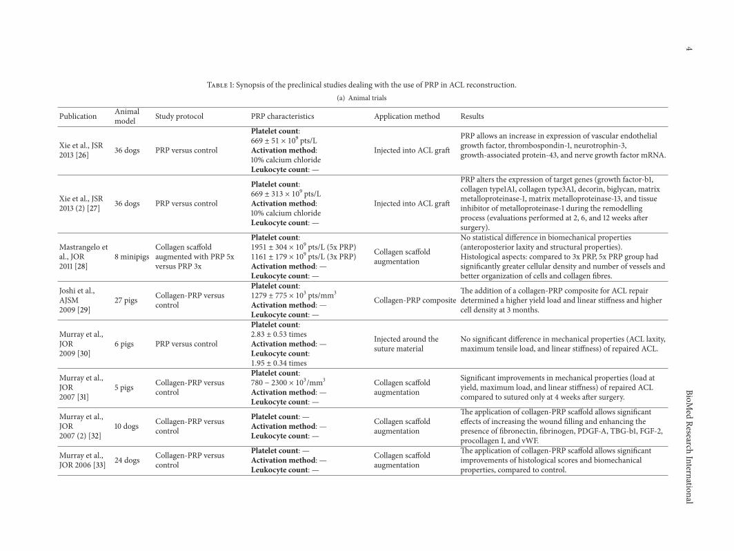
The database search identified 60 records, and the abstractswere screened and selected according to the inclusion/ex-clusion criteria. As shown in Figure 1, a total of 33 full-text articles were assessed for eligibility. Four articles [18–21] did not fulfill the criteria and were further excluded,and 3 articles came from the screening of the reference lists,leading to a total of 32 studies included in the final analysis.A detailed description of preclinical studies is reported inTable 1, whereas clinical studies are summarized in Table 2and discussed in more detail in the following paragraph.
3.1. Clinical Evidence. Fifteen clinical trials were selectedaccording to the inclusion criteria: 11 were randomizedcontrolled trials, 3were prospective comparative studies, and1 was a retrospective comparative trial (Table 2).
With regard to the graft used to performACL reconstruc-tion, in 9 studies, authors used hamstring tendons (gracilisand semitendinosus), in 4 studies bone-patellar tendon-bone(BPB) graft, in 1 study allograft, and in 1 paper authors bothhamstring tendons and BPB graft. Fourteen papers describeda single-bundle reconstruction technique, whereas just in onetrial a double-bundle procedure was used. Concerning PRPdelivery methods, in 2 papers (both dealing with BPB graft)PRP was used to promote harvest site healing, in 1 paper PRPwas applied intra-articularly by a suprapatellar injection atthe very end of the surgical procedure, in 1 paper PRP wasused just to coat the intra-articular portion of the graft, andin 11 papers it was administered both on the graft and in thebony tunnels to enhance the graft-bone integration. Amongthe delivery techniques used, in 2 studies peculiar approacheswere applied. While most of the authors applied PRP on thegraft surface, Sanchez et al. [22] placed PRP inside the graftthrough multiple intratendinous depots, while Radice et al.[23] used a more complex technique: before PRP applicationa bioabsorbable spongy membrane (Gelfoam, Pfizer, NewYork, NY) was carefully sutured on the harvested tendonand around the femoral bone plug in case of the BPB graft.Then, PRP was administered to allow the spongy membraneto absorb the PRP and avoid its dispersion in the articularspace.
In all but one case PRP was activated before administra-tion. Details regarding PRP preparation methods, numberof patients included, patients’ characteristics, and follow-upevaluations are included in Table 2.
To make the results more understandable, they willbe discussed separately according to the specific aspectsanalyzed in the clinical trials:
(i) harvest site healing;(ii) tendon graft maturation and bony tunnel/graft inte-
gration;(iii) clinical results.
3.2. Healing of the Harvest Site. Two papers were specificallyaimed at assessing the contribution of PRP in the healing ofthe harvest site after ACL reconstruction with BPB graft, andboth of them reported positive outcomes with PRP.

BioMed Research International 3
Records identified throughPubMed searching
(n = 60)
Abstracts screened(n = 60)
Abstracts excluded(n = 27)
Additional recordsidentified through references
(n = 3)
Full-text articles assessedfor eligibility
(n = 36)Elig
ibili
tySc
reen
ing
Inclu
ded
Iden
tifica
tion
Studies included inqualitative synthesis
(n = 32)- Clinical study 15- Preclinical study 17
Full-text articles excluded(n = 4)
Reasons:- No results on PRP effects (Biercevicz et al., 2013) 1- No application of PRP (Kuroda et al., 2000,Slater et al., 1995, and Vavken et al., 2010) 3
Figure 1: Flowchart of the systematic review.
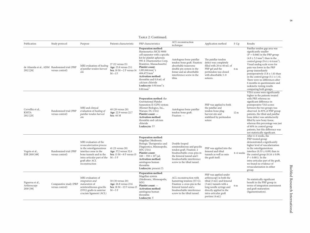
In the study led by de Almeida et al. [24] two groups ofpatients (15 and 12, resp.) were randomized to receive or notreceive PRP after BPB tendonharvesting. Total amount of 20–40mLof PRPwas locally applied and then the peritendineumwas sutured. Clinical and MRI evaluations were performedup to 6 months’ after operation. The authors found thatPRP augmentation determined a significantly smaller patellartendon gap area with respect to the control group and also alower postoperation pain reaction in the PRP group. How-ever, at the final clinical evaluation no statistical intergroupdifference was recorded in the questionnaires used or in theisokinetic test. In the paper authored by Cervellin et al. [25],40 patients were included and divided into two treatmentgroups in the same manner. At 1-year followup, the PRPgroup showed statistically superior clinical outcomes whenevaluated by the VISA-P score and, although not significant,a better bone healing when analyzed byMRI imaging, at bothpatellar and tibial defect sites (85% of patients in PRP versus60% in the control group).
3.3. Tendon GraftMaturation and Bony Tunnel/Graft Integra-tion. Twelve papers examined outcome regarding (a) graft
maturation over time and (b) graft integration in the bonetunnels.
Among the 6 studies reporting data about graft mat-uration, 4 of them were in favour of PRP augmentation,whereas 2 reported no intergroup difference. With respect tobony tunnels/graft area, 9 studies focused on the integration,documenting in 7 cases no advantage after PRP admin-istration. The evolution over time was investigated morespecifically in 3 trials focused on the bony tunnel widening:none demonstrated that PRP was able to prevent tunnels’enlargement over time.
Looking in more detail at the available literature, thefirst paper was published by Orrego et al. [53] in 2008: 108patients were divided into 4 treatment groups due to thefact that the authors also used an autologous spongy bonegraft (placed by interference fit in the femoral tunnel aftergraft fixation) as autologous augmentation. A control groupwas compared to PRP, bone plug, and PRP + bone pluggroups. At 6 months MRI revealed a statistically significantadvantage for the PRP group in terms of graft maturation(lower signal intensity at MRI) with respect to the control
4BioM
edResearch
International
Table 1: Synopsis of the preclinical studies dealing with the use of PRP in ACL reconstruction.(a) Animal trials
Publication Animalmodel Study protocol PRP characteristics Application method Results
Xie et al., JSR2013 [26] 36 dogs PRP versus control
Platelet count:669 ± 51 × 109 pts/LActivation method:10% calcium chlorideLeukocyte count: —
Injected into ACL graftPRP allows an increase in expression of vascular endothelialgrowth factor, thrombospondin-1, neurotrophin-3,growth-associated protein-43, and nerve growth factor mRNA.
Xie et al., JSR2013 (2) [27] 36 dogs PRP versus control
Platelet count:669 ± 313 × 109 pts/LActivation method:10% calcium chlorideLeukocyte count: —
Injected into ACL graft
PRP alters the expression of target genes (growth factor-b1,collagen type1A1, collagen type3A1, decorin, biglycan, matrixmetalloproteinase-1, matrix metalloproteinase-13, and tissueinhibitor of metalloproteinase-1 during the remodellingprocess (evaluations performed at 2, 6, and 12 weeks aftersurgery).
Mastrangelo etal., JOR2011 [28]
8minipigsCollagen scaffoldaugmented with PRP 5xversus PRP 3x
Platelet count:1951 ± 304 × 109 pts/L (5x PRP)1161 ± 179 × 109 pts/L (3x PRP)Activation method: —Leukocyte count: —
Collagen scaffoldaugmentation
No statistical difference in biomechanical properties(anteroposterior laxity and structural properties).Histological aspects: compared to 3x PRP, 5x PRP group hadsignificantly greater cellular density and number of vessels andbetter organization of cells and collagen fibres.
Joshi et al.,AJSM2009 [29]
27 pigs Collagen-PRP versuscontrol
Platelet count:1279 ± 775 × 103 pts/mm3
Activation method: —Leukocyte count: —
Collagen-PRP compositeThe addition of a collagen-PRP composite for ACL repairdetermined a higher yield load and linear stiffness and highercell density at 3months.
Murray et al.,JOR2009 [30]
6 pigs PRP versus control
Platelet count:2.83 ± 0.53 timesActivation method: —Leukocyte count:1.95 ± 0.34 times
Injected around thesuture material
No significant difference in mechanical properties (ACL laxity,maximum tensile load, and linear stiffness) of repaired ACL.
Murray et al.,JOR2007 [31]
5 pigs Collagen-PRP versuscontrol
Platelet count:780 − 2300 × 103/mm3
Activation method: —Leukocyte count: —
Collagen scaffoldaugmentation
Significant improvements in mechanical properties (load atyield, maximum load, and linear stiffness) of repaired ACLcompared to sutured only at 4 weeks after surgery.
Murray et al.,JOR2007 (2) [32]
10 dogs Collagen-PRP versuscontrol
Platelet count: —Activation method: —Leukocyte count: —
Collagen scaffoldaugmentation
The application of collagen-PRP scaffold allows significanteffects of increasing the wound filling and enhancing thepresence of fibronectin, fibrinogen, PDGF-A, TBG-b1, FGF-2,procollagen I, and vWF.
Murray et al.,JOR 2006 [33] 24 dogs Collagen-PRP versus
controlPlatelet count: —Activation method: —Leukocyte count: —
Collagen scaffoldaugmentation
The application of collagen-PRP scaffold allows significantimprovements of histological scores and biomechanicalproperties, compared to control.

BioMed
ResearchInternational
5
(a) Continued.
Publication Animalmodel Study protocol PRP characteristics Application method Results
Weiler et al.,AJSM2004 [34]
48 sheep PRP versus controlPlatelet count: —Activation method: —Leukocyte count: —
Injected into ACL graft
Significantly improvements between PRP group and controlgroup concerning cross-sectional area, failure load, stiffness,and tensile stress after 3, 6, or 12 weeks; however, after 24weeks this difference is not confirmed and mechanical scoresare worse than the intact ACL.
(b) In vitro trials
Publications Purpose PRP characteristics Application method Results
Yoshida et al.,JOR2014 [35]
Does increasing plateletconcentration enhanceACL fibroblast proliferationand collagen production?
Platelet count:PPP: 8 × 10⁶;PRP: 129 × 106 (1x PRP);370 × 106 (3x PRP);615 × 106 pts/mL (5x PRP)Leukocyte count:0.03 × 10⁶ cells/mL
Scaffold’s augmentation(collagen scaffold)
Highest cell metabolism, lowest apoptosis rates, and highestcollagen gene expression with PPP and 1x PRP.
Yoshida andMurray, JOR2013 [36]
How peripheral bloodmononuclear cells(PBMCs) in PRP affectfibroblast behaviour?
Platelet count:PRP: 911 × 106 pts/mLLeukocyte count:PRP: —PBMC: 5 × 10⁶ cells/mL
Scaffold’s augmentation(collagen scaffold)
PRP with PBMCs determined an increasing of type I and typeIII procollagen gene expression, collagen protein expression,and cell proliferation. An increase of IL-6 expression wasdetected in PBMCs exposed to PRP.No effect of PBMCs without PRP.
Cheng et al.,JOR2012 [37]
Does cell’s age influence theACL cell response to PRP?
Platelet count:628 × 106 pts/mLLeukocyte count: —
Scaffold’s augmentation(collagen scaffold)
PRP increases cellular metabolic activity and reducedapoptotic rate and stimulation of collagen production onimmature and adolescent cells, compared to collagen onlyscaffold. Lower effects of PRP on adult cells.
Fallouh et al.,JBJSAm2010 [38]
Effects of autologous PRPon cell viability andcollagen synthesis ofhuman ACL cells.
Platelet count: —Activation method:10% thrombin solutionLeukocyte count: —
PRP clot added toculture environment
PRP group had higher concentration of growth factors, cellnumber, and total collagen production, compared to PPPgroup.
Magarian et al.,Knee2011 [39]
Age dependence of ACLfibroblast response to PRP
Platelet count:684 × 109 pts/LLeukocyte count:2.78 × 10⁹ cells/L Scaffold’s augmentation
(collagen scaffold)
The comparison between immature and adolescent cellsshowed a significantly higher cell migration and proliferationin immature group, whereas no differences were seen inscaffold contraction.
Cheng et al.,TissEngA2010 [40]
Does PRP components (ptsor PPP) independentlyinfluence ACL cellsbehaviour?
Platelet count:911 × 106 pts/mLLeukocyte count: —
Scaffold’s augmentation(collagen scaffold) withPRP, pts, or PPP
The addition of PPP, platelets, or PRP all reduced cell apoptosisand enhanced metabolic activity of fibroblasts.Significantly higher expression of collagen only with PRP.

6BioM
edResearch
International
(b) Continued.
Publications Purpose PRP characteristics Application method Results
Mastrangelo etal., JOR2010 [41]
Age dependence in ACLhealing
Platelet count:915 × 109 pts/L (porcine PRP);369 × 109 pts/L (ovine PRP)Leukocyte count:5.37 × 10⁹ cells/L (porcine PRP);9.6 × 10⁹ cells/L (ovine PRP)
Scaffold’s augmentation(collagen scaffold)
Comparison between adult, adolescent, and immature cellsconcerning cell proliferation and cellular migration: betterresults for immature cells.
Scherping et al.,CTR1997 [42]
Analysis of effects of singlegrowth factors (GF) onfibroblasts harvested frommedial collateral ligamentand ACL of skeletallymature rabbits
Growth factor analysed are asfollows:epidermal GF, basic fibroblast GF,platelet derived GF-BB, acidfibroblast GF, TGF-b1, insulin-likeGF-1, platelet derived GF-AA, andinterleukin-1a
Epidermal GF, basic fibroblast GF, and platelet derived GF-BBdetermined a significantly higher fibroblast’s proliferation thanthe untreated cells. No significant difference reported for theothers GFs analysed.

BioMed
ResearchInternational
7
Table 2: Synopsis of the clinical studies dealing with the use of PRP in ACL reconstruction.
Publication Study protocol Purpose Patients characteristic PRP characteristics ACL reconstructiontechnique Application method F-Up Results
Rupreht et al.,RadioOncol2013 [43]
Randomized trial (PRPversus control)
MRI quantitativeevaluation of tunnel wallcortical bone (TCB)formation
41 (21 versus 20)Age: 37.2 versus 32.6Sex: 13M—8 F versus 15M—5 F
Preparation method: —Platelet count: —Activation method: —Leukocyte: —
Double-loopedsemitendinosus and gracilistendon autograft. Fixation: 2bioabsorbable cross-pins inthe femoral tunnel and onebioabsorbable interferencescrew in the tibial tunnel.
After autograftpositioning into thefemoral and tibialtunnels (1mL in each ofthem), and onto the graftitself (3mL) withoutarthroscopic fluid.
6m
A gradual increase in thepercentage of the tunnel wallconsisting of tunnel wallcortical bone (TCB) during thefollowup was observed. At sixmonths the mean percentage ofTCB was significantly higher(! = 0.003) in the PRP groupthan in the control group.
Seijas et al., JOR2013 [44]
Randomized trial (PRPversus control)
MRI evaluation ofremodelling stages of thegraft
98 (49 versus 49)Age: —Sex: —
Preparation method: PRGFtechnique (BTI SystemsVitoria, Spain)Platelet count: —Activation method: calciumchlorideLeukocyte: N?
Autologous patellar tendongrafts with bone plugs of9mm. Fixation:hydroxylapatite screws inthe femur and tibia.
8mL of PRPpercutaneously injectedinto the suprapatellarjoint after portal suture.
12m
More patients in the PRP groupthan controls attained higherstages of remodelling at month4 (! = 0.003), month 6(! = 0.0001), and month 12(but NS ! = 0.354).
Mirzatolooei et al.,BJJ2013 [45]
Randomized trial (PRPversus control)
Clinical, CT, andarthrometric evaluationof PRP role in preventionof tunnel widening afterACL reconstruction
46 (23 versus 23)Age: 26.4 versus 26.9Sex: 20M—3 F versus 22M—1 F
Preparation method:Double syringe system(Arthrex)Platelet count: —Activation method: noneLeukocyte: few
Single-bundle quadrupledautograft of hamstrings.Fixation: a cross-pin in thefemoral tunnel and abioabsorbable interferencescrew in the tibial tunnel
Graft immersed in thePRP solution for fiveminutes beforeimplantation; 2mL ofPRP injected into thefemoral tunnel and1.5mL into the tibialtunnel at the end of thesurgery.
3m
Despite slightly less tunnelwidening in the PRP group,there were no significantdifferences at any of the sites ofmeasurement betweenimmediately after surgery andthree months postoperatively.
Magnussen et al.,Knee2013 [46]
Retrospectivecomparative study (PRPversus control)
Evaluation of the effect ofintraoperative PRP onpatient-reported clinicaloutcomes
58 (29 versus 29)Age: 35.1 versus 35.3Sex: —
Preparation method: GPSII Platelet ConcentrateSeparation Kit (Biomet, Inc.,Warsaw, IN, USA)Platelet count: —Activation method: calciumchlorideLeukocyte: Y?
Allograft tibial tendon.Fixation: an absorbablecross-pin in the femoraltunnel and an absorbableinterference screw in thetibial tunnel
After graft positioningintra-articular portion ofthe graft was coated withPRP.
24m
Decreased effusions at 10 ± 4days were noted in the PRPgroup, but this differencedisappeared by 8 ± 4 weeks. Nodifferences in patient-reportedoutcomes were noted in the 58patients with two-year outcomedata.
Rupreht et al., JMRI2013 [47]
Randomized trial (PRPversus control)
Evaluate if PRPG has aninfluence on the extent ofthe edema and vascularityin the tibial tunnel thatcan be assessed by DWIand DCE-MRI
41 (21 versus 20)Age: 37.2 versus 32.6Sex: 13M—8 F versus 15M—5 F
Preparation method: —Platelet count: 190 × 109/LActivation method: —Leukocyte: —
Double-loopedsemitendinosus and gracilistendon autograft. Fixation: 2bioabsorbable cross-pins inthe femoral tunnel and onebioabsorbable interferencescrew in the tibial tunnel.
Applied after autograftpositioning, into thefemoral and tibialtunnels (1mL in each ofthem), as well as ontothe graft itself (3mL)without arthroscopicfluid
6m
DWI and DCE-MRImeasurements indicate areduced extent of edema duringthe first postoperative month aswell as an increased vasculardensity and microvesselpermeability in the proximaltibial tunnel at 1 and 2.5postoperative months as theeffect of the application ofPRPG.
Vadala et al., KSSTA2013 [48]
Randomized trial (PRPversus control)
CT evaluation of theefficacy of platelet-richplasma (PRP) in reducingfemoral and tibial tunnelenlargement and clinicalscore
40 (20 versus 20)Age: 34.5Sex: 40M
Preparation method: PRPFast Biotech kit (MyCellsPPT-Platelet PreparationTube).Platelet count: —Activation method:addition of thrombin and10% Ca-gluconateLeukocyte: Y?
ACL reconstruction withhamstrings (Out-Intechnique). Fixation:Swing-Bridge device on thefemoral side and Evolgatescrew on the tibial side.
(i) 5mL of PRP betweenthe peripheral part of thegraft and the femoraltunnel wall;(ii) 5mL of PRP in itssemisolid pattern abovethe graft;(iii) 5mL of liquid andsemisolid PRP on thetibial side.
14.7m
The use of PRP does not seemto be effective in preventingtunnel enlargement. Physicalexamination as well as theevaluation scales used showedno differences between the twogroups.
8BioM
edResearch
International
Table 2: Continued.
Publication Study protocol Purpose Patients characteristic PRP characteristics ACL reconstructiontechnique Application method F-Up Results
de Almeida et al., AJSM2012 [24]
Randomized trial (PRPversus control)
MRI evaluation of healingof patellar tendon harvestsite
27 (12 versus 15)Age: 25.8 versus 23.1Sex: 10M—2 F versus 14M—1 F
Preparation method:Haemonetics MCS1 9000cell separator with a specifickit for platelet apheresis995-E (Haemonetics Corp,Braintree, Massachusetts)Platelet count:1,185,166/mm3±404,472/mm3
Activation method:thrombin and 0.8mL ofcalcium chlorideLeukocyte: 0.91/mm3±0.81/mm3
Autologous bone-patellartendon-bone graft. Fixation:absorbable transversedouble pin system in thefemur and an absorbableinterference screw in thetibia.
The patellar tendondefect was completelyfilled with 20 to 40mL ofPRP gel and theperitendon was closedwith absorbable 3–0sutures.
6m
Patellar tendon gap area wassignificantly smaller(! = 0.046) in the PRP group(4.9 ± 5.3mm2) than in thecontrol group (9.4 ± 4.4mm2).Visual analog scale score forpain was lower in the PRPgroup immediatelypostoperatively (3.8 ± 1.0) thanin the control group (5.1 ± 1.4).There were no differences after6months in questionnaire andisokinetic testing resultscomparing both groups.
Cervellin et al.,KSSTA2012 [25]
Randomized trial (PRPversus control)
MRI and clinicalevaluation of healing ofpatellar tendon harvestsite
40 (20 versus 20)Age: 22.9 versus 22.7Sex: 40M
Preparation method: theGravitational PlateletSeparation II (GPS) system(Biomet Biologics, Inc.,Warsaw, IN, USA)Platelet count: —Activation method:thrombin and calciumchlorideLeukocyte: Y?
Autologous bone-patellartendon-bone graft.Fixation: —
PRP was applied to boththe patellar andtendon-bone plugharvest site andstabilized by peritendonsuture
12m
VISA scores were significantlyhigher in the patients treatedwith PRP, whereas nosignificant difference inpostoperative VAS scoresbetween the two groups wasobserved. In 85% of PRP grouppatients, the tibial and patellarbone defect was satisfactorilyfilled by new bony tissue,whereas this percentage was justof 60% in control grouppatients, but this difference wasnot statistically significant.
Vogrin et al.,ESR 2010 [49]
Randomized trial (PRPversus control)
MRI evaluation of therevascularization processin the osteoligamentousinterface zone in thebone tunnels and in theintra-articular part of thegraft after ACLreconstruction
41 (21 versus 20)Age: 37.2 versus 32.6Sex: 13M—8 F versus 15M—5 F
Preparation method:Magellan (MedtronicBiologicTherapeutics andDiagnostics, Minneapolis,MN, USA)Platelet count:160 − 350 × 103/$LActivation method:autologous humanthrombinLeukocyte: present (?)
Double-loopedsemitendinosus and gracilistendon graft. Fixation: 2bioabsorbable cross-pins inthe femoral tunnel and 1bioabsorbable interferencescrew in the tibial tunnel.
PRP was applied into thefemoral and tibialtunnels as well as ontothe graft itself.
4–6 weeks
After 4–6 weeks, thePRP-treated groupdemonstrated a significantlyhigher level of vascularizationin the osteoligamentousinterface (0.33 ± 0.09) than inthe control group (0.16 ± 0.09;! < 0.001). In theintra-articular part of the graft,we found no evidence ofrevascularization in eithergroup.
Figueroa et al.,Arthroscopy2010 [50]
Comparative study (PRPversus control)
MRI evaluation ofintegration andmaturation ofsemitendinosus-gracilis(STG) grafts in anteriorcruciate ligament (ACL)
50 (30 versus 20)Age: 26.8 versus 23.6Sex: 18M—12 F versus 15M—5 F
Preparation method:Magellan system(Medtronic, Minneapolis,MN)Platelet count: —Activation method:autologous humanthrombinLeukocyte: Y
ACL reconstruction withhamstring tendons (ST-G).Fixation: a cross-pin in thefemoral tunnel and abioabsorbable interferencescrew in the tibial tunnel.
PRP was applied underarthroscopy in both thetibial (3mL) and femoral(3mL) tunnels with along needle syringe anddirectly applied in theintra-articular graftportion (4mL)
6m
No statistically significantbenefit in the PRP group interms of integration assessmentand graftmaturation(ligamentization).

BioMed
ResearchInternational
9
Table 2: Continued.
Publication Study protocol Purpose Patients characteristic PRP characteristics ACL reconstructiontechnique Application method F-Up Results
Sanchez et al.,Arthroscopy2010[22]
Comparative study (PRPversus control)
Macroscopic andhistologic evaluation ofligamentization of tendongrafts
37 (22 versus 15)Age: 28Sex: 26M—11 F
Preparation method: BTISystem II (BTIBiotechnology Institute,Vitoria, Spain)Platelet count: 2- to 3-foldthe platelet count ofperipheral bloodActivation method: calciumchlorideLeukocyte: scarceleukocytes
ACL reconstruction withhamstring tendons.Fixation: transcondylarscrew proximally andPRGF-treated bone plug and2metal staples distally.
Six mL PRP was injectedwithin the tendon graftfascicles with severalpunctures performedalong the graft length,graft soaked in PRP untilimplantation and theremaining aliquots wereapplied at the portalsduring suturing.
15m
Overall, arthroscopicevaluations were not statisticallydifferent between PRGF andcontrol groups (! = 0.051).PRGF treatment influenced thehistologic characteristics of thetendon graft, resulting in tissuethat was more mature than incontrols (! = 0.024).Histologically evident newlyformed connective tissueenveloping the graft was presentin 77.3% of PRGF-treated graftsand 40% of controls.
Radice et al.,Arthroscopy2010 [23]
Comparative study (PRPversus control)
MRI evaluation of PRPGeffect on cell proliferationand collagen productionin the human tendon andplays a key role in theremodeling and repairprocesses of the graftused in ACLreconstruction.
50 (25 versus 25)Age: 30 versus 32Sex: 18M—7 F versus 21M—4 F
Preparation method: GPSsystem of Biomet (Warsaw,IN)Platelet count: —Activation method: calciumchlorideLeukocyte: Y?
BPTB autograft (15 versus10) or hamstring (10 versus15).Fixation: in BPTB autograftmetallic interference screws;in hamstring autograftmetallic or bioabsorbablecross-pin in the distalfemur; and a bioabsorbablescrew with a metallic staplein the proximal tibia.
PRP administered withthe help of a sutured andcompressed Gelfoam;5mL PRP was addedhomogeneously so as tocompletely cover thegraft.
9 versus 12m
ACL reconstruction with theuse of PRPG achieves completehomogeneous grafts assessed byMRI, in 179 days compared with369 days for ACLreconstruction without PRPG.This represents a timeshortening of 48% with respectto ACL reconstruction withoutPRPG.
Valentı Ninet al., 2009Arthroscopy [51]
Randomized trial (PRPversus control)
To evaluate and comparethe clinical andinflammatory parameterswith the addition ofplatelet-derived growthfactor (PDGF) in primaryanterior cruciate ligament(ACL) reconstructionwith bone-patellartendon-bone allograft.
100 (50 versus 50)Age: 26.1 versus 26.6Sex: 40M—10 F versus38M—12 F
Preparation method:40mL of citrated blood wascentrifuged for 8minutes at3,000 rpm (2,217 g) by use ofa standard centrifugePlatelet count:837 × 103/mm3
Activation method: 10%calcium chlorideLeukocyte: present (?)
ACL reconstruction withpatellar tendon allograft.Fixation: 2 biodegradablecross-pins in the femoralbone and a tibialbiodegradable interferencescrew.
Ligament covered withPRP and sutured overitself with PRP in itsinterior.The rest of thegel was introduced afterimplantation of the graftinside the tibial tunnel.
18m
The results did not show anystatistically significantdifferences between the groupsfor inflammatory parameters,magnetic resonance imagingappearance of the graft, andclinical evaluation scores.
Silva and Sampaio,KSSTA2009 [52]
Randomized trial (4groups: group A control;group B PRP in FT;group C with PRP in FTand intra-articular at 2and 4 weeks; group Dwith PRP activated withthrombin in FT)
To assess with magneticresonance (MR) imagingif the PRP acceleratestendon-to-boneintegration in the femoraltunnel (FT) afterhamstring double-bundleACL reconstruction.
40 (10 versus 10 versus 10versus 10)Age: 26.8Sex: 38M—2 F
Preparation method: MiniGPS III Kit (Biomet)Platelet count: —Activation method: calciumchlorideLeukocyte: Y?
Double-bundle arthroscopicACL reconstruction withautologous hamstringtendons.Fixation: 2 Endobutton forthe AM and PL bundle inthe femur, 2 bioabsorbableinterference screw in thetibia.
PRP was placed betweenthe strands of the graft ineach femoral tunnel.
3m
The graft integration is notcomplete at 3months aftersurgery in the PL and AMfemoral tunnel, usingEndobutton CL for fixation, andthe use of PRP isolated or withthrombin seems not toaccelerate tendon integration

10BioM
edResearch
International
Table 2: Continued.
Publication Study protocol Purpose Patients characteristic PRP characteristics ACL reconstructiontechnique Application method F-Up Results
Orrego et al.,Arthroscopy2008 [53]
Randomized trial (lesserquality, 4 groups:control, PC, BP, and PC+ BP)
Determine if the use ofplatelet concentrate (PC)and bone plug (BP) doesaccelerate the healingprocess in anteriorcruciate ligament (ACL)reconstruction, in termsofmaturation of the graft,osteoligamentousinterface, and widening ofthe femoral tunnel.
108 (27 versus 26 versus28 versus 27)Age: 30Sex: 99M—17 F (−8dropout)
Preparation method:Biomet GPS II kit (Biomet,Warsaw, IN)Platelet count: —Activation method: calciumchlorideLeukocyte: Y?
ACL reconstruction withquadruple STG.Fixation: a biodegradabletransfixing pin proximallyand a biodegradableinterference screw distally;the bone plug was placed byinterference fit at the level ofthe femoral tunnel.
Five mL PRP was addedbetween the strands ofthe quadruple STG graftbefore passing into thetunnel. After fixation,1mL of PRP was injectedinto the femoral tunnelbetween the strands ofthe graft.
6m
The use of PC had an enhancingeffect on the graftmaturationprocess evaluated only by MRIsignal intensity, withoutshowing any significant effect inthe osteoligamentous interfaceor tunnel widening evolution.The use of a BP effectivelyprevented tunnel widening.TheBP and PC combination did notshow a synergic effect ascompared to PC or BPindividually.

BioMed Research International 11
group. However, no differences were observed regarding theevaluation of osteoligamentous interface between the tunnelsand the graft and, with regard to tunnel widening, the bestresults were achieved in the bone plug group which showedthe lowest rate of widening, without further advantage fromPRP administration. A positive influence of PRP local admin-istration in graftmaturationwas also shown by other authors.The study authored by Radice et al. [23], who performedACL reconstruction through both hamstring and PBP grafts,found a better maturation of the intra-articular portion ofthe graft in the PRP group. In particular, serial postoperationMRI assessments revealed that the mean time to achievea homogenous “ligamentous-like” signal of the graft was179 days in PRP group versus 362 days in control group.Subgroup analysis proved that the best responding categorywas the one where BPB graft reconstruction was performed.Similarly, Sanchez et al. [22] performed macroscopic andhistologic evaluations in 37ACL patients (22PRP and 15control) who underwent second-look arthroscopies for otherreasons.The authors found that, in patientswho received PRPaugmentation, there was a superior arthroscopic appearanceof the graft (although not significant; # = 0.051) and evenbiopsies revealed a superior tissue quality for PRP, with newlyformed synovial-like tissue enveloping the graft in 77% ofcases versus 40% of the control group. Seijas et al. [44]were the only authors that administered PRP by an intra-articular injection immediately after closing the arthroscopicportals, thus avoiding its selective delivery onto the graftor into the bony tunnels: the authors found that, despitethis unselective administration, therewas significant superiorgraft maturation in PRP group at MRI evaluation both at 3and 6 months. Finally, with regard to positive PRP effects,Rupreht et al. were the only ones documenting benefit inthe bony tunnel/graft area. In a randomized study [47] theyshowed that PRP administration reduced edema aroundthe tibial tunnel during the first postoperation month andalso increased vascular density and microvessel permeabilityin the proximal tibial tunnel at 1 and 2.5 months’ MRIevaluation, thus suggesting that PRP is most effective in theearly phases of healing. In another paper [43], the sameauthors reported that PRP increased cortical bone formationaround the tibial tunnel wall at 6 months followup, thussuggesting that PRPmight effectively contribute to graft-boneintegration.
Among the studies documenting no beneficial effectsof PRP administration, the first one was authored by Silvaet al. [52] who evaluated the effect of PRP augmentationin promoting graft integration within the femoral tunnels.Forty patients were randomly divided into 4 groups: 10 werecontrols, 10 received PRP on the graft and in the femoraltunnels, 10 received the same treatment with thrombin-activated PRP, and 10 received further PRP intra-articularinjections at 2 and 4 weeks after surgery. MRI controls3 months after operation showed no statistical intergroupdifference regarding maturation of fibrous interzone and,therefore, in osteoligamentous integration. Similar results at 6months’ evaluation were reported by Figueroa et al. [50] whotreated 50 patients and analyzed MRI with the purpose ofassessing graftmaturation and osteoligamentous integration:
no significant intergroup difference was found either in thefirst parameter or in the second. Another study by Vogrinet al. [49] investigated the vascularization at the interfacebetween bone tunnels and graft, and along the intra-articularportion of the graft: a superior vascularization was foundin the PRP group only at 4–6 weeks with MRI but not at10–12 weeks, and no difference was observed with respectto the control group in the vascularization of the intra-articular part of the graft. Another randomized study by Ninet al. [51] on 100 patients revealed that PRP augmentationdid not provide superior results in terms of clinical scores,biomechanical tests, andMRI parameters of graftmaturationat 12 months evaluation. Furthermore, the authors found nodifference either in postoperation swelling or in C-reactiveprotein levels (inflammatory index) at 10 days after operationFinally, with regard to the evolution of the bony tendon/graftarea, beside the previously mentioned study by Orrego etel. [53], more recently Vadala et al. in 2013 [48] analyzedthe role of PRP in preventing bone tunnel enlargementafter ACL reconstruction: CT evaluation in 40 patients at amean of 14.7 months’ followup did not reveal any beneficialeffect of PRP and furthermore the clinical scores were notpositively affected by this biological augmentation. In asimilar randomized study on 46 patients led by Mirzatolooeiet al. [45] the same findings were reported regarding bonetunnel widening, without any advantage using PRP.
3.4. Clinical Results. Seven studies reported clinical out-comes after ACL reconstructive surgery with or withoutPRP augmentation, focusing on the short-term outcome,with followup reported from 6 months to 2 years. Noneof them showed any statistical inter-group difference. OnlyMagnussen et al. in a comparative retrospective trial [46]observed a lower swelling reaction in the PRPgroup at ameanof 10 days after operation, but the difference was no longersignificant after 8 weeks.
4. Discussion
The reduction of recovery time and prevention of reinjuryafterACL reconstruction are themain goals of sportmedicinesurgeons. In recent years a number of studies have beenpublished aimed at clarifying the best surgical techniques andconsidering the different types of grafts, the fixation devices,and other biomechanical and technical variables. Biologicalaugmentation through PRP administration is one of thelatest topics for researchers, with the aim of enhancing ACLreconstruction via the help of powerful biological agents. Invitro studies and also animal trials have highlighted overallencouraging results (Table 1), thus confirming the expecta-tions about the potential of platelet-derived GFs in stimulat-ing tissue healing: the use of PRP increased the expression ofprocollagen gene and collagen protein and also contributed toreduce apoptosis and stimulate fibroblast metabolic activity[35–42]. In the animal model it was also observed that PRPwas able to determine superior biomechanical propertiessuch as a higher tensile load and linear stiffness of thegraft [26–34]. In light of these findings, the application

12 BioMed Research International
of PRP augmentation in clinical practice appeared justi-fied. However, this systematic review underlines that resultsare more controversial when looking at clinical publisheddata.
According to the clinical trials currently available, it ispossible to highlight some important findings. The first oneregards the safety of this approach.The intraoperative use ofPRP proved to be safe: in none of the clinical trials consideredadverse events related specifically to its use could be identi-fied, and no infections or other complications were reportedafter PRP administration. PRP actually proved to even reducethe surgical morbidity in two papers [24, 25] specificallyinvestigating its role in promoting graft harvest site healing.In both cases a BPB graft was used for ACL reconstructionand PRP contributed to better healing response evaluatedradiographically and, in one case [25], this difference wasalso reflected in the clinical score (VISA-P). These findingsunderline that PRP is beneficial in the tendon healing processand confirm the results obtained in other studies dealing withPRP treatment of patellar tendon disease [10]. Based on theseresults, PRP could be considered as a valid option to addressthe problemof donor-sitemorbiditywhen the patellar tendonis the surgeon’s choice for graft harvesting.
More controversial are the results in terms of biologi-cal efficacy on the reconstructed ACL. The application ofPRP has been hypothesized to improve graft integrationwithin the tunnels. Concerning the osteoligamentous inte-gration, Rupreht et al. showed increased vascular densityand microvessel permeability in the proximal tibial tunnelat 1 and 2.5 months’ MRI evaluation, as well as reducedbone edema around the tunnel and increased cortical boneformation around the tibial tunnel wall at 6months followup[43, 47]. Vogrin et al. [49] also showed a significantly higherneovascularization 4–6 weeks after PRP administration, butthe advantage of PRP was not confirmed at further fol-lowup, thus suggesting that PRP is most effective in theearly phases of healing. Other studies did not show anydifference in osteoligamentous integration [50, 52], and thethree studies, all randomized trials [45, 48, 53] focused onthe evolution of bony tunnel/graft area, found that biolog-ical augmentation did not contribute to preventing tunnelenlargement with respect to the control group. Therefore,this particular aspect still remains controversial and furtherstudies should better clarify if the role of PRP in bone-graft integration is limited in favouring just the first healingphases.
Also with respect to the graft maturation process, theliterature is not univocal, but in this case PRP seems to havea more positive influence. The studies led by Orrego et al.,Radice et al., and Seijas et. al proved that PRP augmentationis able to determine a faster and better graft maturationwith respect to the control groups [23, 44, 53]. In thesetrials MRI evaluations revealed that at 4 to 12 monthsafter surgery the signal of the graft in the PRP group wassignificantlymore homogenous and comparable to that of theintact posterior cruciate ligament. Radice et al. [23] reportedthat PRP augmentation contributed to reducing the timeneeded to have a homogenous, low-intensity graft signalby almost 50%. Besides MRI findings, Sanchez et al. were
able to perform arthroscopic and histological evaluations ina selected group of patients who underwent second-lookarthroscopies [22].The biopsies from PRP-augmented graftsconfirmed the superior tissue quality thatwas hypothesized atimaging. Two studies did not show a significantly better graftmaturation following PRP augmentation [50, 51]: however, inboth these studies PRP determined superior MRI results andthe lack of statistical significance might be attributed to thelow sample size included in these trials [54]. Based on thisevidence, it is possible to assume that PRP might positivelyinfluence graftmaturation.
Finally, another important aspect regarding the clinicalbenefit that could be related to the observed tissue changesin terms of graft integration and maturation. The literatureanalysis on the clinical outcome of ACL reconstructiondid not show any superior results with PRP augmentation.However, the majority of the published clinical studies didnot consider clinical results as the primary outcome of thebiological augmentation. Clinical evaluations were carriedout in only a few studies and were limited to short-termfollowup, which prevents an accurate analysis of differencein failure rate and revision surgeries between the treatmentgroups, and further studies at longer followup should focusthe real overall clinical benefit of PRP.
The present literature analysis is affected by some limi-tations. First of all each trial involves a different techniquefor ACL reconstruction: different types of graft were used,different tunneling, and also different fixation devices, socomparison of results is difficult and, at least hypothetically,it should be considered that some techniques might have abetter response to biological stimulation than others. Fur-thermore, PRP has been administered in different ways: someauthors applied it by a simple intra-articular injection afterthe procedure, others used it to cover the graft before or afterits intra-articular fixation, and others injected it into the graft,and PRP could be even combined with substrates to increaseACL augmentation. Moreover, besides the intra-articulararea of the graft, some authors also applied PRP inside thebone tunnels: some in both the tibial and femoral one andsome only in the femoral one. Therefore, different applica-tive methods on different specific targets might determinedifferent outcomes. Finally, another confounding factor isdirectly linked to PRP itself, that is, a well-known and debatedtopic, that is, the dramatic variability among different PRPformulations [55]. Currently there are several different PRPsin clinical use, differing in terms of preparation methods,activation, and cell content. These features affect the finalconcentration of GFs and other bioactivemolecules deliveredin situ and might influence the overall regenerative potentialof PRP. In light of these aspects, it is even harder to summarizeresults of clinical trials: each author uses his own formulationand so it is impossible, at present, to establish the bestPRP for improving ACL reconstruction, as well as the realpotential of this biological approach for ACL augmentation.Considering the great interproduct variability and the variousapplicative strategies, further high quality studies are neededto determine the best formulation, administrationmodalities,and the best responding targets of PRP augmentation in ACLreconstructive surgery.
BioMed Research International 13
5. Conclusion
This systematic review underlines that clinical results on PRPuse for ACL augmentation are controversial.The intraopera-tive use of PRP proved to be safe, and PRP actually showed toeven reduce the surgical morbidity promoting graft harvestsite healing. Based on current evidence, PRP seems to playa positive role in the healing mechanisms after ACL surgeryfor what regards graft maturation, whereas the majority ofthe studies showed no benefit in terms of graft integration,especially in preventing bone tunnel widening. Finally, PRPdid not provide a superior clinical outcome at short-termfollowup, whereas data at longer followup are lacking toaddress the overall clinical benefit of PRP augmentation.
Conflict of Interests
This work was partially supported by Italian Ministry ofHealth (Project “Ricerca Finalizzata”-2009-1498841).
References
[1] A. M. Buoncristiani, F. P. Tjoumakaris, J. S. Starman, M. Fer-retti, and F. H. Fu, “Anatomic double-bundle anterior cruciateligament reconstruction,” Arthroscopy, vol. 22, no. 9, pp. 1000–1006, 2006.
[2] K. B. Freedman, M. J. D’Amato, D. D. Nedeff, A. Kaz, and B. R.Bach Jr., “Arthroscopic anterior cruciate ligament reconstruc-tion: a metaanalysis comparing patellar tendon and hamstringtendon autografts,” The American Journal of Sports Medicine,vol. 31, no. 1, pp. 2–11, 2003.
[3] H. S. Kim, J. K. Seon, and A. R. Jo, “Current trends in anteriorcruciate ligament reconstruction,” Knee Surgery & RelatedResearch, vol. 25, no. 4, pp. 165–173, 2013.
[4] S. Abe, M. Kurosaka, T. Iguchi, S. Yoshiya, and K. Hirohata,“Light and electron microscopic study of remodeling andmaturation process in autogenous graft for anterior cruciateligament reconstruction,”Arthroscopy, vol. 9, no. 4, pp. 394–405,1993.
[5] S. Cho, T. Muneta, S. Ito, K. Yagishita, and S. Ichinose,“Electron microscopic evaluation of two-bundle anatomicallyreconstructed anterior cruciate ligament graft,” Journal ofOrthopaedic Science, vol. 9, no. 3, pp. 296–301, 2004.
[6] R. P. Falconiero, V. J. DiStefano, and T. M. Cook, “Revascu-larization and ligamentization of autogenous anterior cruciateligament grafts in humans,” Arthroscopy, vol. 14, no. 2, pp. 197–205, 1998.
[7] S. M. Howell, K. E. Knox, T. E. Farley, and M. A. Taylor,“Revascularization of a human anterior cruciate ligament graftduring the first two years of implantation,” The AmericanJournal of Sports Medicine, vol. 23, no. 1, pp. 42–49, 1995.
[8] L. L. Johnson, “The outcome of a free autogenous semitendi-nosus tendon graft in human anterior cruciate reconstructivesurgery: a histological study,” Arthroscopy, vol. 9, no. 2, pp. 131–142, 1993.
[9] C. Signorelli, T. Bonanzinga, N. Lopomo et al., “Do pre-operative knee laxity values influence post-operative onesafter anterior cruciate ligament reconstruction?” ScandinavianJournal ofMedicine and Science in Sports, vol. 23, no. 4, pp. e219–e224, 2013.
[10] B. Di Matteo, G. Filardo, E. Kon, and M. Marcacci, “Platelet-rich plasma: evidence for the treatment of patellar and Achillestendinopathy—a systematic review,” MUSCULOSKELETALSURGERY, vol. 99, no. 1, pp. 1–9, 2015.
[11] E. Kon, G. Filardo, B. Di Matteo, and M. Marcacci, “PRP forthe treatment of cartilage pathology,” The Open OrthopaedicsJournal, vol. 7, pp. 120–128, 2013.
[12] T. Yuan, C. Q. Zhang, and J. H. Wang, “Augmenting tendonand ligament repair with platelet-rich plasma (PRP),” Muscle,Ligaments and Tendons Journal, vol. 3, no. 3, pp. 139–149, 2013.
[13] G. Filardo, E. Kon, A. Roffi, B. Di Matteo, M. L. Merli, andM. Marcacci, “Platelet-rich plasma: why intra-articular? Asystematic review of preclinical studies and clinical evidence onPRP for joint degeneration,”Knee Surgery, Sports Traumatology,Arthroscopy, 2013.
[14] N. A. Smyth, C. D. Murawski, L. A. Fortier, B. J. Cole, and J. G.Kennedy, “Platelet-rich plasma in the pathologic processes ofcartilage: review of basic science evidence,”Arthroscopy, vol. 29,no. 8, pp. 1399–1409, 2013.
[15] L. Engebretsen, K. Steffen, J. Alsousou et al., “IOC consensuspaper on the use of platelet-rich plasma in sports medicine,”British Journal of Sports Medicine, vol. 44, no. 15, pp. 1072–1081,2010.
[16] N. Baksh, C. P. Hannon, C. D. Murawski, N. A. Smyth, and J. G.Kennedy, “Platelet-rich plasma in tendon models: a systematicreview of basic science literature,”Arthroscopy, vol. 29, no. 3, pp.596–607, 2013.
[17] S. G. Boswell, B. J. Cole, E. A. Sundman, V. Karas, and L.A. Fortier, “Platelet-rich plasma: a milieu of bioactive factors,”Arthroscopy, vol. 28, no. 3, pp. 429–439, 2012.
[18] A. M. Biercevicz, D. L. Miranda, J. T. MacHan, M. M. Murray,and B. C. Fleming, “In situ, noninvasive, T2*-weighted mri-derived parameters predict Ex vivo structural properties ofan anterior cruciate ligament reconstruction or bioenhancedprimary repair in a porcine model,” The American Journal ofSports Medicine, vol. 41, no. 3, pp. 560–566, 2013.
[19] R. Kuroda, M. Kurosaka, S. Yoshiya, and K. Mizuno, “Local-ization of growth factors in the reconstructed anterior cruciateligament: immunohistological study in dogs,” Knee Surgery,Sports Traumatology, Arthroscopy, vol. 8, no. 2, pp. 120–126,2000.
[20] M. Slater, J. Patava, K. Kingham, and R. S. Mason, “Involve-ment of platelets in stimulating osteogenic activity,” Journal ofOrthopaedic Research, vol. 13, no. 5, pp. 655–663, 1995.
[21] P. Vavken, F. A. Saad, and M. M. Murray, “Age dependenceof expression of growth factor receptors in porcine ACLfibroblasts,” Journal of Orthopaedic Research, vol. 28, no. 8, pp.1107–1112, 2010.
[22] M. Sanchez, E. Anitua, J. Azofra, R. Prado, F. Muruzabal, andI. Andia, “Ligamentization of tendon grafts treated with anendogenous preparation rich in growth factors: gross morphol-ogy and histology,” Arthroscopy, vol. 26, no. 4, pp. 470–480,2010.
[23] F. Radice, R. Yanez, V. Gutierrez, J. Rosales, M. Pinedo, and S.Coda, “Comparison of magnetic resonance imaging findings inanterior cruciate ligament grafts with and without autologousplatelet-derived growth factors,” Arthroscopy, vol. 26, no. 1, pp.50–57, 2010.
[24] A. M. de Almeida, M. K. Demange, M. F. Sobrado, M. B.Rodrigues, A. Pedrinelli, and A. J. Hernandez, “Patellar tendonhealing with platelet-rich plasma: a prospective randomized
14 BioMed Research International
controlled trial,” The American Journal of Sports Medicine, vol.40, no. 6, pp. 1282–1288, 2012.
[25] M. Cervellin, L. de Girolamo, C. Bait, M. Denti, and P.Volpi, “Autologous platelet-rich plasma gel to reduce donor-site morbidity after patellar tendon graft harvesting for anteriorcruciate ligament reconstruction: a randomized, controlledclinical study,” Knee Surgery, Sports Traumatology, Arthroscopy,vol. 20, no. 1, pp. 114–120, 2012.
[26] X. Xie, S. Zhao, H. Wu et al., “Platelet-rich plasma enhancesautograft revascularization and reinnervation in a dog modelof anterior cruciate ligament reconstruction,” Journal of SurgicalResearch, vol. 183, no. 1, pp. 214–222, 2013.
[27] X. Xie, H. Wu, S. Zhao, G. Xie, X. Huangfu, and J. Zhao, “Theeffect of platelet-rich plasma on patterns of gene expressionin a dog model of anterior cruciate ligament reconstruction,”Journal of Surgical Research, vol. 180, no. 1, pp. 80–88, 2013.
[28] A. N.Mastrangelo, P. Vavken, B. C. Fleming, S. L. Harrison, andM. M. Murray, “Reduced platelet concentration does not harmPRP effectiveness for ACL repair in a porcine in vivo model,”Journal of Orthopaedic Research, vol. 29, no. 7, pp. 1002–1007,2011.
[29] S. M. Joshi, A. N. Mastrangelo, E. M. Magarian, B. C. Fleming,and M. M. Murray, “Collagen-platelet composite enhancesbiomechanical and histologic healing of the porcine anteriorcruciate ligament,”The American Journal of SportsMedicine, vol.37, no. 12, pp. 2401–2410, 2009.
[30] M. M. Murray, M. Palmer, E. Abreu, K. P. Spindler, D.Zurakowski, and B. C. Fleming, “Platelet-rich plasma alone isnot sufficient to enhance suture repair of the ACL in skeletallyimmature animals: an in vivo study,” Journal of OrthopaedicResearch, vol. 27, no. 5, pp. 639–645, 2009.
[31] M. M. Murray, K. P. Spindler, E. Abreu et al., “Collagen-plateletrich plasma hydrogel enhances primary repair of the porcineanterior cruciate ligament,” Journal ofOrthopaedic Research, vol.25, no. 1, pp. 81–91, 2007.
[32] M. M. Murray, K. P. Spindler, P. Ballard, T. P. Welch, D.Zurakowski, and L. B. Nanney, “Enhanced histologic repair in acentral wound in the anterior cruciate ligament with a collagen-platelet-rich plasma scaffold,” Journal of Orthopaedic Research,vol. 25, no. 8, pp. 1007–1017, 2007.
[33] M.M.Murray, K. P. Spindler, C. Devin et al., “Use of a collagen-platelet rich plasma scaffold to stimulate healing of a centraldefect in the canine ACL,” Journal of Orthopaedic Research, vol.24, no. 4, pp. 820–830, 2006.
[34] A. Weiler, C. Forster, P. Hunt et al., “The influence of locallyapplied platelet-derived growth factor-BB on free tendon graftremodeling after anterior cruciate ligament reconstruction,”TheAmerican Journal of Sports Medicine, vol. 32, no. 4, pp. 881–891,2004.
[35] R. Yoshida, M. Cheng, and M. M. Murray, “Increasing plateletconcentration in platelet-rich plasma inhibits anterior cruciateligament cell function in three-dimensional culture,” Journal ofOrthopaedic Research, vol. 32, no. 2, pp. 291–295, 2014.
[36] R. Yoshida and M. M. Murray, “Peripheral blood mononuclearcells enhance the anabolic effects of platelet-rich plasma onanterior cruciate ligament fibroblasts,” Journal of OrthopaedicResearch, vol. 31, no. 1, pp. 29–34, 2013.
[37] M. Cheng, V. M. Johnson, and M. M. Murray, “Effects of ageand platelet-rich plasma on ACL cell viability and collagen geneexpression,” Journal of Orthopaedic Research, vol. 30, no. 1, pp.79–85, 2012.
[38] L. Fallouh, K. Nakagawa, T. Sasho et al., “Effects of autologousplatelet-rich plasma on cell viability and collagen synthesis ininjured human anterior cruciate ligament,” Journal of Bone andJoint Surgery, vol. 92, no. 18, pp. 2909–2916, 2010.
[39] E.M.Magarian, P. Vavken, andM.M.Murray, “Human anteriorcruciate ligament fibroblasts from immature patients have astronger in vitro response to platelet concentrates than thosefrom mature individuals,” Knee, vol. 18, no. 4, pp. 247–251, 2011.
[40] M. Cheng, H. Wang, R. Yoshida, and M. M. Murray, “Plateletsand plasma proteins are both required to stimulate collagengene expression by anterior cruciate ligament cells in three-dimensional culture,” Tissue Engineering A, vol. 16, no. 5, pp.1479–1489, 2010.
[41] A. N. Mastrangelo, E. M. Magarian, M. P. Palmer, P. Vavken,andM.M.Murray, “The effect of skeletal maturity on the regen-erative function of intrinsic ACL cells,” Journal of OrthopaedicResearch, vol. 28, no. 5, pp. 644–651, 2010.
[42] S. C. Scherping Jr., C. C. Schmidt, H. I. Georgescu, C. K.Kwoh, C. H. Evans, and S. L. Woo, “Effect of growth factors onthe proliferation of ligament fibroblasts from skeletally maturerabbits,” Connective Tissue Research, vol. 36, no. 1, pp. 1–8, 1997.
[43] M. Rupreht, M. Vogrin, and M. Hussein, “MRI evaluation oftibial tunnel wall cortical bone formation after platelet-richplasma applied during anterior cruciate ligament reconstruc-tion,” Radiology and Oncology, vol. 47, no. 2, pp. 119–124, 2013.
[44] R. Seijas, O. Ares, J. Catala, P. Alvarez-Diaz, X. Cusco, andR. Cugat, “Magnetic resonance imaging evaluation of patel-lar tendon graft remodelling after anterior cruciate ligamentreconstruction with or without platelet-rich plasma,” Journal ofOrthopaedic Surgery, vol. 21, no. 1, pp. 10–14, 2013.
[45] F. Mirzatolooei, M. T. Alamdari, and H. R. Khalkhali, “Theimpact of platelet-rich plasma on the prevention of tunnelwidening in anterior cruciate ligament reconstruction usingquadrupled autologous hamstring tendon: a randomised clin-ical trial,” Journal of Bone and Joint Surgery, vol. 95-B, no. 1, pp.65–69, 2013.
[46] R. A. Magnussen, D. C. Flanigan, A. D. Pedroza, K. A. Heinlein,and C. C. Kaeding, “Platelet rich plasma use in allograft ACLreconstructions: two-year clinical results of a MOON cohortstudy,” Knee, vol. 20, no. 4, pp. 277–280, 2013.
[47] M. Rupreht, V. Jevtic, I. Sersa, M. Vogrin, and M. Jevsek,“Evaluation of the tibial tunnel after intraoperatively adminis-tered platelet-rich plasma gel during anterior cruciate ligamentreconstruction using diffusion weighted and dynamic contrast-enhancedMRI,” Journal of Magnetic Resonance Imaging, vol. 37,no. 4, pp. 928–935, 2013.
[48] A. Vadala, R. Iorio, A. De Carli et al., “Platelet-rich plasma:does it help reduce tunnel widening after ACL reconstruction?”Knee Surgery, Sports Traumatology, Arthroscopy, vol. 21, no. 4,pp. 824–829, 2013.
[49] M. Vogrin, M. Rupreht, D. Dinevski et al., “Effects of a plateletgel on early graft revascularization after anterior cruciateligament reconstruction: a prospective, randomized, double-blind, clinical trial,” European Surgical Research, vol. 45, no. 2,pp. 77–85, 2010.
[50] D. Figueroa, P. Melean, R. Calvo et al., “Magnetic reso-nance imaging evaluation of the integration and matura-tion of semitendinosus-gracilis graft in anterior cruciate lig-ament reconstruction using autologous platelet concentrate,”Arthroscopy, vol. 26, no. 10, pp. 1318–1325, 2010.

BioMed Research International 15
[51] J. R. Valentı Nin, G. Mora Gasque, A. Valentı Azcarate, J. D.Aquerreta Beola, and M. Hernandez Gonzalez, “Has platelet-rich plasma any role in anterior cruciate ligament allografthealing?” Arthroscopy, vol. 25, no. 11, pp. 1206–1213, 2009.
[52] A. Silva and R. Sampaio, “Anatomic ACL reconstruction:does the platelet-rich plasma accelerate tendon healing?” KneeSurgery, Sports Traumatology, Arthroscopy, vol. 17, no. 6, pp.676–682, 2009.
[53] M. Orrego, C. Larrain, J. Rosales et al., “Effects of plateletconcentrate and a bone plug on the healing of hamstringtendons in a bone tunnel,” Arthroscopy, vol. 24, no. 12, pp. 1373–1380, 2008.
[54] P. Vavken, P. Sadoghi, and M. M. Murray, “The effect of plateletconcentrates on graft maturation and graft-bone interfacehealing in anterior cruciate ligament reconstruction in humanpatients: a systematic review of controlled trials,” Arthroscopy,vol. 27, no. 11, pp. 1573–1583, 2011.
[55] M. Tschon, M. Fini, R. Giardino et al., “Lights and shadowsconcerning platelet products for musculoskeletal regeneration,”Frontiers in Bioscience, vol. 3, no. 1, pp. 96–107, 2011.

Submit your manuscripts athttp://www.hindawi.com
Stem CellsInternational
Hindawi Publishing Corporationhttp://www.hindawi.com Volume 201
Hindawi Publishing Corporationhttp://www.hindawi.com Volume 201
MEDIATORSINFLAMMATION
of
Hindawi Publishing Corporationhttp://www.hindawi.com Volume 201
Behavioural Neurology
EndocrinologyInternational Journal of
Hindawi Publishing Corporationhttp://www.hindawi.com Volume 2014
Hindawi Publishing Corporationhttp://www.hindawi.com Volume 2014
Disease Markers
Hindawi Publishing Corporationhttp://www.hindawi.com Volume 2014
BioMed Research International
OncologyJournal of
Hindawi Publishing Corporationhttp://www.hindawi.com Volume 201
Hindawi Publishing Corporationhttp://www.hindawi.com Volume 2014
Oxidative Medicine and Cellular Longevity
Hindawi Publishing Corporationhttp://www.hindawi.com Volume 2014
PPAR Research
The Scientific World JournalHindawi Publishing Corporation http://www.hindawi.com Volume 2014
Immunology ResearchHindawi Publishing Corporationhttp://www.hindawi.com Volume 201
Journal of
ObesityJournal of
Hindawi Publishing Corporationhttp://www.hindawi.com Volume 201
Hindawi Publishing Corporationhttp://www.hindawi.com Volume 201
Computational and Mathematical Methods in Medicine
OphthalmologyJournal of
Hindawi Publishing Corporationhttp://www.hindawi.com Volume 201
Diabetes ResearchJournal of
Hindawi Publishing Corporationhttp://www.hindawi.com Volume 2014
Hindawi Publishing Corporationhttp://www.hindawi.com Volume 201
Research and TreatmentAIDS
Gastroenterology Research and Practice
Hindawi Publishing Corporationhttp://www.hindawi.com Volume 2014
Parkinson’s Disease
Evidence-Based Complementary and Alternative Medicine
Volume 201Hindawi Publishing Corporationhttp://www.hindawi.com