Review Article Proniosomal drug delivery systems: An Review Article Proniosomal drug delivery...
13
INTERNATIONAL JOURNAL OF PHARMACEUTICAL AND CHEMICAL SCIENCES ISSN: 22775005 Vol. 1 (3) Jul-Sep 2012 www.ijpcsonline.com 1044 Review Article Proniosomal drug delivery systems: An overview Waghmode Maya* and Shruti Ashar Smt.Kashibai Navale college of Pharmacy, Kondhwa, Pune, Maharashtra, India. _______________________________________________________________________ ABSTRACT Nanotechnology is an advancing technology expected to bring revolutionary changes in the field of life sciences including drug delivery, diagnostics, nutraceuticals and biomedical for implants. The advance in nanotechnology helps in preparing newer formulations. One of the advancement in nanotechnology is the preparation of ‘Proniosome derived’ niosomes. Approaches to stabilize niosomal drug delivery system without affecting its properties of merits have resulted in the development of the promising drug carrier, Proniosome.These proniosomes minimize problems of niosomes physical stability such as aggregation,fusion,leaking and provide additional convenience in transportation, distribution, storage and dosing. Proniosome is dry formulation using suitable carrier coated with non ionic surfactants and can be converted into niosomes immediately before use by hydration. Proniosomal gels have been also reported to enhance the topical drug delivery. Proniosomal gels are the formulations, which on in situ hydration with water from the skin are converted into niosomes. These proniosomes-derived niosomes are as good as or even better than conventional niosomes. The focus of this review is to bring out different aspects related to proniosomes preparation, characterization, entrapment efficiency, in vitro drug release, applications and merits. Keywords: Niosomes, Proniosomes, Nonionic surfactant, Hydration. INTRODUCTION 1 The traditional colloidal systems like micro- spheres and emulsions appeared in 1950’s, out of which emulsions has primarily used by the cosmetic industry in the topical/dermal delivery of cosmetic agents. In 1960’s liposomes were discovered, and the introduction of liposomes in cosmetic market was in 1986 by company Dior. And from a long time liposomes were considered as the main innovative contributors in the dermal area for both pharmaceutical and cosmetic products. Due to some drawbacks like high cost, variable purity of natural phospholipids and unstable nature, surfactant based vesicles ‘Niosomes’ came into existence. Niosomes are microscopic lamellar structures, which are formed on the admixture of non-ionic surfactants with or without incorporation of cholesterol or other lipids. Niosomes are widely studied as an alternative to liposomes. These vesicles appear to be similar to liposomes in terms of their physical properties. From a technical point of view, niosomes are promising drug carriers as they possess greater stability and lack of many disadvantages associate with liposomes. These vesicular delivery systems have attracted considerable attention in topical/transdermal drug delivery for many reasons. These penetration enhancers are biodegradable, non-toxic, amphiphilic in nature, and effective in the modulation of drug release properties. Their effectiveness is strongly dependent on their physiological properties, such as composition, size, charge, lamellarity and application conditions. Most of these methods allow only for a predetermined lot size so material is often wasted if smaller quantities are required for particular dose application .The size of niosomes are microscopic and lies in nanometric scale. The particle size ranges from 10nm-100nm. 1, 2, 3 L’Oreal had brought the first cosmetic product called ‘Niosomes’ containing niosome vesicles into the market. The product also had its successors like 'Niosome Plus' anti-ageing cream by Lancome, which reached the market in the early 1990's. 3 Fig. 1: Representation of Niosomes
Transcript of Review Article Proniosomal drug delivery systems: An Review Article Proniosomal drug delivery...

INTERNATIONAL JOURNAL OF PHARMACEUTICAL AND CHEMICAL SCIENCES ISSN: 22775005
Vol. 1 (3) Jul-Sep 2012 www.ijpcsonline.com 1044
Review Article Proniosomal drug delivery systems: An overview
Waghmode Maya* and Shruti Ashar
Smt.Kashibai Navale college of Pharmacy, Kondhwa, Pune, Maharashtra, India.
_______________________________________________________________________ ABSTRACT Nanotechnology is an advancing technology expected to bring revolutionary changes in the field of life sciences including drug delivery, diagnostics, nutraceuticals and biomedical for implants. The advance in nanotechnology helps in preparing newer formulations. One of the advancement in nanotechnology is the preparation of ‘Proniosome derived’ niosomes. Approaches to stabilize niosomal drug delivery system without affecting its properties of merits have resulted in the development of the promising drug carrier, Proniosome.These proniosomes minimize problems of niosomes physical stability such as aggregation,fusion,leaking and provide additional convenience in transportation, distribution, storage and dosing. Proniosome is dry formulation using suitable carrier coated with non ionic surfactants and can be converted into niosomes immediately before use by hydration. Proniosomal gels have been also reported to enhance the topical drug delivery. Proniosomal gels are the formulations, which on in situ hydration with water from the skin are converted into niosomes. These proniosomes-derived niosomes are as good as or even better than conventional niosomes. The focus of this review is to bring out different aspects related to proniosomes preparation, characterization, entrapment efficiency, in vitro drug release, applications and merits. Keywords: Niosomes, Proniosomes, Nonionic surfactant, Hydration. INTRODUCTION1
The traditional colloidal systems like micro-spheres and emulsions appeared in 1950’s, out of which emulsions has primarily used by the cosmetic industry in the topical/dermal delivery of cosmetic agents. In 1960’s liposomes were discovered, and the introduction of liposomes in cosmetic market was in 1986 by company Dior. And from a long time liposomes were considered as the main innovative contributors in the dermal area for both pharmaceutical and cosmetic products. Due to some drawbacks like high cost, variable purity of natural phospholipids and unstable nature, surfactant based vesicles ‘Niosomes’ came into existence. Niosomes are microscopic lamellar structures, which are formed on the admixture of non-ionic surfactants with or without incorporation of cholesterol or other lipids. Niosomes are widely studied as an alternative to liposomes. These vesicles appear to be similar to liposomes in terms of their physical properties. From a technical point of view, niosomes are promising drug carriers as they possess greater stability and lack of many disadvantages associate with liposomes. These vesicular delivery systems have attracted considerable attention in topical/transdermal drug delivery for many reasons. These penetration enhancers are
biodegradable, non-toxic, amphiphilic in nature, and effective in the modulation of drug release properties. Their effectiveness is strongly dependent on their physiological properties, such as composition, size, charge, lamellarity and application conditions. Most of these methods allow only for a predetermined lot size so material is often wasted if smaller quantities are required for particular dose application .The size of niosomes are microscopic and lies in nanometric scale. The particle size ranges from 10nm-100nm. 1, 2, 3
L’Oreal had brought the first cosmetic product called ‘Niosomes’ containing niosome vesicles into the market. The product also had its successors like 'Niosome Plus' anti-ageing cream by Lancome, which reached the market in the early 1990's.
3Fig. 1: Representation of Niosomes

INTERNATIONAL JOURNAL OF PHARMACEUTICAL AND CHEMICAL SCIENCES ISSN: 22775005
Vol. 1 (3) Jul-Sep 2012 www.ijpcsonline.com 1045
3Disadvantages of Niosomes 1. Physical instability 2. Aggregation 3. Fusion 4. Leaking of entrapped drug 5. Hydrolysis of encapsulated drugs which limiting the shelf life of the dispersion But the advancements in the delivery systems are necessary to produce the better characteristics and simplification of the formulation process. The advancement in the niosomes leads to the evolution of proniosomal delivery systems. Proniosomes are non-ionic based surfactant vesicles, which may be hydrated immediately before use to yield aqueous niosome dispersions. Proniosomes are nowadays used to enhance drug delivery in addition to conventional niosomes. They are converted into niosomes respectively upon simple hydration or by the hydration of skin itself after application. Proniosomes exists in two forms, i.e. semisolid liquid crystal gel and dry granular powder, depending on their method of preparation .Out of these two forms, the proniosome gel is mainly used for topical/transdermal applications. Introduction to the skin1, 11
A brief introduction of skin’s structure and function is required for better understanding of features of drug and cosmetic preparations for topical / transdermal delivery. The skin is the largest human organ covering an area of about 2 sq. m. in an average human adult and consists of three functional layers: epidermis, dermis and subcutaneous as described in. It is also composed of blood vessels, nerve endings, appendages, fat and connective tissue. One major task of the skin is to protect the organism against the loss of endogenous substances and mechanical, chemical, microbial and physical influences. The outermost layer of the skin, the epidermis, provides the protective properties. Out of the five layers of the epidermis, it is mainly the uppermost stratum corneum (SC), which is responsible for the permeation barrier properties of the skin. The SC consists of the keratin-filled dead cells, the corneocyte, which are entirely surrounded by crystalline lamellar lipid region. The cell boundary, the cornified envelope, is a very densely cross-linked protein structure, which reduces absorption of drugs into the cells. The dermis contains a variable amount of fat, collagen and elastin fibres which provide strength and flexibility. Subcutaneous fat is the innermost layer of the skin structure, varies its thickness in different
regions of the body. For these reasons most of the active substances applied onto the skin diffuse along the lipid lamellae in the intercellular region. It is now widely reported that skin is not only a protective membrane but is increasingly becoming popular as a portal for the administration of many drugs. The active ingredients present in the formulation of drugs and cosmetics/cosmeceuticals permeate through the intercellular lipid matrix, i.e. intercellular and transcellular. However, vesicular delivery systems use three pathways for permeation of drugs in the tissues and they are, a) hair follicle associated with sebaceous glands, b) through sweat glands and c) across the continuous stratum corneum (SC) layer.
Fig. 1: Possible pathways for permeation
of drug a through skin Proniosomes an Overview 2, 3
Proniosomes: Hu and Rhodes et al 7 reported that Proniosomes are dry formulations of surfactant-coated carrier, which can be measured out as needed and rehydrated by brief agitation in hot water. These “proniosomes” minimize problems of niosomes physical stability such as aggregation, fusion and leaking and provided additional convenience in transportation, distribution,storage and dosing.Proniosome-derived niosomes are superior to conventional niosomes in convenience of storage,transport and dosing. Stability of dry proniosomes is expected to be more stable than a pre-manufactured niosomal formulation. In release studies proniosomes appear to be equivalent to conventional niosomes. Size distributions of proniosome-derived niosomes are somewhat better that those of conventional niosomes so the release performance in more critical cases turns out to be superior 6- 9. Proniosomes are dry powder, which makes further processing and packaging possible. The powder form provides optimal flexibility, unit dosing, in which the proniosome powder is provided in
INTERNATIONAL JOURNAL OF PHARMACEUTICAL AND CHEMICAL SCIENCES ISSN: 22775005
Vol. 1 (3) Jul-Sep 2012 www.ijpcsonline.com 1046
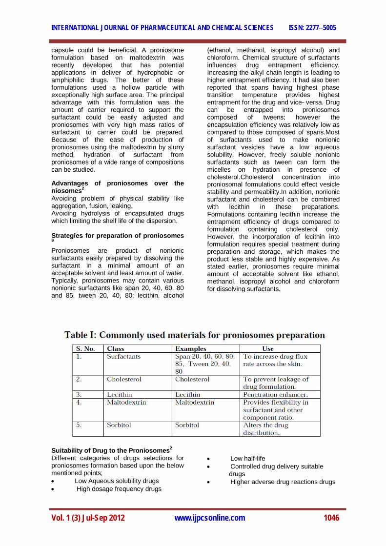
capsule could be beneficial. A proniosome formulation based on maltodextrin was recently developed that has potential applications in deliver of hydrophobic or amphiphilic drugs. The better of these formulations used a hollow particle with exceptionally high surface area. The principal advantage with this formulation was the amount of carrier required to support the surfactant could be easily adjusted and proniosomes with very high mass ratios of surfactant to carrier could be prepared. Because of the ease of production of proniosomes using the maltodextrin by slurry method, hydration of surfactant from proniosomes of a wide range of compositions can be studied.
Advantages of proniosomes over the niosomes2 Avoiding problem of physical stability like aggregation, fusion, leaking. Avoiding hydrolysis of encapsulated drugs which limiting the shelf life of the dispersion. Strategies for preparation of proniosomes 9
Proniosomes are product of nonionic surfactants easily prepared by dissolving the surfactant in a minimal amount of an acceptable solvent and least amount of water. Typically, proniosomes may contain various nonionic surfactants like span 20, 40, 60, 80 and 85, tween 20, 40, 80; lecithin, alcohol
(ethanol, methanol, isopropyl alcohol) and chloroform. Chemical structure of surfactants influences drug entrapment efficiency. Increasing the alkyl chain length is leading to higher entrapment efficiency. It had also been reported that spans having highest phase transition temperature provides highest entrapment for the drug and vice- versa. Drug can be entrapped into proniosomes composed of tweens; however the encapsulation efficiency was relatively low as compared to those composed of spans.Most of surfactants used to make nonionic surfactant vesicles have a low aqueous solubility. However, freely soluble nonionic surfactants such as tween can form the micelles on hydration in presence of cholesterol.Cholesterol concentration into proniosomal formulations could effect vesicle stability and permeability.In addition, nonionic surfactant and cholesterol can be combined with lecithin in these preparations. Formulations containing lecithin increase the entrapment efficiency of drugs compared to formulation containing cholesterol only. However, the incorporation of lecithin into formulation requires special treatment during preparation and storage, which makes the product less stable and highly expensive. As stated earlier, proniosomes require minimal amount of acceptable solvent like ethanol, methanol, isopropyl alcohol and chloroform for dissolving surfactants.
Suitability of Drug to the Proniosomes2 Different categories of drugs selections for proniosomes formation based upon the below mentioned points; Low Aqueous solubility drugs High dosage frequency drugs
Low half-life Controlled drug delivery suitable drugs Higher adverse drug reactions drugs

INTERNATIONAL JOURNAL OF PHARMACEUTICAL AND CHEMICAL SCIENCES ISSN: 22775005
Vol. 1 (3) Jul-Sep 2012 www.ijpcsonline.com 1047
Methodology 2,3,4,5 Preparation of proniosomes: The proniosomes can be prepared by 1. Spraying method. 2. Slurry method. 3. Coacervation phase separation method. Spraying method (Hu and Rhodes et al in 1999) prepared proniosomes by spraying the surfactant in organic solvent onto sorbitol powder and then evaporating the solvent. Because the sorbitol carrier is soluble in the organic solvent, it is necessary to repeat the process until the desired surfactant load has been achieved.The surfactant coating on the carrier comes out to be very thin and hydration of this coating allows multilamellar vesicles to form. Slurry method (Almira.I and Blazek - Walsh et al 8 in 2001) developed slurry method to produce proniosomes using maltodextrin as a carrier. The time required to produce proniosome by this is independent of the ration of surfactant solution to carrier material. In slurry method, the entire volume of surfactant solution is
added to maltodextrin powder in a rotary evaporator and vacuum applied until the powder appears to be dry and free flowing. Drug containing proniosome-derived niosomes can be prepared in manner analogous to that used for the conventional niosomes, by adding drug to the surfactant mixture prior to spraying the solution onto the carrier (sorbitol, maltodextrin) or by addition of drug to the aqueous solution used to dissolve hydrate the proniosomes. Coacervation phase separation method Accurately weighed or required amount of surfactant, carrier (lecithin), cholesterol and drug can be taken in a clean and dry wide mouthed glass vial (5 ml) and solvent should be added to it. All these ingredients has to be heated and after heating all the ingredients should be mixed with glass rod. To prevent the loss of solvent, the open end of the glass vial can be covered with a lid. It has to be warmed over water bath at 60-700 C for 5 minutes until the surfactant dissolved completely. The mixture should be allowed to cool down at room temperature till the dispersion get converted to a proniosomal gel.
Fig. 2: Coacervation phase separation method
Formation of Niosomes from Proniosomes The niosomes can be prepared from the proniosomes by adding the aqueous phase with the drug to the proniosomes with brief agitation at a temperature greater than the
mean transition phase temperature of the surfactant. T > Tm Where, T = Temperature, Tm = mean phase transition temperature

INTERNATIONAL JOURNAL OF PHARMACEUTICAL AND CHEMICAL SCIENCES ISSN: 22775005
Vol. 1 (3) Jul-Sep 2012 www.ijpcsonline.com 1048
Fig 3: Formation of Niosomes from Proniosomes.
Mechanism of drug transport through skin7, 11
As studies performed on the transdermal/topical application of vesicles have rendered conflicting results. It is still not clear which factors influence the vesicle–skin interactions and play an important role in determining the efficiency of drug transport through the skin. But it is clear that Proniosomes should be hydrated to form niosomal vesicles before the drug is released and permeates across the skin. Many scientists proposed different theories/mechanism for vesicle skin interaction. Two types of vesicle–skin interactions observed during in vitro studies using human skin which may induce various effects on dermal or transdermal drug delivery. 1. When vesicles come in contact with stratum corneum aggregate, fuse and adhere to the surface of cell. It is believed that this type of interaction leads to a high thermodynamic activity gradient of the drug at the interface of vesicle and stratum corneum, which is the driving force for penetration of the lipophilic drugs across the stratum corneum. 2. This type of interaction involves the ultra-structural changes in the intercellular lipid regions of the skin and its deeper layers at
maximum depth of about 10 mm as revealed by Freeze Fracture Electron Microscopy (FFEM) and Small Angle X-ray Scattering (SAXS). In addition to these two several other mechanisms which could explain the ability of vesicles to modulate drug transfer across skin, including: Nature of drug The lipid bi-layers of niosomes act as a rate limiting membrane barrier for drugs Dehydration of vesicles The vesicles act as penetration enhancers to reduce the barrier properties of the skin Size and composition of vesicles Biophysical factors Separation of Unentrapped Drug6, 7, 8
The removal of unentrapped solute from the vesicles can be accomplished by various techniques, which include: - 1. Dialysis The aqueous niosomal dispersion is dialyzed in dialysis tubing against suitable dissolution medium at room temperature. The samples are withdrawn from the medium at suitable time intervals, centrifuged and analysed for drug content using suitable method (U.V. spectroscopy, HPLC etc). 2. Gel Filtration The unentrapped drug is removed by gel filtration of niosomal dispersion through a Sephadex-G-50 column and eluted with suitable mobile phase and analyzed with suitable analytical techniques. 3. Centrifugation The proniosome derived niosomal suspension is centrifuged and the supernatant is separated. The pellet is washed and then resuspended to obtain a niosomal suspension free from unentrapped drug.
INTERNATIONAL JOURNAL OF PHARMACEUTICAL AND CHEMICAL SCIENCES ISSN: 22775005
Vol. 1 (3) Jul-Sep 2012 www.ijpcsonline.com 1049
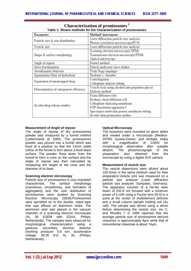
Characterization of proniosomes 2 Table 1: Shows methods for the characterization of proniosomes
Measurement of Angle of repose The angle of repose of dry proniosomes powder was measured by a funnel method (Liebermanet al 1990). The proniosomes powder was poured into a funnel which was fixed at a position so that the 13mm outlet orifice of the funnel is 5cm above a level black surface. The powder flows down from the funnel to form a cone on the surface and the angle of repose was then calculated by measuring the height of the cone and the diameter of its base. Scanning electron microscopy Particle size of proniosomes is very important characteristic. The surface morphology (roundness, smoothness, and formation of aggregates) and the size distribution of proniosomes were studied by Scanning Electron Microscopy (SEM).Proniosomes were sprinkled on to the double- sided tape that was affixed on aluminum stubs. The aluminum stub was placed in the vacuum chamber of a scanning electron microscope (XL 30 ESEM with EDAX, Philips, Netherlands). The samples were observed for morphological characterization using a gaseous secondary electron detector (working pressure: 0.8 torr, acceleration voltage: 30.00 KV) XL 30, (Philips, Netherlands).
Optical Microscopy The niosomes were mounted on glass slides and viewed under a microscope (Medilux-207RII, Kyowa-Getner, and Ambala, India) with a magnification of 1200X for morphological observation after suitable dilution. The photomicrograph of the preparation also obtained from the microscope by using a digital SLR camera. Measurement of vesicle size The vesicle dispersions were diluted about 100 times in the same medium used for their preparation.Vesicle size was measured on a particle size analyzer (Laser diffraction particle size analyzer, Sympatec, Germany). The apparatus consists of a He-Ne laser beam of 632.8 nm focused with a minimum power of 5 mW using a Fourier lens [R-5] to a point at the center of multielement detector and a small volume sample holding cell (Su cell). The sample was stirred using a stirrer before determining the vesicle size. Hu C. and Rhodes 7 in 1999 reported that the average particle size of proniosomes derived niosomes is approximately 6μm while that of conventional niosomes is about 14μm.

INTERNATIONAL JOURNAL OF PHARMACEUTICAL AND CHEMICAL SCIENCES ISSN: 22775005
Vol. 1 (3) Jul-Sep 2012 www.ijpcsonline.com 1050
Entrapment efficiency Entrapment efficiency of the niosomal dispersion in can be done by separating the unentrapped drug by dialysis, centrifugation or gel filtration as described above and the drug remained entrapped in niosomes is determined by complete vesicle disruption using 50% n-propanol or 0.1% Triton X-100 and analyzing the resultant solution by appropriate assay method for the drug. In-vitro methods for the assessment of drug release from proniosomes In vitro drug release can be done by (Chen DB et al, 2001) · Dialysis tubing · Reverse dialysis · Franz diffusion cell Dialysis tubing Muller et al 35 in 2002 studied in vitro drug release could be achieved by using dialysis tubing. The proniosomes is placed in prewashed dialysis tubing which can be hermetically sealed. The dialysis sac is then dialyzed against a suitable dissolution medium at room temperature; the samples are withdrawn from the medium at suitable intervals, centrifuged and analysed for drug content using suitable method (U.V.spectroscopy, HPLC etc). The maintenance of sink condition is essential. Reverse dialysis In this technique a number of small dialysis as containing 1ml of dissolution medium are placed in proniosomes. The proniosomes are then displaced into the dissolution medium. The direct dilution of the proniosomes is possible with this method; however the rapid release cannot be quantified using this method. Franz diffusion cell The in vitro diffusion studies can be performed by using Franz diffusion cell. Proniosomes is placed in the donor chamber of a Franz diffusion cell fitted with a cellophane membrane. The proniosomes is then dialyzed against a suitable dissolution medium at room temperature; the samples are withdrawn from the medium at suitable intervals, and analysed for drug content using suitable method (U.V spectroscopy,HPLC, etc) .the maintenance of sink condition is essential. Drug Release Kinetic Data Analysis The release data obtained from various formulations were studied further for their
fitness of data in different kinetic models like Zero order, Higuchi’s and peppa’s. In order to understand the kinetic and mechanism of drug release, the result of in-vitro drug release study of Niosome were fitted with various kinetic equation like zero order (Equation 1) as cumulative % release vs. time, higuchi’s model (Equation 2) as cumulative % drug release vs. square root of time. r and k values were calculated for the linear curve obtained by regression analysis of the above plots C = k0t …..(1) Where k0 is the zero order rate constant expressed in units of concentration / time and t is time in hours. Q = kHt1/2 …..(2) Where kH is higuchi’s square root of time kinetic drug release constant To understand the release mechanism in-vitro data was analyzed by peppa’s model (Equation 3) as log cumulative % drug release vs. log time and the exponent n was calculated through the slope of the straight line. Mt / M∞ = btn …..(3) Where Mt is amount of drug release at time t, M∞ is the overall amount of the drug, b is constant, and n is the release exponent indicative of the drug release mechanism. If the exponent n = 0.5 or near, then the drug release mechanism is Fickian diffusion, and if n have value near 1.0 then it is non-Fickian diffusion. Osmotic shock The change in the vesicle size can be determined by osmotic studies. Niosomal formulations are incubated with hypotonic, isotonic, hypertonic solutions for 3 hours. Then the changes in the size of vesicles in the formulations are viewed under optical microscopy. Stability studies To determine the stability of proniosomes, the optimized batch was stored in airtight sealed vials at different temperatures. Surface characteristics and percentage drug retained in proniosomes and parameters for evaluation of the stability, since instability of the formulation would reflect in drug leakage and a decrease. In the percentage drug retained. The proniosomes were sample at regular intervals of time (0,1,2,and 3months ),observed for color change, surface characteristics and tested for the percentage drug retained after being hydrated to form niosomes and analyzed by suitable analytical

INTERNATIONAL JOURNAL OF PHARMACEUTICAL AND CHEMICAL SCIENCES ISSN: 22775005
Vol. 1 (3) Jul-Sep 2012 www.ijpcsonline.com 1051
methods(UV spectroscopy, HPLC methods etc). Zeta potential analysis Zeta potential analysis is done for determining the colloidal properties of the prepared formulations. The suitably diluted proniosome derived niosome dispersion was determined using zeta potential analyzer based on electrophoretic light scattering and laser Doppler velocimetry method (Zetaplus™, Brookhaven Instrument Corporation, New York, USA). The temperature was set at 25°C. Charge on vesicles and their mean zeta potential values with standard deviation of 5 measurements were obtained directly from the measurement. Factors affecting the formulation of proniosomes 3, 7, 8
Various processing and formulation variables affect the proniosomes characteristics. They include surfactant chain length, cholesterol content, drug concentration, total lipid concentration, charge of lipids, pH of the dispersion medium and type of alcohol used in the preparation. 1. Surfactant chain length Spans are commonly used in the preparation of proniosomes. All span types have the same head group and different alkyl chain. Increasing the alkyl chain length is leading to higher entrapment efficiency. The entrapment efficiency followed the trend Span60 (C18)>Span40 (C16)>Span20 (C12)>Span80 (C18). Span 60 and Span 80 have the same head groups but Span80 has an unsaturated alkyl chain. De Giere demonstrated that the introduction of double bonds into the paraffin chains causes a marked enhancement of the permeability of liposomes, possibly explaining the lower entrapment efficiency of the Span80 formulation.
Fig. 4: Schematic illustration of niosome formation
2. Cholesterol content Cholesterol increases or decreases the percentage encapsulation efficiency depending on either the type of the surfactant or its concentration within the formulae. 3. pH of the hydration medium The percentage encapsulation efficiency of niosomes prepared by hydration of proniosomal gels of Span 60/cholesterol (9:1) was found to be greatly affected by the pH of the hydrating medium.For example, the fraction of flurbiprofen encapsulated was increased to about 1.5 times as the pH decreased from pH 8 to 5.5. The increase in the percentage encapsulation efficiency of flurbiprofen by decreasing the pH could be attributed to the presence of the ionizable carboxylic group in its chemical structure. Decreasing the pH could increase the proportions of the unionized species of flurbiprofen, which have higher partitioning to the bilayer lipid phase compared to the ionized species. 4. Total lipid concentration The percentage encapsulation efficiency of flurbiprofen was increased as the lipid concentration was increased from 25 to 200mol/ml, respectively. The increase in percentage encapsulation efficiency of flurbiprofen as a function of total lipid concentration was linear. On the other hand, the amount of flurbioprofen entrapped was decreased on increasing the lipid concentration from 25 to 200mol/ml, respectively. This leads to the fact that the fraction of lipid taking part in encapsulation decreases as the concentration of lipid increases. 5. Drug concentration Increasing flurbiprofen concentration from 25 to 75mg/mmol lipids in the Proniosomes prepared from Span 60/cholesterol (9:1) showed an increase in both percentage encapsulation efficiency and the amount of drug encapsulated per mol total lipids upon hydration and formation of niosomes. 6. Charge of the lipids Incorporation of either dicetyl phosphate (DCP) which induces negative charge or stearylamine (SA) which induces positive charge decreased the percentage encapsulation efficiency of flurbiprofen into niosomal vesicles.
INTERNATIONAL JOURNAL OF PHARMACEUTICAL AND CHEMICAL SCIENCES ISSN: 22775005
Vol. 1 (3) Jul-Sep 2012 www.ijpcsonline.com 1052
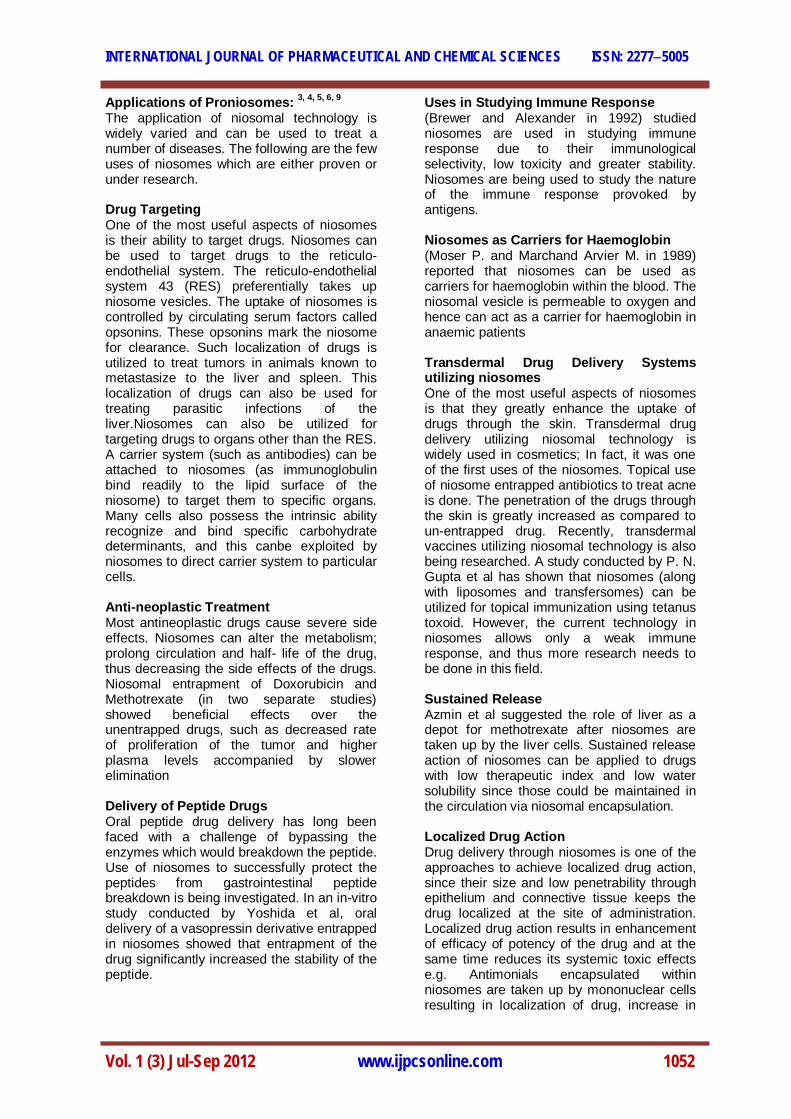
Applications of Proniosomes: 3, 4, 5, 6, 9
The application of niosomal technology is widely varied and can be used to treat a number of diseases. The following are the few uses of niosomes which are either proven or under research. Drug Targeting One of the most useful aspects of niosomes is their ability to target drugs. Niosomes can be used to target drugs to the reticulo-endothelial system. The reticulo-endothelial system 43 (RES) preferentially takes up niosome vesicles. The uptake of niosomes is controlled by circulating serum factors called opsonins. These opsonins mark the niosome for clearance. Such localization of drugs is utilized to treat tumors in animals known to metastasize to the liver and spleen. This localization of drugs can also be used for treating parasitic infections of the liver.Niosomes can also be utilized for targeting drugs to organs other than the RES. A carrier system (such as antibodies) can be attached to niosomes (as immunoglobulin bind readily to the lipid surface of the niosome) to target them to specific organs. Many cells also possess the intrinsic ability recognize and bind specific carbohydrate determinants, and this canbe exploited by niosomes to direct carrier system to particular cells. Anti-neoplastic Treatment Most antineoplastic drugs cause severe side effects. Niosomes can alter the metabolism; prolong circulation and half- life of the drug, thus decreasing the side effects of the drugs. Niosomal entrapment of Doxorubicin and Methotrexate (in two separate studies) showed beneficial effects over the unentrapped drugs, such as decreased rate of proliferation of the tumor and higher plasma levels accompanied by slower elimination Delivery of Peptide Drugs Oral peptide drug delivery has long been faced with a challenge of bypassing the enzymes which would breakdown the peptide. Use of niosomes to successfully protect the peptides from gastrointestinal peptide breakdown is being investigated. In an in-vitro study conducted by Yoshida et al, oral delivery of a vasopressin derivative entrapped in niosomes showed that entrapment of the drug significantly increased the stability of the peptide.
Uses in Studying Immune Response (Brewer and Alexander in 1992) studied niosomes are used in studying immune response due to their immunological selectivity, low toxicity and greater stability. Niosomes are being used to study the nature of the immune response provoked by antigens. Niosomes as Carriers for Haemoglobin (Moser P. and Marchand Arvier M. in 1989) reported that niosomes can be used as carriers for haemoglobin within the blood. The niosomal vesicle is permeable to oxygen and hence can act as a carrier for haemoglobin in anaemic patients Transdermal Drug Delivery Systems utilizing niosomes One of the most useful aspects of niosomes is that they greatly enhance the uptake of drugs through the skin. Transdermal drug delivery utilizing niosomal technology is widely used in cosmetics; In fact, it was one of the first uses of the niosomes. Topical use of niosome entrapped antibiotics to treat acne is done. The penetration of the drugs through the skin is greatly increased as compared to un-entrapped drug. Recently, transdermal vaccines utilizing niosomal technology is also being researched. A study conducted by P. N. Gupta et al has shown that niosomes (along with liposomes and transfersomes) can be utilized for topical immunization using tetanus toxoid. However, the current technology in niosomes allows only a weak immune response, and thus more research needs to be done in this field. Sustained Release Azmin et al suggested the role of liver as a depot for methotrexate after niosomes are taken up by the liver cells. Sustained release action of niosomes can be applied to drugs with low therapeutic index and low water solubility since those could be maintained in the circulation via niosomal encapsulation. Localized Drug Action Drug delivery through niosomes is one of the approaches to achieve localized drug action, since their size and low penetrability through epithelium and connective tissue keeps the drug localized at the site of administration. Localized drug action results in enhancement of efficacy of potency of the drug and at the same time reduces its systemic toxic effects e.g. Antimonials encapsulated within niosomes are taken up by mononuclear cells resulting in localization of drug, increase in

INTERNATIONAL JOURNAL OF PHARMACEUTICAL AND CHEMICAL SCIENCES ISSN: 22775005
Vol. 1 (3) Jul-Sep 2012 www.ijpcsonline.com 1053
potency and hence decrease both in dose and toxicity. The evolution of niosomal drug delivery technology is still at an infancy stage,
but this type of drug delivery system has shown promise in cancer chemotherapy and anti-leishmanial therapy.
REPORTED STUDY IN PRONIOSOMAL APPLICATIONS 2
Table 3: Proniosomal systems studied for Transdermal applications
CASE STUDY 10Design and Development of a Proniosomal Transdermal Drug Delivery System for Captopril. Ankur Gupta*, Sunil Kumar Prajapati, M Balamurugan, Mamta Singh, Daksh Bhatia. College of Pharmacy, ISI-15, Sitapura Institutional Area, Tonk Road, Jaipur-302022, Rajasthan Purpose The aim of the study was to develop a proniosomal carrier system for captopril for the treatment of hypertension that is capable of efficiently delivering entrapped drug over an extended period of time.
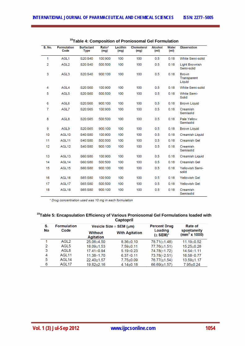
Method The potential of proniosomes as a transdermal drug delivery system for captopril was investigated by encapsulating the drug in various formulations of proniosomal gel composed of various ratios of sorbitan fatty acid esters, cholesterol, lecithin prepared by coacervation-phase separation method. The formulated systems were characterized in vitro for size, vesicle count, drug entrapment, drug release profiles and vesicular stability at different storage conditions. Stability studies for proniosomal gel were carried out for 4 weeks.
INTERNATIONAL JOURNAL OF PHARMACEUTICAL AND CHEMICAL SCIENCES ISSN: 22775005
Vol. 1 (3) Jul-Sep 2012 www.ijpcsonline.com 1054
10Table 4: Composition of Proniosomal Gel Formulation
10Table 5: Encapsulation Efficiency of Various Proniosomal Gel Formulations loaded with Captopril

INTERNATIONAL JOURNAL OF PHARMACEUTICAL AND CHEMICAL SCIENCES ISSN: 22775005
Vol. 1 (3) Jul-Sep 2012 www.ijpcsonline.com 1055
10Fig. 5: Comparative in-vitro release profile of selected proniosomal gel formulation
10Fig. 6: Stability study of AGL2 at different temperatures
10Fig. 7: Electron Micrograph of AGL2

INTERNATIONAL JOURNAL OF PHARMACEUTICAL AND CHEMICAL SCIENCES ISSN: 22775005
Vol. 1 (3) Jul-Sep 2012 www.ijpcsonline.com 1056
Results The method of proniosome loading resulted in an encapsulation yield of 66.7 - 78.7%.Proniosomes were characterized by transmission electron microscopy. In vitro studies showed prolonged release of entrapped captopril. At refrigerated conditions, higher drug retention was observed. CONCLUSION It is evident from this study that proniosomes are a promising prolonged delivery system for captopril and have reasonably good stability characteristics. CONCLUSION Proniosomes are promising drug carriers for the future with greater physical, chemical stability and potentially scalable for commercial viability. Proniosomal delivery system holds effective delivery for amphiphilic drugs. Due to advantages of nontoxicity & penetration enhancing effect of surfactants & effective modification of drug release, proniosomes has attracted a greater deal of attention for delivery of drugs through transdermal route.Proniosomes in dry form makes the possibility of convenient unit dosing as they further converted into beads,tablets,capsules.The findings of the studies on proniosomes opens the door for the future use of different carriers materials with biocompatibity and suitability for the preparation of proniosomes. The future experiments would explore the suitability of proniosomes with more drugs having defined drawbacks for improved & effective intended therapy. REFERENCES
1. Vyas SP and Khar RK. Niosomes: Targeted and controlled drug delivery novel carrier system, C.B.S. Publication, 1st edition, 2001;23:173-248.
2. Thejaswi C, Gobinath M and Radharni J. A review on design and characterization of Proniosomes As a drug carrier,Dept.of Pharmaceutics,Ratnam Institute of pharmacy, Nellore.
3. Sudhamani T. Proniosomes-A promising Drug carriers, dept. Of Pharmaceutics, Erode I , Ijptr, India.
4. Sankar V. Proniosomes as drug carriers, Dept. Of Pharmaceutics PSG college of pharmacy, Coimbatore-641004.
5. Deepti Annakula. Provesicular drug delivery systems: An overview and appraisal, St.Peters Institute of Pharmaceutical sciences, vidyanagar, Hanamkonda, A.p, INDIA.
6. Almira I. Blazek-Welsh1 and David G. Rhodes, SEM Imaging Predicts Quality of Niosomes from Maltodextrin-Based Proniosomes
7. Kiran Yadav and Deepak Yadav. Proniosomal Gel: A provesicular approach for transdermal drug delivery Institute of Pharmaceutical Sciences, Kurukshetra University, Kurukshetra, Haryana, India,National Institute of Pharmaceutical Education and Research (NIPER), S.A.S. Nagar, Punjab
8. Surender Verma, Singh SK; Navneet Syan, Pooja Mathur and Vinay Valecha. Institute of Pharmaceutical Sciences, Kurukshetra University, Kurukshetra, Haryana, India:Nanoparticle vesicular systems: A versatile tool for drug delivery, www.jocpr.com.
9. Rishu Kakar. Proniosomes: An Emerging Vesicular System in Drug Delivery and cosmetics.M.M. College of Pharmacy, M.M. University, Mullana, Ambala 133001, India
10. Ankur Gupta, Sunil Kumar Prajapati and Balamurugan M. Design and Development of a Proniosomal Transdermal Drug Delivery System for Captopril” College of Pharmacy, ISI-15, Sitapura Institutional Area, Tonk Road,Jaipur-302022, Rajasthan, Tropical Journal of Pharmaceutical Research, June 2007; 6 (2): 687-693.
11. Kamal-kuk, Sanju nanda and nand kishor. Proniosome Gel: Potential Carrier System in Topical/Transdermal Delivery for Drugs and Cosmetics/Cosmeceuticals.
12. www.google.com 13. S.S. Biju, Sushama Talegaonkar,
P.R. Mishra, R.K. Khar; Indian journal of pharmaceutical sciences.2006;68:144-156.
14. www.wikipedia.com 15. www.sciencedirect.com 16. www.scholaesearch.com.