Review Article on Targeted Polymeric Nanoparticles : An ... · Review Article on Nanoparticles : An...
20
American Jo American Journal of Advanced Review Article Nanoparticles : Kumar Ganesh, Dhyani Archa Sri Guru Ram Rai Institute of Tec AB In car bee Du risk act now cel exi cel Ke Bio INTRODUCTION Nanoparticles can be de colloidal particles having size ra 10 to 1000 nm. The adv nanotechnology is to provide th the effective medicine (Nanomed to substantially influence the la both pharmaceutical and bio industries .1 They have application fields of life sciences such as technologies, histological studi diagnostic assays and drug deliv (DDS). The application of the for the DDS is of particular inte they have some advantages su purification and sterilization, dr possibilities, and sustained rel Date of Receipt- 06/06/2013 Date of Revision- 19/06/2013 Date of Acceptance- 12/07/2013 Address for Correspondence Sri Guru Ram Rai Institute ofTechnology and Sciences Patel Nagar, Dehradun, Uttarakhand, India. Tel: +919456315495, E-mail: archana.dhyani89@ gmail.com ournal of Advanced Drug Deliver www.ojadd.com d Drug Delivery w Re on Targeted Polymeric An Overview ana, Kothiyal Preeti chnology and Sciences Patel Nagar, Dehradun, Utt BSTRACT the recent years, the potential use of polymeric rriers for a wide range of drugs for therapeutic en increased due to their versatility and wide ran ue to limitations in the conventional drug thera k of adverse reactions will occur. Nanopartic tion at the desired sites and thus gain wadays.With these nanoparticles the specific tar lls or receptors can be achieved.There are vari ists which are responsible for cellular int llular uptake. eywords: Asialoglycoprotein, Phagocytosis, Gal odegradable polymers, Ligands efined as the anging from vantages of he safe and dicine) is set andscape of otechnology in various s separation ies, clinical very systems nanoparticle erest because uch as easy rug targeting lease action 2. However, they have som such as they can be easily r reticuloendothelial system seconds or minutes after in due to phagocytosis b present in the liver and spl modification of nanoparticle hydrophilic poly(ethylene have reduced the reco nanaoparticles and thus th for the more prolong increased half-life in blood rate of uptake by the liver .E targeting of carriers to spe antibodies , or sugars has be A large number of drugs c using nanoparticulate carrie ry www.ojadd.com eview Article c tarakhand, India. c nanoparticles as c applications has nge of properties. apy the increased cles provides the ning importance rgeting to various ious mechanisms ternalization and lactose/Mannose; me disadvantages recognized by the m (RES) within njection, which is by macrophages leen. The surface es by albumin or oxide) chains ognition of hey are available ged circulation, d and a reduced Especially, active ecific cells using een attempted .3 can be delivered ers via a number
Transcript of Review Article on Targeted Polymeric Nanoparticles : An ... · Review Article on Nanoparticles : An...

American Journal of Advanced Drug Delivery
American Journal of Advanced Drug Delivery
Review Article onNanoparticles : An Overview
Kumar Ganesh, Dhyani ArchanaSri Guru Ram Rai Institute of Technology and Sciences
ABSTRACT
In the recent years, the potential use of polymeric nanoparticles as carriers for a wide range of drugs for therapeutic applications has been increased due to their versatility and wide range of properties. Due to risk of adverse reactions will occur. Nanoparticles provides the action at the desired sites and thus gaining importance nowadays.With these nanoparticles the specific targeting to various cells orexists which are responsible for cellular internalization and cellular uptake.
KeywordsBiodegradable polymers
INTRODUCTION
Nanoparticles can be defined as the colloidal particles having size ranging from 10 to 1000 nm. The advantages of nanotechnology is to provide the the effective medicine (Nanomedicine) is set to substantially influence the landscape of both pharmaceutical and biotechnology industries.1
They have application in various fields of life sciences such as separation technologies, histological studies, clinical diagnostic assays and drug delivery systems (DDS). The application of the nanoparticle for the DDS is of particular interest because they have some advantages such as easy purification and sterilization, drug targeting possibilities, and sustained release action
Date of Receipt- 06/06/2013 Date of Revision- 19/06/2013 Date of Acceptance- 12/07/2013
Address for Correspondence Sri Guru Ram Rai Institute ofTechnology and Sciences Patel Nagar, Dehradun, Uttarakhand, India. Tel: +919456315495,
E-mail: archana.dhyani89@ gmail.com
American Journal of Advanced Drug Delivery
www.ojadd.com
Advanced Drug Delivery www.ojadd.com
Review
Review Article on Targeted Polymeric Nanoparticles : An Overview
Kumar Ganesh, Dhyani Archana, Kothiyal Preeti Sri Guru Ram Rai Institute of Technology and Sciences Patel Nagar, Dehradun, Uttarakhand, India.
ABSTRACT
In the recent years, the potential use of polymeric nanoparticles as carriers for a wide range of drugs for therapeutic applications has been increased due to their versatility and wide range of properties. Due to limitations in the conventional drug therapy the increased risk of adverse reactions will occur. Nanoparticles provides the action at the desired sites and thus gaining importance nowadays.With these nanoparticles the specific targeting to various cells or receptors can be achieved.There are various mechanisms exists which are responsible for cellular internalization and cellular uptake.
Keywords: Asialoglycoprotein, Phagocytosis, Galactose/MannoseBiodegradable polymers, Ligands
Nanoparticles can be defined as the colloidal particles having size ranging from 10 to 1000 nm. The advantages of nanotechnology is to provide the safe and the effective medicine (Nanomedicine) is set to substantially influence the landscape of both pharmaceutical and biotechnology
They have application in various fields of life sciences such as separation
ies, clinical diagnostic assays and drug delivery systems (DDS). The application of the nanoparticle for the DDS is of particular interest because they have some advantages such as easy purification and sterilization, drug targeting
ained release action
2.However, they have some disadvantages such as they can be easily recognized by the reticuloendothelial system (RES) within seconds or minutes after injection, which is due to phagocytosis by macrophages present in the liver and splmodification of nanoparticles by albumin or hydrophilic poly(ethylene oxide) chains have reduced the recognition of nanaoparticles and thus they are available for the more prolonged circulation, increased half-life in blood and a reducrate of uptake by the liver .Especially, active targeting of carriers to specific cells using antibodies , or sugars has been attemptedA large number of drugs can be delivered using nanoparticulate carriers via a number
American Journal of Advanced Drug Delivery
www.ojadd.com
Review Article
Targeted Polymeric
Uttarakhand, India.
In the recent years, the potential use of polymeric nanoparticles as carriers for a wide range of drugs for therapeutic applications has been increased due to their versatility and wide range of properties.
limitations in the conventional drug therapy the increased risk of adverse reactions will occur. Nanoparticles provides the action at the desired sites and thus gaining importance nowadays.With these nanoparticles the specific targeting to various
receptors can be achieved.There are various mechanisms exists which are responsible for cellular internalization and
Galactose/Mannose;
However, they have some disadvantages such as they can be easily recognized by the reticuloendothelial system (RES) within seconds or minutes after injection, which is due to phagocytosis by macrophages present in the liver and spleen. The surface modification of nanoparticles by albumin or hydrophilic poly(ethylene oxide) chains have reduced the recognition of nanaoparticles and thus they are available for the more prolonged circulation,
life in blood and a reduced rate of uptake by the liver .Especially, active targeting of carriers to specific cells using antibodies , or sugars has been attempted.3 A large number of drugs can be delivered using nanoparticulate carriers via a number

Archana et al__________________________________________________________________
AJADD[3][3][2015] 196-215
of routes. These include many hydrophilic drugs, hydrophobic drugs as well as for proteins, vaccines, biological macromolecules, etc. They can be formulated for targeted delivery to the lymphatic system, brain, arterial walls, lungs, liver, spleen, or made for long-term systemic circulation. 4
METHOD OF PREPARATION OF NANOPARTICLES
The various methods can be used for preparation of nanoparticles..For example, drugs can be entrapped in the polymer matrix, encapsulated in a nanoparticle core, surrounded by a shell- like polymer membrane, chemically conjugated to the polymer, or either it may be bound to the particle’s surface by adsorption.
a. Emulsification solvent evaporation
technique One of the methods used for the
preparation of nanoparticles is emulsification solvent evaporation technique. It is basically used for encapsulating hydrophobic drugs, but shows the poor results for incorporation of bioactive agents of a hydrophilic nature. The solvent evaporation is carried out the polymer and the compound are dissolved in an organic solvent such as chloroform, ethyl acetate, or methylene chloride and then it is emulsified in an aqueous phase containing a stabilizer (e.g., PVA). Just after formation of the nanoemulsion the solvent diffuses to the external phase until saturation. The solvent molecules that reach the water-air interphase evaporate, which leads to continuous diffusion of the solvent molecules from the inner droplets of the emulsion to the external phase; simultaneously, the precipitation of the polymer leads to the formation of nanospheres.
In many cases, the induction of nanosized polymer droplets can be done by sonication or homogenization. The organic solvent is then evaporated and the nanoparticles are usually collected by centrifugation and lyophilization .5
The small changes in this method are used for encapsulating hydrophilic compounds and proteins, which can be done by, the double, or multiple emulsion technique. As the name signifies firstly, a hydrophilic drug and a stabilizer dissolve in water.The primary emulsion is prepared by dispersing the aqueous phase into an organic solvent containing a dissolved polymer. This is then emulsified in an outer aqueous phase also containing stabilizer.The nanoparticles can be achieved by the solvent evaporation method. One of the main problems associated for the encapsulation of a hydrophilic molecule like a protein or peptide-drug is the rapid diffusion of the molecule into the outer aqueous phase during the emulsification. This can result in poor encapsulation efficiency, i.e. drug loading.6 b. Emulsification Diffusion method
Another method which can be used for preparation of nanoparticles is the emulsification diffusion method. The method utilizes a partially water-soluble solvent like acetone or propylene carbonate. The polymer and the drug are dissolved in the solvent and it is emulsified in the aqueous phase containing the stabilizer. The role of stabilizer prevents the aggregation of emulsion droplets by adsorbing of the surface of the droplets. Addition of water to the emulsion, allow the diffusion of the solvent into the water. The solution is stirred leading to the nano precipitation of the particles. Further, it can be collected by centrifugation, or the solvent can be removed effectively by dialysis. The main problem with this method is that the water soluble drugs tend to leak out from the

Archana et al__________________________________________________________________
AJADD[3][3][2015] 196-215
polymer phase during diffusion steps. So, in order to avoid this problem the dispersing medium changed from aqueous medium to medium chain triglycerides and a small amount of surfactant is added into it. The nanpoarticles are collected from the oily suspension by centrifugation.7
c. Nanoprecipitation method
Nanoparticles can be synthesized by the nanoprecipitation method. In this method, polymer and drug are dissolved in acetone, ethanol, or methanol and incorporated under magnetic stirring into an aqueous solution of the surfactant. The organic solvent diffuses instantaneously to the external aqueous phase, followed by precipitation of the polymer and drug. After formation of the nanoparticles, the solvent is eliminated and the suspension concentrated under reduced pressure. The advantage of this method is that no surfactant is employed; however, the method is limited to drugs that are highly soluble in a polar solvent.8
d. Salting-out method
The salting-out process is another method for the preparation of nanoparticles. This technique is based on the precipitation of a hydrophobic polymer, is useful for the encapsulation of either hydrophilic or hydrophobic drugs because a variety of solvents, including polar (e.g., acetone or methanol) and non-polar (methylene chloride or chloroform) solvents can be chosen for dissolving the drug.7-8
POLYMERICNANOPARTICLES
Controlled release systems generally refer to technologies or the sytems in which the drugs are released at predetermined and/or tuneable rates, or in response to some external stimuli and triggers. Polymers can be used as a controlled release system since they have unique physicochemical, synthetic, biocompatibility, and degradation properties.
Additionally, polymeric nanoparticles also have advantages over lipidic carriers such as liposomes. For example some of the limitations of liposomes include: their propensity to burst release cargo in vivo, a lack of compatibility with various active agents, a limited drug loading volume, the oxidation of liposomal phospholipids, and poor shelf-life stability. In contrast, polymeric drug delivery systems are comparably stable in vivo, having high drug loading capacities, and can employ both controlled and triggered release of drugs. Due to these properties, polymeric nanomaterials are well preferred than the lipid nanoparticles..8,9
a. Biodegradable polymers
Nanoparticles can be synthesized from the biodegradable as well as non-biodegradable polymers. A wide range of the natural and the synthetic polymers can be used for the preparation. Today, the most commonly used polymers for controlled drug release applications include poly (D,L-lactide-co-glycolide) (PLGA), poly(lactic acid) (PLA), poly (glutamic acid) (PGA), poly(caprolactone) (PCL), N-(2-hydroxypropyl)-methacrylate copolymers (HPMA), and poly(amino acids). In particular, PLGA, PGA and PLA have been widely used in an impressive number of controlled release products, particularly due to their favourable biocompatibility and biodegradability properties.10
PLGA has been the most common polymer used to make biodegradable nanoparticles; however, these are clearly not the optimal carrier for all drug delivery applications. For each application, one must evaluate the properties of the system (drug and particle) .For example, poly (butyl cyanoacrylate) double emulsion solvent evaporation technique have been successful in delivering drugs to the brain11. Other cyanoacrylate-based nanoparticles such as polyalkylcyanoacrylate (PACA) and

Archana et al__________________________________________________________________
AJADD[3][3][2015] 196-215
polyethylcyanoacrylate (PECA) have also been prepared. The cyanoacrylates may be considered as the promising drug delivery systems due to their various properties like mucoadhesive properties and ability to entrap biologically active compounds. These polymers are biodegradable, biocompatible, as well as compatible with a wide range of compatible drugs. Further these polymers having a faster degradation rate than PLGA, which in some cases may be more desirable. PECA nanoparticles have been prepared by emulsion polymerization technique.10,11
The nanoparticles can also be prepared from the functionalized PLGA polymers.Jung et al. synthesized nanoparticles made of a branched, poly(2-sulfobutyl-vinyl alcohol)-g- PLGA . The purpose of using sulfobutyl groups attached to the hydrophilic backbone was to provide a higher affinity to proteins by electrostatic interactions.In another case, a carboxylic end group of PLGA was conjugated to a hydroxyl group of doxorubicin and formulated into nanoparticles. This modification produced a sustained release of the drug that was approximately six times longer than with unconjugated drug.12
The main advantages of the controlled release systems were principally they enable the potential use of molecules that had short half-lives as therapeutics so that their availability in the systemic circulation can be increased, to enhance patient compliance, to improve drug efficacy and reduce side effects by delivering agents locally, and to simplify dosing in cases where prolonged drug exposure was necessary.13. For example, Zoladex, a PLGA copolymer impregnated with Goserelin acetate can be used for treatment of breast and prostate cancers and was approved by the FDA in 1998. It is basically injected subcutaneously so that the active agent could be released slowly into systemic circulation and reach its target sites14.In the same year, Lupron Depot,
launched a PLGA microsphere formulation of leuprolide acetate, which was used the to treat advanced prostate cancers later it was approved by FDA15. Gliadel, a biodegradable Polifeprosan 20 carmustine-embedded wafer for the treatment of gliomas that became the first new treatment for gliomas in 20 years on its approval in 199616and Atridox, a polylactide and N-methyl-2-pyrrolidone (NMP) polymer blend containing doxycycline hyclate for subgingival delivery, which was FDA approved in 1998 to treat periodontal disease17.
Another controlled release formulations of include Sandostatin LAR, a PLGA slow release formulation of octreotide acetate for tumor control in neuroendocrine disorders approved by FDA in 1998.Trelstar Depot, a PLGA based microparticle formulation of triptorelin pamoatea can be used for prostate cancer and other indications, was FDA approved in 2000. 18 The ability to manufacture the polymeric formulations leads to the growing interest in the of nanotechnology.
The molecular weight and concentration of the polymer will also affect the nanoparticles. The molecular weight of the polymer has opposite effects of nanoparticles and encapsulation efficiency. 10Smaller size nanoparticles, approximately 100nm, can be prepared with lower molecular weight polymer; however, it reduces the drug encapsulation efficiency. On the other hand, increase in polymer concentration increases encapsulation efficiency and the size of the nanoparticles. When considering a particular polymeric drug delivery application, particle size and the encapsulation efficiency are two of the most important characteristics of nanoparticles. Another, important characteristics of nanoparticles are zeta potential. It represents the particle stability. 11

Archana et al__________________________________________________________________
AJADD[3][3][2015] 196-215
SURFACE MODIFICATIONS OF NANOPARTICLES
Phagocytosis plays a critical and important physiological role in the defense mechanism. It protects the body against the infectious agents (most bacteria and some viruses) as well exogenous inert particles—including drug delivery nanoparticles.
a. Mechanism of opsonization and
phagocytosis Phagocytosis occurs primarily in
specialized cells, also called professional phagocytes and non-professional phagocytes: macrophages, monocytes, neutrophils and dendritic cell. These are professional phagocytes19.Other types of cells (fibroblasts, epithelial and endothelial cells), referred to as para and non- professional phagocytes, which shows, lower extent of phagocytosis.20. The phagocytic pathway of entry into cells can be described using three distinct steps: recognition by opsonization in the bloodstream; adhesion of the opsonized particles to the macrophages; ingestion of the particle.
Opsonization is the process of attaching of the foreign nanoparticles by proteins called opsonins, so that the it can be recognise by macrophages. Major opsonins include immunoglubulins (IgG and M) as well as complement components (C3, C4, C5),[21,6] in addition to other blood serum proteins (including laminin, fibronectin, C-reactive protein, type-I collagen).22 The opsonized particles then attach to the macrophage surfaces through the specific receptor-ligand interactions .The major and best-studied receptors for this purpose include the Fc receptors (FcR) and the complement receptors (CR). FcRs bind to the constant fragment of particle-adsorbed immunoglobulins, the best understood interaction involving IgG and FcR; CRs mostly bind to C3fragments 19,22. Other
receptors, involving in the phagocytosis are the mannose/ fructose and scavenger receptors, while new opsono-receptors like CD44 are still being discovered .23
However ,in order to avoid opsonization , several methods of surface modifications have been developed so that polymeric colloidal particles can not recognized by the Reticuloendothelial systems(RES).So, the nanoparticles can be coated with hydrophilic molecules in order to hide the hydrophobicity as the body considers the hydrophobic particles as the foreign and thus the RES can easily eliminates from the blood stream.The most common moiety for surface modification is the hydrophilic and non-ionic polymer polyethylene glycol (PEG) . It has been largely demonstrated that the “PEGylation” increases their blood circulation and half-life by several orders of magnitude. Moreover, PEG exhibits an excellent biocompatibility. Poloxamer, poloxamines or Chitosan have also been studied for surface modification.24,10 These groups can block the electrostatic and hydrophobic interactions that help opsonins to bind to particle surfaces.
On the other hand, the other application of surface modification is the targeting of tumors or organs of nanoparticles to increase selective cellular binding and internalization through receptor-mediated endocytosis. 25 Ligands need to be optimally conjugated on nanoparticles to maintain their affinity for receptors binding. As a sufficient PEG coating is essential for avoiding recognition by the RES, ligands should be extended away from the nanoparticle surfaces to avoid shielding by the PEG chains.26
Surface charges of nanoparticles also have an important influence on their interaction with cells and on their uptake. Positively charged nanoparticles seem to allow higher extent of internalization, apparently as a result of the ionic interactions established between

Archana et al__________________________________________________________________
AJADD[3][3][2015] 196-215
positively charged particles and negatively charged cell membranes27,28. Moreover, positively charged nanoparticles seem to be able to escape from lysosomes after being internalized and exhibit perinuclear localization, whereas the negatively and neutrally charged nanoparticles prefers to colocalize with lysosomes29 . PLGA nanoparticles have negative charges which can be shifted to neutral or positive charges by surface modification, for example by PEGylation of the PLGA polymer30 or chitosan coating31 respectively. LOCALISATION OF NANOPARTICLES: BY PASSIVE OR ACTIVE TARGETING
a. Passive targeting
The majority of these nanoparticles exhibit prolonged circulation times in vivo and thus accumulate at particular sites simply due to blood hemodynamic forces and diffusive mechanisms. The advantages of utilizing passive targeting of nanoparticles in the field of oncology due to its widely reported ‘‘enhanced permeation and retention’’ (EPR) effect. Maeda et al., in the 1980’s demonstrated the principle of passive targeting of colloidal particles to tumors.[32] In their initial studies, significantly higher concentrations of the cytotoxic drug neocarzinostatin was discovered in tumor tissue postal administration of the polymer-drug conjugate poly(styreneco- maleic acid)-neocarzinostatin (SMANCS), in comparison to control experiments where the drug was administered in its free form.[32] This led Maeda et al. to postulate that the enhanced accumulation of the colloidal particles in the tumor was attributed to the structural features of the tumor vasculature, an observation, which was termed the EPR effect.[33]The EPR effect has been observed with a wide range of macromolecular agents
such as proteins; including immunoglobulin G (IgG),drug-polymer conjugates, micelles, liposomes, polymeric nanoparticles and many other types of nanoparticles..34,35 Current, the observations of EPR are the main premise for the design of tumor specific nanomedicines for drug delivery or imaging applications, however there are a number of factors that need to be considered.36,37 The majority of passively targeted nanoparticles should be surface modified ,that is ,it's coated with PEG polymer for biocompatibility; to avoid the endocytic uptake by cancer cells within the tumour. This problem which has been referred to by some as the ‘‘PEG dilemma’’38,39 has been suggested to hamper efficient drug delivery in tumors as passively targeted nanoparticles end up releasing their therapeutic pay load into the tumor milieu rather than within cancer cells. However, in the case of cytotoxic drugs—many have been shown to have longer elimination half-lives in tumors than the normal tissue. Therefore, the delivery of higher amounts of drugs to tumors can lead to longer durations of drug exposure at higher concentrations and enhanced efficacy. For example, docetaxel (Dtxl) has an elimination half-life of 2.2–4.5 h in normal tissue and 22 h in tumours, demonstrating long tumor site retention relative to non-tumoral tissues.40
b. Active targeting
Active targeting is based on the affinity of the ligands to direct the binding of nanoparticles to antigens, differently over expressed on the plasma membrane of diseased cells or to the extra-cellular matrix proteins that are differentially over expressed in the diseased tissue.
Targeting molecules can be either antibody or non-antibody ligands: The advantages of the non-antibody ligands, like peptides, sugars or vitamins, they are readily

Archana et al__________________________________________________________________
AJADD[3][3][2015] 196-215
available, inexpensive to manufacture and easy to handle. 41,42Actively targeted nanoparticles can be utilized in applications where drug release is either extracellular or intracellular. Therapies that act on intracellular sites of action are most effectively delivered with targeted nanoparticles.43 The various mechanisms for the internalization actively targeted NPs can be clathrin-dependent endocytosis pathways, caveolin-assisted, cell adhesion molecule directed, or lipid raft associated mechanisms, leading to endosome formation, which ultimately leads to lysosomes.44 For hydrophobic small molecule drugs that can readily permeate through the lipid bilayer of the endosomal membrane, drug release within the endosome will result in permeation within the intracellular compartments. For delivery of bioactive macromolecules such as nucleic acids (DNA, siRNA, miRNA) or charged hydrophilic small molecules that are relatively impermeable to the endosomal membrane, the nanoparticles need to escape the endosome prior to fusion with lysosomes if NPs are to reach their desired subcellular compartments.44
Antibodies, have a higher specificity and a wide range of binding affinities as compared to the non –antibody ligands.
Ligand mediated targeting is also beneficial in the case of vascular endothelial targeting for both oncology and cardiovascular applications as well as other various diseases, and the identification of high affinity ligands for this purpose is an active area of research.42
Typically, the ligand receptor binding involves the exploitation of a binding interaction of either a ligand at that site for an introduced receptor (targeting receptor) or of a receptor at that site for an introduced ligand (targeting ligand). The feasibility of using carbohydrate ligands to target protein receptors at sites of
localization, termed 'glycotargeting', was first demonstrated in 1971 .[45,46].During this year, the potential of using carbohydrates to create or actively-targeted drug delivery system has been made clear. However, despite 30 years of research, there is no general therapeutic system on the market, and many challenges still exist.
The asialoglycoprotein receptor (ASGP-R) which is particularly presents on mammilain hepatocytes can be utilize for active targeting by using its natural and synthetic ligands.By utilizing this receptors can provides an unique means for the development of liver-specific carriers, such as liposomes, recombinant lipoproteins, and polymers for drug or gene delivery to the liver, especially to hepatocytes.47 These receptors recognize the ligands with terminal galactose or N-acetylgalactosamine residues, and endocytose the ligands for an intracellular degradation process. The use of its natural ligand, i.e. asialofetuin, 48or synthetic ligands with galactosylated or lactosylated residues, such as galactosylated cholesterol, glycolipids, or galactosylated polymers has achieved significant targeting efficacy to the liver.49 There are several examples of successful targeted therapy for acute liver injury with asialofetuin-labeled and vitamin E-associated liposomes .50
Asialoglycoprotein receptor(ASGP-R) Asialoglycoprotein receptor (ASGP-
R), also called hepatic lectin, is responsible for the clearance of glycoproteins with desialylated galactose or acetylgalactosamine residues from the circulation by receptor-mediated endocytosis. It also involves the clearance of lipoproteins, and apoptotic cells. It is an integral transmembrane glycoprotein heteroligomer with an apparent molecular mass of 41 kD, which is composed of two structurally different subunits, H1 and H2, in human hepatocytes. H1 is the major species of the receptor and is seven times more

Archana et al__________________________________________________________________
AJADD[3][3][2015] 196-215
abundant than H2. Both subunits are similar in molecular weight, and share 57% sequence homology.51Subunit H1 and H2 has an amino-terminal cytoplasmic tails, transmembrane domains that function as internal signal sequences, and carboxyl terminal extracellular domains, which contain N-linked oligosaccharide binding sites. The galactose-binding extracellular domain belongs to the long-form subfamily with three conserved intramolecular disulphide bonds. It isnable to bind terminal non-reducing galactose residues and N-acetylgalactosamine residues of desialylated tri or tetraantennary N-linked glycans.52 Chronic ethanol exposure leads to impairment of receptors in rat hepatocytes due to hyperphosphorylation of ASGP-R .53
Carbohydrate recognition domain (CRD)
The specificity of the receptor for D-galactose or D-mannose is accomplished by specific hydrogen bonding of the 3 and 4-hydroxyl groups with carboxylate and amide side-chains. Therefore, mutation of the amino acid sequence in the carbohydrate recognition domain (CRD) results in a conversion of its specificity54. The structure also provides a direct confirmation for the conversion of the ligand-binding site of mannose-binding protein to an ASGP-R-like specificity.54
Internalization of the ligands by ASGP-R
The internalization of the receptor-ligand complex occurs once the ligands bind to the extracellular domains of the receptors. The carbohydrate recognition domain binds to the specific carbohydrate residues in the extracellular space, and after endocytosis, the ligands are released into endosomes (lower pH).55 This process is pH dependent . 55The acidification in endosomes leads to segregation of ligand from the receptor, with receptor molecules recycling back to the plasma membrane. Two subunits of the receptor are endocytosed at different average
rates, and ligand binding increases the turnover rates of both subunits .56 Nanometer particles, generated from poly (λ- benzyl L-glutamine) (PBLG) or poly(lactic acid) (PLA), were conjugated with poly(vinyl benzyl-lactonamide) (PVLA) as the carbohydrate-carrying polystyrene (PS). The particles were loaded with colchicine, cytochalasin B and taxol for testing their feasibility in delivering the drugs to primary hepatocytes in vitro. It has been shown by confocal microscopy that the drug-loaded particles coated with sugar-carrying polymers were internalized by the hepatocytes after one hour of incubation, and that the internalization process occurs via a receptor-mediated mechanism 49
THE BIOPHYSICOCHEMICAL CHARACTERISTICS OF NANOPARTICLES
The physicochemical properties of
nanoparticles, such as size, geometry/shape,surface charge, surface chemistry, hydrophobicity, roughness, rigidity, and degree of composition , can affect in the differential uptake and/or targeting to certain organs, tissues or cells.57
Influence of Nanoparticles size
One of the parameters affecting the cellular uptake rate of nanoparticles is its size as it influences their internalization mechanism, and thus affects the in vivo circulation half-life. The two major endocytic mechanisms by which cells take up particles and macromolecules, and these are referred to as phagocytosis and pinocytosis (or fluid-phase uptake)58.The phagocytosis mechanisms are responsible for internalization of large particles(41mm), which are present only on phagocytic cells, such as macrophages, neutrophils, or dendritic cells. Therefore, pinocytosis is more relevant to nanoparticle cellular uptake

Archana et al__________________________________________________________________
AJADD[3][3][2015] 196-215
and can occur either via adsorptive pinocytosis (non-specific adsorption of nanoparticle or macromolecules to the cell membrane followed by internalization) or via receptor-mediated endocytosis (RME, which describes the interaction of nanoparticles and macromolecules with receptors, followed by their internalization).59,60 Pinocytic mechanisms of uptake can be further divided into caveolae mediated endocytosis or clathrin-mediated endocytosis, as well as clathrin-independent or caveolin-independent endocytosis (smaller nanoparticles can be internalized through a number of these pathways). 61-63 Cellular internalization of nanoparticles is majorly dependent on the size of the nanoparticles, and in general, particles in the 40–50 nm range exhibit maximal uptake in vitro.62
The accepted size range for the development of nanoparticles is 10–100 nm for in vivo applications which relates to their in vivo clearance and biodistribution patterns. The main problem with the large nanoparticles is their interactions with the opsonins. The nanoparticles, smaller than approximately 5.5 nm have been shown to be rapidly cleared by glomerular filtration in the kidneys.
Influence of nanoparticles shape The majority of nanoparticles
developed for drug delivery have a spherical shape. In some cases, it was found that spherical nanoparticles had a higher and faster rate of endocytosis compared to rods or disks shaped nanoparticles.64
Recent studies have shown that particle shape may be an important factor in the rate of nanoparticle cellular internalization. This is mainly due to the fact that nanoparticle shapes that can accommodate cellular membrane wrapping processes become more effective at cellular uptake.64
Nanoparticles, shape are also an
important factor for the biodistribution and circulation of nanoparticles, in vivo. Geng and Decuzzi et al. have reported that non-spherical particles with longitudinal lengths reaching cellular diameters and discoidal shapes can exhibit long circulation times than spherical particles..65
Influence of Nanoparticles surface charge Surface charges of nanoparticles also
have an important influence on their interaction with cells and on their uptake. Positively charged nanoparticles have higher extent of internalization, apparently as a result of the ionic interactions established between positively charged particles and negatively charged cell membranes.58
Moreover, positively charged nanoparticles seem to be able to escape from lysosomes after being internalized and exhibit perinuclear localization, whereas the negatively and neutrally charged nanoparticles prefer to colocalize with lysosomes.66
Influence of Nanoparticles hydrophobicity Hydrophobic surfaces of
nanoparticles can be easily binds with opsonins so in order to avoid this interaction by surface modification with hydrophilic polymers. In addition, surface effects such as smooth versus rough surfaces also influence the degree of nanoparticles, surface binding to cells.67
Influence of nanoparticles PEGylation The body recognizes hydrophobic
particles as foreign. The reticulo-endothelial system (RES) eliminates these from the blood stream and takes them up in the liver or the spleen. This process is one of the most important biological barriers to nanoparticles-based controlled drug delivery 10The binding of opsonin proteins present in

Archana et al__________________________________________________________________
AJADD[3][3][2015] 196-215
the blood serum to inject nanoparticles leads to the attachment of opsonized particles to macrophages and subsequently to their internalization by phagocytosis .25
In order to avoid these problems, several methods of surface modifications have been developed to produce nanoparticles not recognized by the RES. Nanoparticles can be coated with molecules that hide the hydrophobicity by providing a hydrophilic layer at the surface. The most common moiety for surface modification is the hydrophilic and non-ionic polymer polyethylene glycol (PEG). It has been largely demonstrated that the “PEGylation” increases their blood circulation half-life by several orders of magnitude.25 Moreover, PEG exhibits an excellent biocompatibility. PEG is a highly hydrophilic polymer that ensures prolonged in vivo half-lives.
Indeed, uncoated NPs have been observed to be rapidly cleared by the macrophages. The density and thickness of this PEG masking layer have also been found to affect opsonization and distribution of injected nanoparticles, and should be studied with more high-throughput and combinatorial approaches that can reproducible manner, together with a comprehensive study that combinatorially investigates the interrelation of nanoparticles, PEG lengths and densities leading to reduced clearance.
Another application of surface modification is the targeting of tumors or organs to increase selective cellular binding and internalization through receptor-mediated endocytosis. Targeting ligands are often grafted at the nanoparticles surface via a linkage on PEG chains.26 Ligands need to be optimally conjugated on nanoparticles to maintain their affinity for receptors binding. As a sufficient PEG coating is essential for avoiding recognition by the RES, ligands should be extended away from the nanoparticle surfaces to avoid shielding by
the PEG chains.
APPLICATIONS OF NANOPARTICULATE DELIVERY SYSTEMS
a. Tumor targeting using nanoparticulate delivery systems
The rationale of using nanoparticles for tumor targeting is based on : 1. Nanoparticles will be able to deliver a
concentrate dose of drug in the vicinity of the tumor targets via the enhanced permeability and retention effect or active targeting by ligands on the surface of nanoparticles;
2. Nanoparticles will reduce the drug exposure of health tissues by limiting drug distribution to target organ. 81
Long circulating nanoparticles To be successful as a drug delivery
system, nanoparticles must be able to target tumors which are localized outside mononuclear phagocytic system -rich organs. In the past decade, a great deal of work has been devoted to developing so-called “stealth particles or PEGylated nanoparticles, which are invisible to macrophages or phagocytes. A major breakthrough in the field came when the use of hydrophilic polymers (such as polyethylene glycol, poloxamines, poloxamers, and polysaccharides) to efficiently coat conventional nanoparticle surface produced an opposing effect to the uptake by the MPS. These coatings provide a dynamic “cloud” of hydrophilic and neutral chains at the particle surface which repel plasma proteins. As a result, those coated nanoparticles become invisible to MPS, therefore, remained in the circulation for a longer period of time. Extensive efforts have been devoted to achieving “active targeting” of nanoparticles in order to

Archana et al__________________________________________________________________
AJADD[3][3][2015] 196-215
deliver drugs to the right targets, based on molecular recognition processes such as ligand-receptor or antigen-antibody interaction. Considering that fact that folate receptors are over expressed on the surface of some human malignant cells and the cell adhesion molecules such as selectins and integrins are involved in metastatic events, nanoparticles bearing specific ligands such as folate may be used to target ovarian carcinoma while specific peptides or carbohydrates may be used to target integrins and selectins.82,83
b. Nanoparticles for oral delivery of peptides and proteins
Significant advances in biotechnology and biochemistry have led to the discovery of a large number of bioactive molecules and vaccines based on peptides and proteins. Development of suitable carriers remains a challenge due to the fact that bioavailability of these molecules is limited by the epithelial barriers of the gastrointestinal tract and their susceptibility to gastrointestinal degradation by digestive enzymes. Polymeric nanoparticles allow encapsulation of bioactive molecules and protec them against enzymatic and hydrolytic degradation. For instance, it has been found that insulin-loaded nanoparticles have preserved insulin activity and produced blood glucose reduction in diabetic rats for up to 14 days following the oral administration. 84
c. Targeting of nanoparticles to epithelial cells in the GI tract using ligands
Targeting strategies to improve the interaction of nanoparticles with adsorptive enterocytes and M-cells of Peyer’s patches in the GI tract can be classified into those utilizing specific binding to ligands or receptors and those based on nonspecific adsorptive mechanism. The surface of enterocytes and M cells display cell-specific
carbohydrates, which may serve as binding sites to colloidal drug carriers containing appropriate ligands. Certain glycoproteins and lectins bind selectively to this type of surface structure by specific receptor-mediated mechanism. Different lectins, such as bean lectin and tomato lectin, have been studied to enhance oral peptide adsorption .Vitamin B12 absorption from the gut under physiological conditions occurs via receptor-mediated endocytosis. The ability to increase oral bioavailability of various peptides (e.g., granulocyte colony stimulating factor, erythropoietin) and particles by covalent coupling to vitamin B-12 has been studied. 85 d. Nanoparticles for gene delivery
Nanoparticles loaded with plasmid DNA could also serve as an efficient sustained release gene delivery system due to their rapid escape from the degradative endo-lysosomal compartment to the cytoplasmic compartment. Hedley et al. reported that following their intracellular uptake and endolysosomal escape, nanoparticles could release DNA at a sustained rate resulting in sustained gene expression. This gene delivery strategy could be applied to facilitate bone healing by using PLGA nanoparticles containing therapeutic genes such as bone morphogenic protein.86 e. Nanoparticles for drug delivery into the
brain Strategies for nanoparticle targeting
to the brain rely on the presence of nanoparticle interaction with specific receptor-mediated transport systems in the BBB (blood brain barrier). For example polysorbate 80/LDL, transferrin receptor binding antibody (such as OX26), lactoferrin, cell penetrating peptides and melanotransferrin have been shown capable of delivery of a self non transportable drug

Archana et al__________________________________________________________________
AJADD[3][3][2015] 196-215
into the brain via the chimeric construct that can undergo receptor-mediated transcytosis. It has been reported poly(butylcyanoacrylate) nanoparticles was able to deliver hexapeptide dalargin, doxorubicin and other agents into the brain which is significant because of the great difficulty for drugs to cross the BBB . Despite some reported success with polysorbate 80 coated NPs, this system does have many shortcomings including desorption of polysorbate coating, rapid NP degradation and toxicity caused by presence of high concentration of polysorbate 80.87
CONCLUSION Polymeric nanoparticles have
therapeutic potential at both research and clinical levels. In order to successfully prepare and biofunctionalise nanoparticles for a given biomedical application, a wide range of physical, chemical, biological and physiological factors and conditions must be taken into account. However, by tuning the nature of the core, shell and ligands, these factors can be taken advantage of to provide the desired, biocompatibility and biofunctionality, making nanocrystals suitable for a very wide range of applications in diagnostics and therapy for numerous medications. By utilizing carbohydrate ligand –receptors binding can also be beneficial for the for the future prospects of the many therapeutic applications.
We have confidence that with a well characterized system including: safe, effective, and specific targeting ligands, biocompatible, biodegradable and bioeliminable materials, and appropriate choice of therapeutics and disease models, targeted polymeric nanoparticles could yield more effective treatments of important human diseases.
REFERENCES 1. Takami A.,Masanori B,Mitsuru
A.,Preparation of nanoparticles by self –organisation of polymers consisting of hydrophobic and hydrophilic segments:Potential applications,Polymer ,2007,48,6729-6747.
2. Hagan S.A,Coombes A.G.A Garnett, M.C,Dunn S.E;Davies M.C,Illum L.,Davis, S.S., Polylactide}poly(ethylene glycol) copolymers as drug delivery systems,Langmuir,1996,12,2153-61.
3. Fabienne Danhier,Eduardo Ansorena Joana,M. Silva.,Régis Coco,Aude Le, Breton,VéroniquePréat;PLGA based nanoparticles:An Overview of biomedical applications,J.Control.Release,2012,161,505-522.
4. Burgess P.,Hutt,P.B.,Farokhzad O.C.,Langer R.,Minick S.,Zale S.,Reshaping the future of Nanopharmaceuticals,Nat. Biotechnol., 2010, 28, 1267–70.
5. Torche A-M, Ex vivo and in situ PLGA microspheres uptake by pig ileal Peyer’s patch segment., Int J Pharm 2000,201,15–27.
6. .Majeti N.V., Kumar Ravi, Kumar Neeraj,Domb. A.J., Arora Meenakshi,Pharmaceutical polymeric controlled drug delivery systems,Adv.in polymer Sci.,2012,160,47-108.
7. Kwon H-Y.,Preparation of PLGA nanoparticles containing estrogen by emulsification–diffusion method, Colloids Surf. Release, 2001,182,123–30.
8. York A.W.,.Kirkland S.E.,McCormick C.L.,Adv,Drug.Delivery,2008,60,1018-1036.
9. Bae Y,Kataoka S.E.,Adv.Drug.Delievery,Rev.2009,61,768-84.
10. Kumari A.,YadavS.K..,Yadav S.C..,Biodegradable polymeric nanoparticles based drug delivery systems,Colloids and Surfaces B:Biointerfaces,2010,75,1-18.
11. Fabienne Danhier,EduardoAnsorena, Joana M. Silva,Régis Coco, Aude Le Breton,Véronique Préat,PLGA based nanoparticles:An Overview of biomedical

Archana et al__________________________________________________________________
AJADD[3][3][2015] 196-215
applications,J.Control.Release,2012,161,505-522.
12. Behan N.,Bukinshow C.Clarke.,n-butyl cyanoacrylate nanoparticles:A mechanistic study of polymerization and particle formation ,Biomaterials,2001,22,1335-44.
13. West C.P.,Lumsden M.A.,Lawson S.,Willaimson J.,Baird D.T.,Shrinkage of uterine fibroids during therapy with goserelin (Zoladex): a luteinizing hormone-releasing hormone agonist administered as a monthly subcutaneous depot,Fertil.Steril.,1987,48,45-51.
14. Sethi R., Sanfilippo N; Six-month depot formulation of leuprorelin acetate in the treatment of prostate cancer, Clin Interv Aging.,2009, 4, 259–267.
15. Karan Gulati, Moom Sinn Aw, Dusan Losic,Nanoengineered drug-releasing Ti wires as an alternative for local delivery of chemotherapeutics in the brain, Int J Nanomedicine, 2012, 7, 2069–2076.
16. Johnson L.R, Stoller N.H, Rationale for the use of Atridox therapy for managing periodontal patients. Compend Contin Educ Dent.,1999,2,19-25,quiz 35.
17. Anthony L.,Freda P.U.,From somatostatin to octreotide LAR: evolution of a somatostatin analogue, Curr Med Res Opin., 2009 ,25,2989-99.
18. David E.,Crawford Jason,Phillips M.,Six-month gonadotropin releasing hormone (GnRH) agonist depots provide efficacy, safety, convenience, and comfort, Cancer Manag Res., 2011, 3, 201–209.
19. .Aderem A.,Underhill D.,Mechanisms of phagocytosis in macrophages,Annu.Rev.,Immunol, 1999,17,593-623.
20. Rabinovitch M.,Professional and non-professional phagocytes-An introduction,Trends Cell Biol,1995,5,85-87.
21. Vonarburg A.,Passirani C.,Sualnier P.,Benoit J.,Parameters influencing the stealthinessof colloidal drug delivery systems,Biomaterials,2006,27,4356-4373.
22. Groves E.,Dart A., Covarelli V.,Caron E.,Molecular mechanisms of phagocytic uptake in mammalian cells,Cell. Mol. Life. Sci., 65,2008,1957-1976.
23. VachonE.,MartinR.,PlumbJ.,KwokV.,VandivirR.,GlogauerM.,KapusA.,WangX,ChowC.,GrinstinS.,Daney G ,CD44 is aphagocytic receptor, Blood,2006,107,4149-4158.
24. HansM.l,LowmanA.M.,Biodegradable nanoparticles for drug delievery and targeting,Curr.Opin,Solid State Matter,Sci,2002,6,319-327.
25. OwensD.E.,Peppas N.A.,Opsonization,biodistribution and pharmacokinetics of polymeric nanoparticles,Int.J.Pharm.,2006,307,93-102.
26. BetarcourtT.,ByrneJ.D.,SunaryoN.,CrowderS.W,KadapakkamM,PatelS.,CasciatoS.,PeppasL.,Pegylation strategies for active targeting of PLA/PLGA nanoparticles,J.Biomed,Mater Res.,2009,91,263-276.
27. Foged C.,Brodin B.,Frokjaer S.,Sundblad S.,Particle size and surface charge affect particle uptake by human dendritic cells in an in vitro model, Int. J. Pharm, 2005,298, 315–322.
28. Vasir J.K.,Labhasetwar V., Quantification of the force of nanoparticle–cell membrane interactions and its influence on intracellular trafficking of nanoparticles,Biomaterials, 2008,29 4244–4252.
29. Yue Z.G.,Wei W,Lv P.P.,Yue H.,Wang L .Y.,Su Z.G.,MaG.H., Surface charge affects cellular uptake and intracellular trafficking of chitosan-based nanoparticles,Biomacromolecules, 2011,12, 2440–2446.
30. Danhier F.,Feron O.,Preat V.,To exploit the tumor microenvironment: passive and active tumor targeting of nanocarriers for anti-cancer drug delivery, J. Control. Release, 2010,148,135–146.
31. Tahara K.,Sakai T.,Yamamoto H.,Takeuchi H.,Hirashima N.,Kawashima Y.,Improved cellular uptake of chitosan-modified PLGA nanospheres by A549 cells, Int. J. Pharm. ,2009,382,198–204.
32. Maeda H.,Tumour selective delievey of Macromolecular drugs via EPR,Bioconjugate Chem,2010,21,797-802.
33. Matsumura Y.,Maeda H.,A new concept for macromolecular therapeutics in cancer chemotherapy ,Cancer Research,1986,46,6387-6392.

Archana et al__________________________________________________________________
AJADD[3][3][2015] 196-215
34. Maeda H.,Bhorate G.Y.,Daruwalla J.,Polymeric drugs for efficient tumour targeted drug delievery based on EPR effect,Eur.J.Pharm Biopharm,2009,71,409-419.
35. TorchilinV.P.,Passive and active targeting,Handb.Exp.Pharmacol,2010,197,3-53.
36. Akinori Nagamitsu,Khaled Greish,Hiroshi Maeda,Elevating Blood Pressure as a stategy to increase tumour targeted delievery of Macromolecular drugs SMANCS,Japanese J.Clin.Oncol.,2009,39,756-766.
37. ChratinaA.,MasseyK.A.,SchnitzerJ.E.,WileyInterdiscip.Rev.,Nanomedicine.Nanobiotechnol.,2011,3,421-27.
38. Romberg B.,Hennik W.E.,Storm G.,Effective delievery of PEGylated siRNA,Pharm.,Res.,2008,25,55-71.
39. Hatakeyama H.,AkitaH.,HarashimaH.,Drug Delievery in Oncology,Adv.Drug Deliev.Rev.,63,152-160.
40. ZamboniW.C.,StrychorS.,JosephE.,PariseR.A.,EgorinM.J.,EisemannJ.L;Tumour,tissueand plasma pharmacokinetic studies and antitumours responses studies of Docetaxel,Cancer,Chemother.Pharmacol.,2008,62.417-26.
41. WinterP.M.,MoraswkiA.M.,CaruthersS.D.,FuhrhopR.W.,ZhangH.,WillaimsT.A.,AllenJ.S.,Lacy E.K.,Robertson J.D.,Lanza G.M.,Wickline S.A.,Molecular imaging of angiogenesis in early stage artherosclerosis with aipha(v)bety(3)-integrin targeted nanoparticles,Circulation,2003,108,2270-2274.
42. Pasaqualini R.,Koivumen E.,Rusolethi E.,Aipha(v) integrin as receptor for tumour targeting by circulating ligands,Nature Biotechnology,1997,131,542-46.
43. Sugano M., Equilmez N.K., Yokota S.J., ChenF.A., HardingJ., HuangS.K., BanketK.B, Antibody Targeting of doxorubicin –loaded liposomes suppresses the growth and metastatic spread of established human lung tumour xenografts in severe combined immunodeficient mice, Cancer,Res.,2000,60,6942-6949.
44. Bareford L.M.,Swaan P.W.,Nanomedicine,Adv.Drug.Deliev.Rev.2007,59,748-58.
45. Monsigny M.,Roche A.C.,Midoux P.,Mayer R;Glcoconjugates as carrier for specific delievery of therapeutic drugs and genes,Adv.Drug Deliev,Rev,1994,14,1-24.
46. Rogers J.C.,Kornfeld S.,Hepatic uptake of proteins coupled to fetuin glycopeptides,Biochem Biophys Res Commun,1971,45,622-672.
47. WuJ.,ZernM.A.,Hepatic stellate cells,a target for treatment of liver fibrosis,J.Gastroenterol,35,2000,665-72.
48. BilderM.C.,CescatoR.,NenoP.,SpiessM.,High affinity to subunit h1 of the asialoglycoprotein receptor in the absence of subunit H2, Eur.J.Biochem,230,1997,207-212.
49. Cho C.S.,Kabayashi A.,Takei R.,Ishihara A.,Maruyama A.,Akaike T.,Receptor –mediated cell modulated delivery to hepatocytes using nanoparticles coated with carbohydrates carrying polymers,Biomaterials,22,2001,45-51.
50. WuJ.,LiuP.,ZhuL.,Maddukuri S.,ZernM.A.,Increased liver uptake of liposomes and improved targeting efficacy by labelling with asialofeutin in rodents,Hepatology,1998,27,772-778.
51. Sorenson A.L.T.,Clausen H.,Wanderl H.Hans.,Carbohydrate clearance receptorin transfusion medicines,Biochimica Biophysica,2012,1797-1808.
52. McAbee D.D.,Jiang X.,Copper and zinc ionsdifferntially block asialoglycoprotein receptor-mediated endocytosisin isolated rat hepatocytes,J.Biol Chem,1999,274,14750-14758.
53. McVickerB.L.,TumaD.J.,CaseyC.A.,Hyperphosphorylation of the asialoglycoprotein receptor in isolated rat hepatocytes following ethanol administration,Biochem Pharmacol,2000,60,343-51.
54. MeierM.,BiderM.D.,MalashkevichV.N.,SpiessM.,BurkhardP.,Cystal structure of the carbohydrate recognition domain of H1 Subunit of asialoglycoprotein receptor,J.Mol.Biol.,2000,300,857-65.
55. Wragg S,Drickamer K,Identification of amino acid residues that determine pH dependence of ligand bindng to the asialoglycoprotein receptor during

Archana et al__________________________________________________________________
AJADD[3][3][2015] 196-215
endocytosis,J.Biol.Chem.1994,274,35400-406.
56. Fukeena T.,Wu G.Y.,WuC.H.,Liver selective nucleic acid targeting using asialoglcoprotein receptor,Gene Ther.Reg.,2000,1,79-93.
57. GrislainL.,CouvreurP.,LenaertsV.,RolandM.,DeprezdecampeneereD,SpeiserP,pharmacokinetics and distribution of a biodegradable drug carrier ,Int.J.Pharm.,1983,15,335-345.
58. Zhao F.,ZhaoY.,Liu Y.,Chang X.,Chang C.,Cellularuptake,IntracellularTrafficking and cytotoxicity of nanomaterials,Small,2011,7,1322-1337.
59. Mukherjee S.,Ghosh R.N.,Maxfield F.R.;Cell-based, High content screen for receptor internalization,Recycling and Intracellular Trafficking,Endocytosis,Physiol.Rev.,1997,77,759-803.
60. Wang J.,ByrneJ.D.,Napier M.E.,Desimone J.M.,More effective nanomedicine through particle design,Small,2011,7,1919-1931.
61. Verma A.,Stellai F.,Effet of surface properties on nanoparticles cell-interactions ,Small,2010,6,12-21.
62. Gratton S.E.,Ropp P.A.,Pohlhaus P.D.,Luft J.C.,MaddenV.J.,Derisome J.E.,The effect of particle design on cellular internalization pathways,Proc.Natl.Acad.Sci.USA,2008,105,11613-618.
63. He C.,HuY.,Yin L.,Tang C.,Yin C.,Effects of particle size and surface charge on cellular uptake and biodistribution of polymeric nanoparticles,Biomaterial,2010,31,3657-66.
64. Devika C.,Ghazani A.,ChanW.,Size and shape dependence of nanoparticles on cellular uptake,Nano lett,2006,6,662-68.
65. GengY.,Dalhaimer P.,Tsai R.,Tewari M.,Minko T.,Discher D.E.,Shape effects of filaments versus spherical particlesin flow and drug delivery,Nanotechnol.,2007,2,249-255.
66. Foged C.,Brodin B.,Frokjaer S.,Sundbard A.,Particle size and surface charge affect particle uptake by human dendritic cells in an vitro model,Int.J.Pharm.2005,298,315-322.
67. Vonaburg A.,Passirani C.,Saulnier P.,Benoit J.P.,Particle size and surface charge affect
particle uptake by human dendritic cells in an in an vitro model,Int.J.Pharm.,2006,47,4356-73.
68. Puglisi G.,Fresta M.,Giammona G.,Ventura C.A.,Influence of the preparation conditions on poly(ethylcyanoacrylate) nanocapsule formation,Int. J. Pharm. ,1995,125, 283-287.
69. Verger ML-L, Preparation and characterization of nanoparticles containing an antihypertensive agent. Eur J Pharm Biopharm Control Release, 1998,46,137–43
70. Govender T.,PLGA nanoparticles prepared by nanoprecipitation: drug loading and release studies of a water soluble drug. J Control Release 1999,57,171–85.
71. Matsumoto J.,Preparation of nanoparticles consisted of poly(L-lactide)-poly(ethylene glycol)-poly(L-lactide) and their evolution in vehicle for the improvement of the delivery of drugs to the ocular vitro. Int J Pharm 1999,185,93–101.
72. Cheng Y-H, Illum L, Davis S.S., A poly(D,L-lactide-co-glycolide) microsphere depot system for delivery of haloperidol. J Control Release 1998,55,203–12.
73. Leroux J-C.,Biodegradable nanoparticles-from sustained release formulations to improved site specific drug delivery. J Control Release 1996,39,339–50.
74. Fishbein I .,Nanoparticulate delivery system of a tyrphostin for the treatment of restenosis. J Control Release 2000,65,221–9.
75. JungT., Breitenbach A.,Kissel T., Sulfobutylated poly(vinyl alcohol graft-poly(lactide-co-glycolide)s facilitate the preparation of small negatively charged biodegradable nanospheres, J. Control Release,2000,67,157-69.
76. Davis M.E.,The first targeted delivery of siRNA in humans via a self-assembling, cyclodextrin polymer-based nanoparticle: from concept to clinic, Mol Pharmaceutics.,2009,6,659-68.
77. Sarris A.H.,Hagemeister F.,Romaguera J.,Rodriguez M.A.,McLaughlin P.,Tsimberidou A.M., Liposomal vincristine in relapsed non-Hodgkin's lymphomas: early results of an ongo-ing phase II trial, Ann Oncol. ,2000,11, 69-72.
78. Lim W.T.,Tan E.H,Toh C.K., Hee S.W.,Leong S.S.,Ang P.C., Phase I

Archana et al__________________________________________________________________
AJADD[3][3][2015] 196-215
pharmacokinetic study of a weekly liposomal paclitaxel formulation (Genexol-PM) in patients with solid tumors, Ann Oncol, 2010, 21, 382-8.
79. Petrelli F.,Borgonovo K.,Barni S., Targeted delivery for breast cancer therapy: the history of nanoparticle-albumin-bound paclitaxel, Expert Opin Pharmacother.,2010,11, 1413-32.
80. Sankhala K.K.,Mita A.C.,Adinin R.,Wood L.,Beeram M.,Bullock S., A phase I pharmacokinetic (PK) study of MBP-426, a novel liposome encapsulated oxaliplatin, J Clin Oncol.,2009,27 S2535.
81. Panyam J.,Williams D.,Dash A.,Leslie-Pelecky D.,Labhasetwar V.,Solid-state solubility influences encapsulation and release of hydrophobic drugs from PLGA/PLA nanoparticles. J Pharm Sci ,93,2004, 1804-14.
82. Couvreur P., Kante B., Lenaerts V., Scailteur V., Roland M., Speiser P., Tissue distribution of antitumor drugs associated with polyalkylcyanoacrylate nanoparticles. J Pharm Sci 69,1980, 199-202
83. Krishna R., Mayer L.,Multidrug resistance (MDR) in cancer-mechanisms, reversal using modulators of MDR and the role of MDR modulators in influencing the pharmacokinetics of anticancer drugs. Eur. J. Cancer Sci , 11,2000, 265-283.
84. Damge C.,Michel C.,Aprahamian M.,Couvreur P.,Devissaguet J.P.,Nanocapsules as carriers for oral peptide delivery. J. Control. Release ,13,1990, 233-239.
85. Haltner E.,Easson J.,Lehr C.,Lectins and bacterial invasion factors for controlling endo- and transcytosis of bioadhesive drug carrier systems. Eur. J. Pharm. Biopharm ,44,1997, 3-13.
86. Hedley M.,Curley J.,Urban R.,Microspheres containing plasmid-encoded antigens elicit cytotoxic T-cell responses. Nat Med ,4,1998, 365-368.
87. Kreuter J.,Influence of the surface properties on nanoparticle-mediated transport of drugs to the brain. J Nanosci Nanotechnol ,4,2004, 484-8.

Archana et al__________________________________________________________________
AJADD[3][3][2015] 196-215
Table 1. Overview of Nanoparticles for drug delivery: Synthesis and material 68-75
PACA poly (aminocyanoacrylate), PLGA poly (lacticcoglycolicacid); PLA, poly (lacticacid); PEG, poly (ethyleneglycol); PHDCA, poly(hexadecylcyanoacrylate);
Process Examples of Materials Solvent Stabilizer Remarks References
Polymeri
zation
PECA
Pluronic F68
Monomers are polymerized in an aqueous solution . Drug is incorporated
either by being dissolved in the polymerization medium or by
adsorption onto the nanoparticles after polymerization
completed.
68
Nanoprec
ipitation
PLGA/PLA/
PCL
Acetone
Pluronic F-68
Polymer and drug dissolved in acetone and added in aqueous solution
containing Pluronic F-68.The acetone is evaporated under
reduced pressure
69
PLGA
Acetonitrile
70
Solvent
evaporati
on
PLA-PEG-
PLA
DCM
PVA
Polymer and the compound in an organic solvent is emulsified in an
aqueous phase containing a stabilizer and the organic solvent is evaporated.
71
PLGA
DCM
72
Salting
out
PLA
Acetone
PVA
W/O emulsion is formed containing polymer,acetone, stabilizer,drug. Water
is added until volume is sufficient to allow for diffusion of acetone into the
water.
73
Solvent
displace
ment
PLA
Acetone/MC
Pluronic F68
Drug was dissolved in a mixture of two organic solvents (2 : 1) and then the polymer was added and completely dissolved in this organic phase. The polymeric drug solution is added in
aqueous phase containing surfactant, and water is added under moderate
magnetic stirring
74
SB-PVA-G-PLA
Acetone/ethyl acetate
Poloxamer188
75
Archana et al__________________________________________________________________
AJADD[3][3][2015] 196-215
PCL,poly(caprolactone); PLGA; DCM,dichloromethane;
Table 2. Targeted Nanoparticles in clinical development
PSMA: prostate specific membrane antigen
Identity Ligand Target
BIND-014
Small molecule
PSMA
CALAA-01
Transferrin
Transferrin receptor
MCC-465
Antibody fragment
Tumour antigen
MBP-426
Transferrin
Transferrin receptor
SGT53-01 Antibody fragment
Transferrin receptor
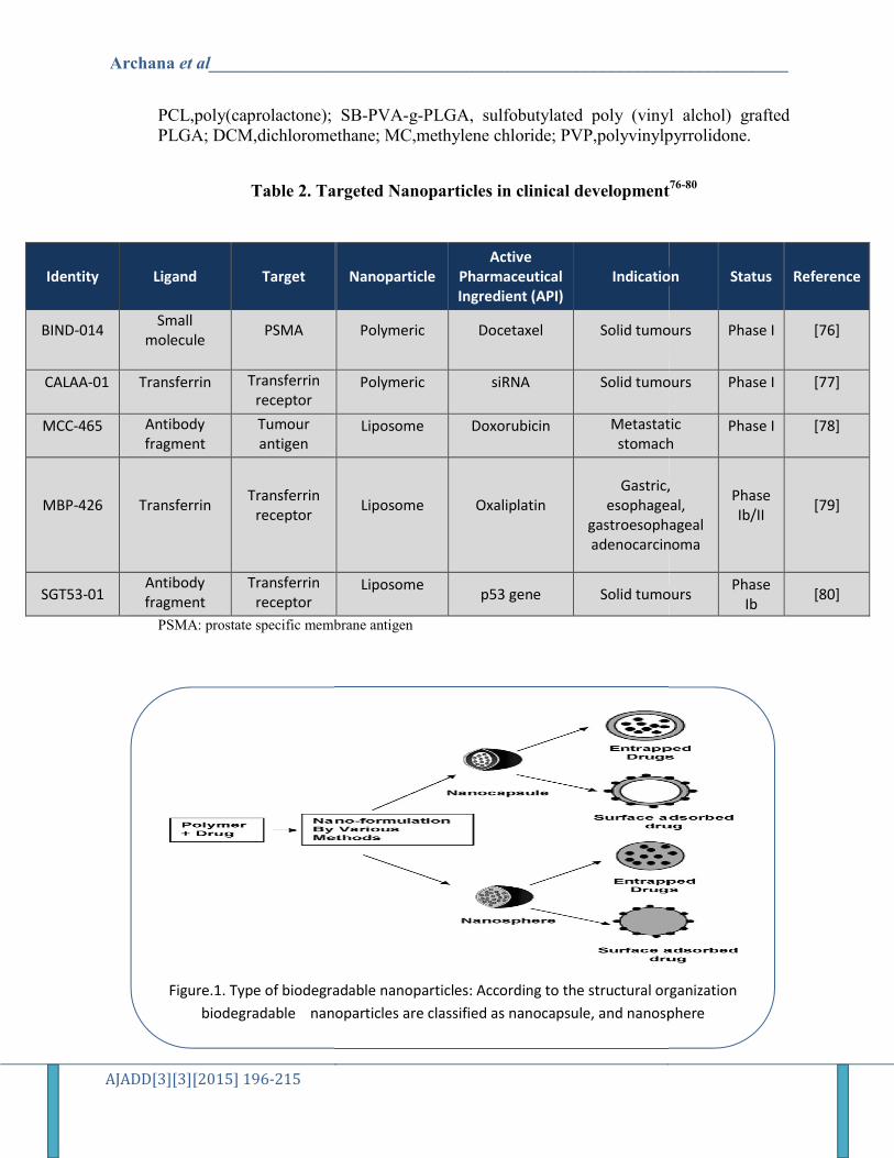
Figure.1. Type of biodegradable nanoparticles: According to the structural organization
biodegradable nanoparticles are classified as nanocapsule, and nanosphere
________________________________________________________
SB-PVA-g-PLGA, sulfobutylated poly (vinyl alchol)DCM,dichloromethane; MC,methylene chloride; PVP,polyvinylpyrrolidone.
Targeted Nanoparticles in clinical development
membrane antigen
Nanoparticle Active
Pharmaceutical Ingredient (API)
Indication
Polymeric
Docetaxel
Solid tumours
Polymeric
siRNA
Solid tumours
Liposome
Doxorubicin
Metastatic stomachCancer
Liposome
Oxaliplatin
Gastric, esophageal,
gastroesophagealadenocarcinoma
Liposome
p53 gene Solid tumours
Type of biodegradable nanoparticles: According to the structural organization
biodegradable nanoparticles are classified as nanocapsule, and nanosphere
________________________________________________________
(vinyl alchol) grafted PVP,polyvinylpyrrolidone.
Targeted Nanoparticles in clinical development76-80
Indication Status Reference
Solid tumours Phase I
[76]
Solid tumours Phase I
[77]
Metastatic stomach
Phase I
[78]
Gastric, esophageal,
gastroesophageal adenocarcinoma
Phase Ib/II
[79]
Solid tumours Phase
Ib [80]
Type of biodegradable nanoparticles: According to the structural organization
biodegradable nanoparticles are classified as nanocapsule, and nanosphere

Archana et al__________________________________________________________________
AJADD[3][3][2015] 196-215
PLGA ( poly(D,L-lactide-co-glycolide)) PLA
PGA (poly(glycolic acid))
Figure.2. Chemical structures of various biodegradable polymers
Figure.1. Schematic representation of the endosomal pathway
________________________________________________________
glycolide)) PLA (poly(lactic
PGA (poly(glycolic acid)) PCLpoly(caprolactone)
Chemical structures of various biodegradable polymers
Schematic representation of the endosomal pathway
________________________________________________________
poly(lacticacid))
poly(caprolactone))
Chemical structures of various biodegradable polymers

Archana et al__________________________________________________________________
AJADD[3][3][2015] 196-215
Figure.4. Schematic representation of active and passive targeting
________________________________________________________
Schematic representation of active and passive targeting
________________________________________________________
Schematic representation of active and passive targeting


![The Five Dhyani Buddhas[2]](https://static.fdocuments.in/doc/165x107/55285eb549795917048b481a/the-five-dhyani-buddhas2.jpg)