Review Article Movement Disorders Associated with COVID-19
11
Review Article Movement Disorders Associated with COVID-19 Mehri Salari, 1 Bahareh Zaker Harofteh, 1 Masoud Etemadifar, 2 Nahad Sedaghat, 2,3 and Hosein Nouri 2,3 1 Department of Neurology, Shahid Beheshti University of Medical Sciences, Tehran, Iran 2 Alzahra Research Institute, Alzahra University Hospital, Isfahan University of Medical Sciences, Isfahan, Iran 3 Network of Immunity in Infection Malignancy and Autoimmunity (NIIMA), Universal Scientific Education and Research Network (USERN), Isfahan, Iran Correspondence should be addressed to Hosein Nouri; [email protected] Received 14 September 2021; Revised 27 September 2021; Accepted 27 October 2021; Published 8 November 2021 Academic Editor: H´ elio Teive Copyright © 2021 Mehri Salari et al. is is an open access article distributed under the Creative Commons Attribution License, which permits unrestricted use, distribution, and reproduction in any medium, provided the original work is properly cited. As neurological complications associated with COVID-19 keep unfolding, the number of cases with COVID-19-associated de novo movement disorders is rising. Although no clear pathomechanistic explanation is provided yet, the growing number of these cases is somewhat alarming. is review gathers information from 64 reports of de novo movement disorders developing after/ during infection with SARS-CoV-2. ree new cases with myoclonus occurring shortly after a COVID-19 infection are also presented. Treatment resulted in partial to complete recovery in all three cases. Although the overall percentage of COVID-19 patients who develop movement disorders is marginal, explanations on a probable causal link have been suggested by numerous reports; most commonly involving immune-mediated and postinfectious and less frequently hypoxic-associated and ischemic- related pathways. e current body of evidence points myoclonus and ataxia out as the most frequent movement disorders occurring in COVID-19 patients. Some cases of tremor, chorea, and hypokinetic-rigid syndrome have also been observed in association with COVID-19. In particular, parkinsonism may be of dual concern in the setting of COVID-19; some have linked viral infections with Parkinson’s disease (PD) based on results from cerebrospinal fluid analyses, and PD is speculated to impact the outcome of COVID-19 in patients negatively. In conclusion, the present paper reviewed the demographic, clinical, and treatment-associated information on de novo movement disorders in COVID-19 patients in detail; it also underlined the higher incidence of myoclonus and ataxia associated with COVID-19 than other movement disorders. 1. Introduction Coronavirus disease 2019 (COVID-19), a lower respi- ratory infection with severe acute respiratory syndrome coronavirus 2 (SARS-CoV-2), is associated with a wide range of neurological manifestations. Most frequent neurological symptoms are nonspecific and include headache, dizziness, disturbance of smell and taste, and myalgia [1, 2]. Among the less common yet more severe complications are cerebrovascular diseases, encephalitis, encephalopathy, and inflammatory central and periph- eral nervous systems (CNS and PNS) disorders [3]. Movement abnormalities such as ataxia and opsoclonus- myoclonus have also been observed in COVID-19 pa- tients [4, 5]. Hypothetically, there are multiple entry routes for the virus into the CNS (e.g., trans-synaptic, hematogenous, and lymphatic pathways) [6]. However, the expression of an- giotensin-converting enzyme 2 (ACE2) in the human brain parenchyma and whether SARS-CoV-2 infects the CNS neuronal cells is not established yet [7]. Although low to very-low concentrations of viral RNA have been detected by reverse transcription-polymerase chain reaction (RT-PCR) in some autopsied brain samples—especially those from the olfactory bulb and medulla; lack of correlation with microglial activation and nodules in those specimens is against the speculated CNS tropism [8, 9]. Nevertheless, direct neuronal invasion is not the only way SARS-CoV-2 may contribute to neurodegeneration and neuro- inflammation. Meanwhile, several observations of Hindawi Parkinson’s Disease Volume 2021, Article ID 3227753, 11 pages https://doi.org/10.1155/2021/3227753
Transcript of Review Article Movement Disorders Associated with COVID-19

Review ArticleMovement Disorders Associated with COVID-19
Mehri Salari,1 Bahareh Zaker Harofteh,1 Masoud Etemadifar,2 Nahad Sedaghat,2,3
and Hosein Nouri 2,3
1Department of Neurology, Shahid Beheshti University of Medical Sciences, Tehran, Iran2Alzahra Research Institute, Alzahra University Hospital, Isfahan University of Medical Sciences, Isfahan, Iran3Network of Immunity in Infection Malignancy and Autoimmunity (NIIMA),Universal Scientific Education and Research Network (USERN), Isfahan, Iran
Correspondence should be addressed to Hosein Nouri; [email protected]
Received 14 September 2021; Revised 27 September 2021; Accepted 27 October 2021; Published 8 November 2021
Academic Editor: Helio Teive
Copyright © 2021 Mehri Salari et al. *is is an open access article distributed under the Creative Commons Attribution License,which permits unrestricted use, distribution, and reproduction in any medium, provided the original work is properly cited.
As neurological complications associated with COVID-19 keep unfolding, the number of cases with COVID-19-associated denovo movement disorders is rising. Although no clear pathomechanistic explanation is provided yet, the growing number of thesecases is somewhat alarming. *is review gathers information from 64 reports of de novo movement disorders developing after/during infection with SARS-CoV-2. *ree new cases with myoclonus occurring shortly after a COVID-19 infection are alsopresented. Treatment resulted in partial to complete recovery in all three cases. Although the overall percentage of COVID-19patients who develop movement disorders is marginal, explanations on a probable causal link have been suggested by numerousreports; most commonly involving immune-mediated and postinfectious and less frequently hypoxic-associated and ischemic-related pathways. *e current body of evidence points myoclonus and ataxia out as the most frequent movement disordersoccurring in COVID-19 patients. Some cases of tremor, chorea, and hypokinetic-rigid syndrome have also been observed inassociation with COVID-19. In particular, parkinsonism may be of dual concern in the setting of COVID-19; some have linkedviral infections with Parkinson’s disease (PD) based on results from cerebrospinal fluid analyses, and PD is speculated to impactthe outcome of COVID-19 in patients negatively. In conclusion, the present paper reviewed the demographic, clinical, andtreatment-associated information on de novo movement disorders in COVID-19 patients in detail; it also underlined the higherincidence of myoclonus and ataxia associated with COVID-19 than other movement disorders.
1. Introduction
Coronavirus disease 2019 (COVID-19), a lower respi-ratory infection with severe acute respiratory syndromecoronavirus 2 (SARS-CoV-2), is associated with a widerange of neurological manifestations. Most frequentneurological symptoms are nonspecific and includeheadache, dizziness, disturbance of smell and taste, andmyalgia [1, 2]. Among the less common yet more severecomplications are cerebrovascular diseases, encephalitis,encephalopathy, and inflammatory central and periph-eral nervous systems (CNS and PNS) disorders [3].Movement abnormalities such as ataxia and opsoclonus-myoclonus have also been observed in COVID-19 pa-tients [4, 5].
Hypothetically, there are multiple entry routes for thevirus into the CNS (e.g., trans-synaptic, hematogenous, andlymphatic pathways) [6]. However, the expression of an-giotensin-converting enzyme 2 (ACE2) in the human brainparenchyma and whether SARS-CoV-2 infects the CNSneuronal cells is not established yet [7]. Although low tovery-low concentrations of viral RNA have been detected byreverse transcription-polymerase chain reaction (RT-PCR)in some autopsied brain samples—especially those from theolfactory bulb and medulla; lack of correlation withmicroglial activation and nodules in those specimens isagainst the speculated CNS tropism [8, 9]. Nevertheless,direct neuronal invasion is not the only way SARS-CoV-2may contribute to neurodegeneration and neuro-inflammation. Meanwhile, several observations of
HindawiParkinson’s DiseaseVolume 2021, Article ID 3227753, 11 pageshttps://doi.org/10.1155/2021/3227753

movement abnormalities development or worsening inCOVID-19 patients have been reported (reviewed later inthis paper), which sounds alarming. We review all publishedreports on COVID-19-associated movement disorders andpresent three new patients with myoclonus after contractingCOVID-19.
2. Literature Search Strategy andInclusion Criteria
We conducted a comprehensive systematic literature searchin PubMed and Scopus databases using the following terms:“coronavirus disease 2019,” “COVID,” “COVID-19,”“SARS-CoV-2,” “Neurologic,” “Movement disorders,”“Tremor,” “Myoclonus,” “Parkinsonism,” “ataxia,” “dysto-nia,” and “chorea”. *e Mendeley application was used todetect, scan, and remove duplicates. *e remaining studieswere scanned for relevant titles/abstracts. All relevant arti-cles that reported patients who developed a movementdisorder during/after a SARS-CoV-2 infection were in-cluded. Reference lists of the obtained articles were searchedfor additional relevant results. Any movement disorders inCOVID-19 patients were reviewed; demographic informa-tion and data on clinical characteristics, treatments applied,and the outcome of patients were extracted. We includedcase reports, case series, and observational studies publishedin peer-reviewed journals and preprints available in English.Articles without full texts and studies without laboratoryconfirmation (RT-PCR) of COVID-19 diagnoses wereexcluded.
3. Results
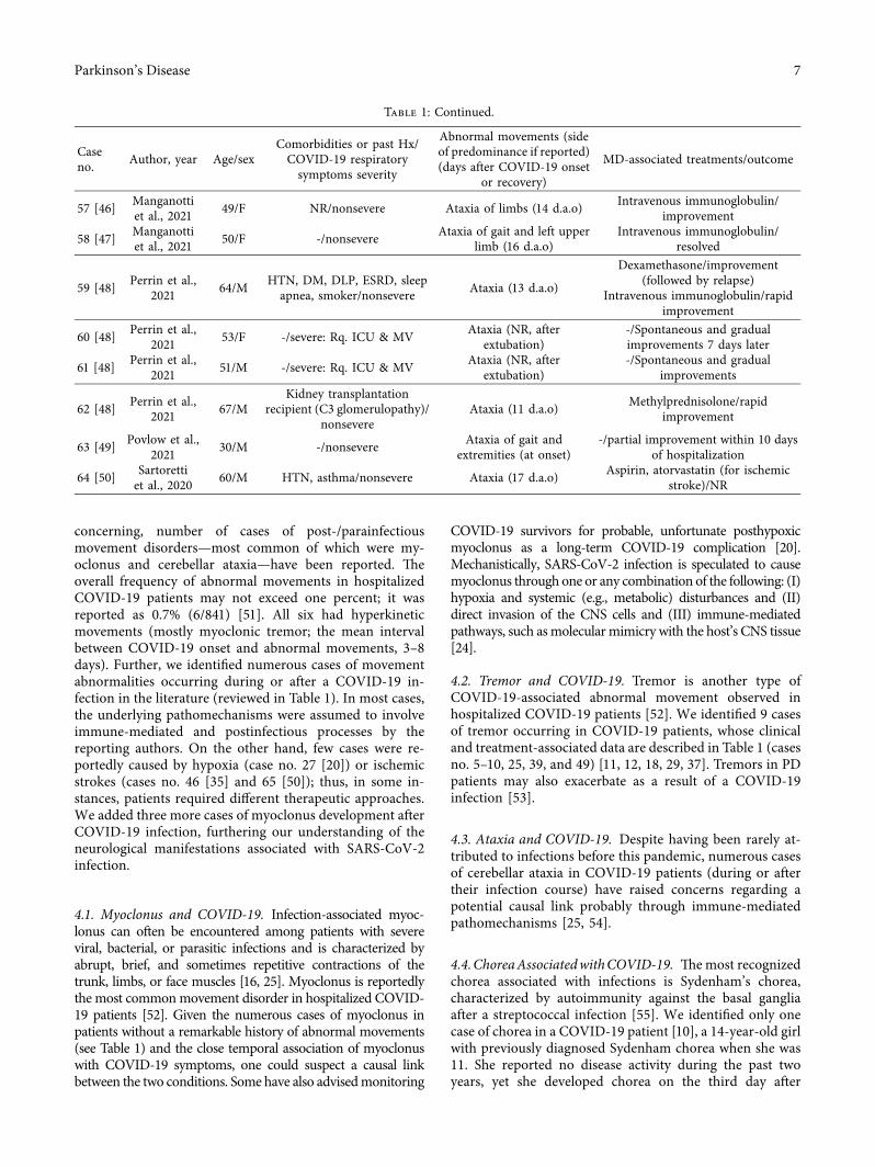
Our extensive search in the literature yielded a final numberof 43 publications reporting movement disorders in 64patients with SARS-CoV-2 infection (Table 1). Demographicinformation and data on their medical history, COVID-19infection severity, movement disorders, treatment, andoutcome are presented in Table 1. Furthermore, we added 3cases of movement disorders in COVID-19 patients fromour center to the literature, reported below.
3.1. Case 1. A previously healthy 42-year-old man presentedwith difficulty speaking, cognitive deficits, ataxia, and pro-gressive disabling jerks in upper and lower limbs, leading tofalls (see Supplementary video-1). Neurological examinationdemonstrated spontaneous, action-induced, and tactile-sensitive myoclonus on the face, upper extremities, andlower extremities. He had dysmetria and dysdiadochoki-nesia with superimposed action-induced myoclonus in theupper and lower extremities. Myoclonus prevented himfrom standing independently, and he had a wide-based gait.*e rest of the neurological examination was normal.Laboratory tests including renal function, liver function,ammonium, urea, and CO2 were normal, except for thenasopharyngeal RT-PCR test for SARS-CoV-2, which waspositive. Computed tomography (CT) of the head and brainmagnetic resonance imaging (MRI) were normal.
Electroencephalogram (EEG) and electromyography (EMG)were performed but did not reveal any abnormalities. Giventhese findings and the positive SARS-CoV-2 PCR, myoc-lonus was considered postinfectious. No other symptomssuch as fever, myalgia, cough, fatigue, hyposmia, or hypo-geusia were present. Cerebrospinal fluid (CSF) analysis wasunremarkable, RT-PCR test for SARS-CoV-2 and autoan-tibodies were negative. He was treated with methylpred-nisolone 1000mg IV daily for five days, bringing about apartial resolution of myoclonus. Complete recovery wasobserved on a follow-up session one month later.
3.2. Case 2. A previously healthy 52-year-old man presentedwith progressive imbalance and generalized jerks (seeSupplementary video-2). A week before, he had experiencedmyalgia and tested positive for SARS-CoV-2. Upon ad-mission, he had generalized stimulus-sensitive myoclonusaffecting his face, hands, and legs, confining him to awheelchair. Other neurological findings, including eyemovements, muscle tone, and deep tendon reflexes, werenormal. He did not have any coughing, fever, dyspnea, andO2 saturation was more than 97% without nasal O2.Ground-glass opacities suggestive of SARS-CoV-2 infectionwere detected on chest CT. Laboratory studies showed noincrease in inflammatory markers in peripheral blood; CRPand white blood cell count were normal. Brain CT and MRIwere normal. Results from CSF evaluations, including au-toantibody investigations, were unremarkable. *e cere-bellar syndrome improved after high-dose IVmethylprednisolone (1 gr/day for five days); on a follow-upsession after three weeks, the patient could walkindependently.
3.3. Case 3. A 38-year-old man was referred to our clinic withgeneralized jerks in his hands (see Supplementary video-3). Aweek before, he had been discharged from the hospital after fourdays of hospitalization due to a positive nasopharyngeal RT-PCR test and evident involvement of his lungs. Four days afterdischarge, he developed fine jerks in his hands and could notwrite; three days later, he was referred to our clinic withprogression and generalization of jerks. He had generalizedmyoclonus in upper and lower limbs and bizarre abnormalmovements that diminished with distraction; he was alsorestless and seemed anxious. *e rest of the neurological ex-aminations were normal. Lab tests were unremarkable. Labo-ratory investigations and brain MRI scans were normal. CSFanalyses, including cell count, protein, and autoimmune panel,were normal. Clonazepam (2mg/day), levetiracetam (1000mg/day), and IV methylprednisolone for 5 days (1 g/day) werestarted, resulting in partial improvement. After two weeks,myoclonus was resolved; functional movements improved withsome residual deficits, but he was still suffering from anxiety.
4. Discussion
Our understanding of COVID-19-associated neurologicalmanifestations is evolving. With about 200 million con-firmed cases of COVID-19, a relatively limited, yet
2 Parkinson’s Disease
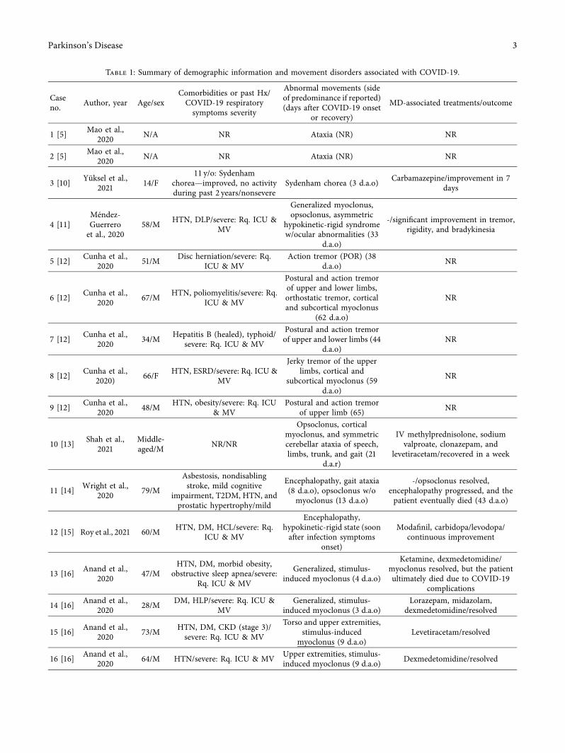
Table 1: Summary of demographic information and movement disorders associated with COVID-19.
Caseno. Author, year Age/sex
Comorbidities or past Hx/COVID-19 respiratorysymptoms severity
Abnormal movements (sideof predominance if reported)(days after COVID-19 onset
or recovery)
MD-associated treatments/outcome
1 [5] Mao et al.,2020 N/A NR Ataxia (NR) NR
2 [5] Mao et al.,2020 N/A NR Ataxia (NR) NR
3 [10] Yuksel et al.,2021 14/F
11 y/o: Sydenhamchorea—improved, no activityduring past 2 years/nonsevere
Sydenham chorea (3 d.a.o) Carbamazepine/improvement in 7days
4 [11]Mendez-Guerreroet al., 2020
58/M HTN, DLP/severe: Rq. ICU &MV
Generalized myoclonus,opsoclonus, asymmetric
hypokinetic-rigid syndromew/ocular abnormalities (33
d.a.o)
-/significant improvement in tremor,rigidity, and bradykinesia
5 [12] Cunha et al.,2020 51/M Disc herniation/severe: Rq.
ICU & MVAction tremor (POR) (38
d.a.o) NR
6 [12] Cunha et al.,2020 67/M HTN, poliomyelitis/severe: Rq.
ICU & MV
Postural and action tremorof upper and lower limbs,orthostatic tremor, corticaland subcortical myoclonus
(62 d.a.o)
NR
7 [12] Cunha et al.,2020 34/M Hepatitis B (healed), typhoid/
severe: Rq. ICU & MV
Postural and action tremorof upper and lower limbs (44
d.a.o)NR
8 [12] Cunha et al.,2020) 66/F HTN, ESRD/severe: Rq. ICU &
MV
Jerky tremor of the upperlimbs, cortical and
subcortical myoclonus (59d.a.o)
NR
9 [12] Cunha et al.,2020 48/M HTN, obesity/severe: Rq. ICU
& MVPostural and action tremor
of upper limb (65) NR
10 [13] Shah et al.,2021
Middle-aged/M NR/NR
Opsoclonus, corticalmyoclonus, and symmetriccerebellar ataxia of speech,limbs, trunk, and gait (21
d.a.r)
IV methylprednisolone, sodiumvalproate, clonazepam, and
levetiracetam/recovered in a week
11 [14] Wright et al.,2020 79/M
Asbestosis, nondisablingstroke, mild cognitive
impairment, T2DM, HTN, andprostatic hypertrophy/mild
Encephalopathy, gait ataxia(8 d.a.o), opsoclonus w/omyoclonus (13 d.a.o)
-/opsoclonus resolved,encephalopathy progressed, and thepatient eventually died (43 d.a.o)
12 [15] Roy et al., 2021 60/M HTN, DM, HCL/severe: Rq.ICU & MV
Encephalopathy,hypokinetic-rigid state (soonafter infection symptoms
onset)
Modafinil, carbidopa/levodopa/continuous improvement
13 [16] Anand et al.,2020 47/M
HTN, DM, morbid obesity,obstructive sleep apnea/severe:
Rq. ICU & MV
Generalized, stimulus-induced myoclonus (4 d.a.o)
Ketamine, dexmedetomidine/myoclonus resolved, but the patientultimately died due to COVID-19
complications
14 [16] Anand et al.,2020 28/M DM, HLP/severe: Rq. ICU &
MVGeneralized, stimulus-
induced myoclonus (3 d.a.o)Lorazepam, midazolam,
dexmedetomidine/resolved
15 [16] Anand et al.,2020 73/M HTN, DM, CKD (stage 3)/
severe: Rq. ICU & MV
Torso and upper extremities,stimulus-induced
myoclonus (9 d.a.o)Levetiracetam/resolved
16 [16] Anand et al.,2020 64/M HTN/severe: Rq. ICU & MV Upper extremities, stimulus-
induced myoclonus (9 d.a.o) Dexmedetomidine/resolved
Parkinson’s Disease 3

Table 1: Continued.
Caseno. Author, year Age/sex
Comorbidities or past Hx/COVID-19 respiratorysymptoms severity
Abnormal movements (sideof predominance if reported)(days after COVID-19 onset
or recovery)
MD-associated treatments/outcome
17 [16] Anand et al.,2020 66/M HTN, DM, obesity/severe: Rq.
ICU & MV
Upper extremities and face,spontaneous myoclonus (2
d.a.o)Dexmedetomidine/resolved
18 [16] Anand et al.,2020 72/M DM/severe: Rq. ICU & MV
Generalized, multifocal,stimulus-induced
myoclonus (7 d.a.o)
Valproic acid, levetiracetam,lorazepam, dexmedetomidine/
continued, but improved myoclonus
19 [16] Anand et al.,2020 62/M HTN/severe: Rq. ICU & MV Generalized, stimulus-
induced myoclonus (7 d.a.o)Valproic acid, primidone,
clonazepam, lorazepam/resolved
20 [16] Anand et al.,2020 71/M HTN, CKD (stage 2)/
nonsevere
Generalized, action-induced;lingual myoclonus, gaitdisturbance (7 d.a.o)
Levetiracetam, valproic acid/resolved
21 [17]Rabano-
Suarez et al.,2020
63/M
Generalized anxiety disorder/nonsevere, but required ICUadmission due to a myoclonic
storm
Asynchronous generalizedmyoclonus (face and limbs),worsening with action andauditory/tactile stimuli (9
d.a.o)
Levetiracetam, valproic acid,clonazepam/scarce improvement
Methylprednisolone/slightimprovement
PLEX after myoclonus worsenedagain/improvement
22 [17]Rabano-
Suarez et al.,2020
88/F
HTN, hypothyroidism,nonfunctioning pituitaryadenoma, mild cognitivedecline, no Hx of MDs
Generalized myoclonus,similar to case no. 22, but
milder (∼21 d.a.o)Methylprednisolone/resolved
23 [17]Rabano-
Suarez et al.,2020
76/M -/nonsevereGeneralized myoclonus,similar to case no. 22, but
milder (11 d.a.o)
Levetiracetam, clonazepam/nobenefit- spontaneous progressiveimprovement after two weeks
24 [18] Piscitelli et al.,2020 39/F -/nonsevere
Lower limb tremor,abnormal movements at restand while sitting or walking
(functional) (7 d.a.o)
Benzodiazepines/no significantimprovement, spontaneous gradual
resolution later on
25 [19] Hewan et al.,2021 57/F
Morbid obesity, obstructivesleep apnea, chronic
obstructive pulmonary disease,heart failure, and
schizoaffective disorder (forwhich she was taking valproicacid)/severe: Rq. ICU & MV
Mental status deterioration,myoclonus in all extremities
and upper torso thatoccurred spontaneously atrest and would intensifyupon passive movements
(∼26 d.a.o)
Levetiracetam, clonazepam/nosignificant improvement
Ketamine was added/myoclonusimproved but she was heavily sedated/myoclonus returned after ketamine
was haltedMethylprednisolone/recovery
26 [20] Ros-Castelloet al., 2020 72/F HTN, asthma/severe: Rq. ICU
and oxygen therapy
Progressively disablingmyoclonus in upper limbsand negative myoclonus inlower limbs (leading to falls;
∼30 d.a.o)
Low-dose clonazepam/resolved
27 [21] Faber et al.,2020 35/F Unremarkable/mild
Akinetic-rigid parkinsonism(10 d.a.o) (Parkinson’s
disease rating scale part IIIscore: 49)
Levodopa/benserazide/significantimprovement (Parkinson’s diseaserating scale part III score: 32)
28 [22] Werner et al.,2021 62/M Unremarkable/nonsevere
Cerebellar syndrome withslightly scanning speech andlimb, truncal, and gait ataxia(16 d.a.o) (SARA ataxia
score: 14)
High-dose methylprednisolone/gradual improvement (SARA 6 days
after treatment: 5, and SARA 4months after treatment: 1)
29 [23]Urrea-
Mendozaet al., 2021
32/M -/nonsevere Opsoclonus, myoclonus,ataxia (12 d.a.o)
Clonazepam, divalproex, oralmethylprednisolone/substantial
improvement
4 Parkinson’s Disease

Table 1: Continued.
Caseno. Author, year Age/sex
Comorbidities or past Hx/COVID-19 respiratorysymptoms severity
Abnormal movements (sideof predominance if reported)(days after COVID-19 onset
or recovery)
MD-associated treatments/outcome
30 [24] El Otmaniet al., 2021 59/M Recent under control DM/
asymptomatic
Generalized myoclonus intorso, limbs, face, tongue,and larynx, intensified bymovement and acoustic
stimuli (not clear, but likely aclose temporal association
was present)
Levetiracetam/no improvementVeinoglobulins/only slight
improvement
Methylprednisolone/rapid,substantial improvement
31 [25] Chan et al.,2021 44/M -/nonsevere
Spontaneous, action-induced, posture-induced,and tactile stimuli sensitivemyoclonus in the face, upper
extremities, and lowerextremities, wide-based,ataxic gait (12 d.a.o)
Methylprednisolone/partialimprovement of spontaneousmyoclonus; action-induced
myoclonus persistedClonazepam/partial improvementLevetiracetam/major improvement,
discharged
32 [26] Chaumontet al., 2020 62/M HTN, DM/severe: Rq. ICU &
MV
Postural and actionmyoclonus in upper limbs,
ataxia (∼19 d.a.o)
Intravenous immunoglobulin/partialimprovement of other non-MD-
associated psychiatric and cognitivesymptoms; myoclonus and ataxia
persisted 3 weeks after discharge (finaloutcome NR)
33 [26] Chaumontet al., 2020 72/M
HTN, DM, obesity, urothelialcarcinoma in remission/severe:
Rq. ICU & MV
Postural and actionmyoclonus in upper limbs,
ataxia (∼34 d.a.o)
Intravenous immunoglobulin/partialimprovement of other non-MD-
associated psychiatric and cognitivesymptoms; myoclonus and ataxia
persisted 3 weeks after discharge (finaloutcome NR)
34 [26] Chaumontet al., 2020 50/M DM/severe: Rq. ICU & MV
Postural and actionmyoclonus in upper limbs,
ataxia (∼48 d.a.o)
Intravenous immunoglobulin/partialimprovement of other non-MD-
associated psychiatric and cognitivesymptoms; myoclonus and ataxia
persisted 3 weeks after discharge (finaloutcome NR)
35 [26] Chaumontet al., 2020 66/M Obstructive sleep apnea/severe:
Rq. ICU & MV
Postural and actionmyoclonus in upper limbs,
ataxia (∼40 d.a.o)
Intravenous immunoglobulin/partialimprovement of other non-MD-
associated psychiatric and cognitivesymptoms; myoclonus and ataxia
persisted 3 weeks after discharge (finaloutcome NR)
36 [27] Delorme et al.,2020 72/M NR/nonsevere Upper limbs myoclonus, and
cerebellar ataxia (15 d.a.o)Intravenous immunoglobulin/
resolved
37 [28] Dijkstra et al.,2020 44/M Unremarkable/mild
Action-induced myoclonicjerks in the face, arms, andtrunk, intensified by tactile
and acoustic stimuli,stuttering speech, prominent
gait ataxia (∼14 d.a.o)
Methylprednisolone, intravenousimmunoglobulin/gradually resolved
within 2 months
38 [29] Grimaldiet al., 2020 72/M
One episode of transient globalamnesia (10 years ago)/
nonsevere
Upper and lower limbs andtrunk action tremor, ataxia,action- and stimuli-induced
diffuse (but worse inproximal limbs) myoclonus
(17 d.a.o)
Intravenous immunoglobulin/nosignificant improvement
Intravenous methylprednisolone/considerable improvement
Parkinson’s Disease 5

Table 1: Continued.
Caseno. Author, year Age/sex
Comorbidities or past Hx/COVID-19 respiratorysymptoms severity
Abnormal movements (sideof predominance if reported)(days after COVID-19 onset
or recovery)
MD-associated treatments/outcome
39 [30] Sanguinettiet al., 2021 57/M HTN, T2DM, DLP/nonsevere
Action-induced myoclonusin upper and lower
extremities, opsoclonus, gaitataxia (at least 5 d.a.o)
Clonazepam, methylprednisolone/improved
40 [31] Schellekenset al., 2020 48/M
Asymptomatic HIV+ withnormal CD4+ T-cell count/
nonsevere
Generalized myoclonus intrunk and limbs, present atrest but worsening both
posturally and with action,cerebellar ataxia of arms andlegs, and an ataxic gait (13
d.a.o)
Levetiracetam/improved
41 [32] Borroni et al.,2020 54/F HTN/nonsevere
Posture-induced myoclonusin diaphragm and leftextremities (∼14 d.a.o)
Clonazepam/significant improvement
42 [32] Borroni et al.,2020 80/M -/nonsevere Diaphragmatic myoclonus
(23 d.a.o) Levetiracetam/resolved
43 [33] Khoo et al.,2020 65/F
Alzheimer’s disease,osteoarthritis,
gastroesophageal refluxdisease/nonsevere
Upper and lower limbs, face,and tongue myoclonus,induced by tactile, visual,
and auditory stimuli (7 d.a.o)
Levetiracetam, clonazepam/partialimprovement
Intravenous methylprednisolone/progressive
improvement—discharged after 10days
44 [34] Muccioli et al.,2020 58/M HTN/severe: Rq. ICU & MV
Multifocal myoclonusinduced by action and tactilestimuli (at least 23 d.a.o)
Clonazepam, levetiracetam/significant improvement within 5
days
45 [35] Ashraf et al.,2020 26/F Obesity, asthma, PTSD,
depression/asymptomaticRight limb ataxia, ataxic gait
(N/A)Aspirin, clopidogrel, statin (for
ischemic stroke)/NR
46 [36] Balestrinoet al., 2020 73/M HTN, T2DM/nonsevere Ataxic gait (at onset)
No additional treatment other thanantiviral agents/resolved within 6
weeks
47 [27] Delorme et al.,2020 60/F
Temporal lobe epilepsy(hippocampal sclerosis)/
nonsevere
Limbs and gait ataxia (atonset) Corticosteroid pulse therapy/resolved
48 [37]Diezma-
Martın et al.,2020
70/M COPD/mildAtaxia/tremor of voice,extremities, and gait,
orthostatic tremor (17 d.a.o)
Clonazepam/slightly improved-discharged and continuously
improved
49 [38] Fadakar et al.,2020 47/M -/nonsevere Ataxia of extremities, gait,
and trunk (3 d.a.o)
No additional treatment other thanantiviral agents/significant
improvement within two weeks
50 [39]Fernandez-Domınguezet al., 2020
74/F HTN, follicular lymphoma/nonsevere Gait ataxia (12–15 d.a.o) Intravenous immunoglobulin/
improvement
51 [40]Gutierrez-Ortiz et al.,
202050/M Asthma/nonsevere Gait ataxia (3 d.a.o) Intravenous immunoglobulin/
resolved within two weeks
52 [41] Hayashi et al.,2020 75/M Alzheimer’s disease/nonsevere Ataxia of limbs and gait (at
onset) -/resolved after 2 days
53 [42] Kopscik et al.,2020 31/M -/nonsevere Ataxia of limbs and gait (at
onset)Intravenous immunoglobulin/
improvement
54 [43] Lahiri et al.,2020 72/M NR/NR Ataxia (at onset) NR/NR
55 [44] Lantos et al.,2020 36/M Remote strabismus/nonsevere Gait ataxia (4 d.a.o) Intravenous immunoglobulin/
improvement
56 [45] Lowery et al.,2020 45/M HTN, DLP, Crohn’s disease/
severe: Rq. ICU & MV Gait ataxia (14 d.a.o) Intravenous immunoglobulin/delayedimprovement
6 Parkinson’s Disease
concerning, number of cases of post-/parainfectiousmovement disorders—most common of which were my-oclonus and cerebellar ataxia—have been reported. *eoverall frequency of abnormal movements in hospitalizedCOVID-19 patients may not exceed one percent; it wasreported as 0.7% (6/841) [51]. All six had hyperkineticmovements (mostly myoclonic tremor; the mean intervalbetween COVID-19 onset and abnormal movements, 3–8days). Further, we identified numerous cases of movementabnormalities occurring during or after a COVID-19 in-fection in the literature (reviewed in Table 1). In most cases,the underlying pathomechanisms were assumed to involveimmune-mediated and postinfectious processes by thereporting authors. On the other hand, few cases were re-portedly caused by hypoxia (case no. 27 [20]) or ischemicstrokes (cases no. 46 [35] and 65 [50]); thus, in some in-stances, patients required different therapeutic approaches.We added three more cases of myoclonus development afterCOVID-19 infection, furthering our understanding of theneurological manifestations associated with SARS-CoV-2infection.
4.1. Myoclonus and COVID-19. Infection-associated myoc-lonus can often be encountered among patients with severeviral, bacterial, or parasitic infections and is characterized byabrupt, brief, and sometimes repetitive contractions of thetrunk, limbs, or face muscles [16, 25]. Myoclonus is reportedlythe most commonmovement disorder in hospitalized COVID-19 patients [52]. Given the numerous cases of myoclonus inpatients without a remarkable history of abnormal movements(see Table 1) and the close temporal association of myoclonuswith COVID-19 symptoms, one could suspect a causal linkbetween the two conditions. Some have also advisedmonitoring
COVID-19 survivors for probable, unfortunate posthypoxicmyoclonus as a long-term COVID-19 complication [20].Mechanistically, SARS-CoV-2 infection is speculated to causemyoclonus through one or any combination of the following: (I)hypoxia and systemic (e.g., metabolic) disturbances and (II)direct invasion of the CNS cells and (III) immune-mediatedpathways, such asmolecularmimicry with the host’s CNS tissue[24].
4.2. Tremor and COVID-19. Tremor is another type ofCOVID-19-associated abnormal movement observed inhospitalized COVID-19 patients [52]. We identified 9 casesof tremor occurring in COVID-19 patients, whose clinicaland treatment-associated data are described in Table 1 (casesno. 5–10, 25, 39, and 49) [11, 12, 18, 29, 37]. Tremors in PDpatients may also exacerbate as a result of a COVID-19infection [53].
4.3. Ataxia and COVID-19. Despite having been rarely at-tributed to infections before this pandemic, numerous casesof cerebellar ataxia in COVID-19 patients (during or aftertheir infection course) have raised concerns regarding apotential causal link probably through immune-mediatedpathomechanisms [25, 54].
4.4.ChoreaAssociatedwithCOVID-19. *emost recognizedchorea associated with infections is Sydenham’s chorea,characterized by autoimmunity against the basal gangliaafter a streptococcal infection [55]. We identified only onecase of chorea in a COVID-19 patient [10], a 14-year-old girlwith previously diagnosed Sydenham chorea when she was11. She reported no disease activity during the past twoyears, yet she developed chorea on the third day after
Table 1: Continued.
Caseno. Author, year Age/sex
Comorbidities or past Hx/COVID-19 respiratorysymptoms severity
Abnormal movements (sideof predominance if reported)(days after COVID-19 onset
or recovery)
MD-associated treatments/outcome
57 [46] Manganottiet al., 2021 49/F NR/nonsevere Ataxia of limbs (14 d.a.o) Intravenous immunoglobulin/
improvement
58 [47] Manganottiet al., 2021 50/F -/nonsevere Ataxia of gait and left upper
limb (16 d.a.o)Intravenous immunoglobulin/
resolved
59 [48] Perrin et al.,2021 64/M HTN, DM, DLP, ESRD, sleep
apnea, smoker/nonsevere Ataxia (13 d.a.o)
Dexamethasone/improvement(followed by relapse)
Intravenous immunoglobulin/rapidimprovement
60 [48] Perrin et al.,2021 53/F -/severe: Rq. ICU & MV Ataxia (NR, after
extubation)-/Spontaneous and gradualimprovements 7 days later
61 [48] Perrin et al.,2021 51/M -/severe: Rq. ICU & MV Ataxia (NR, after
extubation)-/Spontaneous and gradual
improvements
62 [48] Perrin et al.,2021 67/M
Kidney transplantationrecipient (C3 glomerulopathy)/
nonsevereAtaxia (11 d.a.o) Methylprednisolone/rapid
improvement
63 [49] Povlow et al.,2021 30/M -/nonsevere Ataxia of gait and
extremities (at onset)-/partial improvement within 10 days
of hospitalization
64 [50] Sartorettiet al., 2020 60/M HTN, asthma/nonsevere Ataxia (17 d.a.o) Aspirin, atorvastatin (for ischemic
stroke)/NR
Parkinson’s Disease 7

COVID-19 symptoms onset. *e authors could not con-clude whether this was a recurrence of her previously silentdisease or an immune-mediated newly developed choreawith a causal link to COVID-19 [10]. However, we assumethe latter was less likely, with the very short interval betweenCOVID-19 onset and chorea manifestation (3 days) arguingagainst an autoimmune scenario.
4.5. Hypokinesia and Parkinsonism: Considerations inCOVID-19. *e acute hypokinetic-rigid syndrome has beenreported in two cases (cases no. 5 [11] and 13 [15]); bothdeveloped a severe course of infection and required intensivecare and mechanical ventilation.
Parkinson’s disease (PD) is a neurodegenerativedisease caused by the destruction of dopaminergicneurons in substantia nigra pars compacta (SNpc). Ev-idence of viral infections associated with PD exists; thus,vigilant monitoring and investigation of probable linksbetween SARS-CoV-2 infection and parkinsonism maybe necessary [7, 56]. On this matter, the presence ofantibodies targeting coronaviruses in cerebrospinal fluidof PD patients was more frequently observed whencompared to individuals without PD. Further, the pro-posed ability of coronaviruses in breaching into the CNSvia nasal cavity and nasal neuroepithelium can give usmore reasons to be concerned [57, 58]. Interestingly, ithas been speculated that PD-associated α-synucleinretrograde spreading and aggregation from the olfactorybulb to the midbrain and deeper CNS structures—leadingto cellular death of the affected tissue—could offer someprotection against SARS-CoV-2 breaching into the CNSvia the olfactory route [59]. However, the aforementionedneuropathological data from autopsy studies call thishypothesis into question [8, 9]. Regardless of such hy-pothesized protective effects, PD patients are neverthelessmore vulnerable to complications following respiratoryinfections, and odds for their hospitalization or devel-opment of other comorbidities are higher than thegeneral population [60]. Furthermore, parkinsoniansymptoms after viral respiratory infections may hint ustoward the less-known sides of the disease etiology andpathogenesis [61].
A community-based case-control study on 12 PD pa-tients who contracted COVID-19 documented a substantialrisk of motor and nonmotor symptoms worsening associ-ated with mild to moderate degrees of COVID-19, regardlessof the patients’ age and disease duration [7].
Lo Monaco and colleagues reported a patient with an 8-year history of PD who developed severe generalized dys-tonia shortly after contracting COVID-19. Improvementwas achieved after an increase in her daily dose of levodopaand clozapine [62].
Inversely, PD and its associated disabilities maynegatively impact the outcome of COVID-19 illness,along with other known risk factors such as age andhypertension. Indeed, Fasano et al. reported a higherCOVID-19 mortality rate in community-dwelling PDpatients (i.e., not living in nursing homes) than the
general population [63], and Salari et al. showed PDpatients have a higher proportion of COVID-19 mortalityin comparison with other patients hospitalized forCOVID-19 [64].
Some limitations apply to the present review article,such as the keywords selection; some studies that have notused the keywords incorporated in our search strategy,e.g., those that only mentioned “neurological manifes-tations” without further specification, might have beenmissed. *e literature search has not thoroughly adheredto the standards of a systematic review, albeit being asexhaustive and comprehensive as possible. Furthermore,cross-reference checking for some reviewed articles mayhave been incomplete. Review papers bear the limitationof their reviewed articles; as discussed earlier, myoclonusmay stem from metabolic comorbidities in some cases.*us, the attribution of myoclonus to COVID-19 by somecase studies is a potential limitation of such reportsapplying to the present review as well. Another limitationwas that electrophysiological data were not obtained fromtwo of our cases (i.e., Cases 2 and 3).
5. Conclusion
*is review highlighted the higher incidence of myoclonusand ataxia after COVID-19 infection. Other movementdisorders were not common after COVID-19, implying thatSARS-CoV-2 mainly affects the brain through immune-mediated pathways rather than a direct invasion of the CNS.Nonetheless, the COVID-19 pandemic can affect the out-come of patients with established movement disorders.
Abbreviations
CKD: Chronic kidney diseaseCOPD: Chronic obstructive pulmonary diseased.a.o: Days after onsetd.a.r: Days after recoveryDLP: DyslipidemiaDM: Diabetes mellitusESRD: End-stage renal diseaseF: FemaleHLP: HyperlipidemiaHTN: HypertensionHx: HistoryICU: Intensive care unit (admission)M: MaleMD: Movement disorderMV: Mechanical ventilationN/A: Not availablePLEX: Plasma exchangePOL: Predominant on the leftPOR: Predominant on the rightPTSD: Posttraumatic stress disorderRq: RequiredSARA: *e scale for the assessment and rating of ataxiaT2DM: Type II diabetes mellitusw/: With.
8 Parkinson’s Disease

Data Availability
All relevant data are provided in the manuscript and thesupplemental files; there are no additional data to bepresented.
Conflicts of Interest
*e authors declare that there are no conflicts of interestrelevant to this work.
Supplementary Materials
A direct link to video files of the three patients reported areincluded as supplementary data of this manuscript. (Sup-plementary Materials)
References
[1] T. T. Favas, P. Dev, R. N. Chaurasia et al., “Neurologicalmanifestations of COVID-19: a systematic review and meta-analysis of proportions,” Neurological Sciences, vol. 41, no. 12,pp. 3437–3470, 2020.
[2] E. M. Liotta, A. Batra, J. R. Clark et al., “Frequent neurologicmanifestations and encephalopathy-associated morbidity inCovid-19 patients,” Annals of Clinical and TranslationalNeurology, vol. 7, no. 11, pp. 2221–2230, 2020.
[3] J. Almqvist, T. Granberg, and A. Tzortzakakis, “Neurologicalmanifestations of coronavirus infections—a systematic re-view,” Annals of Clinical and Translational Neurology, vol. 7,no. 10, pp. 2057–2071, 2020.
[4] V. Montalvan, J. Lee, T. Bueso, J. De Toledo, and K. Rivas,“Neurological manifestations of COVID-19 and other coro-navirus infections: a systematic review,” Clinical Neurologyand Neurosurgery, vol. 194, Article ID 105921, 2020.
[5] L. Mao, H. Jin, M. Wang et al., “Neurologic manifestations ofhospitalized patients with coronavirus disease 2019 inWuhan,China,” JAMA Neurology, vol. 77, no. 6, p. 683, 2020.
[6] A. Sepehrinezhad, A. Shahbazi, and S. S. Negah, “COVID-19virus may have neuroinvasive potential and cause neuro-logical complications: a perspective review,” Journal ofNeurovirology, vol. 26, no. 3, pp. 324–329, 2020.
[7] D. Sulzer, A. Antonini, V. Leta et al., “COVID-19 and possiblelinks with Parkinson’s disease and parkinsonism: from benchto bedside,” npj Parkinson’s Disease, vol. 6, no. 1, p. 18, 2020.
[8] K. T. *akur, E. H. Miller, and M. D. Glendinning, “COVID-19 neuropathology at Columbia university irving medicalcenter/New York presbyterian hospital,” Brain, vol. 144, no. 9,pp. 2696–2708, 2021.
[9] I. H. Solomon, E. Normandin, S. Bhattacharyya et al.,“Neuropathological features of Covid-19,” New EnglandJournal of Medicine, vol. 383, no. 10, pp. 989–992, 2020.
[10] M. F. Yuksel, M. Yıldırım, O. Bektas, S. Sahin, and S. Teber, “Asydenham chorea attack associated with COVID-19 infec-tion,” Brain, Behavior Immunity Health, vol. 13, Article ID100222, 2021.
[11] A. Mendez-Guerrero, M. I. Laespada-Garcıa, and A. Gomez-Grande, “Acute hypokinetic-rigid syndrome following SARS-CoV-2 infection,” Neurology, vol. 95, no. 15, pp. e2109–e2118,2020.
[12] P. Cunha, B. Herlin, K. Vassilev et al., “Movement disorders asa new neurological clinical picture in severe SARS-CoV-2
infection,” European Journal of Neurology, vol. 27, no. 12,pp. e88–e90, 2020.
[13] P. B. Shah and S. D. Desai, “Opsoclonus myoclonus ataxiasyndrome in the setting of COVID-19 infection,” Neurology,vol. 96, no. 1, p. 33, 2021.
[14] D. Wright, R. Rowley, P. Halks-Wellstead, T. Anderson, andT. Y. Wu, “Abnormal saccadic oscillations associated withsevere acute respiratory syndrome coronavirus 2 encepha-lopathy and ataxia,” Movement Disorder Clinical Practice,vol. 7, no. 8, pp. 980–982, 2020.
[15] D. Roy, J. Song, N. Awad, and P. Zamudio, “Treatment ofunexplained coma and hypokinetic-rigid syndrome in a pa-tient with COVID-19,” BMJ Case Reports, vol. 14, no. 3,Article ID e239781, 2021.
[16] P. Anand, A. Zakaria, K. Benameur et al., “Myoclonus inpatients with coronavirus disease 2019: a multicenter caseseries,” Critical Care Medicine, vol. 48, no. 11, pp. 1664–1669,2020.
[17] P. Rabano-Suarez, L. Bermejo-Guerrero, and A. Mendez-Guerrero, “Generalized myoclonus in COVID-19,” Neurol-ogy, vol. 95, no. 6, pp. e767–e772, 2020.
[18] D. Piscitelli, C. Perin, L. Tremolizzo, F. Peroni, C. G. Cerri,and C. M. Cornaggia, “Functional movement disorders in apatient with COVID-19,” Neurological Sciences, vol. 41, no. 9,pp. 2343-2344, 2020.
[19] H. Hewan, A. Yang, A. Vaddiparti, and B. Keung, “Non-epileptic myoclonus in COVID-19: case report,” 9e Neu-rohospitalist, Article ID 194187442110043, 2021.
[20] V. Ros-Castello, C. Quereda, J. Lopez-Sendon, and I. Corral,“Post-hypoxic myoclonus after COVID-19 infection recov-ery,” Movement Disorders Clinical Practice, vol. 7, no. 8,pp. 983-984, 2020.
[21] I. Faber, P. R. P. Brandão, F. Menegatti, D. D. Carvalho Bispo,F. B. Maluf, and F. Cardoso, “Coronavirus disease 2019 andparkinsonism: a non-post-encephalitic case,” MovementDisorders, vol. 35, no. 10, pp. 1721-1722, 2020.
[22] J. Werner, I. Reichen, M. Huber, I. A. Abela, M. Weller, andI. Jelcic, “Subacute cerebellar ataxia following respiratorysymptoms of COVID-19: a case report,” BMC InfectiousDiseases, vol. 21, no. 1, p. 298, 2021.
[23] E. Urrea-Mendoza, K. Okafor, S. Ravindran, J. Absher,V. Chaubal, and F. J. Revilla, “Opsoclonus-myoclonus-ataxiasyndrome (OMAS) associated with SARS-CoV-2 infection:post-infectious neurological complication with benignprognosis,” Tremor and other Hyperkinetic Movements,vol. 11, no. 1, 2021.
[24] H. El Otmani, F. Moutaouakil, M. Ouazzani, and K. Mjahed,“Isolated generalized myoclonus immune-mediated by SARS-CoV-2: an illustrative videotaped case,” Neurology Science,vol. 42, no. 8, pp. 3411–3413, 2021.
[25] J. L. Chan, K. A. Murphy, and J. R. Sarna, “Myoclonus andcerebellar ataxia associated with COVID-19: a case report andsystematic review,” Journal of Neurology, vol. 268, 2021.
[26] H. Chaumont, A. San-Galli, F. Martino et al., “Mixed centraland peripheral nervous system disorders in severe SARS-CoV-2 infection,” Journal of Neurology, vol. 267, no. 11,pp. 3121–3127, 2020.
[27] C. Delorme, O. Paccoud, A. Kas et al., “COVID-19-relatedencephalopathy: a case series with brain FDG-positron-emission tomography/computed tomography findings,” Eu-ropean Journal of Neurology, vol. 27, no. 12, pp. 2651–2657,2020.
[28] F. Dijkstra, T. Van den Bossche, B. Willekens, P. Cras, andD. Crosiers, “Myoclonus and cerebellar ataxia following
Parkinson’s Disease 9
COVID-19,” Movement Disorder Clinical Practice, vol. 7,no. 8, pp. 974–976, 2020.
[29] S. Grimaldi, S. Lagarde, J. R. Harle, J. Boucraut, and E. Guedj,“Autoimmune encephalitis concomitant with SARS-CoV-2infection: insight from 18 F-FDG PET imaging and neuronalautoantibodies,” Journal of Nuclear Medicine, vol. 61, no. 12,pp. 1726–1729, 2020.
[30] S. Y. Sanguinetti and R. A. Ramdhani, “Opsoclonus-myoc-lonus-ataxia syndrome related to the novel coronavirus(COVID-19),” Journal of Neuro-Ophthalmology, vol. 41, 2021.
[31] M. M. I. Schellekens, C. P. Bleeker-Rovers, P. A. J. Keurlings,C. J. Mummery, and B. R. Bloem, “Reversible myoclonus-ataxia as a postinfectious manifestation of COVID-19,”Movement Disorder Clinical Practice, vol. 7, no. 8, pp. 977–979, 2020.
[32] B. Borroni, S. Gazzina, F. Dono et al., “Diaphragmatic my-oclonus due to SARS-CoV-2 infection,”Neurological Sciences,vol. 41, no. 12, pp. 3471–3474, 2020.
[33] A. Khoo, B. McLoughlin, S. Cheema et al., “Postinfectiousbrainstem encephalitis associated with SARS-CoV-2,” Journalof Neurology, Neurosurgery & Psychiatry, vol. 91, no. 9,pp. 1013-1014, 2020.
[34] L. Muccioli, F. Rondelli, L. Ferri, G. Rossini, P. Cortelli, andM. Guarino, “Subcortical myoclonus in coronavirus disease2019: comprehensive evaluation of a patient,” MovementDisorder Clinical Practice, vol. 7, no. 8, pp. 971–973, 2020.
[35] M. Ashraf and S. Sajed, “Acute stroke in a young patient withcoronavirus disease 2019 in the presence of patent foramenovale,” Cureus, vol. 12, 2020.
[36] R. Balestrino, M. Rizzone, M. Zibetti et al., “Onset of Covid-19with impaired consciousness and ataxia: a case report,”Journal of Neurology, vol. 267, no. 10, pp. 2797-2798, 2020.
[37] A. M. Diezma-Martın, M. I. Morales-Casado, N. Garcıa-Alvarado, A. Vadillo Bermejo, N. Lopez-Ariztegui, andM. A. Sepulveda Berrocal, “Tremor and ataxia in COVID-19,”Neurologia, vol. 35, no. 6, pp. 409-410, 2020.
[38] N. Fadakar, S. Ghaemmaghami, S. M. Masoompour et al., “Afirst case of acute cerebellitis associated with coronavirusdisease (COVID-19): a case report and literature review,”9eCerebellum, vol. 19, no. 6, pp. 911–914, 2020.
[39] J. Fernandez-Domınguez, E. Ameijide-Sanluis, C. Garcıa-Cabo, R. Garcıa-Rodrıguez, and V. Mateos, “Miller–fisher-like syndrome related to SARS-CoV-2 infection (COVID19),” Journal of Neurology, vol. 267, no. 9, pp. 2495-2496,2020.
[40] C. Gutierrez-Ortiz, A. Mendez-Guerrero, and S. Rodrigo-Rey,“Miller Fisher syndrome and polyneuritis cranialis inCOVID-19,” Neurology, vol. 95, no. 5, pp. e601–e605, 2020.
[41] M. Hayashi, Y. Sahashi, Y. Baba, H. Okura, and T. Shimohata,“COVID-19-associated mild encephalitis/encephalopathywith a reversible splenial lesion,” Journal of the NeurologicalSciences, vol. 415, Article ID 116941, 2020.
[42] M. Kopscik, B. Giourgas, and B. Presley, “A case report ofacute motor and sensory polyneuropathy as the presentingsymptom of SARS-CoV-2,” Clinical Practice and Cases inEmergency Medicine, vol. 4, no. 3, pp. 352–354, 2020.
[43] D. Lahiri and A. Ardila, “COVID-19 pandemic: a neurologicalperspective,” Cureus, vol. 12, no. 4, 2020.
[44] J. E. Lantos, S. B. Strauss, and E. Lin, “COVID-19–associatedmiller fisher syndrome: MRI findings,” American Journal ofNeuroradiology, vol. 41, no. 7, pp. 1184–1186, 2020.
[45] M. M. Lowery, M. Taimur Malik, J. Seemiller, and C. S. Tsai,“Atypical variant of guillain barre syndrome in a patient with
COVID-19,” Journal of Critical Care Medicine, vol. 6, no. 4,pp. 231–236, 2020.
[46] P. Manganotti, G. Bellavita, L. D’Acunto et al., “Clinicalneurophysiology and cerebrospinal liquor analysis to detectguillain-barre syndrome and polyneuritis cranialis inCOVID-19 patients: a case series,” Journal of Medical Vi-rology, vol. 93, no. 2, pp. 766–774, 2021.
[47] P. Manganotti, V. Pesavento, A. Buoite Stella et al., “Millerfisher syndrome diagnosis and treatment in a patient withSARS-CoV-2,” Journal of NeuroVirology, vol. 26, no. 4,pp. 605-606, 2020.
[48] P. Perrin, N. Collongues, S. Baloglu et al., “Cytokine releasesyndrome-associated encephalopathy in patients withCOVID-19,” European Journal of Neurology, vol. 28, no. 1,pp. 248–258, 2021.
[49] A. Povlow and A. J. Auerbach, “Acute cerebellar ataxia inCOVID-19 infection: a case report,” American Journal ofEmergency Medicine, vol. 60, no. 1, pp. 73–76, 2021.
[50] E. Sartoretti, T. Sartoretti, R. Imoberdorf, J. Drackle, andS. Sartoretti-Schefer, “Long-segment arterial cerebral vesselthrombosis after mild COVID-19,” BMJ Case Reports, vol. 13,no. 9, Article ID e236571, 2020.
[51] C. M. Romero-Sanchez, I. Dıaz-Maroto, and E. Fernandez-Dıaz, “Neurologic manifestations in hospitalized patients withCOVID-19,” Neurology, vol. 95, no. 8, pp. e1060–e1070, 2020.
[52] J. R. Clark, E. M. Liotta, N. J. Reish et al., “Abnormalmovements in hospitalized COVID-19 patients: a case series,”Journal of Neurology Science, vol. 423, 2021.
[53] A. Van Der Heide, M. J. Meinders, B. R. Bloem, andR. C. Helmich, “*e impact of the COVID-19 pandemic onpsychological distress, physical activity, and symptom severityin parkinson’s disease,” Journal of Parkinson’s Disease, vol. 10,no. 4, pp. 1355–1364, 2020.
[54] C. Foucard, A. San-Galli, C. Tarrano, H. Chaumont,A. Lannuzel, and E. Roze, “Acute cerebellar ataxia and my-oclonus with or without opsoclonus: a parainfectious syn-drome associated with COVID-19,” European Journal ofNeurology, vol. 28, no. 10, pp. 3533–3536, 2021.
[55] R. C. Dale, P. M. Candler, A. J. Church, R. Wait, J. M. Pocock,and G. Giovannoni, “Neuronal surface glycolytic enzymes areautoantigen targets in post-streptococcal autoimmune CNSdisease,” Journal of Neuroimmunology, vol. 172, no. 1–2,pp. 187–197, 2006.
[56] L. C. Beauchamp, D. I. Finkelstein, A. I. Bush, A. H. Evans,and K. J. Barnham, “Parkinsonism as a third wave of theCOVID-19 pandemic?” Journal of Parkinson’s Disease, vol. 10,no. 4, pp. 1343–1353, 2020.
[57] M. Buccafusca, C. Micali, M. Autunno, A. G. Versace,G. Nunnari, and O.Musumeci, “Favourable course in a cohortof parkinson’s disease patients infected by SARS-CoV-2: asingle-centre experience,” Neurology Science, vol. 42, no. 3,pp. 811–816, 2021.
[58] L. Ferini-Strambi and M. Salsone, “COVID-19 and neuro-logical disorders: are neurodegenerative or neuro-immunological diseases more vulnerable?” Journal ofNeurology, vol. 268, no. 2, pp. 409–419, 2020.
[59] F. Spina Tensini, T. Spina Tensini, G. L. Franklin, andH. A. G. Teive, “May Parkinson’s disease be a protective factoragainst CNS involvement by SARS-CoV2?” Parkinsonism &Related Disorders, vol. 77, p. 20, 2020.
[60] B. Sennott, K. Woo, S. Hess et al., “Novel outreach programand practical strategies for patients with parkinsonism in theCOVID-19 pandemic,” Journal of Parkinson’s Disease, vol. 10,no. 4, pp. 1383–1388, 2020.
10 Parkinson’s Disease
[61] A. V. Boika, “A post-COVID-19 parkinsonism in the future?”Movement Disorders, vol. 35, pp. 1094–7, 2020.
[62] M. R. Lo Monaco, A. R. Bentivoglio, D. Fusco, P. Calabresi,and C. Piano, “Subacute onset dystonia in a woman affectedby parkinson’s disease following SARS-COV-2 infection,”Clinical Parkinsonism and Related Disorder, vol. 4, no. 2020,Article ID 100082, 2021.
[63] A. Fasano, A. E. Elia, C. Dallocchio et al., “Predictors ofCOVID-19 outcome in parkinson’s disease,” Parkinsonism &Related Disorders, vol. 78, pp. 134–137, 2020.
[64] M. Salari, M. Etemadifar, F. Ashrafi, D. Ommi, Z. Aminzade,and S. Tehrani Fateh, “Parkinson’s disease patients may havehigher rates of Covid-19 mortality in Iran,” Parkinsonism andRelated Disorders, vol. 89, pp. 90–92, 2021.
Parkinson’s Disease 11