
Review Articledownloads.hindawi.com/journals/ad/2012/502813.pdfReview Article...
10
Hindawi Publishing Corporation Autoimmune Diseases Volume 2012, Article ID 502813, 9 pages doi:10.1155/2012/502813 Review Article Heat Shock Proteins: Pathogenic Role in Atherosclerosis and Potential Therapeutic Implications Arman Kilic and Kaushik Mandal Division of Cardiac Surgery, Department of Surgery, Johns Hopkins Hospital, Baltimore, MD 21287, USA Correspondence should be addressed to Kaushik Mandal, [email protected] Received 15 June 2012; Revised 15 September 2012; Accepted 24 September 2012 Academic Editor: Boel De Paepe Copyright © 2012 A. Kilic and K. Mandal. This is an open access article distributed under the Creative Commons Attribution License, which permits unrestricted use, distribution, and reproduction in any medium, provided the original work is properly cited. Heat shock proteins (HSPs) are a highly conserved group of proteins that are constitutively expressed and function as molecular chaperones, aiding in protein folding and preventing the accumulation of misfolded proteins. In the arterial wall, HSPs have a protective role under normal physiologic conditions. In disease states, however, HSPs expressed on the vascular endothelial cell surface can act as targets for detrimental autoimmunity due to their highly conserved sequences. Developing therapeutic strategies for atherosclerosis based on HSPs is challenged by the need to balance such physiologic and pathologic roles of these proteins. This paper summarizes the role of HSPs in normal vascular wall processes as well as in the development and progression of atherosclerosis. The potential implications of HSPs in clinical therapies for atherosclerosis are also discussed. 1. Introduction Heat shock proteins (HSPs) were first discovered as being expressed in response to increased temperature, as the name suggests [1]. This family of proteins is highly conserved, displaying high sequence homology between prokaryotes and eukaryotes and between different species [2]. The highly conserved nature of HSPs is a reflection of their essential role in protective mechanisms from stress conditions. At the intracellular level, HSPs act as molecular chaperones and assist in the folding of misfolded proteins, thereby preventing their aggregation. At the extracellular level, HSPs can elicit immunogenic responses. These basic functions of HSPs are evident in the human arterial wall, where HSPs have been shown to be important mediators of protective pathways as well as targets for autoimmunity leading to atherosclerosis [3, 4]. Although the diverse roles of HSPs in normal arterial physiology as well as in atherosclerosis have been discussed in prior reviews, the body of literature at both the basic science and clinical levels has expanded exponentially in this field in recent years [3–5]. As such, the purpose of this paper is to provide an updated overview of our understanding of the role of HSPs in atherosclerosis. In addition, an updated review of the potential clinical implications of HSPs in atherosclerosis-directed therapy is provided as well. 2. Methods We performed our literature search using MEDLINE, with no limits regarding date of publication. The search terms used were “heat shock proteins” and “atherosclerosis”. Limits included articles in English only. 3. Results 3.1. Role of HSPs in Normal Physiologic Processes of the Arterial Wall. The arterial wall is undoubtedly a dynamic structure that continually responds to stresses in its environment [6]. HSPs, which are classified according to their molecular weight, have been implicated in a variety of physiologic processes in the normal arterial wall that are aimed at protecting these structures from such stresses (Table 1). The principal function of HSPs is in protein folding and unfolding. Also, by modulating misfolded proteins, HSPs prevent their aggregation within the cell. Specific subtypes of HSPs, however, exhibit different secondary functions or mechanisms of function.
-
Upload
phungkhanh -
Category
Documents
-
view
214 -
download
0
Transcript of Review Articledownloads.hindawi.com/journals/ad/2012/502813.pdfReview Article...
Hindawi Publishing CorporationAutoimmune DiseasesVolume 2012, Article ID 502813, 9 pagesdoi:10.1155/2012/502813
Review Article
Heat Shock Proteins: Pathogenic Role in Atherosclerosis andPotential Therapeutic Implications
Arman Kilic and Kaushik Mandal
Division of Cardiac Surgery, Department of Surgery, Johns Hopkins Hospital, Baltimore, MD 21287, USA
Correspondence should be addressed to Kaushik Mandal, [email protected]
Received 15 June 2012; Revised 15 September 2012; Accepted 24 September 2012
Academic Editor: Boel De Paepe
Copyright © 2012 A. Kilic and K. Mandal. This is an open access article distributed under the Creative Commons AttributionLicense, which permits unrestricted use, distribution, and reproduction in any medium, provided the original work is properlycited.
Heat shock proteins (HSPs) are a highly conserved group of proteins that are constitutively expressed and function as molecularchaperones, aiding in protein folding and preventing the accumulation of misfolded proteins. In the arterial wall, HSPs have aprotective role under normal physiologic conditions. In disease states, however, HSPs expressed on the vascular endothelial cellsurface can act as targets for detrimental autoimmunity due to their highly conserved sequences. Developing therapeutic strategiesfor atherosclerosis based on HSPs is challenged by the need to balance such physiologic and pathologic roles of these proteins.This paper summarizes the role of HSPs in normal vascular wall processes as well as in the development and progression ofatherosclerosis. The potential implications of HSPs in clinical therapies for atherosclerosis are also discussed.
1. Introduction
Heat shock proteins (HSPs) were first discovered as beingexpressed in response to increased temperature, as the namesuggests [1]. This family of proteins is highly conserved,displaying high sequence homology between prokaryotesand eukaryotes and between different species [2]. The highlyconserved nature of HSPs is a reflection of their essentialrole in protective mechanisms from stress conditions. At theintracellular level, HSPs act as molecular chaperones andassist in the folding of misfolded proteins, thereby preventingtheir aggregation. At the extracellular level, HSPs can elicitimmunogenic responses. These basic functions of HSPs areevident in the human arterial wall, where HSPs have beenshown to be important mediators of protective pathways aswell as targets for autoimmunity leading to atherosclerosis[3, 4].
Although the diverse roles of HSPs in normal arterialphysiology as well as in atherosclerosis have been discussed inprior reviews, the body of literature at both the basic scienceand clinical levels has expanded exponentially in this fieldin recent years [3–5]. As such, the purpose of this paperis to provide an updated overview of our understanding ofthe role of HSPs in atherosclerosis. In addition, an updated
review of the potential clinical implications of HSPs inatherosclerosis-directed therapy is provided as well.
2. Methods
We performed our literature search using MEDLINE, withno limits regarding date of publication. The search termsused were “heat shock proteins” and “atherosclerosis”. Limitsincluded articles in English only.
3. Results
3.1. Role of HSPs in Normal Physiologic Processes of the ArterialWall. The arterial wall is undoubtedly a dynamic structurethat continually responds to stresses in its environment[6]. HSPs, which are classified according to their molecularweight, have been implicated in a variety of physiologicprocesses in the normal arterial wall that are aimed atprotecting these structures from such stresses (Table 1).The principal function of HSPs is in protein folding andunfolding. Also, by modulating misfolded proteins, HSPsprevent their aggregation within the cell. Specific subtypesof HSPs, however, exhibit different secondary functions ormechanisms of function.
2 Autoimmune Diseases
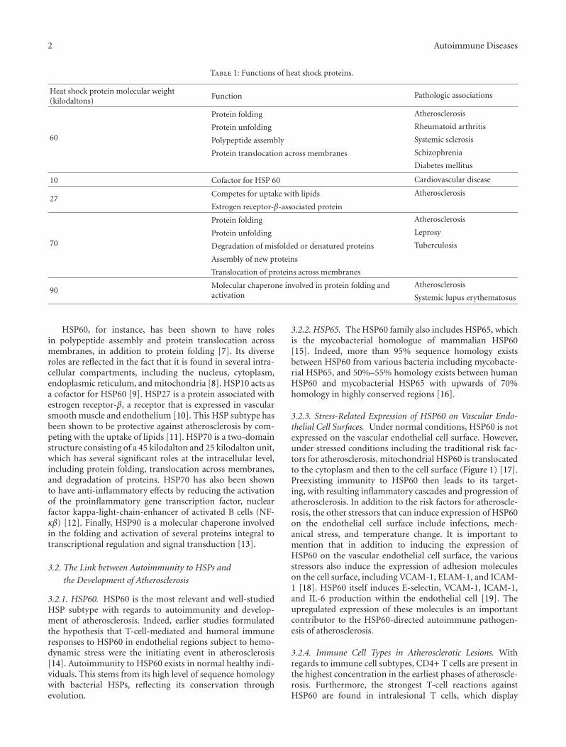
Table 1: Functions of heat shock proteins.
Heat shock protein molecular weight(kilodaltons)
Function Pathologic associations
60
Protein folding Atherosclerosis
Protein unfolding Rheumatoid arthritis
Polypeptide assembly Systemic sclerosis
Protein translocation across membranes Schizophrenia
Diabetes mellitus
10 Cofactor for HSP 60 Cardiovascular disease
27Competes for uptake with lipids Atherosclerosis
Estrogen receptor-β-associated protein
70
Protein folding Atherosclerosis
Protein unfolding Leprosy
Degradation of misfolded or denatured proteins Tuberculosis
Assembly of new proteins
Translocation of proteins across membranes
90Molecular chaperone involved in protein folding andactivation
Atherosclerosis
Systemic lupus erythematosus
HSP60, for instance, has been shown to have rolesin polypeptide assembly and protein translocation acrossmembranes, in addition to protein folding [7]. Its diverseroles are reflected in the fact that it is found in several intra-cellular compartments, including the nucleus, cytoplasm,endoplasmic reticulum, and mitochondria [8]. HSP10 acts asa cofactor for HSP60 [9]. HSP27 is a protein associated withestrogen receptor-β, a receptor that is expressed in vascularsmooth muscle and endothelium [10]. This HSP subtype hasbeen shown to be protective against atherosclerosis by com-peting with the uptake of lipids [11]. HSP70 is a two-domainstructure consisting of a 45 kilodalton and 25 kilodalton unit,which has several significant roles at the intracellular level,including protein folding, translocation across membranes,and degradation of proteins. HSP70 has also been shownto have anti-inflammatory effects by reducing the activationof the proinflammatory gene transcription factor, nuclearfactor kappa-light-chain-enhancer of activated B cells (NF-κβ) [12]. Finally, HSP90 is a molecular chaperone involvedin the folding and activation of several proteins integral totranscriptional regulation and signal transduction [13].
3.2. The Link between Autoimmunity to HSPs and
the Development of Atherosclerosis
3.2.1. HSP60. HSP60 is the most relevant and well-studiedHSP subtype with regards to autoimmunity and develop-ment of atherosclerosis. Indeed, earlier studies formulatedthe hypothesis that T-cell-mediated and humoral immuneresponses to HSP60 in endothelial regions subject to hemo-dynamic stress were the initiating event in atherosclerosis[14]. Autoimmunity to HSP60 exists in normal healthy indi-viduals. This stems from its high level of sequence homologywith bacterial HSPs, reflecting its conservation throughevolution.
3.2.2. HSP65. The HSP60 family also includes HSP65, whichis the mycobacterial homologue of mammalian HSP60[15]. Indeed, more than 95% sequence homology existsbetween HSP60 from various bacteria including mycobacte-rial HSP65, and 50%–55% homology exists between humanHSP60 and mycobacterial HSP65 with upwards of 70%homology in highly conserved regions [16].
3.2.3. Stress-Related Expression of HSP60 on Vascular Endo-thelial Cell Surfaces. Under normal conditions, HSP60 is notexpressed on the vascular endothelial cell surface. However,under stressed conditions including the traditional risk fac-tors for atherosclerosis, mitochondrial HSP60 is translocatedto the cytoplasm and then to the cell surface (Figure 1) [17].Preexisting immunity to HSP60 then leads to its target-ing, with resulting inflammatory cascades and progression ofatherosclerosis. In addition to the risk factors for atheroscle-rosis, the other stressors that can induce expression of HSP60on the endothelial cell surface include infections, mech-anical stress, and temperature change. It is important tomention that in addition to inducing the expression ofHSP60 on the vascular endothelial cell surface, the variousstressors also induce the expression of adhesion moleculeson the cell surface, including VCAM-1, ELAM-1, and ICAM-1 [18]. HSP60 itself induces E-selectin, VCAM-1, ICAM-1,and IL-6 production within the endothelial cell [19]. Theupregulated expression of these molecules is an importantcontributor to the HSP60-directed autoimmune pathogen-esis of atherosclerosis.
3.2.4. Immune Cell Types in Atherosclerotic Lesions. Withregards to immune cell subtypes, CD4+ T cells are present inthe highest concentration in the earliest phases of atheroscle-rosis. Furthermore, the strongest T-cell reactions againstHSP60 are found in intralesional T cells, which display
Autoimmune Diseases 3
Normal condition
Stressors
Stressed condition
Inflammation /atherosclerosis
Vascular endothelial cell
Heat shock protein 60
Adhesion molecules
Macrophages
T lymphocyte
Figure 1: Concept of autoimmunity towards heat shock protein 60and the development of atherosclerosis. Under normal conditions,heat shock protein 60 is located intracellularly and is not expressedon the vascular endothelial cell surface. Under stressed conditions,heat shock protein 60 and various adhesion molecules are upregu-lated and expressed on the cell surface. This leads to inflammationand the development of atherosclerosis.
an oligoclonally restricted receptor phenotype, as comparedto extralesional peripheral T cells which have weaker reac-tions to HSP60 and display polyclonal phenotypes [20]. Astudy of young clinically healthy males found that T-cellreactivity against HSP60 was an independent risk factor forearly intima-media thickening (Table 2) [21].
The role of B cells in the development of atherosclerosisis less clearly understood. Several studies have demonstratedprogression of atherosclerotic disease with B-cell depletion,whereas other studies demonstrated reduction in the diseasewith B-cell depletion [22–24]. These differing results may bedue to the presence of both atherogenesis-promoting as wellas atherogenesis-inhibiting antibodies/mediators. Similar tothe different roles of these antibodies, other inflammatorymediators have also been shown to either promote or inhibitatherosclerosis. IL-1β, IL-8, IL-12, IL-18, MCP-1, leukotrieneP4, and IFN-γ have been demonstrated as proatherogenicmediators whereas IL-4, IL-10, PDGF-β, and TGF-β havebeen shown to be antiatherogenic [25].
Table 2: Logistic regression analysis for the impact of various riskfactors on high vascular intima-media thickness in a study of 141young (17- or 18-year old) white males (see [21]).
Risk factor Odds ratio (95% CI) P value
Cigarette smoking 3.58 (1.34–9.54) 0.0108
High-density lipoprotein level 0.56 (0.36–0.89) 0.0144
Alcohol consumption 0.51 (0.30–0.87) 0.0133
Diastolic blood pressure 1.61 (1.03–2.52) 0.0374
Maximum expiratory flow at50% vital capacity
0.52 (0.33–0.82) 0.0047
HSP60 stimulation index 2.18 (1.32–3.60) 0.0023
HSP60 antibody titer 1.52 (1.00–2.31) 0.0514
Odds ratios were calculated based on a 1 standard deviation unit change inthe given variable.
3.2.5. Soluble HSP60. In addition to being expressed on theendothelial cell surface, HSP60 can be shed into the circu-lation in a soluble form under stressed conditions. A study of826 human patients found that levels of soluble HSP60 weresignificantly elevated in patients with carotid atherosclerosis[26]. The authors postulated that the release of HSP60 fromcells may be mediated by infectious agents. More specifically,chlamydiae are known to exhibit both nonlytic and lyticinfective phases, and during the latter, the human host cellreleases both its own HSP60 as well as chlamydial HSP60.This postulate is supported by evidence that both HSP60subtypes exist in high concentrations in atheroscleroticlesions, and that soluble HSP60 levels correlate with anti-Chlamydial antibody titers [27]. More recent studies havealso shown the association between elevated levels of solubleHSP subtypes and various cardiovascular diseases (Table 3)[15, 28–32].
3.2.6. Serum Antibodies to HSP60 and HSP65. Similar tosoluble HSP60, prior studies have also demonstrated elevatedserum antibody levels to HSP65, which is the mycobacterialhomolog of human HSP60 [33]. A followup study alsoshowed that anti-HSP65 antibodies remained consistentlyelevated over several years in humans with progressiveatherosclerosis [34]. Furthermore, levels of anti-HSP65 anti-bodies correlated strongly with antibody titers to Chlamydiapneumoniae and Helicobacter pylori, suggesting an infectiousrole [35]. Coronary events were observed more frequently inpatients that had high HSP60 IgA levels coupled with hightiters of antibodies to Chlamydia pneumoniae and high C-reactive protein levels [36]. However, a polymerase chainreaction study of 40 atherosclerotic human patients and20 nonatherosclerotic human controls found similar detec-tion rates of Chlamydia pneumoniae, Mycoplasma pneumo-niae, Helicobacter pylori, herpes simplex virus, and cyto-megalovirus in the aortic wall, which does not support theinfectious etiology hypothesis for atherosclerosis [37].
3.2.7. Establishing Causality in HSP60/65 Antibody-MediatedAtherosclerosis. Although soluble HSP60 and antibodies toHSP60/65 have been shown to be elevated in human patients

4 Autoimmune Diseases
Table 3: Soluble heat shock proteins and their association with cardiovascular diseases.
Soluble heat shockprotein subtype
Number ofpatients
Cardiovasculardisease
Study finding Reference
HSP70 24Acute myocardial
infarctionSoluble HSP70 is released into the circulation after anacute myocardial infarction
[28]
HSP60 684Carotid
atherosclerosisLevels of soluble HSP60 are associated with earlycarotid atherosclerosis
[29]
HSP7052 cases
20 controlsAcute myocardial
infarctionLevels of soluble HSP70 are associated with progressionof heart failure after acute myocardial infarction
[30]
HSP60, HSP7288 cases
44 controls
Idiopathic leftventriculardysfunction
Levels of soluble HSP60 and HSP72 correlate withseverity of cardiac and microvascular dysfunction inpatients with idiopathic left ventricular dysfunction
[15]
HSP70 167Congestive heart
failureLevels of soluble HSP70 are associated with severity ofheart failure in patients with congestive heart failure
[31]
HSP601003 cases
1003 controlsCoronary artery
diseaseLevels of soluble HSP60 correlate with the presence ofcoronary artery disease
[32]
with atherosclerosis, it is unclear whether this simply repre-sents an association or whether a causal relationship exists.Administration of a murine monoclonal antibody (II-13) toamino acid residues 288 to 366 of HSP60 induced atheroscle-rosis in apolipoprotein E-deficient mice [38]. II-13 injectionresulted in endothelial cell damage, leukocyte attachment,and accumulation of macrophages and smooth muscle cellsin lesions. The same study demonstrated that isolating anti-HSP60 antibodies from humans with coronary atherosclero-sis and injecting them into apolipoprotein E-deficient micecaused significant increases in aortic atherosclerotic lesions[38]. Passive transfer of T cells from mice immunized withmycobacterial HSP65 to nonimmunized mice led to thedevelopment of atherosclerosis in the nonimmunized cohort[39].
A study of 120 normocholesterolemic rabbits found thatthose immunized with recombinant mycobacterial HSP65had increased atherosclerosis [40]. In the rabbits that werefed a cholesterol-rich diet in addition to being immunizedwith HSP65, the atherosclerotic lesions were even moresevere. A followup investigation by the same group demon-strated that the early atherosclerotic lesions induced byHSP65 could be inhibited by T-cell depletion using an anti-CD3 monoclonal antibody [41].
3.2.8. HSP10. HSP10 is an important cofactor for HSP60[9]. The significant interplay between these HSP subtypes isfurther evidenced by the fact that their genes are localizedin a head-to-head manner on chromosome 2, separated by abidirectional promoter [42]. Similar to HSP60, the overex-pression of HSP10 is met with an overexpression of BcL-2 and Bcl-xL [43]. These molecules protect vascular endo-thelial cells from TNF-mediated apoptosis in addition toinhibiting activation of NF-κβ and thereby inhibiting theupregulation of proinflammatory genes [44]. The antiapop-totic roles of HSP10 are evidenced by the fact that trans-fecting doxorubicin-treated cardiomyocytes with HSP10 andHSP60 by an adenoviral vector suppresses apoptosis andresulting cardiomyopathy [43]. A study of antibodies toHSP10 of Chlamydia pneumoniae in patients with coronary
artery disease failed to demonstrate significant differencesin levels versus controls; however, the importance of HSP10to the development of atherosclerosis may indeed lie in itsgenetic and physiologic link to HSP60 [45].
3.2.9. HSP27. Emerging data has implicated HSP27 in thepathogenesis of atherosclerosis. A study of human athero-sclerotic plaques revealed an increase in expression of HSP27in normal-appearing vessel adjacent to the plaque, withdecreased levels in the plaque itself [46]. HSP27 phospho-rylation was decreased in both plaque and adjacent vesselcompared to reference vessel. And finally, when the inves-tigators examined HSP27 levels in plasma, they found thatin patients with acute coronary syndrome, levels of HSP27were increased and found to correlate with levels of HSP70,C-reactive protein, and CD40L [46].
Another study similarly found that HSP27 release wassignificantly decreased in atherosclerotic plaques [47]. Cir-culating levels of soluble HSP27 were also significantlydecreased in patients with carotid stenosis compared withhealthy controls [47]. A study of 22 heart transplant recip-ients found that those with cardiac allograft vasculopathyhad significantly reduced levels of phosphorylated HSP27in biopsy samples as compared to those recipients withoutvasculopathy [48]. The decreased expression of HSP27within plaques may be related to its degradation by enhancedproteolytic pathways which are known to be important con-tributors to vascular remodeling [49].
HSP27 may also offer protection from atherosclerosis dueto its role in plaque stability. A proteomic analysis of stableversus unstable human carotid artery atherosclerotic plaquesfound reduced levels of HSP27 in unstable lesions [50].Moreover, at the molecular level, phosphorylated HSP27 isknown to be a regulator of actin filament dynamics [51]. Fur-thermore, HSP27 may modulate the effects of plasmin andother extracellular mediators of apoptosis in vascular smoothmuscle cells, a process which has been shown to lead toplaque instability through the weakening of the fibrous capof the atheroma with potential plaque rupture and resultantatherothrombosis [52].
Autoimmune Diseases 5
In addition, HSP27 has been demonstrated in vitroto be released into the extracellular space in response tovarious stimuli, including estrogen or acetylated low-densitylipoprotein, where it binds the scavenger receptor A to pre-vent low-density lipoprotein uptake and foam cell formation[53]. HSP27, which is an estrogen receptor-β associated pro-tein, also modulates estrogen signaling and may have addi-tional atheroprotective functions via this mechanism [54].Increased estrogen receptor-β expression has indeed beennoted in both males as well as pre- and postmenopausalfemales with atherosclerosis [55, 56].
In an apolipoprotein E-deficient animal model, overex-pression of human HSP27 resulted in a 35% reduction inaortic atherosclerosis in female, but not male, mice [53].Serum levels of HSP27 were over tenfold higher in femalesas compared to males, again using the model of HSP27-overexpressing apolipoprotein-E deficient mice. CirculatingHSP27 levels demonstrated a strong inverse correlation withatherosclerotic lesion area in both female and male mice [53].
3.2.10. HSP70. In early atherosclerosis, dendritic cells exclu-sively overexpress HSP70 as well as HLA-DR and CD1d, thelatter being a unique molecule used in lipid antigen pre-sentation [57]. These HSP70-expressing dendritic cells alsofrequently interact with T cells within the arterial wall andtherefore may be responsible for presenting lipid antigensto them. Unlike the early stages, several immune cell types,including macrophages, smooth muscle cells, monocytes,and dendritic cells, have been shown to overexpress HSP70 inadvanced atherosclerosis [57]. Furthermore, a gene expres-sion profiling analysis revealed that two HSP70 family mem-bers were expressed within aortic atherosclerotic lesions butnot within nonlesional tissue [58].
Another study found that oxidized low-density lipopro-tein stimulated the expression of HSP70 and that superna-tants from oxidized low-density lipoprotein-treated macro-phages could induce both IL-1β and IL-12 secretion in naıvemacrophages [59]. Furthermore, this latter effect on cytokineproduction was inhibited by inhibiting HSP70 transcriptionor secretion. Extracellular HSP70 could therefore be animportant inducer of cytokine expression and inflamma-tion.
HSP70 may also have anti-inflammatory roles. In oneimmunization study, a peptide sequence of myobacterialHSP70 was found to induce the production of IL-10 bypeptide-specific T cells, a phenomenon that was also seenwith T cells responsive to the whole HSP70 protein [60]. IL-10 is known to be a potent anti-inflammatory cytokine, andindeed, its production was found in the prior study to pre-vent arthritis. Another study found that HSP70 attenuatedNF-κβ activation and its associated proinflammatory geneupregulation [61]. The potentially protective roles of HSP70were further supported by a study of 421 blood samplesfrom human subjects which found that high levels of HSP70were associated with low risk of coronary artery disease[62]. Another study also found low plasma levels of HSP70in patients with atherosclerosis, with activated neutrophilsbeing a potential source for proteases involved in HSP70degradation [63].
HSP70 may also be implicated in the calcification ofblood vessels. HSP70 was found to enhance bone morpho-genetic protein-4-induced proliferation in endothelial cellsand to enhance bone morphogenetic protein-induced cal-cium deposition in vascular cells [64]. The same study foundthat HSP70 mediated the IL-6 procalcific effect on vascularcells. Levels of HSP70, bone morphogenetic protein-4, andIL-6 were all elevated within the aortic wall as well as theserum in a mouse model of atherosclerosis [64]. Antibodiesto HSP70 diminished this procalcific effect.
3.2.11. HSP90. A study of human carotid atherosclerosisdemonstrated overexpression of HSP90 in both plaque andserum as compared to healthy controls [65]. Moreover,plaque-derived and circulating T cells from patients withatherosclerosis proliferated in response to HSP90 whereascells from controls did not. Finally, HSP90-specific T cellsexpressed both proinflammatory and anti-inflammatorycytokines, implying a dichotomous role [65].
Another study of human atherosclerotic plaques foundthat the expression of HSP90 was associated with plaqueinstability in advanced lesions [66]. Inhibitors of HSP90also reduced atherosclerosis-related inflammation in theiranalysis. Another investigation by the same group found thatHSP90 inhibitors interfere with oxidative stress by reducingpro-oxidative factors in experimental atherosclerosis [67].
The potential anti-inflammatory therapeutic benefits ofHSP90 inhibitors have been demonstrated in other diseasesas well. In a mouse model of systemic lupus erythematosus,HSP90 was found to have a potential role in regulating T-celldifferentiation and activation, and its inhibition was asso-ciated with reduced inflammation [68]. In a murine sepsismodel, the administration of HSP90 inhibitors resulted inreduced systemic and pulmonary inflammatory markerscompared to controls as well as improved lung function andsurvival [69].
3.3. Clinical Implications
3.3.1. Screening, Diagnosis, and Prognosis. There are severalclinical implications related to HSPs and their role in athero-sclerosis. One potential clinical application would be toexploit the presence of HSP antibodies for screening at-risk patients to detect significant atherosclerosis. A study of750 human subjects demonstrated the correlation betweenHSP65 antibody titers and advanced carotid atheroscleroticlesions [29]. Another study similarly showed that anti-HSP65 antibody titers correlated strongly with severity ofcoronary atherosclerosis [70]. In both of these studies, thesefindings persisted after adjusting for potential confounderssuch as patient age and smoking history. In addition toidentifying atherosclerosis, there may be a role in identifyingpatients who have suffered from myocardial infarction.HSP70, for instance, was found to be rapidly released insignificant quantities following an acute myocardial infarc-tion in 24 patients, highlighting its potential as a marker formyocardial damage [28].
Screening patients based on titers could be a useful strat-egy for the detection of significant atherosclerosis, although
6 Autoimmune Diseases
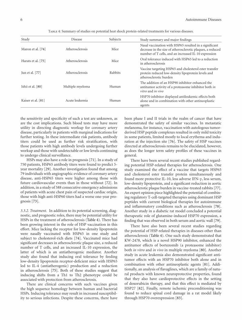
Table 4: Summary of studies on potential heat shock protein-related treatments for various diseases.
Study Disease Subjects Study summary and major findings
Maron et al. [74] Atherosclerosis MiceNasal vaccination with HSP65 resulted in a significantdecrease in the size of atherosclerotic plaques, a reducednumber of T cells, and an increased IL-10 expression
Harats et al. [75] Atherosclerosis Mice Oral tolerance induced with HSP65 led to a reductionin atherosclerosis
Jun et al. [77] Atherosclerosis RabbitsVaccine targeting HSP65 and cholesterol ester transferprotein reduced low-density lipoprotein levels andatherosclerotic burden
Ishii et al. [80] Multiple myeloma HumanThe addition of an HSP90 inhibitor enhanced theantitumor activity of a proteasome inhibitor both invitro and in vivo
Kaiser et al. [81] Acute leukemia HumanHSP70 inhibitor displayed antileukemic effects bothalone and in combination with other antineoplasticagents
the sensitivity and specificity of such a test are unknown, asare the cost implications. Such blood tests may have moreutility in directing diagnostic workup for coronary arterydisease, particularly in patients with marginal indications forfurther testing. In these intermediate risk patients, antibodytiters could be used as further risk stratification, withthose patients with high antibody levels undergoing furtherworkup and those with undetectable or low levels continuingto undergo clinical surveillance.
HSPs may also have a role in prognosis [71]. In a study of750 patients, HSP65 antibody titers were found to predict 5-year mortality [29]. Another investigation found that among79 individuals with angiographic evidence of coronary arterydisease, anti-HSP65 titers were higher among those withfuture cardiovascular events than in those without [72]. Inaddition, in a study of 588 consecutive emergency admissionsof patients with acute chest pain of suspected cardiac origin,those with high anti-HSP60 titers had a worse one-year pro-gnosis [73].
3.3.2. Treatment. In addition to its potential screening, diag-nostic, and prognostic roles, there may be potential utility forHSPs in the treatment of atherosclerosis (Table 4). There hasbeen growing interest in the role of HSP vaccination in thiseffort. Mice lacking the receptor for low-density lipoproteinwere nasally vaccinated with HSP65 in one study andsubject to cholesterol-rich diets [74]. Vaccinated mice hadsignificant decreases in atherosclerotic plaque size, a reducednumber of T cells, and an increased IL-10 expression, thelatter of which is an antiatherogenic mediator. Anotherstudy also found that inducing oral tolerance by feedinglow-density lipoprotein receptor-deficient mice with HSP65led to IL-4 (antiatherogenic) production and a reductionin atherosclerosis [75]. Both of these studies suggest thatinducing shifts from a Th1 to Th2 phenotype could beassociated with protection from atherosclerosis.
There are clinical concerns with such vaccines giventhe high sequence homology between human and bacterialHSPs. Inducing tolerance may result in increased susceptibil-ity to serious infections. Despite these concerns, there have
been phase I and II trials in the realm of cancer that havedemonstrated the safety of similar vaccines. In metastaticmelanoma, for instance, vaccination with autologous tumor-derived HSP peptide complexes resulted in only mild toxicityin some patients, limited mostly to local erythema and indu-ration at the injection site [76]. The safety of HSP vaccinesdirected at atherosclerosis remains to be elucidated, however,as does the longer term safety profiles of these vaccines ingeneral.
There have been several recent studies published regard-ing potential HSP-related therapies for atherosclerosis. Onestudy examined the effect of a vaccine that targets HSP65and cholesterol ester transfer protein simultaneously andfound more protective IL-10, less adverse IFN-γ, less serum,low-density lipoprotein, and a significant reduction in aorticatherosclerotic plaque burden in vaccine-treated rabbits [77].An expert opinion piece highlighted the potential of combin-ing regulatory T-cell-targeted therapies using dominant HSPpeptides with current biological therapies for autoimmuneand inflammatory conditions such as atherosclerosis [78].Another study in a diabetic rat model concluded a potentialtherapeutic role of glutamine-induced HSP70 expression, afinding that was observed in both serum and aortic wall [79].
There have also been several recent studies regardingthe potential of HSP-related therapies in diseases other thanatherosclerosis (Table 4). One such study demonstrated thatKW-2478, which is a novel HSP90 inhibitor, enhanced theantitumor effects of bortezomib (a proteasome inhibitor)both in vitro and in vivo in multiple myeloma [80]. Anotherstudy in acute leukemia also demonstrated significant anti-tumor effects with an HSP70 inhibitor both alone and incombination with other antineoplastic agents [81]. Addi-tionally, an analysis of flavaglines, which are a family of natu-ral products with known neuroprotective properties, foundthat they also have cardioprotective effects in the settingof doxorubicin therapy, and that this effect is mediated byHSP27 [82]. Finally, remote ischemic preconditioning wasfound to reduce spinal cord damage in a rat model likelythrough HSP70 overexpression [83].
Autoimmune Diseases 7
4. Conclusions
A growing body of evidence in both animal models andhuman subjects has implicated autoimmunity towards HSPsas a potential pathogenic mechanism for the developmentof atherosclerosis. Ongoing and future studies that furtherelucidate the mechanisms whereby HSPs, infection, andimmune response pathways interact and lead to the commonpathway of atherosclerosis will be essential to developingmore specific and potentially safer novel therapies for thisdevastating disease process. A better understanding of thefunctions of HSPs in other pathologies such as cancer mayalso be useful in advancing our knowledge of the role of thisimportant family of molecules in atherosclerosis and theirpotential therapeutic utility.
References
[1] R. Milkman, “Temperature effects on day old Drosophila pu-pae,” The Journal of General Physiology, vol. 45, pp. 777–799,1962.
[2] R. I. Morimoto, “Cells in stress: transcriptional activation ofheat shock genes,” Science, vol. 259, no. 5100, pp. 1409–1410,1993.
[3] J. Madrigal-Matute, J. L. Martin-Ventura, L. M. Blanco-Colio,J. Egido, J. B. Michel, and O. Meilhac, “Heat-shock proteinsin cardiovascular disease,” Advances in Clinical Chemistry, vol.54, pp. 1–43, 2011.
[4] Q. Xu, B. Metzler, M. Jahangiri, and K. Mandal, “Molecularchaperones and heat shock proteins in atherosclerosis,” Amer-ican Journal of Physiology, vol. 302, no. 3, pp. H506–H514,2012.
[5] A. Bielecka-Dabrowa, M. Barylski, D. P. Mikhailidis, J. Rysz,and M. Banach, “HSP 70 and atherosclerosis—protector oractivator?” Expert Opinion on Therapeutic Targets, vol. 13, no.3, pp. 307–317, 2009.
[6] Q. Xu, “Biomechanical-stress-induced signaling and geneexpression in the development of arteriosclerosis,” Trends inCardiovascular Medicine, vol. 10, no. 1, pp. 35–41, 2000.
[7] F. U. Hartl, “Heat shock proteins in protein folding and mem-brane translocation,” Seminars in Immunology, vol. 3, no. 1,pp. 5–16, 1991.
[8] P. Roma and A. L. Catapano, “Stress proteins and atheroscle-rosis,” Atherosclerosis, vol. 127, no. 2, pp. 147–154, 1996.
[9] K. L. Nielsen and N. J. Cowan, “A single ring is sufficient forproductive chaperonin-mediated folding in vivo,” MolecularCell, vol. 2, no. 1, pp. 93–99, 1998.
[10] M. R. Voss, J. N. Stallone, M. Li, R. N. M. Cornelussen, P.Knuefermann, and A. A. Knowlton, “Gender differences inthe expression of heat shock proteins: the effect of estrogen,”American Journal of Physiology, vol. 285, no. 2, pp. H687–H692, 2003.
[11] K. Rayner, Y. X. Chen, T. Siebert, and E. R. O’Brien, “Heatshock protein 27: clue to understanding estrogen-mediatedatheroprotection?” Trends in Cardiovascular Medicine, vol. 20,no. 2, pp. 54–58, 2010.
[12] M. Shimizu, M. Tamamori-Adachi, H. Arai, N. Tabuchi, H.Tanaka, and M. Sunamori, “Lipopolysaccharide pretreatmentattenuates myocardial infarct size: a possible mechanisminvolving heat shock protein 70-inhibitory κBα complex andattenuation of nuclear factor κB,” Journal of Thoracic andCardiovascular Surgery, vol. 124, no. 5, pp. 933–941, 2002.
[13] W. B. Pratt and D. O. Toft, “Regulation of signaling proteinfunction and trafficking by the hsp90/hsp70-based chaperonemachinery,” Experimental Biology and Medicine, vol. 228, no.2, pp. 111–133, 2003.
[14] G. Wick, G. Schett, A. Amberger, R. Kleindienst, and Q. Xu, “Isatherosclerosis an immunologically mediated disease?” Immu-nology Today, vol. 16, no. 1, pp. 27–33, 1995.
[15] D. Giannessi, C. Colotti, M. Maltinti et al., “Circulating heatshock proteins and inflammatory markers in patients withidiopathic left ventricular dysfunction: their relationships withmyocardial and microvascular impairment,” Cell Stress andChaperones, vol. 12, no. 3, pp. 265–274, 2007.
[16] R. A. Young and T. J. Elliott, “Stress proteins, infection, andimmune surveillance,” Cell, vol. 59, no. 1, pp. 5–8, 1989.
[17] G. Wick, R. Kleindienst, G. Schett, A. Amberger, and Q. Xu,“Role of heat shock protein 65/60 in the pathogenesis of athe-rosclerosis,” International Archives of Allergy and Immunology,vol. 107, no. 1–3, pp. 130–131, 1995.
[18] A. Amberger, C. Maczek, G. Jurgens et al., “Co-expression ofICAM-1, VCAM-1, ELAM-1 and Hsp60 in human arterial andvenous endothelial cells in response to cytokines and oxidizedlow-density lipoproteins,” Cell Stress and Chaperones, vol. 2,no. 2, pp. 94–103, 1997.
[19] A. Kol, T. Bourcier, A. H. Lichtman, and P. Libby, “Chlamydialand human heat shock protein 60s activate human vascularendothelium, smooth muscle cells, and macrophages,” TheJournal of Clinical Investigation, vol. 103, no. 4, pp. 571–577,1999.
[20] A. Rossmann, B. Henderson, B. Heidecker et al., “T-cells fromadvanced atherosclerotic lesions recognize hHSP60 and havea restricted T-cell receptor repertoire,” Experimental Geronto-logy, vol. 43, no. 3, pp. 229–237, 2008.
[21] M. Knoflach, S. Kiechl, M. Kind et al., “Cardiovascular riskfactors and atherosclerosis in young males: ARMY study(atherosclerosis risk-factors in male youngsters),” Circulation,vol. 108, no. 9, pp. 1064–1069, 2003.
[22] A. S. Major, S. Fazio, and M. F. Linton, “B-lymphocyte defi-ciency increases atherosclerosis in LDL receptor-null mice,”Arteriosclerosis, Thrombosis, and Vascular Biology, vol. 22, no.11, pp. 1892–1898, 2002.
[23] G. Caligiuri, A. Nicoletti, B. Poirierand, and G. K. Hansson,“Protective immunity against atherosclerosis carried by B cellsof hypercholesterolemic mice,” The Journal of Clinical Invest-igation, vol. 109, no. 6, pp. 745–753, 2002.
[24] T. Kyaw, C. Tay, A. Khan et al., “Conventional B2 B cell deple-tion ameliorates whereas its adoptive transfer aggravates athe-rosclerosis,” The Journal of Immunology, vol. 185, no. 7, pp.4410–4419, 2010.
[25] D. R. Greaves and K. M. Channon, “Inflammation and im-mune responses in atherosclerosis,” Trends in Immunology, vol.23, no. 11, pp. 535–541, 2002.
[26] Q. Xu, G. Schett, H. Perschinka et al., “Serum soluble heatshock protein 60 is elevated in subjects with atherosclerosis ina general population,” Circulation, vol. 102, no. 1, pp. 14–20,2000.
[27] A. Kol, G. K. Sukhova, A. H. Lichtman, and P. Libby, “Chlamy-dial heat shock protein 60 localizes in human atheroma andregulates macrophage tumor necrosis factor-α and matrixmetalloproteinase expression,” Circulation, vol. 98, no. 4, pp.300–307, 1998.
[28] B. Dybdahl, S. A. Slørdahl, A. Waage, P. Kierulf, T. Espevik,and A. Sundan, “Myocardial ischaemia and the inflammatoryresponse: release of heat shock protein 70 after myocardialinfarction,” Heart, vol. 91, no. 3, pp. 299–304, 2005.

8 Autoimmune Diseases
[29] Q. Xiao, K. Mandal, G. Schett et al., “Association of serum-soluble heat shock protein 60 with carotid atherosclerosis:clinical significance determined in a follow-up study,” Stroke,vol. 36, no. 12, pp. 2571–2576, 2005.
[30] M. Satoh, Y. Shimoda, T. Akatsu, Y. Ishikawa, Y. Minami, andM. Nakamura, “Elevated circulating levels of heat shock pro-tein 70 are related to systemic inflammatory reaction throughmonocyte Toll signal in patients with heart failure after acutemyocardial infarction,” European Journal of Heart Failure, vol.8, no. 8, pp. 810–815, 2006.
[31] T. Gombos, Z. Forhecz, Z. Pozsonyi, L. Janoskuti, and Z.Prohaszka, “Interaction of serum 70-kDa heat shock proteinlevels and HspA1B (+1267) gene polymorphism with diseaseseverity in patients with chronic heart failure,” Cell Stress andChaperones, vol. 13, no. 2, pp. 199–206, 2008.
[32] X. Zhang, M. He, L. Cheng et al., “Elevated heat shock protein60 levels are associated with higher risk of coronary heartdisease in Chinese,” Circulation, vol. 118, no. 25, pp. 2687–2693, 2008.
[33] Q. Xu, J. Willeit, M. Marosi et al., “Association of serum anti-bodies to heat-shock protein 65 with carotid atherosclerosis,”The Lancet, vol. 341, no. 8840, pp. 255–259, 1993.
[34] Q. Xu, S. Kiechl, M. Mayr et al., “Association of serum anti-bodies to heat-shock protein 65 with carotid atherosclerosis:clinical significance determined in a follow-up study,” Circula-tion, vol. 100, no. 11, pp. 1169–1174, 1999.
[35] M. Mayr, S. Kiechl, J. Willeit, G. Wick, and Q. Xu, “Infec-tions, immunity, and atherosclerosis: associations of antibod-ies to Chlamydia pneumoniae, Helicobacter pylori, and cyto-megalovirus with immune reactions to heat-shock protein 60and carotid or femoral atherosclerosis,” Circulation, vol. 102,no. 8, pp. 833–839, 2000.
[36] T. Huittinen, M. Leinonen, L. Tenkanen et al., “Autoimmunityto human heat shock protein 60, Chlamydia pneumoniae infec-tion, and inflammation in predicting coronary risk,” Arte-riosclerosis, Thrombosis, and Vascular Biology, vol. 22, no. 3, pp.431–437, 2002.
[37] E. Reszka, B. Jegier, W. Wasowicz, M. Lelonek, M. Banach, andR. Jaszewski, “Detection of infectious agents by polymerasechain reaction in human aortic wall,” Cardiovascular Pathol-ogy, vol. 17, no. 5, pp. 297–302, 2008.
[38] G. Foteinos, A. R. Afzal, K. Mandal, M. Jahangiri, and Q. Xu,“Anti-heat shock protein 60 autoantibodies induce atheroscle-rosis in apolipoprotein E-deficient mice via endothelial dam-age,” Circulation, vol. 112, no. 8, pp. 1206–1213, 2005.
[39] J. George, A. Afek, B. Gilburd, Y. Shoenfeld, and D. Harats,“Cellular and humoral immune responses to heat shock pro-tein 65 are both involved in promoting fatty-streak formationin LDL-receptor deficient mice,” Journal of the AmericanCollege of Cardiology, vol. 38, no. 3, pp. 900–905, 2001.
[40] Q. Xu, H. Dietrich, H. J. Steiner et al., “Induction of arte-riosclerosis in normocholesterolemic rabbits by immunizationwith heat shock protein 65,” Arteriosclerosis and Thrombosis,vol. 12, no. 7, pp. 789–799, 1992.
[41] B. Metzler, M. Mayr, H. Dietrich et al., “Inhibition of arterio-sclerosis by T-cell depletion in normocholesterolemic rabbitsimmunized with heat shock protein 65,” Arteriosclerosis, Thro-mbosis, and Vascular Biology, vol. 19, no. 8, pp. 1905–1911,1999.
[42] J. J. Hansen, P. Bross, M. Westergaard et al., “Genomic struc-ture of the human mitochondrial chaperonin genes: HSP60and HSP10 are localised head to head on chromosome 2separated by a bidirectional promoter,” Human Genetics, vol.112, no. 1, pp. 71–77, 2003.
[43] Y. X. Shan, T. J. Liu, H. F. Su, A. Samsamshariat, R. Mestril,and P. H. Wang, “Hsp10 and Hsp60 modulate Bcl-2 familyand mitochondria apoptosis signaling induced by doxorubicinin cardiac muscle cells,” Journal of Molecular and CellularCardiology, vol. 35, no. 9, pp. 1135–1143, 2003.
[44] A. Z. Badrichani, D. M. Stroka, G. Bilbao, D. T. Curiel, F. H.Bach, and C. Ferran, “Bcl-2 and Bcl-X(L) serve an anti-inflam-matory function in endothelial cells through inhibition of NF-κB,” The Journal of Clinical Investigation, vol. 103, no. 4, pp.543–553, 1999.
[45] A. Ciervo, A. Petrucca, U. Villano, G. Fioroni, and A. Cassone,“Low prevalence of antibodies against heat shock protein 10of Chlamydophila pneumoniae in patients with coronary heartdisease,” Journal of Microbiological Methods, vol. 63, no. 3, pp.248–253, 2005.
[46] H. K. Park, E. C. Park, S. W. Bae et al., “Expression ofheat shock protein 27 in human atherosclerotic plaques andincreased plasma level of heat shock protein 27 in patientswith acute coronary syndrome,” Circulation, vol. 114, no. 9,pp. 886–893, 2006.
[47] J. L. Martin-Ventura, M. C. Duran, L. M. Blanco-Colio et al.,“Identification by a differential proteomic approach of heatshock protein 27 as a potential marker of atherosclerosis,” Cir-culation, vol. 110, no. 15, pp. 2216–2219, 2004.
[48] A. I. De Souza, R. Wait, A. G. Mitchell, N. R. Banner, M. J.Dunn, and M. L. Rose, “Heat shock protein 27 is associatedwith freedom from graft vasculopathy after human cardiactransplantation,” Circulation Research, vol. 97, no. 2, pp. 192–198, 2005.
[49] H. R. Lijnen, “Plasmin and matrix metalloproteinases in vas-cular remodeling,” Thrombosis and Haemostasis, vol. 86, no. 1,pp. 324–333, 2001.
[50] A. J. Lepedda, A. Cigliano, G. M. Cherchi et al., “A proteomicapproach to differentiate histologically classified stable andunstable plaques from human carotid arteries,” Atherosclerosis,vol. 203, no. 1, pp. 112–118, 2009.
[51] J. Guay, H. Lambert, G. Gingras-Breton, J. N. Lavoie, J. Huot,and J. Landry, “Regulation of actin filament dynamics by p38map kinase-mediated phosphorylation of heat shock protein27,” Journal of Cell Science, vol. 110, no. 3, pp. 357–368, 1997.
[52] J. L. Martin-Ventura, V. Nicolas, X. Houard et al., “Biologicalsignificance of decreased HSP27 in human atherosclerosis,”Arteriosclerosis, Thrombosis, and Vascular Biology, vol. 26, no.6, pp. 1337–1343, 2006.
[53] K. Rayner, Y. X. Chen, M. McNulty et al., “Extracellular releaseof the atheroprotective heat shock protein 27 is mediatedby estrogen and competitively inhibits acLDL binding toscavenger receptor-a,” Circulation Research, vol. 103, no. 2, pp.133–141, 2008.
[54] H. Miller, S. Poon, B. Hibbert, K. Rayner, Y. X. Chen, and E.R. O’Brien, “Modulation of estrogen signaling by the novelinteraction of heat shock protein 27, a biomarker for athero-sclerosis, and estrogen receptor beta: mechanistic insight intothe vascular effects of estrogens,” Arteriosclerosis, Thrombosis,and Vascular Biology, vol. 25, no. 3, pp. e10–e14, 2005.
[55] P. Y. Liu, R. C. Christian, M. Ruan, V. M. Miller, and L. A. Fitz-patrick, “Correlating androgen and estrogen steroid receptorexpression with coronary calcification and atherosclerosis inmen without known coronary artery disease,” Journal of Clin-ical Endocrinology and Metabolism, vol. 90, no. 2, pp. 1041–1046, 2005.
[56] R. C. Christian, P. Y. Liu, S. Harrington, M. Ruan, V. M. Miller,and L. A. Fitzpatrick, “Intimal estrogen Receptor (ER)β, butNot ERα expression, is correlated with coronary calcification
Autoimmune Diseases 9
and atherosclerosis in pre- and postmenopausal women,” Jour-nal of Clinical Endocrinology and Metabolism, vol. 91, no. 7, pp.2713–2720, 2006.
[57] Y. V. Bobryshev and R. S. A. Lord, “Expression of heat shockprotein-70 by dendritic cells in the arterial intima and itspotential significance in atherogenesis,” Journal of VascularSurgery, vol. 35, no. 2, pp. 368–375, 2002.
[58] Z. Han, Q. A. Truong, S. Park, and J. L. Breslow, “Two Hsp70family members expressed in atherosclerotic lesions,” Pro-ceedings of the National Academy of Sciences of the United Statesof America, vol. 100, no. 3, pp. 1256–1261, 2003.
[59] P. A. Svensson, A. Asea, M. C. O. Englund et al., “Major roleof HSP70 as a paracrine inducer of cytokine production inhuman oxidized LDL treated macrophages,” Atherosclerosis,vol. 185, no. 1, pp. 32–38, 2006.
[60] U. Wendling, L. Paul, R. Van Der Zee, B. Prakken, M. Singh,and W. Van Eden, “A conserved mycobacterial heat shock pro-tein (hsp) 70 sequence prevents adjuvant arthritis upon nasaladministration and induces IL-10-producing T cells thatcross-react with the mammalian self-hsp70 homologue,” TheJournal of Immunology, vol. 164, no. 5, pp. 2711–2717, 2000.
[61] M. Shimizu, M. Tamamori-Adachi, H. Arai, N. Tabuchi, H.Tanaka, and M. Sunamori, “Lipopolysaccharide pretreatmentattenuates myocardial infarct size: a possible mechanisminvolving heat shock protein 70-inhibitory κBα complex andattenuation of nuclear factor κB,” Journal of Thoracic andCardiovascular Surgery, vol. 124, no. 5, pp. 933–941, 2002.
[62] J. Zhu, A. A. Quyyumi, H. Wu et al., “Increased serum levelsof heat shock protein 70 are associated with low risk of coro-nary artery disease,” Arteriosclerosis, Thrombosis, and VascularBiology, vol. 23, no. 6, pp. 1055–1059, 2003.
[63] J. L. Martin-Ventura, A. Leclercq, L. M. Blanco-Colio et al.,“Low plasma levels of HSP70 in patients with carotid athero-sclerosis are associated with increased levels of proteolyticmarkers of neutrophil activation,” Atherosclerosis, vol. 194, no.2, pp. 334–341, 2007.
[64] Y. Yao, A. D. Watson, S. Ji, and K. I. Bostrom, “Heat shock pro-tein 70 enhances vascular bone morphogenetic protein-4 sig-naling by binding Matrix Gla protein,” Circulation Research,vol. 105, no. 6, pp. 575–584, 2009.
[65] R. Businaro, E. Profumo, A. Tagliani et al., “Heat-shock pro-tein 90: a novel autoantigen in human carotid atherosclerosis,”Atherosclerosis, vol. 207, no. 1, pp. 74–83, 2009.
[66] J. Madrigal-Matute, O. Lopez-Franco, L. M. Blanco-Colio etal., “Heat shock protein 90 inhibitors attenuate inflammatoryresponses in atherosclerosis,” Cardiovascular Research, vol. 86,no. 2, pp. 330–337, 2010.
[67] J. Madrigal-Matute, C. E. Fernandez-Garcia, C. Gomez-Guerrero et al., “HSP90 inhibition by 17-DMAG attenuatesoxidative stress in experimental atherosclerosis,” Cardiovascu-lar Research, vol. 95, no. 1, pp. 116–123, 2012.
[68] S. K. Shimp III, C. B. Chafin, N. L. Regna et al., “Heatshock protein 90 inhibition by 17-DMAG lessens disease inthe MRL/lpr mouse model of systemic lupus erythematosus,”Cellular and Molecular Immunology, vol. 9, no. 3, pp. 255–266,2012.
[69] A. Chatterjee, C. Dimitropoulou, F. Drakopanayiotakis et al.,“Heat shock protein 90 inhibitors prolong survival, attenuateinflammation, and reduce lung injury in murine sepsis,”American Journal of Respiratory and Critical Care Medicine,vol. 176, no. 7, pp. 667–675, 2007.
[70] D. H. Birnie, E. R. Holme, I. C. McKay, S. Hood, K. E. L.McColl, and W. S. Hillis, “Association between antibodies toheat shock protein 65 and coronary atherosclerosis. Possible
mechanism of action of Helicobacter pylori and other bacterialinfections in increasing cardiovascular risk,” European HeartJournal, vol. 19, no. 3, pp. 387–394, 1998.
[71] M. Barylski, J. Małyszko, J. Rysz, M. Mysliwiec, and M.Banach, “Lipids, blood pressure, kidney—what was new in2011?” Archives of Medical Science, vol. 7, no. 6, pp. 1055–1066,2011.
[72] F. Hoppichler, T. Koch, A. Dzien, G. Gschwandtner, and M.Lechleitner, “Prognostic value of antibody titre to heat-shockprotein 65 on cardiovascular events,” Cardiology, vol. 94, no.4, pp. 220–223, 2000.
[73] D. H. Birnie, L. E. Vickers, W. S. Hillis, J. Norrie, and S. M.Cobbe, “Increased titres of anti-human heat shock protein 60predict an adverse one year prognosis in patients with acutecardiac chest pain,” Heart, vol. 91, no. 9, pp. 1148–1153, 2005.
[74] R. Maron, G. Sukhova, A. M. Faria et al., “Mucosal admin-istration of heat shock protein-65 decreases atherosclerosisand inflammation in aortic arch of low-density lipoproteinreceptor-deficient mice,” Circulation, vol. 106, no. 13, pp.1708–1715, 2002.
[75] D. Harats, N. Yacov, B. Gilburd, Y. Shoenfeld, and J.George, “Oral tolerance with heat shock protein 65 attenu-ates Mycobacterium tuberculosis-induced and high-fat-diet-driven atherosclerotic lesions,” Journal of the American Collegeof Cardiology, vol. 40, no. 7, pp. 1333–1338, 2002.
[76] L. Pilla, R. Patuzzo, L. Rivoltini et al., “A phase II trial of vac-cination with autologous, tumor-derived heat-shock proteinpeptide complexes Gp96, in combination with GM-CSF andinterferon-α in metastatic melanoma patients,” Cancer Immu-nology, Immunotherapy, vol. 55, no. 8, pp. 958–968, 2006.
[77] L. Jun, L. Jie, Y. Dongping et al., “Effects of nasal immuniza-tion of multi-target preventive vaccines on atherosclerosis,”Vaccine, vol. 30, no. 6, pp. 1029–1037, 2012.
[78] C. Keijzer, L. Wieten, M. Van Herwijnen, R. Van Der Zee,W. Van Eden, and F. Broere, “Heat shock proteins are ther-apeutic targets in autoimmune diseases and other chronicinflammatory conditions,” Expert Opinion on TherapeuticTargets, vol. 16, no. 9, pp. 849–857, 2012.
[79] M. Ugurlucan, D. Erer, Y. Kalko et al., “Aortic stiffness indiabetes mellitus—association with glutamine and heat shockprotein 70 expression: a pilot study based on an experimentalrodent model,” Expert Opinion on Therapeutic Targets, vol. 13,no. 3, pp. 267–274, 2009.
[80] T. Ishii, T. Seike, T. Nakashima et al., “Anti-tumor activityagainst multiple myeloma by combination of KW-2478, anHsp90 inhibitor, with bortezomib,” Blood Cancer Journal, vol.2, no. 4, e68, 2012.
[81] M. Kaiser, A. Kuhnl, J. Reins et al., “Antileukemic activity ofthe HSP70 inhibitor pifithrin-μ in acute leukemia,” Blood Can-cer Journal, vol. 1, article e28, 2011.
[82] Y. Bernard, N. Ribeiro, F. Thuaud et al., “Flavaglines alleviatedoxorubicin cardiotoxicity: implication of Hsp27,” PLoS ONE,vol. 6, no. 10, Article ID e25302, 2011.
[83] O. Selimoglu, M. Ugurlucan, M. Basaran et al., “Efficacy ofremote ischaemic preconditioning for spinal cord protectionagainst ischaemic injury: association with heat shock proteinexpression,” Folia Neuropathologica, vol. 46, no. 3, pp. 204–212, 2008.

Submit your manuscripts athttp://www.hindawi.com
Stem CellsInternational
Hindawi Publishing Corporationhttp://www.hindawi.com Volume 2014
Hindawi Publishing Corporationhttp://www.hindawi.com Volume 2014
MEDIATORSINFLAMMATION
of
Hindawi Publishing Corporationhttp://www.hindawi.com Volume 2014
Behavioural Neurology
EndocrinologyInternational Journal of
Hindawi Publishing Corporationhttp://www.hindawi.com Volume 2014
Hindawi Publishing Corporationhttp://www.hindawi.com Volume 2014
Disease Markers
Hindawi Publishing Corporationhttp://www.hindawi.com Volume 2014
BioMed Research International
OncologyJournal of
Hindawi Publishing Corporationhttp://www.hindawi.com Volume 2014
Hindawi Publishing Corporationhttp://www.hindawi.com Volume 2014
Oxidative Medicine and Cellular Longevity
Hindawi Publishing Corporationhttp://www.hindawi.com Volume 2014
PPAR Research
The Scientific World JournalHindawi Publishing Corporation http://www.hindawi.com Volume 2014
Immunology ResearchHindawi Publishing Corporationhttp://www.hindawi.com Volume 2014
Journal of
ObesityJournal of
Hindawi Publishing Corporationhttp://www.hindawi.com Volume 2014
Hindawi Publishing Corporationhttp://www.hindawi.com Volume 2014
Computational and Mathematical Methods in Medicine
OphthalmologyJournal of
Hindawi Publishing Corporationhttp://www.hindawi.com Volume 2014
Diabetes ResearchJournal of
Hindawi Publishing Corporationhttp://www.hindawi.com Volume 2014
Hindawi Publishing Corporationhttp://www.hindawi.com Volume 2014
Research and TreatmentAIDS
Hindawi Publishing Corporationhttp://www.hindawi.com Volume 2014
Gastroenterology Research and Practice
Hindawi Publishing Corporationhttp://www.hindawi.com Volume 2014
Parkinson’s Disease
Evidence-Based Complementary and Alternative Medicine
Volume 2014Hindawi Publishing Corporationhttp://www.hindawi.com


![Research Articledownloads.hindawi.com/journals/geofluids/2019/9484657.pdf · cing the saltwater and reducing the groundwater salinity [9–11]. Due to density differences between](https://static.fdocuments.in/doc/165x107/604bf95e69928e7e884d2671/research-cing-the-saltwater-and-reducing-the-groundwater-salinity-9a11-due.jpg)


![Review Articledownloads.hindawi.com/journals/aorth/2012/218385.pdf · 2019. 7. 31. · spinal deformity are inappropriate candidates for Dynesys implantation [2]. Bordes-Monmeneu](https://static.fdocuments.in/doc/165x107/60d0d8c2f4ecc86fea7b81f5/review-2019-7-31-spinal-deformity-are-inappropriate-candidates-for-dynesys.jpg)

