Review Article Gastrointestinal absorption and biological ... · Review on absorption of serine and...
18
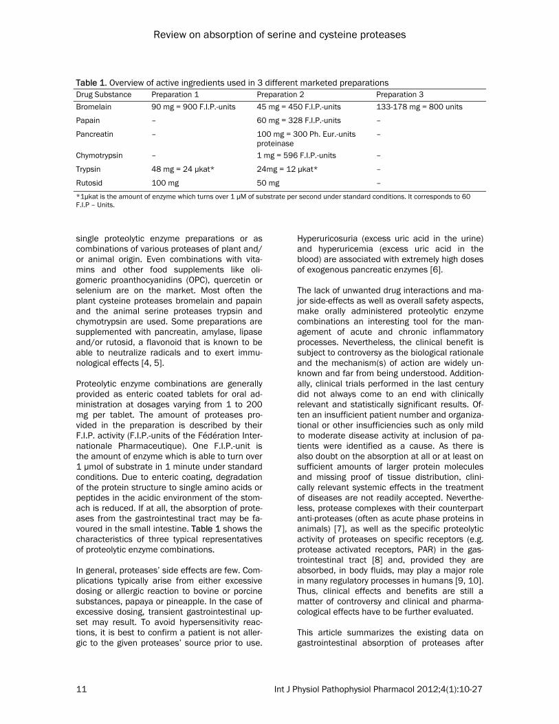
Introduction Single orally administered proteolytic enzymes of plant and animal origin are widely used as a medical treatment for a variety of digestive, ab- sorption and pancreatic disorders. Historically, porcine and bovine pancreatic enzymes have been the preferred form of supplementation for exocrine pancreatic insufficiency. Plant-based enzymes, such as bromelain from pineapple, also serve as effective digestive aids in the breakdown of proteins [1]. However, orally administered proteolytic enzyme combinations often supplemented with rutosid are widely used as an alternative or a supple- mentary treatment of different disease condi- tions such as acute and post surgical trauma, phlebitis, rheumatoid and osteoarthritis, as well as in an adjunctive therapy in cancer treatment [2, 3]. The terms proteolytic enzymes, proteases or proteinases are often synonymously used and the medical treatment is often described as systemic enzyme therapy, which implies a gas- trointestinal absorption for being active in the organism by distribution in body fluids. In the United States and in Europe various pro- teolytic enzyme preparations are available on the market. Their marketing status is different and varies from prescription drug to OTC or food supplementation. They are marketed either as Int J Physiol Pathophysiol Pharmacol 2012;4(1):10-27 www.ijppp.org /ISSN:1944-8171/IJPPP1112001 Review Article Gastrointestinal absorption and biological activities of serine and cysteine proteases of animal and plant origin: review on absorption of serine and cysteine proteases Gerhard Lorkowski GL Pharma Consulting Research & Development (GL Pharma CR&D), D-82131 Gauting/Munich, Germany Received December 19, 2011; accepted February 12, 2012; Epub February 28, 2012; Published March 15, 2012 Abstract: Research has confirmed that peptides and larger protein molecules pass through the mucosal barrier of the gastrointestinal tract. Orally administered serine and cysteine proteases of plant and animal origin also reach blood and lymph as intact, high molecular weight and physiologically active protein molecules. Their absorption may be supported by a self-enhanced paracellular transport mechanism resulting in sub-nanomolar concentration of tran- siently free protease molecules or, in a complex with anti-proteases, at higher concentrations. Data from pharmacoki- netic investigations reveals dose linearity for maximum plasma levels of free proteases not unusual for body prote- ases and a high inter-individual variability. There is no interference with each other after oral administration of prote- ase combinations, and absorption follows an unusual invasion and elimination kinetic due to slow velocity of absorp- tion and a fast 100% protein binding to anti-proteases. Oral application of proteases leads to increased proteolytic serum activity and increased plasma concentrations of the corresponding anti-proteases. Their biological activity is determined by their proteolytic activity as free proteases on soluble peptides/proteins or cell surface receptors (e.g. protease activated receptors) and their activity in the complex formed with their specific and/or unspecific anti- proteases. The anti-protease-complexes, during immune reaction and injuries often loaded with different cytokines, are cleared from body fluids and tissue by receptor mediated endocytosis on hepatocytes and/or blood cells. Oral administration of enteric coated tablets containing proteolytic enzymes of plant and animal origin may be a safe method to stabilize, positively influence or enhance physiological and immunological processes during disease proc- esses and in healthy consumers. Keywords: Gastrointestinal absorption, serine and cysteine protease, proteinase, proteolytic enzyme activity, anti- protease, protease activated receptors
Transcript of Review Article Gastrointestinal absorption and biological ... · Review on absorption of serine and...
Introduction Single orally administered proteolytic enzymes of plant and animal origin are widely used as a medical treatment for a variety of digestive, ab-sorption and pancreatic disorders. Historically, porcine and bovine pancreatic enzymes have been the preferred form of supplementation for exocrine pancreatic insufficiency. Plant-based enzymes, such as bromelain from pineapple, also serve as effective digestive aids in the breakdown of proteins [1]. However, orally administered proteolytic enzyme combinations often supplemented with rutosid are widely used as an alternative or a supple-
mentary treatment of different disease condi-tions such as acute and post surgical trauma, phlebitis, rheumatoid and osteoarthritis, as well as in an adjunctive therapy in cancer treatment[2, 3]. The terms proteolytic enzymes, proteases or proteinases are often synonymously used and the medical treatment is often described as systemic enzyme therapy, which implies a gas-trointestinal absorption for being active in the organism by distribution in body fluids. In the United States and in Europe various pro-teolytic enzyme preparations are available on the market. Their marketing status is different and varies from prescription drug to OTC or food supplementation. They are marketed either as
Int J Physiol Pathophysiol Pharmacol 2012;4(1):10-27 www.ijppp.org /ISSN:1944-8171/IJPPP1112001
Review Article Gastrointestinal absorption and biological activities of serine and cysteine proteases of animal and plant origin: review on absorption of serine and cysteine proteases Gerhard Lorkowski GL Pharma Consulting Research & Development (GL Pharma CR&D), D-82131 Gauting/Munich, Germany Received December 19, 2011; accepted February 12, 2012; Epub February 28, 2012; Published March 15, 2012 Abstract: Research has confirmed that peptides and larger protein molecules pass through the mucosal barrier of the gastrointestinal tract. Orally administered serine and cysteine proteases of plant and animal origin also reach blood and lymph as intact, high molecular weight and physiologically active protein molecules. Their absorption may be supported by a self-enhanced paracellular transport mechanism resulting in sub-nanomolar concentration of tran-siently free protease molecules or, in a complex with anti-proteases, at higher concentrations. Data from pharmacoki-netic investigations reveals dose linearity for maximum plasma levels of free proteases not unusual for body prote-ases and a high inter-individual variability. There is no interference with each other after oral administration of prote-ase combinations, and absorption follows an unusual invasion and elimination kinetic due to slow velocity of absorp-tion and a fast 100% protein binding to anti-proteases. Oral application of proteases leads to increased proteolytic serum activity and increased plasma concentrations of the corresponding anti-proteases. Their biological activity is determined by their proteolytic activity as free proteases on soluble peptides/proteins or cell surface receptors (e.g. protease activated receptors) and their activity in the complex formed with their specific and/or unspecific anti-proteases. The anti-protease-complexes, during immune reaction and injuries often loaded with different cytokines, are cleared from body fluids and tissue by receptor mediated endocytosis on hepatocytes and/or blood cells. Oral administration of enteric coated tablets containing proteolytic enzymes of plant and animal origin may be a safe method to stabilize, positively influence or enhance physiological and immunological processes during disease proc-esses and in healthy consumers. Keywords: Gastrointestinal absorption, serine and cysteine protease, proteinase, proteolytic enzyme activity, anti-protease, protease activated receptors
Review on absorption of serine and cysteine proteases
11 Int J Physiol Pathophysiol Pharmacol 2012;4(1):10-27
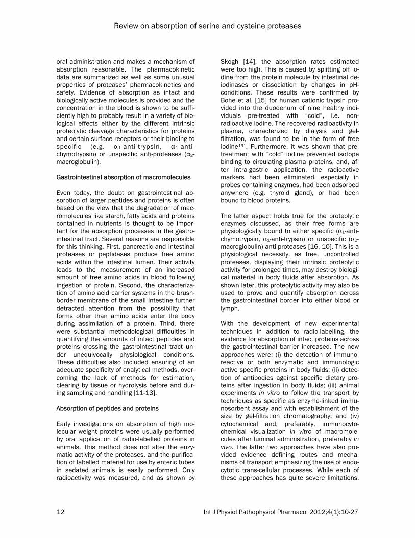
single proteolytic enzyme preparations or as combinations of various proteases of plant and/or animal origin. Even combinations with vita-mins and other food supplements like oli-gomeric proanthocyanidins (OPC), quercetin or selenium are on the market. Most often the plant cysteine proteases bromelain and papain and the animal serine proteases trypsin and chymotrypsin are used. Some preparations are supplemented with pancreatin, amylase, lipase and/or rutosid, a flavonoid that is known to be able to neutralize radicals and to exert immu-nological effects [4, 5]. Proteolytic enzyme combinations are generally provided as enteric coated tablets for oral ad-ministration at dosages varying from 1 to 200 mg per tablet. The amount of proteases pro-vided in the preparation is described by their F.I.P. activity (F.I.P.-units of the Fédération Inter-nationale Pharmaceutique). One F.I.P.-unit is the amount of enzyme which is able to turn over 1 μmol of substrate in 1 minute under standard conditions. Due to enteric coating, degradation of the protein structure to single amino acids or peptides in the acidic environment of the stom-ach is reduced. If at all, the absorption of prote-ases from the gastrointestinal tract may be fa-voured in the small intestine. Table 1 shows the characteristics of three typical representatives of proteolytic enzyme combinations. In general, proteases’ side effects are few. Com-plications typically arise from either excessive dosing or allergic reaction to bovine or porcine substances, papaya or pineapple. In the case of excessive dosing, transient gastrointestinal up-set may result. To avoid hypersensitivity reac-tions, it is best to confirm a patient is not aller-gic to the given proteases’ source prior to use.
Hyperuricosuria (excess uric acid in the urine) and hyperuricemia (excess uric acid in the blood) are associated with extremely high doses of exogenous pancreatic enzymes [6]. The lack of unwanted drug interactions and ma-jor side-effects as well as overall safety aspects, make orally administered proteolytic enzyme combinations an interesting tool for the man-agement of acute and chronic inflammatory processes. Nevertheless, the clinical benefit is subject to controversy as the biological rationale and the mechanism(s) of action are widely un-known and far from being understood. Addition-ally, clinical trials performed in the last century did not always come to an end with clinically relevant and statistically significant results. Of-ten an insufficient patient number and organiza-tional or other insufficiencies such as only mild to moderate disease activity at inclusion of pa-tients were identified as a cause. As there is also doubt on the absorption at all or at least on sufficient amounts of larger protein molecules and missing proof of tissue distribution, clini-cally relevant systemic effects in the treatment of diseases are not readily accepted. Neverthe-less, protease complexes with their counterpart anti-proteases (often as acute phase proteins in animals) [7], as well as the specific proteolytic activity of proteases on specific receptors (e.g. protease activated receptors, PAR) in the gas-trointestinal tract [8] and, provided they are absorbed, in body fluids, may play a major role in many regulatory processes in humans [9, 10]. Thus, clinical effects and benefits are still a matter of controversy and clinical and pharma-cological effects have to be further evaluated. This article summarizes the existing data on gastrointestinal absorption of proteases after
Table 1. Overview of active ingredients used in 3 different marketed preparations Drug Substance Preparation 1 Preparation 2 Preparation 3 Bromelain 90 mg = 900 F.I.P.-units 45 mg = 450 F.I.P.-units 133-178 mg = 800 units
Papain – 60 mg = 328 F.I.P.-units –
Pancreatin – 100 mg = 300 Ph. Eur.-units proteinase
–
Chymotrypsin – 1 mg = 596 F.I.P.-units –
Trypsin 48 mg = 24 μkat* 24mg = 12 μkat* –
Rutosid 100 mg 50 mg –
*1μkat is the amount of enzyme which turns over 1 μM of substrate per second under standard conditions. It corresponds to 60 F.I.P – Units.
Review on absorption of serine and cysteine proteases
12 Int J Physiol Pathophysiol Pharmacol 2012;4(1):10-27
oral administration and makes a mechanism of absorption reasonable. The pharmacokinetic data are summarized as well as some unusual properties of proteases’ pharmacokinetics and safety. Evidence of absorption as intact and biologically active molecules is provided and the concentration in the blood is shown to be suffi-ciently high to probably result in a variety of bio-logical effects either by the different intrinsic proteolytic cleavage characteristics for proteins and certain surface receptors or their binding to specific (e.g. α1-anti-trypsin, α1-anti-chymotrypsin) or unspecific anti-proteases (α2-macroglobulin). Gastrointestinal absorption of macromolecules Even today, the doubt on gastrointestinal ab-sorption of larger peptides and proteins is often based on the view that the degradation of mac-romolecules like starch, fatty acids and proteins contained in nutrients is thought to be impor-tant for the absorption processes in the gastro-intestinal tract. Several reasons are responsible for this thinking. First, pancreatic and intestinal proteases or peptidases produce free amino acids within the intestinal lumen. Their activity leads to the measurement of an increased amount of free amino acids in blood following ingestion of protein. Second, the characteriza-tion of amino acid carrier systems in the brush-border membrane of the small intestine further detracted attention from the possibility that forms other than amino acids enter the body during assimilation of a protein. Third, there were substantial methodological difficulties in quantifying the amounts of intact peptides and proteins crossing the gastrointestinal tract un-der unequivocally physiological conditions. These difficulties also included ensuring of an adequate specificity of analytical methods, over-coming the lack of methods for estimation, clearing by tissue or hydrolysis before and dur-ing sampling and handling [11-13]. Absorption of peptides and proteins Early investigations on absorption of high mo-lecular weight proteins were usually performed by oral application of radio-labelled proteins in animals. This method does not alter the enzy-matic activity of the proteases, and the purifica-tion of labelled material for use by enteric tubes in sedated animals is easily performed. Only radioactivity was measured, and as shown by
Skogh [14], the absorption rates estimated were too high. This is caused by splitting off io-dine from the protein molecule by intestinal de-iodinases or dissociation by changes in pH-conditions. These results were confirmed by Bohe et al. [15] for human cationic trypsin pro-vided into the duodenum of nine healthy indi-viduals pre-treated with “cold”, i.e. non-radioactive iodine. The recovered radioactivity in plasma, characterized by dialysis and gel-filtration, was found to be in the form of free iodine131. Furthermore, it was shown that pre-treatment with “cold” iodine prevented isotope binding to circulating plasma proteins, and, af-ter intra-gastric application, the radioactive markers had been eliminated, especially in probes containing enzymes, had been adsorbed anywhere (e.g. thyroid gland), or had been bound to blood proteins. The latter aspect holds true for the proteolytic enzymes discussed, as their free forms are physiologically bound to either specific (α1-anti-chymotrypsin, α1-anti-trypsin) or unspecific (α2-macroglobulin) anti-proteases [16, 10]. This is a physiological necessity, as free, uncontrolled proteases, displaying their intrinsic proteolytic activity for prolonged times, may destroy biologi-cal material in body fluids after absorption. As shown later, this proteolytic activity may also be used to prove and quantify absorption across the gastrointestinal border into either blood or lymph. With the development of new experimental techniques in addition to radio-labelling, the evidence for absorption of intact proteins across the gastrointestinal barrier increased. The new approaches were: (i) the detection of immuno-reactive or both enzymatic and immunologic active specific proteins in body fluids; (ii) detec-tion of antibodies against specific dietary pro-teins after ingestion in body fluids; (iii) animal experiments in vitro to follow the transport by techniques as specific as enzyme-linked immu-nosorbent assay and with establishment of the size by gel-filtration chromatography; and (iv) cytochemical and, preferably, immunocyto-chemical visualization in vitro of macromole-cules after luminal administration, preferably in vivo. The latter two approaches have also pro-vided evidence defining routes and mecha-nisms of transport emphasizing the use of endo-cytotic trans-cellular processes. While each of these approaches has quite severe limitations,
Review on absorption of serine and cysteine proteases
13 Int J Physiol Pathophysiol Pharmacol 2012;4(1):10-27
the fact that all approaches point to the same conclusion (i.e. transport of small, but poten-tially biologically significant amounts of biologi-cally active macromolecules) greatly strength-ens the validity of this conclusion [13]. Immunological investigations confirming the absorption of not degraded dietary proteins in healthy humans include those on human β-lactoglobulin by Jakobsson et al. [17], on oval-bumin by Husby et al. [18], on bovine serum albumin, beta-lactoglobulin and ovalbumin by Paganelli and Levinsky [19], and on horseradish peroxidase [20]. However, the majority of ex-perimental studies on macromolecule transport have been undertaken on animal intestine. Bockmann and Winbom [21] visualized by elec-tron microscopy intracellular vesicles containing absorbed ferritin in hamster intestine and Walker and Isselbacher [22] these containing horseradish peroxidase. Studies on absorption of proteins across biop-sies of human intestine, although generally from children or from initially abnormal (e.g. malnour-ished) intestine, have provided generally similar information (e.g. [20, 23] and other references in [12]). Therefore, there is no reason to sus-pect major intra-species differences in the gas-trointestinal handling of intact proteins. In animal experiments, generally in rats, similar developments in knowledge have occurred con-cerning absorption of intact (or incompletely digested) peptides: as for large protein mole-cules, it was firmly believed that peptides are wholly hydrolyzed before absorption. This dogma has also had to be revised to account for transport of small but significant amounts of intact, biologically active peptides. The mecha-nisms for their trans-intestinal transport have now been characterized (e.g. [11, 12]) as de-scribed below. For example, in rats, biologically effective amounts of luteinizing hormone releas-ing hormone can be absorbed [24, 25], in ani-mals and man vasopressin and analogues [26-30], and responses to oral thyreotropin-releasing hormone have been recorded in man [31]. Within the last four decades the view on the absorption of high molecular weight molecules (e.g. proteins and peptides) across the gastro-intestinal barrier has completely changed. It is now accepted beyond reasonable doubt that
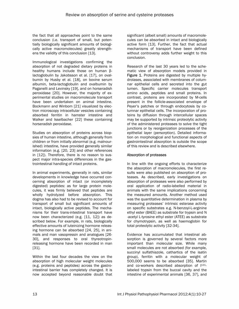
significant (albeit small) amounts of macromole-cules can be absorbed in intact and biologically active form [13]. Further, the fact that actual mechanisms of transport have been defined without controversy adds further weight to this conclusion. Research of the last 30 years led to the sche-matic view of absorption models provided in Figure 1. Proteins are digested by multiple hy-drolases, associated with membranes of colum-nar epithelial cells and secreted into the gut lumen. Specific carrier molecules transport amino acids, peptides and small proteins. In contrast, proteins are incorporated by M-cells present in the follicle-associated envelope of Peyer’s patches or through endocytosis by co-lumnar epithelial cells. The incorporation of pro-teins by diffusion through intercellular spaces may be supported by intrinsic proteolytic activity of the administered proteases to solve the tight junctions or by reorganization processes of the epithelial layer (persorption). Detailed informa-tion on morphological and functional aspects of gastrointestinal absorption is outside the scope of this review and is described elsewhere. Absorption of proteases In line with the ongoing efforts to characterize the absorption of macromolecules, the first re-sults were also published on absorption of pro-teases. As described, early investigations on absorption of proteases were also performed by oral application of radio-labelled material in animals with the same implications concerning the measured amounts. Another method used was the quantitative determination in plasma by measuring proteases’ intrinsic esterase activity on specific substrates e.g. N-benzoyl-L-arginine ethyl ester (BAEE) as substrate for trypsin and N-acetyl L-tyrosine ethyl ester (ATEE) as substrate for chymotrypsin, as well as haemoglobin for total proteolytic activity [32-34]. Evidence has accumulated that intestinal ab-sorption is governed by several factors more important than molecular size. While many small molecules are not absorbed (for example, succinyl sulfathiazole, cathartics of the isatin group), ferritin with a molecular weight of 500,000 seems to be absorbed [35]. Martin and co-workers described absorption of I131-labeled trypsin from the buccal cavity and the intestine of experimental animals [36, 37], and
Review on absorption of serine and cysteine proteases
14 Int J Physiol Pathophysiol Pharmacol 2012;4(1):10-27
Miller [38] reported similar studies in man. Bogner and colleagues obtained comparable results with I131-labelled trypsin in rats and I131-labeled chymotrypsin in man [39]. Kabacoff and co-workers found ATEE activity in the blood of rabbits after intestinal or rectal administration of chymotrypsin [40]. Using the same method, Avakian demonstrated oral chymotrypsin ab-sorption in man [41]. The same conclusion was drawn by Kabacoff (quoted by Bodi [42] on the basis of comparative oral and parenteral toxicity studies in mice. In 1964, Megel et al. [43] de-veloped a sensitive method for detecting trypsin-like activity in rat plasma. For the first time they were able to determine that a minimal effective dose of 500 mg/kg of orally fed trypsin signifi-cantly increased plasma trypsin in rats [43]. First investigations with isolated rat intestines started in 1972, when Faudemay et al. [44] studied the transport of trypsin across the intes-tinal wall, using pieces of isolated rat jejunum and ileum. The intestine was filled with trypsin solution and was incubated in buffer. Aliquots of the acceptor buffer (serosal fluid) were taken and trypsin activity was determined by enzy-matic reaction. Trypsin was found in the exter-
nal medium in amounts increasing with time. Detailed studies concerning the oral bioavail-ability of chymotrypsin were carried out by Mo-riya et al. in 1967 [45]. Radio-iodinated chy-motrypsin was administered into rat intestine and the tissue distribution was determined. As a control, I131-labeled potassium iodide was used, resulting in a different radioactive distribution. In addition, protein-bound radioactivity was proven as well as the stability of chymotrypsin after incubation with intestinal juices and in-creased serum esterase activity. Absorption of the plant hydrolase bromelain (Ananas comosus) has been shown by Miller and Opher in 1964 [46]. They demonstrated an increased ability of the blood serum to digest casein after oral administration of enteric coated bromelain tablets. The bioavailability of bromelain has been studied in detail by White et al. in 1988 [47]. The total plasma radioactivity, TCA-precipitable I125-compounds and the mo-lecular weight profile of I125-proteins, was meas-ured after oral administration in rats. A maxi-mum level, equivalent to 270 ng/ml bromelain, was found at 1 h after administration. Approxi-
Figure 1. Schematic view on absorption mechanisms of the gastrointestinal tract for amino acids, peptides and pro-tein molecules.
Review on absorption of serine and cysteine proteases
15 Int J Physiol Pathophysiol Pharmacol 2012;4(1):10-27
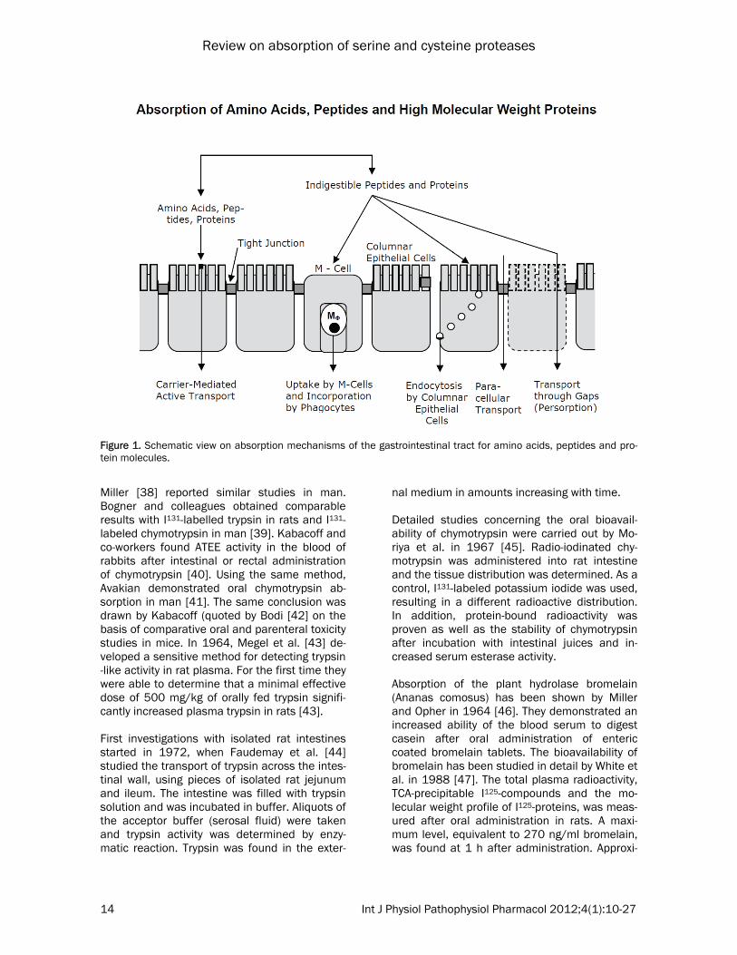
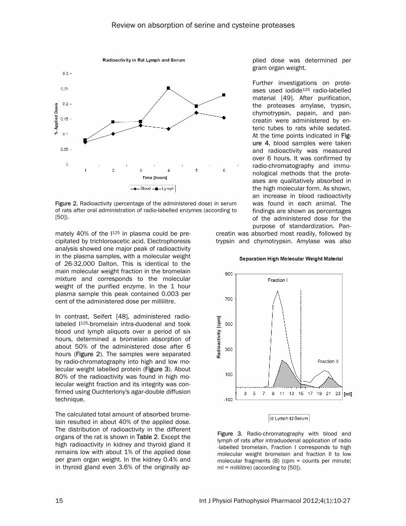
mately 40% of the I125 in plasma could be pre-cipitated by trichloroacetic acid. Electrophoresis analysis showed one major peak of radioactivity in the plasma samples, with a molecular weight of 26-32,000 Dalton. This is identical to the main molecular weight fraction in the bromelain mixture and corresponds to the molecular weight of the purified enzyme. In the 1 hour plasma sample this peak contained 0.003 per cent of the administered dose per millilitre. In contrast, Seifert [48], administered radio-labeled I125-bromelain intra-duodenal and took blood und lymph aliquots over a period of six hours, determined a bromelain absorption of about 50% of the administered dose after 6 hours (Figure 2). The samples were separated by radio-chromatography into high and low mo-lecular weight labelled protein (Figure 3). About 80% of the radioactivity was found in high mo-lecular weight fraction and its integrity was con-firmed using Ouchterlony's agar-double diffusion technique. The calculated total amount of absorbed brome-lain resulted in about 40% of the applied dose. The distribution of radioactivity in the different organs of the rat is shown in Table 2. Except the high radioactivity in kidney and thyroid gland it remains low with about 1% of the applied dose per gram organ weight. In the kidney 0.4% and in thyroid gland even 3.6% of the originally ap-
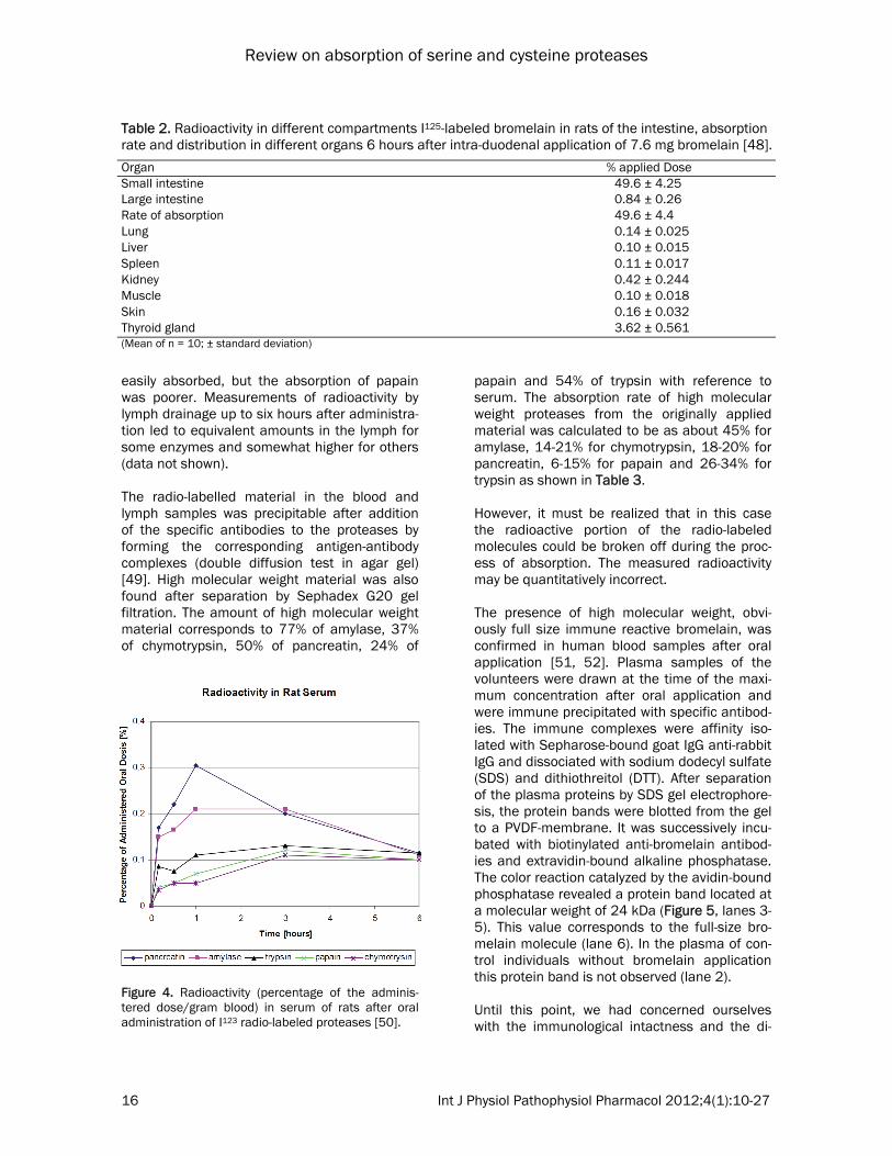
plied dose was determined per gram organ weight. Further investigations on prote-ases used iodide125 radio-labelled material [49]. After purification, the proteases amylase, trypsin, chymotrypsin, papain, and pan-creatin were administered by en-teric tubes to rats while sedated. At the time points indicated in Fig-ure 4, blood samples were taken and radioactivity was measured over 6 hours. It was confirmed by radio-chromatography and immu-nological methods that the prote-ases are qualitatively absorbed in the high molecular form. As shown, an increase in blood radioactivity was found in each animal. The findings are shown as percentages of the administered dose for the purpose of standardization. Pan-
creatin was absorbed most readily, followed by trypsin and chymotrypsin. Amylase was also
Figure 2. Radioactivity (percentage of the administered dose) in serum of rats after oral administration of radio-labelled enzymes (according to [50]).
Figure 3. Radio-chromatography with blood and lymph of rats after intraduodenal application of radio-labelled bromelain. Fraction I corresponds to high molecular weight bromelain and fraction II to low molecular fragments (B) (cpm = counts per minute; ml = millilitre) (according to [50]).
Review on absorption of serine and cysteine proteases
16 Int J Physiol Pathophysiol Pharmacol 2012;4(1):10-27
easily absorbed, but the absorption of papain was poorer. Measurements of radioactivity by lymph drainage up to six hours after administra-tion led to equivalent amounts in the lymph for some enzymes and somewhat higher for others (data not shown). The radio-labelled material in the blood and lymph samples was precipitable after addition of the specific antibodies to the proteases by forming the corresponding antigen-antibody complexes (double diffusion test in agar gel) [49]. High molecular weight material was also found after separation by Sephadex G20 gel filtration. The amount of high molecular weight material corresponds to 77% of amylase, 37% of chymotrypsin, 50% of pancreatin, 24% of
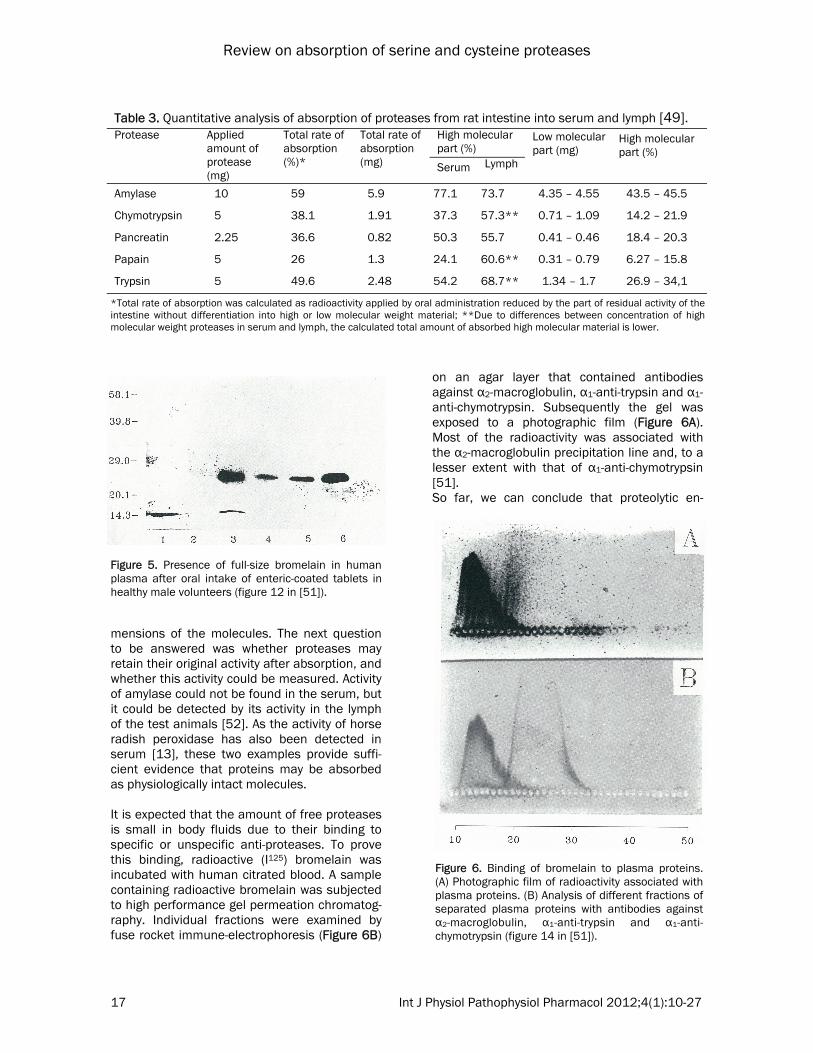
papain and 54% of trypsin with reference to serum. The absorption rate of high molecular weight proteases from the originally applied material was calculated to be as about 45% for amylase, 14-21% for chymotrypsin, 18-20% for pancreatin, 6-15% for papain and 26-34% for trypsin as shown in Table 3. However, it must be realized that in this case the radioactive portion of the radio-labeled molecules could be broken off during the proc-ess of absorption. The measured radioactivity may be quantitatively incorrect. The presence of high molecular weight, obvi-ously full size immune reactive bromelain, was confirmed in human blood samples after oral application [51, 52]. Plasma samples of the volunteers were drawn at the time of the maxi-mum concentration after oral application and were immune precipitated with specific antibod-ies. The immune complexes were affinity iso-lated with Sepharose-bound goat IgG anti-rabbit IgG and dissociated with sodium dodecyl sulfate (SDS) and dithiothreitol (DTT). After separation of the plasma proteins by SDS gel electrophore-sis, the protein bands were blotted from the gel to a PVDF-membrane. It was successively incu-bated with biotinylated anti-bromelain antibod-ies and extravidin-bound alkaline phosphatase. The color reaction catalyzed by the avidin-bound phosphatase revealed a protein band located at a molecular weight of 24 kDa (Figure 5, lanes 3-5). This value corresponds to the full-size bro-melain molecule (lane 6). In the plasma of con-trol individuals without bromelain application this protein band is not observed (lane 2). Until this point, we had concerned ourselves with the immunological intactness and the di-
Figure 4. Radioactivity (percentage of the adminis-tered dose/gram blood) in serum of rats after oral administration of I123 radio-labeled proteases [50].
Table 2. Radioactivity in different compartments I125-labeled bromelain in rats of the intestine, absorption rate and distribution in different organs 6 hours after intra-duodenal application of 7.6 mg bromelain [48].
Organ % applied Dose Small intestine 49.6 ± 4.25 Large intestine 0.84 ± 0.26 Rate of absorption 49.6 ± 4.4 Lung 0.14 ± 0.025 Liver 0.10 ± 0.015 Spleen 0.11 ± 0.017 Kidney 0.42 ± 0.244 Muscle 0.10 ± 0.018 Skin 0.16 ± 0.032 Thyroid gland 3.62 ± 0.561 (Mean of n = 10; ± standard deviation)
Review on absorption of serine and cysteine proteases
17 Int J Physiol Pathophysiol Pharmacol 2012;4(1):10-27
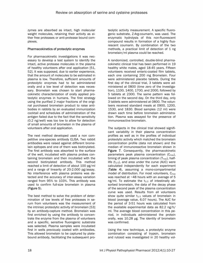
mensions of the molecules. The next question to be answered was whether proteases may retain their original activity after absorption, and whether this activity could be measured. Activity of amylase could not be found in the serum, but it could be detected by its activity in the lymph of the test animals [52]. As the activity of horse radish peroxidase has also been detected in serum [13], these two examples provide suffi-cient evidence that proteins may be absorbed as physiologically intact molecules. It is expected that the amount of free proteases is small in body fluids due to their binding to specific or unspecific anti-proteases. To prove this binding, radioactive (I125) bromelain was incubated with human citrated blood. A sample containing radioactive bromelain was subjected to high performance gel permeation chromatog-raphy. Individual fractions were examined by fuse rocket immune-electrophoresis (Figure 6B)
on an agar layer that contained antibodies against α2-macroglobulin, α1-anti-trypsin and α1-anti-chymotrypsin. Subsequently the gel was exposed to a photographic film (Figure 6A). Most of the radioactivity was associated with the α2-macroglobulin precipitation line and, to a lesser extent with that of α1-anti-chymotrypsin [51]. So far, we can conclude that proteolytic en-
Table 3. Quantitative analysis of absorption of proteases from rat intestine into serum and lymph [49]. Protease Applied
amount of protease (mg)
Total rate of absorption (%)*
Total rate of absorption (mg)
High molecular part (%)
Low molecular part (mg)
High molecular part (%)
Serum Lymph
Amylase 10 59 5.9 77.1 73.7 4.35 – 4.55 43.5 – 45.5
Chymotrypsin 5 38.1 1.91 37.3 57.3** 0.71 – 1.09 14.2 – 21.9
Pancreatin 2.25 36.6 0.82 50.3 55.7 0.41 – 0.46 18.4 – 20.3
Papain 5 26 1.3 24.1 60.6** 0.31 – 0.79 6.27 – 15.8
Trypsin 5 49.6 2.48 54.2 68.7** 1.34 – 1.7 26.9 – 34,1
*Total rate of absorption was calculated as radioactivity applied by oral administration reduced by the part of residual activity of the intestine without differentiation into high or low molecular weight material; **Due to differences between concentration of high molecular weight proteases in serum and lymph, the calculated total amount of absorbed high molecular material is lower.
Figure 5. Presence of full-size bromelain in human plasma after oral intake of enteric-coated tablets in healthy male volunteers (figure 12 in [51]).
Figure 6. Binding of bromelain to plasma proteins. (A) Photographic film of radioactivity associated with plasma proteins. (B) Analysis of different fractions of separated plasma proteins with antibodies against α2-macroglobulin, α1-anti-trypsin and α1-anti-chymotrypsin (figure 14 in [51]).
Review on absorption of serine and cysteine proteases
18 Int J Physiol Pathophysiol Pharmacol 2012;4(1):10-27
zymes are absorbed as intact, high molecular weight molecules, retaining their activity as ei-ther free proteases or anti-protease bound com-plexes. Pharmacokinetics of proteolytic enzymes For pharmacokinetic investigations it was nec-essary to develop a test system to identify the intact, active protease molecules in the plasma of healthy volunteers after oral application [51, 52]. It was supposed, due to earlier experience, that the amount of molecules to be estimated in plasma is low. Therefore, sufficient amounts of proteolytic enzymes had to be administered orally and a low level of detection was neces-sary. Bromelain was chosen to start pharma-cokinetic characterization of orally applied pro-teolytic enzymes in humans. The first strategy using the purified 2 major fractions of the origi-nal purchased bromelain product to raise anti-bodies in rabbits by an evaluated immunization cocktail and schedules of administration of the antigen failed due to the fact that the sensitivity (0.2 ng/well) was too low to allow for detection of small amounts of bromelain in the plasma of volunteers after oral application. The next method developed used a non com-petitive one-species antibody ELISA. Two rabbit antibodies were raised against different brome-lain epitopes and one of them was biotinylated. The first antibody was absorbed to the surface of the well, incubated with plasma probes con-taining bromelain and then incubated with the second biotinylated antibody. This method reached a limit of detection of about 100 pg/ml and a range of linearity of 20-2.000 pg/assay. No interference with plasma proteins was de-tected and the accuracy of inter-assay variation ranged from 95% to 103%. This antibody was used to confirm full-size bromelain in plasma (Figure 5). The best method to solve the problem of deter-mination of low levels of free proteases in se-rum from volunteers was the measurement of the intrinsic proteolytic activity of bromelain [53] by an antibody-capture method. Bromelain was first enriched by using the antibody to concen-trate the enzyme from the plasma of volunteers and a specific, sensitive fluorogenic substrate was selected. Plasma samples were incubated first in wells previously coated with antibodies. This allowed bromelain to be captured by plate-bound antibody, facilitating the subsequent pro-
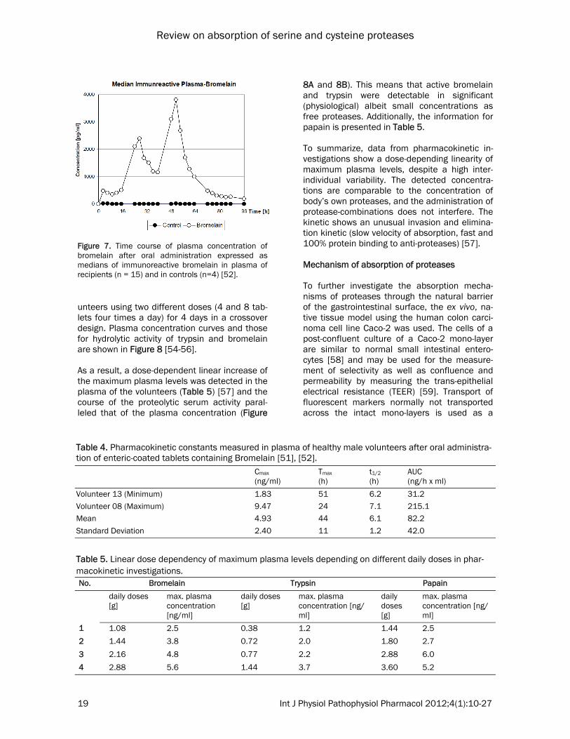
teolytic activity measurement. A specific fluoro-genic substrate, Z-Arg-coumarin, was used. The enzymatic hydrolysis of this non-fluorescent compound results in formation of a highly fluo-rescent coumarin. By combination of the two methods, a practical limit of detection of 1 ng bromelain/ml plasma could be reached. A randomized, controlled, double-blind pharma-cokinetic clinical trial has been performed in 19 healthy white males, aged 18-45 years. Fifteen volunteers received enteric-coated film tablets, each one containing 200 mg Bromelain. Four were administered placebo tablets. During the first day of the clinical trial, 3 tablets were ad-ministered at 0800 (time zero of the investiga-tion), 1100, 1400, 1700, and 2000, followed by 5 tablets at 2300. The same pattern was fol-lowed on the second day. On the third day, only 3 tablets were administered at 0800. The volun-teers received standard meals at 0900, 1200, 1530, and 1830. Blood samples (12 ml) were drawn each time before bromelain administra-tion. Plasma was assayed for the presence of immunoreactive bromelain. The subjects in the clinical trial showed signifi-cant variability in their plasma concentration profiles as well as in the profiles of individual proteolytic activity which matched the bromelain concentration profile (data not shown) and the median of immunoreactive bromelain shown in Figure 7. Consequently, the pharmacokinetic parameters peak plasma concentration (Cmax), timing of peak plasma concentration (Tmax), half-life (t1/2), and area under the curve (AUC) were calculated independently for each experiment (Table 4), assuming a mono-compartmental model of distribution. For most volunteers, Cmax
was reached at ~48 hours with an average of 5 ng/ml. To estimate the t1/2 of intestinally ab-sorbed bromelain, the data of the decay phase of the second peak of the plasma concentration curve was used. Results from all volunteers show quite similar t1/2 values of bromelain in blood (average value, 6.07 hours). The AUC for the period of 3-51 hours was calculated from the available experimental data as 82.2 ng/h/ml. The average blood concentration in this pe-riod, in individuals administered the protein orally, was 10.28 μg. The identity of bromelain was confirmed. Using the new technique, a proteolytic enzyme combination consisting of trypsin, bromelain and rutosid was investigated in 20 healthy vol-
Review on absorption of serine and cysteine proteases
19 Int J Physiol Pathophysiol Pharmacol 2012;4(1):10-27
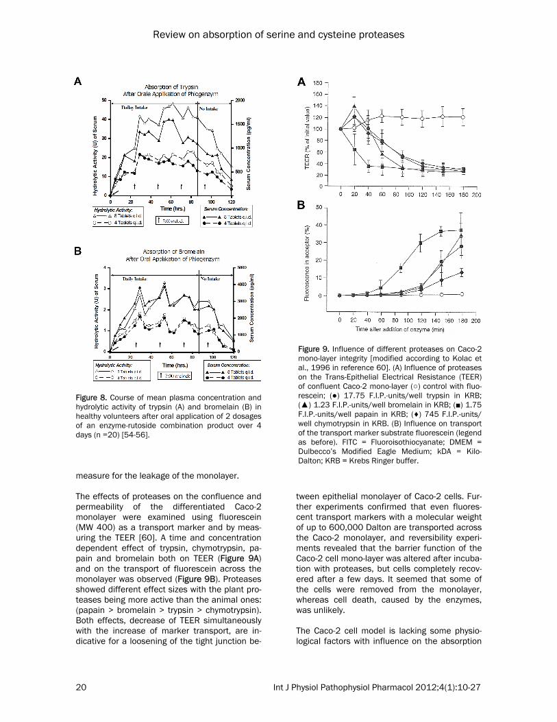
unteers using two different doses (4 and 8 tab-lets four times a day) for 4 days in a crossover design. Plasma concentration curves and those for hydrolytic activity of trypsin and bromelain are shown in Figure 8 [54-56]. As a result, a dose-dependent linear increase of the maximum plasma levels was detected in the plasma of the volunteers (Table 5) [57] and the course of the proteolytic serum activity paral-leled that of the plasma concentration (Figure
8A and 8B). This means that active bromelain and trypsin were detectable in significant (physiological) albeit small concentrations as free proteases. Additionally, the information for papain is presented in Table 5. To summarize, data from pharmacokinetic in-vestigations show a dose-depending linearity of maximum plasma levels, despite a high inter-individual variability. The detected concentra-tions are comparable to the concentration of body’s own proteases, and the administration of protease-combinations does not interfere. The kinetic shows an unusual invasion and elimina-tion kinetic (slow velocity of absorption, fast and 100% protein binding to anti-proteases) [57]. Mechanism of absorption of proteases To further investigate the absorption mecha-nisms of proteases through the natural barrier of the gastrointestinal surface, the ex vivo, na-tive tissue model using the human colon carci-noma cell line Caco-2 was used. The cells of a post-confluent culture of a Caco-2 mono-layer are similar to normal small intestinal entero-cytes [58] and may be used for the measure-ment of selectivity as well as confluence and permeability by measuring the trans-epithelial electrical resistance (TEER) [59]. Transport of fluorescent markers normally not transported across the intact mono-layers is used as a
Figure 7. Time course of plasma concentration of bromelain after oral administration expressed as medians of immunoreactive bromelain in plasma of recipients (n = 15) and in controls (n=4) [52].
Table 4. Pharmacokinetic constants measured in plasma of healthy male volunteers after oral administra-tion of enteric-coated tablets containing Bromelain [51], [52]. Cmax
(ng/ml) Tmax (h)
t1/2 (h)
AUC (ng/h x ml)
Volunteer 13 (Minimum) 1.83 51 6.2 31.2 Volunteer 08 (Maximum) 9.47 24 7.1 215.1 Mean 4.93 44 6.1 82.2 Standard Deviation 2.40 11 1.2 42.0
Table 5. Linear dose dependency of maximum plasma levels depending on different daily doses in phar-macokinetic investigations. No. Bromelain Trypsin Papain
daily doses [g]
max. plasma concentration [ng/ml]
daily doses [g]
max. plasma concentration [ng/ml]
daily doses [g]
max. plasma concentration [ng/ml]
1 1.08 2.5 0.38 1.2 1.44 2.5 2 1.44 3.8 0.72 2.0 1.80 2.7
3 2.16 4.8 0.77 2.2 2.88 6.0
4 2.88 5.6 1.44 3.7 3.60 5.2
Review on absorption of serine and cysteine proteases
20 Int J Physiol Pathophysiol Pharmacol 2012;4(1):10-27
measure for the leakage of the monolayer. The effects of proteases on the confluence and permeability of the differentiated Caco-2 monolayer were examined using fluorescein (MW 400) as a transport marker and by meas-uring the TEER [60]. A time and concentration dependent effect of trypsin, chymotrypsin, pa-pain and bromelain both on TEER (Figure 9A) and on the transport of fluorescein across the monolayer was observed (Figure 9B). Proteases showed different effect sizes with the plant pro-teases being more active than the animal ones: (papain > bromelain > trypsin > chymotrypsin). Both effects, decrease of TEER simultaneously with the increase of marker transport, are in-dicative for a loosening of the tight junction be-
tween epithelial monolayer of Caco-2 cells. Fur-ther experiments confirmed that even fluores-cent transport markers with a molecular weight of up to 600,000 Dalton are transported across the Caco-2 monolayer, and reversibility experi-ments revealed that the barrier function of the Caco-2 cell mono-layer was altered after incuba-tion with proteases, but cells completely recov-ered after a few days. It seemed that some of the cells were removed from the monolayer, whereas cell death, caused by the enzymes, was unlikely. The Caco-2 cell model is lacking some physio-logical factors with influence on the absorption
Figure 8. Course of mean plasma concentration and hydrolytic activity of trypsin (A) and bromelain (B) in healthy volunteers after oral application of 2 dosages of an enzyme-rutoside combination product over 4 days (n =20) [54-56].
Figure 9. Influence of different proteases on Caco-2 mono-layer integrity [modified according to Kolac et al., 1996 in reference 60]. (A) Influence of proteases on the Trans-Epithelial Electrical Resistance (TEER) of confluent Caco-2 mono-layer (○) control with fluo-rescein; (●) 17.75 F.I.P.-units/well trypsin in KRB; (▲) 1.23 F.I.P.-units/well bromelain in KRB; (■) 1.75 F.I.P.-units/well papain in KRB; (♦) 745 F.I.P.-units/well chymotrypsin in KRB. (B) Influence on transport of the transport marker substrate fluorescein (legend as before). FITC = Fluoroisothiocyanate; DMEM = Dulbecco’s Modified Eagle Medium; kDA = Kilo-Dalton; KRB = Krebs Ringer buffer.
Review on absorption of serine and cysteine proteases
21 Int J Physiol Pathophysiol Pharmacol 2012;4(1):10-27
of macromolecules, e.g. the unstirred water layer, the presence of mucus, food, and compo-nents of blood plasma (albumin, antiproteases). The addition of mucin and albumin to the Caco-2 mono-layer had different effects on the activ-ity of proteases on TEER and fluorescein trans-port, suggesting different mechanisms of action of proteases. The activity of trypsin and chy-motrypsin was widely unaffected, that of papain was decreased by mucin and that of bromelain was increased by albumin. Albumin did not have any inhibitory effect on the proteases. The absorption of proteases across the intesti-nal mucosa could be interpreted as self-enhanced paracellular diffusion through locally widened intercellular junctions. This hypothesis is further supported by the already known strong mucolytic activity of bromelain, trypsin and papain, based upon cleavage of amino acid binding sequence of mucus glycoproteins. Pro-teases are widely used in cell isolation for their ability to degrade extra-cellular matrix compo-nents. As shown above, the tightness of intra-cellular junctions is affected, opening the paracellular route across epithelium for non absorbable compounds. Within some tissues, papain has proved to be less damaging and more effective than other proteases. Enhance-ment of further low molecular weight com-pounds through the small intestine mucosa could also be shown [61]. Orally applied proteolytic enzymes reach con-centration in the gastrointestinal tract several folds higher than in the used in vitro experi-ment, making the mechanism of paracellular transport even more reasonable, and the regen-eration of the effects further support the safety of orally applied proteolytic enzyme in clinical use. During the last decade, a possible role of prote-ases as signalling molecules has been empha-sized with the discovery of a novel class of G-protein coupled receptors located on cell mem-branes that may be activated by proteolytic cleavage of their N-terminal extra cellular do-main. Type 2 protease-activated receptors (PAR-2) are cleaved by serine-proteases such as tryp-sin and tryptase. PAR-2 is present in many intes-tinal cell types and particularly on epithelial cells. Multiple functions have been demon-strated in the gut for PAR-2, including epithelial permeability, mainly the intercellular permeabil-
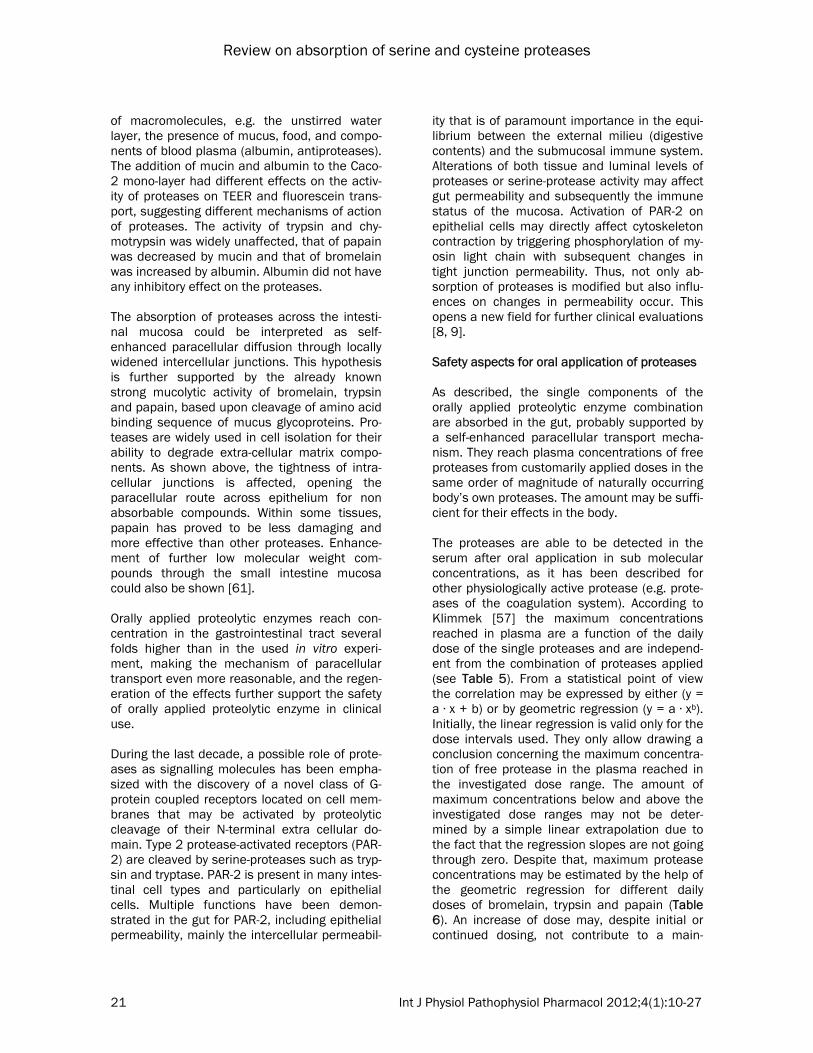
ity that is of paramount importance in the equi-librium between the external milieu (digestive contents) and the submucosal immune system. Alterations of both tissue and luminal levels of proteases or serine-protease activity may affect gut permeability and subsequently the immune status of the mucosa. Activation of PAR-2 on epithelial cells may directly affect cytoskeleton contraction by triggering phosphorylation of my-osin light chain with subsequent changes in tight junction permeability. Thus, not only ab-sorption of proteases is modified but also influ-ences on changes in permeability occur. This opens a new field for further clinical evaluations [8, 9]. Safety aspects for oral application of proteases As described, the single components of the orally applied proteolytic enzyme combination are absorbed in the gut, probably supported by a self-enhanced paracellular transport mecha-nism. They reach plasma concentrations of free proteases from customarily applied doses in the same order of magnitude of naturally occurring body’s own proteases. The amount may be suffi-cient for their effects in the body. The proteases are able to be detected in the serum after oral application in sub molecular concentrations, as it has been described for other physiologically active protease (e.g. prote-ases of the coagulation system). According to Klimmek [57] the maximum concentrations reached in plasma are a function of the daily dose of the single proteases and are independ-ent from the combination of proteases applied (see Table 5). From a statistical point of view the correlation may be expressed by either (y = a · x + b) or by geometric regression (y = a · xb). Initially, the linear regression is valid only for the dose intervals used. They only allow drawing a conclusion concerning the maximum concentra-tion of free protease in the plasma reached in the investigated dose range. The amount of maximum concentrations below and above the investigated dose ranges may not be deter-mined by a simple linear extrapolation due to the fact that the regression slopes are not going through zero. Despite that, maximum protease concentrations may be estimated by the help of the geometric regression for different daily doses of bromelain, trypsin and papain (Table 6). An increase of dose may, despite initial or continued dosing, not contribute to a main-
Review on absorption of serine and cysteine proteases
22 Int J Physiol Pathophysiol Pharmacol 2012;4(1):10-27
tained increase of the concentration of free pro-teases in serum. Therefore, the risk of a strong increase of the concentration of free proteases is low. The safety of orally applied protease combina-tions is also documented by calculation. The occurrence of free proteases in the blood is lim-ited by time and derives from a rapid complex formation with anti-proteases (e.g. α2-macroglobulin). The complex formation follows the law of mass action and reaches for the ab-sorbed protease 100% with time. Not only the foreign protease but also body’s own proteases verifiably have only a tiny chance to escape the catching by α2-macroglobulin. The amount and the occurrence of α2-macroglobulin have been excessively investi-gated without detecting a complete deficiency of this macroglobulin. It indicates that α2-macroglobulin is of vital importance in man. Depending on the velocity, about 0.2% (being very rational from a practical point of view) and after ingestion of 800 mg protease about 16% (practically very improbable) of the naturally occurring α2-macroglobulin reservoir might be irreversibly and cumulatively occupied and made available for a rapid elimination in the liver. A complete occupation of the body's α2-macroglobulin reserve by orally applied prote-ases is more than unlikely. Therefore, the com-ponents of the protease combinations are safe even in high doses due to these pharmacologi-
cal mechanisms. Biological activity of proteolytic enzymes The biological activity of proteases is deriving either from their site-specific hydrolytic cleavage activity or their binding to anti-proteases. The hydrolytic activity is essential in biological processes, and thousands of different prote-ases are already known. Their activity can be differentiated by their substrate specificity, pH-dependency, confirmation and modification (e.g. anti-protease binding). After oral administration and absorption, free proteases may be active in body fluids and even damaged tissue despite their low concentrations (pmol - nmol) as long as the complex with anti-proteases is not formed [13, 50, 62]. The binding to anti-proteases is required to pro-tect the organism from self destruction. If prote-ases’ concentration, from whatever source, in-creases locally, their activity must be controlled. Mostly, proteases are activated as a response to changes in the environment e.g. metabolic changes, injury, invading pathogens or negative chronic effects like oxidative stress or inflamma-tion, either acute or chronic by auto reactive antibodies. This makes them an ideal signal molecule to participate in regulatory processes. As all free proteases are harmful to cells, tis-sues and even organisms, they are all con-trolled by unique mechanisms such as inactive
Table 6. Point estimates for the plasma concentrations y [ng/ml] of free soluble bromelain, trypsin and papain dependent from daily dose x [g] in humans deriving from corresponding geometric regression equation [57].
Daily dose x [g] Bromelain Trypsin Papain y = 2,5588x0.7863 2.7061x0.8466 1.6916x0.9867 0 0 0 0 0.1 0.41854 0.38525 0.17442 0.2 0.72183 0.69278 0.34564 0.3 0.99288 0.97651 0.51567 0.4 1.24491 1.24580 0.68494 0.5 1.48367 1.50485 0.85363 0.6 1.71237 1.75601 1.02188 0.8 2.14702 2.24027 1.35730 1.0 2.55880 2.70610 1.69160 1.5 3.51963 3.81437 2.52375 2.0 4.41302 4.86627 3.35215 3.0 6.07011 6.85922 5.00119 4.0 7.61090 8.75080 6.64279
Review on absorption of serine and cysteine proteases
23 Int J Physiol Pathophysiol Pharmacol 2012;4(1):10-27
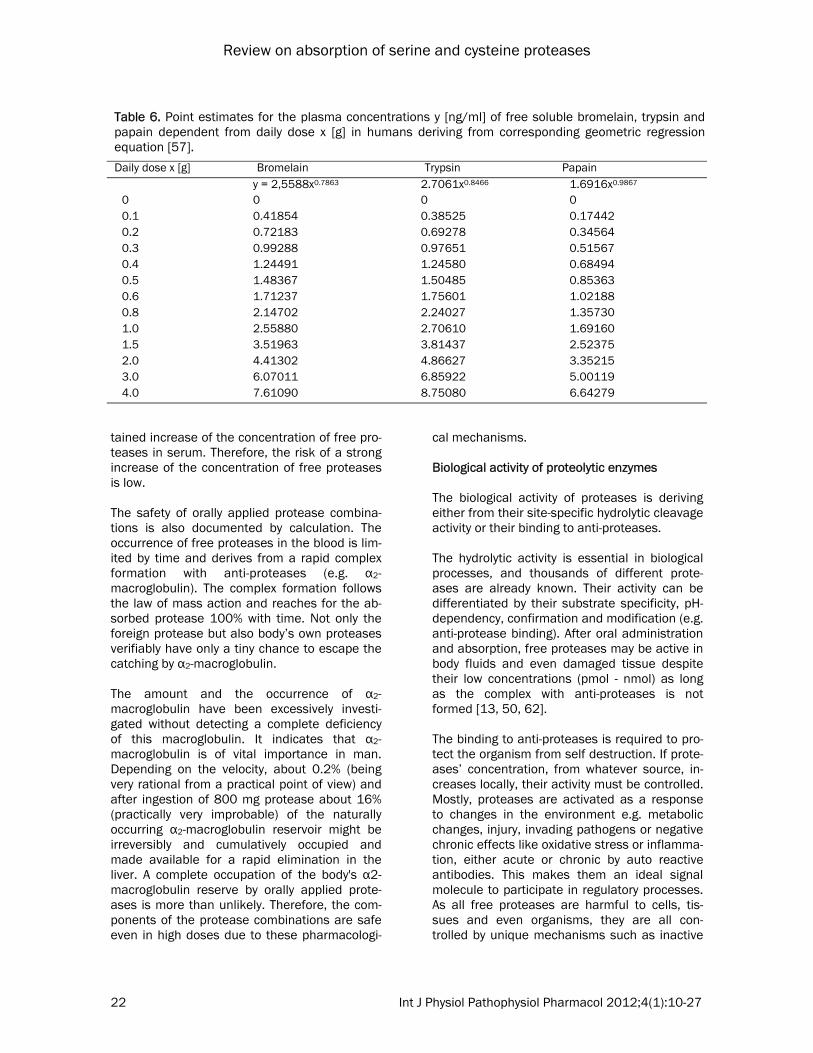
precursor generation or binding to anti-proteases. Obviously the organism does not differentiate proteases according to their origin (foreign plant or animal as well as body’s own proteases). The anti-proteases can bind a vari-ety of proteases [63, 7]. Biological activity of anti-protease complexes It is not known which residual hydrolytic activity is expressed by anti-protease bound proteases. Low molecular weight substrates and probably peptides may be hydrolysed. Figure 10 shows protease molecules entrapped by α2-macroglobulin [63]. The resulting com-plexes are recognized by low density lipoprotein receptor-related proteins on the surface of blood cells and hepatocytes for rapid elimina-tion and activating the production of anti-proteases; a necessity, if their consumption due to acute or chronic processes is increased. Anti-proteases have a half life of several days. Structural changes of the α2-macroglobulin molecules after binding of 1 or 2 protease mole-cules in both cases generate a “fast”-form, which is, in contrast to the “slow”-form, elimi-nated within minutes by macrophages and hepatocytes [64, 65].
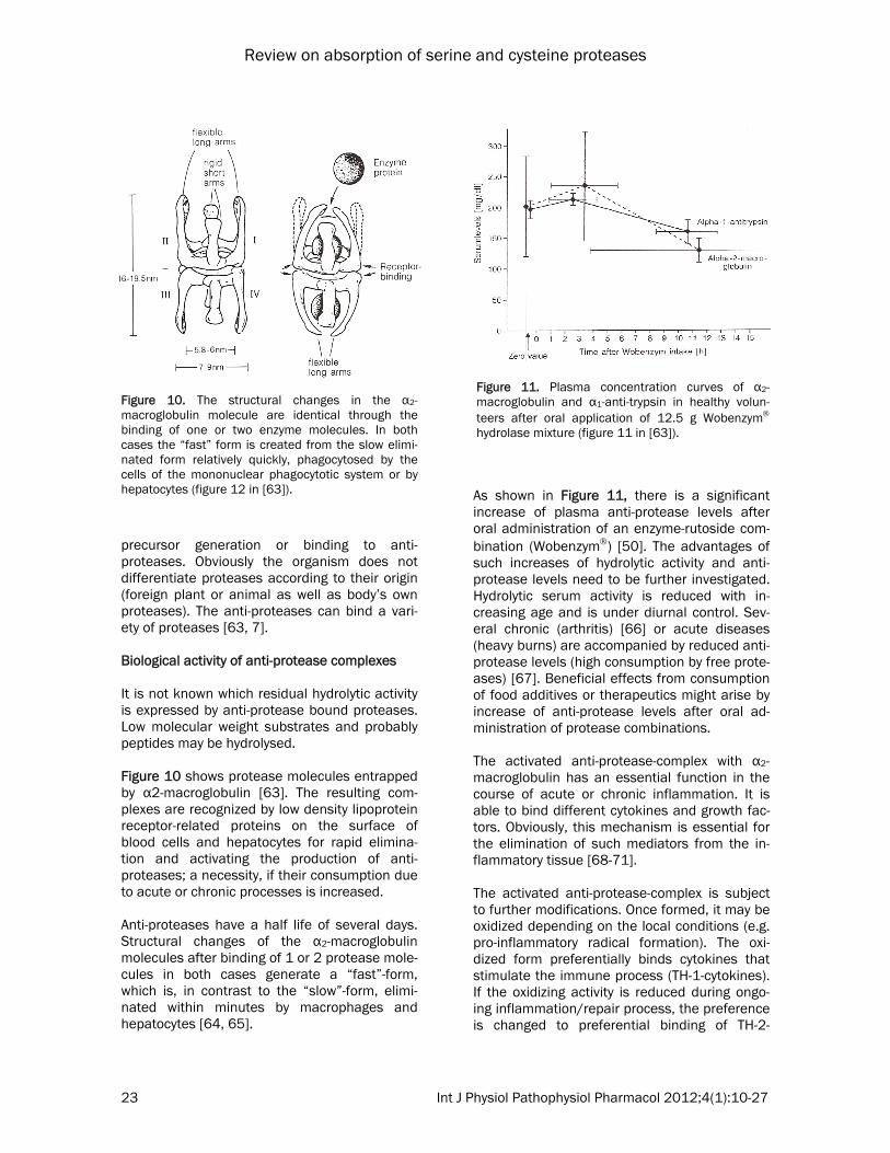
As shown in Figure 11, there is a significant increase of plasma anti-protease levels after oral administration of an enzyme-rutoside com-bination (Wobenzym) [50]. The advantages of such increases of hydrolytic activity and anti-protease levels need to be further investigated. Hydrolytic serum activity is reduced with in-creasing age and is under diurnal control. Sev-eral chronic (arthritis) [66] or acute diseases (heavy burns) are accompanied by reduced anti-protease levels (high consumption by free prote-ases) [67]. Beneficial effects from consumption of food additives or therapeutics might arise by increase of anti-protease levels after oral ad-ministration of protease combinations. The activated anti-protease-complex with α2-macroglobulin has an essential function in the course of acute or chronic inflammation. It is able to bind different cytokines and growth fac-tors. Obviously, this mechanism is essential for the elimination of such mediators from the in-flammatory tissue [68-71]. The activated anti-protease-complex is subject to further modifications. Once formed, it may be oxidized depending on the local conditions (e.g. pro-inflammatory radical formation). The oxi-dized form preferentially binds cytokines that stimulate the immune process (TH-1-cytokines). If the oxidizing activity is reduced during ongo-ing inflammation/repair process, the preference is changed to preferential binding of TH-2-
Figure 10. The structural changes in the α2-macroglobulin molecule are identical through the binding of one or two enzyme molecules. In both cases the “fast” form is created from the slow elimi-nated form relatively quickly, phagocytosed by the cells of the mononuclear phagocytotic system or by hepatocytes (figure 12 in [63]).
Figure 11. Plasma concentration curves of α2-macroglobulin and α1-anti-trypsin in healthy volun-teers after oral application of 12.5 g Wobenzym hydrolase mixture (figure 11 in [63]).
Review on absorption of serine and cysteine proteases
24 Int J Physiol Pathophysiol Pharmacol 2012;4(1):10-27
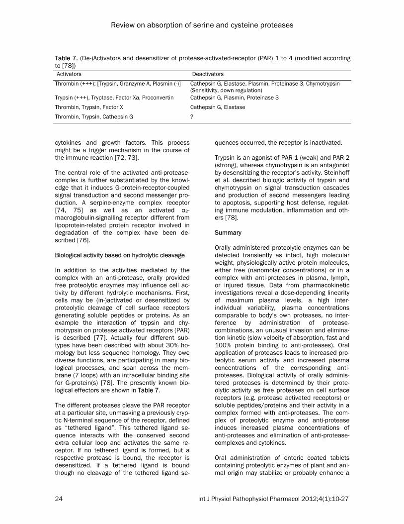
cytokines and growth factors. This process might be a trigger mechanism in the course of the immune reaction [72, 73]. The central role of the activated anti-protease-complex is further substantiated by the knowl-edge that it induces G-protein-receptor-coupled signal transduction and second messenger pro-duction. A serpine-enzyme complex receptor [74, 75] as well as an activated α2-macroglobulin-signalling receptor different from lipoprotein-related protein receptor involved in degradation of the complex have been de-scribed [76]. Biological activity based on hydrolytic cleavage In addition to the activities mediated by the complex with an anti-protease, orally provided free proteolytic enzymes may influence cell ac-tivity by different hydrolytic mechanisms. First, cells may be (in-)activated or desensitized by proteolytic cleavage of cell surface receptors generating soluble peptides or proteins. As an example the interaction of trypsin and chy-motrypsin on protease activated receptors (PAR) is described [77]. Actually four different sub-types have been described with about 30% ho-mology but less sequence homology. They owe diverse functions, are participating in many bio-logical processes, and span across the mem-brane (7 loops) with an intracellular binding site for G-protein(s) [78]. The presently known bio-logical effectors are shown in Table 7. The different proteases cleave the PAR receptor at a particular site, unmasking a previously cryp-tic N-terminal sequence of the receptor, defined as “tethered ligand”. This tethered ligand se-quence interacts with the conserved second extra cellular loop and activates the same re-ceptor. If no tethered ligand is formed, but a respective protease is bound, the receptor is desensitized. If a tethered ligand is bound though no cleavage of the tethered ligand se-
quences occurred, the receptor is inactivated. Trypsin is an agonist of PAR-1 (weak) and PAR-2 (strong), whereas chymotrypsin is an antagonist by desensitizing the receptor’s activity. Steinhoff et al. described biologic activity of trypsin and chymotrypsin on signal transduction cascades and production of second messengers leading to apoptosis, supporting host defense, regulat-ing immune modulation, inflammation and oth-ers [78]. Summary Orally administered proteolytic enzymes can be detected transiently as intact, high molecular weight, physiologically active protein molecules, either free (nanomolar concentrations) or in a complex with anti-proteases in plasma, lymph, or injured tissue. Data from pharmacokinetic investigations reveal a dose-depending linearity of maximum plasma levels, a high inter-individual variability, plasma concentrations comparable to body’s own proteases, no inter-ference by administration of protease-combinations, an unusual invasion and elimina-tion kinetic (slow velocity of absorption, fast and 100% protein binding to anti-proteases). Oral application of proteases leads to increased pro-teolytic serum activity and increased plasma concentrations of the corresponding anti-proteases. Biological activity of orally adminis-tered proteases is determined by their prote-olytic activity as free proteases on cell surface receptors (e.g. protease activated receptors) or soluble peptides/proteins and their activity in a complex formed with anti-proteases. The com-plex of proteolytic enzyme and anti-protease induces increased plasma concentrations of anti-proteases and elimination of anti-protease-complexes and cytokines. Oral administration of enteric coated tablets containing proteolytic enzymes of plant and ani-mal origin may stabilize or probably enhance a
Table 7. (De-)Activators and desensitizer of protease-activated-receptor (PAR) 1 to 4 (modified according to [78]) Activators Deactivators
Thrombin (+++); [Trypsin, Granzyme A, Plasmin (-)] Cathepsin G, Elastase, Plasmin, Proteinase 3, Chymotrypsin (Sensitivity, down regulation)
Trypsin (+++), Tryptase, Factor Xa, Proconvertin Cathepsin G, Plasmin, Proteinase 3
Thrombin, Trypsin, Factor X Cathepsin G, Elastase
Thrombin, Trypsin, Cathepsin G ?
Review on absorption of serine and cysteine proteases
25 Int J Physiol Pathophysiol Pharmacol 2012;4(1):10-27
variety of physiological and immunological proc-esses even in healthy consumers. Acknowledgements Figures 5, 6, 10, 11 have been kindly author-ized by Springer Science and Business Media and the authors. The author wishes to thank Sonja Raum, Munich, for critical reading of the manuscript. Address correspondence to: Dr. Gerhard Lorkowski, GL Pharma Consulting Research & Development (GL Pharma CR&D), Hubertusstr. 77a, D-82131 Gauting, Germany; Tel: +49-89-803696; Fax: +49-89-803644; Mobile: +49-171-641 99 75; E-mail: [email protected] References [1] Roxas M. The role of enzyme supplementation
in digestive disorders. Alt Med Rev 2008; 13: 307-314.
[2] Leipner J, Iten F, Saller R. Therapy with prote-olytic enzymes in rheumatic disorders. Biodrugs 2001; 15: 779-789.
[3] Leipner J, Saller R. Therapy with proteolytic enzymes in oncology. Drugs 2001; 59: 769-780.
[4] Middleton E, Kandaswami C. The impact of plant flavonoids on mammalian biology: impli-cations for immunity, inflammation and cancer. In: Harborne JB, editor. The flavonoids: ad-vances in research since 1986. London: Chap-man & Hall, 1993, pp. 619-652.
[5] Boots AW, Haenen GRMM, Bast A. Health ef-fects of Quercetin: From antioxidant to neutraceutical. Europ J Pharmacol 2008; 585: 325-337.
[6] Cichoke AJ. Pancreatic enzymes. In: Pizzorno J and Murray M, editors. Textbook of Natural Medicine. St Louis, MO: Churchill Livingstone, 2006; pp. 1131-1146.
[7] Barrett AJ, Starkey PM. The Interaction of α2-macroglobulin with proteinases. Biochem J 1973; 133: 709-724.
[8] Bueno L, Fiomonti J. Protease-activated recep-tor 2 and gut permeability: a review. Neurogas-troenterol Motil 2008; 20: 580-587.
[9] Hollenberg MD, Houle S. Proteinases as hor-mone-like signal messengers - proteinase-activated receptors and the pathophysiology of inflammation, pain, cardiovascular disease and cancer. Swiss Med Wkly 2005; 135: 425-436.
[10] Borth W. α2-macroglobulin, a multifunctional binding protein with targeting characteristics. FASEB 1994; 6: 3345-3353.
[11] Gardner MLG. Intestinal assimilation of intact peptides and proteins - a neglected field? Biol Rev 1984; 59: 289-331.
[12] Gardner MLG. Absorption of intact proteins and peptides. In: Johnson LR, editor. The physiology of the gastrointestinal tract. New York: Raven Press 1994; pp. 1795-1820.
[13] Gardner MLG. A review of current knowledge of gastrointestinal absorption of intact proteins including medicinal preparations of proteolytic enzymes. In: Gardner MLG and Steffens KJ, editors. Absorption of orally administered En-zymes. Berlin: Springer Verlag 1995; pp. 1-7.
[14] Skogh T. Overestimate of 125J-protein uptake from adult mouse gut. Gut 1982; 23: 1077-1080.
[15] Bohe M, Borgström A, Genell S, Ohlsson K. Characterization of radioactivity in plasma after intraduodenal administration of 131I-labelled human cationic trypsin. Scand J Gastroenterol Suppl 1986; 126: 21-24.
[16] Silverman GA, Bird PI, Carrell RW, Church FC, Coughlin PB, Gettins PG, Irving JA, Lomas DA, Luke CJ, Moyer RW, Pemberton PA, Remold-O’Donnell E, Salvesen GS, Travis J, Whisstock JC. The serpins are an expanding superfamily of structurally similar but functionally diverse pro-teins. Evolution, mechanism of inhibition, novel functions, and a revised nomenclature. J Biol Chem 2001; 276: 33293-33296.
[17] Jakobsson I, Lindberg T, Lothe L, Axelson I, Benediktsson B. Human ß-Iactoglobulin as a marker of macromolecular absorption. Gut 1986; 27: 1029-1034.
[18] Husby S, Jensenius SC, Svehag SE. Passage of undegraded dietary antigen into the blood of healthy adults. Further characterisation of the kinetics of uptake and the size distribution of the antigen. Scand J Imrnunol 1986; 24: 447-455.
[19] Paganelli R, Levinsky RI. Solid phase radioim-munoassay for detection of circulating food protein antigen in human serum. I Immunol Methods 1980; 37: 333-341.
[20] Heyman M, Boudraa G, Sarrut S, Giraud M, Evans L, Touhami M, Desjeux JF. Macromolecu-lar transport in jejunal mucosa of children with severe malnutrition: a quantitative study. J Pe-diatr Gastroenterol Nutr 1984; 3: 357-363.
[21] Bockman DE, Winbom WB. Light and electron microscopy of intestinal ferritin absorption. Observations in sensitized and non-sensitized hamsters (Mesocricetus auratus). Anat Rec 1966; 155: 603-662.
[22] Walker WA, Isselbacher KJ. Uptake and trans-port of macromolecules by the intestine. Possi-ble role in clinical disorders. Gastroenterol 1974; 67: 531-550.
[23] Heyman M, Desjeux JF. Significance of intesti-nal food protein transport. J Pediatr Gastroen-terol Nutr 1992; 15: 48-57.
[24] Nishi N, Arimura A, Coy DH, Vilchez-Martinez JA, Schally AV. The effect of oral and vaginal ad-ministration of synthetic LHRH and [D-ALA6, DES GLY-10-NH2]-LH-RH ethylamide on serum
Review on absorption of serine and cysteine proteases
26 Int J Physiol Pathophysiol Pharmacol 2012;4(1):10-27
LH levels in ovariectomized, steroid blocked rats. Proc Soc Exp Biol Med 1975; 148: 1009-1012.
[25] Amoss M, Rivier J, Guillemin R. Release of go-nadotrophins by oral administration of synthetic LRF or a tripeptide fragment of LRF. Clin Endo-crinol Metab 1972; 35: 175-177.
[26] Lundin S, Vilhardt, H. Absorption of 1-deamino-8-D-arginine vasopressin from different regions of the gastrointestinal tract in rabbits. Acta Endocrinol (Copenh) 1986; 112: 457-460.
[27] Lundin S, Vilhardt H. Absorption of intragastri-cally administered DDAVP in conscious dogs. Life Sci 1986; 38: 703-709.
[28] Vilhardt H, Lundin S. In vitro intestinal transport of vasopressin and its analogues. Acta Physiol Scand 1986; 126: 601-607.
[29] Vilhardt H, Lundin S. Biological effect and plasma concentrations of DDAVP after intrana-sal and peroral administration to humans. Gen Pharmacol 1986; 17: 481-483.
[30] Williams TDM, Dunger DB, Lyon CC, Lewis RJ, Taylor F, Lightman SL. Antidiuretic effect and pharmacokinetics of oral l-desamino-8-D-arginine vasopressin. 1. Studies in adults and children. J Clin Endocrinol Metab 1986; 63: 129-132.
[31] Ormiston RJ. Clinical effects of TRH on TSH after i.v. and oral administration in normal vol-unteers and patients with thyroid disease. In: Hall H, Wemer I, Holgate H, editors. Thyreotro-pin releasing hormone. Basel: Karger 1972; pp. 45-52.
[32] Schwert GW, Takenaka Y. A spectrophotometrie determination of trypsin and chymotrypsin. Biochim Biophys Acta 1955; 16: 570-575.
[33] Kabacoff BL, Umkey M, Wohlman A, Avakian S. Sensitive und reproducible assay method for chymotrypsin. J Pharm Sci 1963; 52: 1188-1190.
[34] Pryty B, Kabacoff L, Umhey M, Wohlman A, Avakian S. Assay of chymotrypsin. Abstracts of papers presented at the Cincinnati, Ohio, Meet-ing of the American Chemical Society Jan; 13-17, 1963.
[35] Ambrus JL, Lassman HB, DeMarchi JJ. Absorp-tion of exogenous and endogenous proteolytic enzymes. Clin Pharmacol Ther 1967; 8: 362-368.
[36] Martin GJ, Brendel R, Beiler M. Absorption of enzymes from the intestinal tract. Am J Pharm 1957; 127: 194-197.
[37] Martin GJ, Bogner RL, Edelman A. Further in vivo observations with radioactive trypsin. Am J Pharm 1957; 129: 386-392.
[38] Miller JM, Williard RF, Polachek A. An investiga-tion of trypsin I-131 in patients. Exp Med Surg 1960; 18: 352-370.
[39] Bogner R, Snyder C. High dosage oral chy-motrypsin as an adjunct in plastic surgery. J Internat Coll Surgeons 1962; 37: 289-295.
[40] Kabacoff BL, Wohlman A, Umhey M, Avakian S.
Absorption of Chymotrypsin from the intestinal tract. Nature 1963; 199: 815-817.
[41] Avakian S. Further studies on the absorption of Chymotrypsin. Clin Pharmacol Ther 1964; 5: 712-715.
[42] Kabacoff BL. In Bodi T. Modifications of tissue permeability by orally administered proteolytic enzymes in man. Exp Med Surg 1965; 23: 51-62.
[43] Megel H, Strauss R, Ho R, Beiler M. Detection of trypsin-like activity in the plasma of rats after oral administration of trypsin. Arch Biochem Biophys 1964; 108: 193-199.
[44] Faudemay F, Laporte JC, Trémolières J. Passa-ge de la trypsine à travers la paroi intestinale de rat in vitro. Nutr Metab 1973; 15: 207-212.
[45] Moriya H, Moriwaki C, Akimoto S, Yamaguchi K, Iwadare M, Studies on the passage of α-chymotrypsin across the intestine. Chem Pharm Bull 1967; 15: 1662-1668.
[46] Miller JM, Opher AW. The increased proteolytic activity of human blood serum after the oral administration of bromelain. Exp Med Surg 1964; 22: 277-280.
[47] White RR, Crawley FEA, Vellini M, Rovati LA. Bioavailability of 125I-bromelain after oral ad-ministration to rats. Biopharmaceut Drug Dis-pos 1988; 9: 397-403.
[48] Seifert J, Ganser R, Brendel W. Absorption of a proteolytic enzyme originating from plants out of the gastro-intestinal tract into blood and lymph of rats. Z Gastroenterol 1979; 17: 1-8.
[49] Seifert J, Siebrecht P, Lange JP. Quantitative Untersuchungen zur Resorption von Trypsin, Chymotrypsin, Amylase, Papain und Pankreatin aus dem Magen-Darm-Trakt nach oraler Applikation. Allgemeinmedizin 1990; 19: 132-137.
[50] Seifert J, Siebrecht D, Lange JP, Axt G, Bambas FB. The quantitative absorption or orally admin-istered proteins and histological evidence of enzymes in the wound. In: Gardner MLG, Steffens KJ, editors. Absorption of orally admin-istered Enzymes. Berlin: Springer Verlag, 1995; pp. 29-38.
[51] Castell JV. Intestinal absorption of undegraded Bromelain in humans. In: Gardner MLG, Steffens KJ, editors. Absorption of orally admin-istered Enzymes. Berlin: Springer Verlag, 1995; pp. 47-60.
[52] Castell JV, Friedrich G, Kuhn CS, Poppe GE. Intestinal absorption of undegraded proteins in men: presence of bromelain in plasma after oral intake. Am J Physiol 1997; 273: G139-146.
[53] Rowan AD, Buttle DJ, Barrett AJ, The cysteine proteinases of the pineapple plant. Biochem J 1990; 266: 869-875.
[54] Roots I, Donath F, Rex A, Mai I. Pilotstudie zur Untersuchung der relativen Bioverfügbarkeit von Trypsin aus zwei Peroralia. Institut für Klinische Pharmakologie, Berlin 1995.
Review on absorption of serine and cysteine proteases
27 Int J Physiol Pathophysiol Pharmacol 2012;4(1):10-27
[55] Roots I. Bioverfügbarkeit von Trypsin, Bromelain und Rutin-Metaboliten nach oraler Gabe von Phlogenzym® bei gesunden Probanden. Randomisierte doppelblinde Crossover-Studie gemäß GCP. Study No MU-695 427. Institut für Klinische Pharmakologie der Med. Fakultät Humboldt-Universität Berlin, Germany 1997.
[56] Donath F, Roots I, Mai I, Maurer A, Wood G, Kuhn CS and Friedrich G. Dose-related bioavail-ability of bromelain and trypsin after repeated oral administration. Eur J Pharmacol 1997; 52: A146.
[57] Klimmek R. Expert Report on Wobenzym N. 2006
[58] Pinto M. Enterocyte-like differentiation and polarization of the human colon carcinoma cell line Caco-2 in culture. Biol Cell 1983; 47: 323-330.
[59] Powell DW. Barrier function of epithelia. Am J Physiol 1981; 241: G275-G288.
[60] Kolac C, Streichhan P, Lehr CM. Oral bioavail-ability of proteolytic enzymes. Europ J Pharma Biopharm 1996; 42: 222-232.
[61] Grabovac V, Schmitz T, Föger F, Bernkop-Schnürch A. Papain: an effective permeation enhancer for orally administered low molecular weight heparin. Pharm Res 2007; 24: 1001-1006.
[62] Siebrecht D, Lange PJ, Seifert J. The absorption of enzymes from the gut and their effects on tissue repair. Eur Surg Res 1994; 26: 37.
[63] Streichhan W, van Schaik W, Stauder G. Bioavailability of therapeutically used hydrolytic enzymes. In: Gardner MLG, Steffens KJ, editors. Absorption of orally administered Enzymes. Berlin: Springer Verlag, 1995; pp. 83-94.
[64] Birkenmeier G, Usbeck E, Schaffer A, Otto A, Glander HJ. Prostate-specific antigen triggers transformation of seminal alpha-2-macroglobulin (alpha-2-M) and its binding to alpha-2-macroglobulin receptor/low-density lipoprotein receptor-related protein (α2-M-R/LRP) on human spermatozoa. The Prostate 1998; 36: 219-225.
[65] Kounnas MZ, Church F, Argraves WS, Strickland DK. Cellular internalization and degradation of antithrombin III-thrombin, heparin cofactor II-thrombin, and α1-antitrypsin-trypsin complexes is mediated by the low density lipoprotein re-ceptor-related Protein. J Biol Chem 1996; 271: 6523-5429.
[66] Okonski M, Gregosiewicz A, Gil L. The protease- antiprotease system in the pathogenesis of transient arthritis of a single joint in children. Chir Narzadow Ruchu Ortop Pol 1990; 55: 205-208.
[67] Faymonville ME, Micheels J, Bodson L, Jacque-min D, Lamy N, Adam J, Duchateau J. Biochemi-cal investigations after burning injury: comple-ment system, protease-antiprotease balance and acute-phase reactants. Burns including
thermal injury 1987; 13: 26-33. [68] Garber TR, Gonias, SL, Webb DJ. Interleukin-4
and IL-10 bind covalently to activated human α2-macroglobulin by a mechanism that re-quires Cys949. J Interferon Cytokine Res 2000; 20: 125-131.
[69] LaMarre J, Wollenberg GK, Gonias SL, Hayes MA. Biology of disease. Cytokine binding and clearance properties of proteinase–activated α2-macroglobulins. Lab Invest 1991; 65: 3-14.
[70] LaMarre J, Hayes MA, Wollenberg GK, Hussaini I, Hall SW, Gonias SL. A α2-macroglobulin re-ceptor-dependent mechanism for the plasma clearance of transforming growth factor β1 in mice. J Clin Invest 1991; 87: 39-44.
[71] Bhattacharjee G, Asplin AR, Wu SM, Gawdi G, Pizzo SV. The conformation-dependent interac-tion of α2-macroglobulin with vascular endothe-lial growth factor. J Biol Chem 2000; 275: 26806-26811.
[72] Wu SM, Boyer CM, Pizzo SV. The binding of receptor-recognized α2-macroglobulin to the low density lipoprotein receptor-related protein and the α2M signaling receptor is decoupled by oxidation. J Biol Chem 1997; 272: 20627-20635.
[73] Wu SM, Dhavalkumar DP, Pizzo SV. Oxidized α2-macroglobulin (α2-M) differentially regulates receptor binding by cytokines/growth factors: Implications for tissue injury and repair mecha-nisms in inflammation. J Immunol 1998; 161: 4356-4365.
[74] Perlmutter DH, Glover GI, Rivetna M, Schasteen CS, Fallon RJ. Identification of a serpin-enzyme complex receptor on human hepatoma cells and human monocytes. PNAS 1990; 87: 3753-3757.
[75] Moraga F, Lindgren S, Janciauskiene S. Effects of noninhibitory α1-antitrypsin on primary hu-man monocyte activation in vitro. Arch Biochem Biophys 2001; 386: 221-226.
[76] Misra UK, Chu CT, Gawdi G, Pizzo SV. Evidence for a second alpha-2-macroglobulin receptor. J Biol Chem 1994; 269: 12541-12547.
[77] Déry O, Bunnett NW: Proteinase-activated Re-ceptors: A Growing Family of Heptahelical Re-ceptors for Thrombin, Trypsin and Tryptase. Biochem Soc Trans 1999; 27: 246-254.
[78] Steinhoff M, Buddenkotte J, Shpacovitch V, Rattenholl A, Moormann C, Vergnolle N, Luger TA, Hollenberg MD. Proteinase-activated recep-tors: Transducers of Proteinase-mediated sig-naling in inflammation and immune response. Endocrine Rev 2005; 26: 1-43.