REVIEW ARTICLE DOI: Diagnosis and treatment of mast cell ... · RESULTADOS: A apresentação...
11
264 Sao Paulo Med J. 2013; 131(4):264-74 REVIEW ARTICLE DOI: 10.1590/1516-3180.2013.1314590 Diagnosis and treatment of mast cell disorders: practical recommendations Diagnóstico e tratamento de doenças mastocitárias: recomendações práticas Alex Freire Sandes I , Raphael Salles Scortegagna Medeiros II , Edgar Gil Rizzatti I Grupo Fleury, São Paulo, Brazil ABSTRACT CONTEXT AND OBJECTIVE: The term mastocytosis covers a group of rare disorders characterized by neoplastic proliferation and accumulation of clonal mast cells in one or more organs. The aim of this study was to assess the principal elements for diagnosing and treating these disorders. DESIGN AND SETTING: Narrative review of the literature conducted at Grupo Fleury, São Paulo, Brazil. METHODS: This study reviewed the scientific papers published in the PubMed, Embase (Excerpta Medica Database), Lilacs (Literatura Latino-Americana e do Caribe em Ciências da Saúde) and Cochrane Library databases that were identified using the search term “mastocytosis.” RESULTS: The clinical presentation of mastocytosis is remarkably heterogeneous and ranges from skin lesions that may regress spontaneously to aggressive forms associated with organ failure and short survival. Currently, seven subtypes of mastocytosis are recognized through the World Health Organization classification system for hematopoietic tumors. These disorders are diagnosed based on clinical manifestations and on identification of neoplastic mast cells using morphological, immunophenotypic, genetic and molecular methods. Abnormal mast cells display atypical and frequently spindle-shaped morphology, and aberrant expression of the CD25 and CD2 antigens. Elevation of serum tryptase is a common finding in some subtypes, and more than 90% of the patients present the D816V KIT mutation in mast cells. CONCLUSION: Here, we described the most common signs and symptoms among patients with mastocytosis and suggested a practical approach for the diagnosis, classification and initial clinical treatment of mastocytosis. RESUMO CONTEXTO E OBJETIVO: O termo mastocitose abrange um grupo de raras doenças caracterizado por proliferação neoplásica e acúmulo de mastócitos clonais em um ou mais órgãos. O objetivo do presente estudo foi avaliar os principais elementos para o diagnóstico e tratamento dessas desordens. TIPO DE ESTUDO E LOCAL: Revisão narrativa da literatura realizada no Grupo Fleury, São Paulo, Brasil. MÉTODOS: O presente estudo revisou artigos científicos publicados nas bases de dados PubMed, Embase (Excerpta Medica Database), Lilacs (Literatura Latino-Americana e do Caribe em Ciências da Saúde) e Cochrane Library, que foram identificados com o termo de busca “mastocitose”. RESULTADOS: A apresentação clínica da mastocitose é marcadamente heterogênea, variando de lesões cutâneas que podem regredir espontaneamente, até formas agressivas associadas a falência de órgãos e curta sobrevida. Atualmente, sete subtipos de mastocitose são reconhecidos pela classificação de tumores hematopoéticos da Organização Mundial de Saúde; o diagnóstico é realizado com base nas manifestações clínicas e na identificação de mastócitos neoplásicos por métodos morfológicos, imunofenotípicos, genéticos e moleculares. Mastócitos anômalos apresentam morfologia atípica, frequentemente fusiforme, e expressão aberrante dos antígenos CD25 e CD2. Aumento de triptase sérica é um achado comum em alguns subtipos; e mais que 90% dos pacientes apresentam mastócitos com a mutação KIT D816V. CONCLUSÃO: No presente artigo, descrevemos os sintomas e sinais mais comuns em pacientes com mastocitose e sugerimos uma prática abordagem para o diagnóstico, classificação e tratamento clínico inicial. I MD, PhD. Medical Consultant in Hematology, Division of Laboratory Medicine and Pathological Anatomy, Grupo Fleury, São Paulo, Brazil. II MD. Medical Consultant in Pathological Anatomy, Division of Laboratory Medicine and Pathological Anatomy, Grupo Fleury, São Paulo, Brazil. KEY WORDS: Mastocytosis. Diagnosis. Anatomy. Flow cytometry. Therapeutics. PALAVRAS-CHAVE: Mastocitose. Diagnóstico. Anatomia. Citometria de fluxo. Terapêutica.
Transcript of REVIEW ARTICLE DOI: Diagnosis and treatment of mast cell ... · RESULTADOS: A apresentação...

264 Sao Paulo Med J. 2013; 131(4):264-74
REVIEW ARTICLE DOI: 10.1590/1516-3180.2013.1314590
Diagnosis and treatment of mast cell disorders: practical recommendationsDiagnóstico e tratamento de doenças mastocitárias: recomendações práticasAlex Freire SandesI, Raphael Salles Scortegagna MedeirosII, Edgar Gil RizzattiI
Grupo Fleury, São Paulo, Brazil
ABSTRACTCONTEXT AND OBJECTIVE: The term mastocytosis covers a group of rare disorders characterized by neoplastic proliferation and accumulation of clonal mast cells in one or more organs. The aim of this study was to assess the principal elements for diagnosing and treating these disorders. DESIGN AND SETTING: Narrative review of the literature conducted at Grupo Fleury, São Paulo, Brazil.METHODS: This study reviewed the scientific papers published in the PubMed, Embase (Excerpta Medica Database), Lilacs (Literatura Latino-Americana e do Caribe em Ciências da Saúde) and Cochrane Library databases that were identified using the search term “mastocytosis.”RESULTS: The clinical presentation of mastocytosis is remarkably heterogeneous and ranges from skin lesions that may regress spontaneously to aggressive forms associated with organ failure and short survival. Currently, seven subtypes of mastocytosis are recognized through the World Health Organization classification system for hematopoietic tumors. These disorders are diagnosed based on clinical manifestations and on identification of neoplastic mast cells using morphological, immunophenotypic, genetic and molecular methods. Abnormal mast cells display atypical and frequently spindle-shaped morphology, and aberrant expression of the CD25 and CD2 antigens. Elevation of serum tryptase is a common finding in some subtypes, and more than 90% of the patients present the D816V KIT mutation in mast cells.CONCLUSION: Here, we described the most common signs and symptoms among patients with mastocytosis and suggested a practical approach for the diagnosis, classification and initial clinical treatment of mastocytosis.
RESUMO CONTEXTO E OBJETIVO: O termo mastocitose abrange um grupo de raras doenças caracterizado por proliferação neoplásica e acúmulo de mastócitos clonais em um ou mais órgãos. O objetivo do presente estudo foi avaliar os principais elementos para o diagnóstico e tratamento dessas desordens. TIPO DE ESTUDO E LOCAL: Revisão narrativa da literatura realizada no Grupo Fleury, São Paulo, Brasil.MÉTODOS: O presente estudo revisou artigos científicos publicados nas bases de dados PubMed, Embase (Excerpta Medica Database), Lilacs (Literatura Latino-Americana e do Caribe em Ciências da Saúde) e Cochrane Library, que foram identificados com o termo de busca “mastocitose”.RESULTADOS: A apresentação clínica da mastocitose é marcadamente heterogênea, variando de lesões cutâneas que podem regredir espontaneamente, até formas agressivas associadas a falência de órgãos e curta sobrevida. Atualmente, sete subtipos de mastocitose são reconhecidos pela classificação de tumores hematopoéticos da Organização Mundial de Saúde; o diagnóstico é realizado com base nas manifestações clínicas e na identificação de mastócitos neoplásicos por métodos morfológicos, imunofenotípicos, genéticos e moleculares. Mastócitos anômalos apresentam morfologia atípica, frequentemente fusiforme, e expressão aberrante dos antígenos CD25 e CD2. Aumento de triptase sérica é um achado comum em alguns subtipos; e mais que 90% dos pacientes apresentam mastócitos com a mutação KIT D816V.CONCLUSÃO: No presente artigo, descrevemos os sintomas e sinais mais comuns em pacientes com mastocitose e sugerimos uma prática abordagem para o diagnóstico, classificação e tratamento clínico inicial.
IMD, PhD. Medical Consultant in Hematology, Division of Laboratory Medicine and Pathological Anatomy, Grupo Fleury, São Paulo, Brazil.IIMD. Medical Consultant in Pathological Anatomy, Division of Laboratory Medicine and Pathological Anatomy, Grupo Fleury, São Paulo, Brazil.
KEY WORDS:Mastocytosis. Diagnosis. Anatomy.Flow cytometry. Therapeutics.
PALAVRAS-CHAVE:Mastocitose. Diagnóstico. Anatomia. Citometria de fluxo. Terapêutica.

Diagnosis and treatment of mast cell disorders: practical recommendations | REVIEW ARTICLE
Sao Paulo Med J. 2013; 131(4):264-74 265
INTRODUCTIONMastocytosis is currently defined as a heterogeneous group of dis-orders characterized by clonal expansion and accumulation of mast cells in one or more tissues, such as skin, bone marrow, liver, spleen, gastrointestinal tract and lymph nodes, among others.1 Clonal mast cells are detected in most cases and in all subtypes of the disease. These clonal mast cells are most often characterized by the presence of the D816V-activating KIT mutation; however, other classes of KIT mutations have been detected.2 Mastocytosis is considered to be one of the eight subcategories of myeloproliferative neoplasms, accord-ing to the 2008 World Health Organization classification system.3
There are two ages of peak onset for this disease: in the first decade of life and between the fourth and fifth decades of life.4,5 The skin is the organ most affected in this class of disorders; in fact, the skin is infiltrated in virtually all children and around 85% of adults with mastocytosis.4 The clinical presentation of mastocytosis is heterogeneous and ranges from disease limited to the skin (i.e. cutaneous mastocytosis) to cases with extracu-taneous involvement (i.e. systemic mastocytosis). Cutaneous mastocytosis occurs mainly during infancy and childhood (i.e. most cases appear within the first year of life) and is commonly associated with spontaneous regression of skin lesions, whereas systemic mastocytosis is usually diagnosed in adults and ranges from indolent to aggressive forms of mastocytosis. The latter is associated with multiorgan failure and decreased survival.3
Mastocytosis is considered to be a rare disorder, but the true incidence and prevalence in the general population are unknown. In Brazil, it can reasonably be assumed that the diagnosis of mastocyto-sis is greatly underestimated, given the low clinical awareness of the disease and the existence of very few specialized diagnostic centers.
OBJECTIVESThe aim of the present study was to review the main signs and symptoms of mastocytosis and suggest a practical approach for diagnosis, classification and initial management of patients with mast cell disorders.
METHODSWe conducted a narrative review by means of a systematic literature search using the PubMed, Embase (Excerpta Medica Database), Lilacs (Literatura Latino-Americana e do Caribe em Ciências da Saúde ) and Cochrane Library databases to identify published sci-entific papers relating to the search term “mastocytosis.” Moreover, the papers identified needed to focus on human subjects (Table 1).
RESULTSClinical manifestationsThe clinical symptoms of mastocytosis are caused by acute and chronic release of intracytoplasmic mast cell mediators (i.e.
histamines, tryptase, prostaglandins and leukotrienes) and tissue infiltration by neoplastic mast cells. Common symp-toms include pruritus (i.e. generalized or restricted to cutane-ous lesions), skin redness and swelling, sweating, palpitations, thoracic pain and headache.1,5,6 Recurrent abdominal pain and diarrhea are present in a significant number of cases and may evolve to severe malabsorption, thereby leading to weight loss and hypoalbuminemia. Osteoporosis can also be found in a small number of cases.
Anaphylactic reactions associated with vascular collapse and risk of death occur in approximately 20% of adults and 6% of children, and these extreme symptoms are more frequent in males.4 In some cases, the allergic triggering factor is not identi-fied, whereas in others, a history of medication use (e.g. aspirin, nonsteroidal anti-inflammatory drugs and iodinated radiocon-trast agents) or insect bites has been linked to the onset of symp-toms. In mast cell leukemia, repeated severe episodes of massive release of mast cell mediators are typical, and these patients must be treated in intensive care units. In clinical subtypes other than mast cell leukemia, however, there may not be any direct relation-ship between mast cell mass, mast cell release and symptoms.7
In many patients, the first sign of the disease is a character-istic maculopapular rash known as urticaria pigmentosa. The symptoms associated with these lesions are exacerbated with fric-tion (Darier’s sign).7,8 In aggressive forms, the massive tissue infil-tration can lead to signs and symptoms secondary to the exis-tence of hepatic or bone marrow infiltration. These additional signs and symptoms include abdominal pain, portal hyperten-sion and ascites (i.e. relating to hepatic infiltration) and pancyto-penia (i.e. relating to bone marrow infiltration).9
Diagnosis and classification of mastocytosisThe diagnosis of mastocytosis is based on identification of neo-plastic mast cells by means of morphological, immunopheno-typic and/or genetic methods.10 The World Health Organization
Table 1. Database search resultsDatabase Search Results
PubMedMastocytosis
[Mesh]2,747
papers
1,144 case reports440 reviews
52 clinical trials2 meta-analyses
1,109 others
Embase (Excerpta Medica Database)
Mastocytosis[Emtree]
2,701 papers
962 case reports554 reviews
168 clinical trials1,017 others
Lilacs (Literatura Latino-Americana e do Caribe em Ciências da Saúde)
Mastocytosis81
papers45 case reports
36 others
Cochrane Library Mastocytosis18
papers18 clinical trials
REVIEW ARTICLE | Sandes AF, Medeiros RSS, Rizzatti EG
266 Sao Paulo Med J. 2013; 131(4):264-74
classification system defines the following seven subtypes of the disease: cutaneous mastocytosis, indolent systemic mastocytosis, systemic mastocytosis associated with other clonal hematologi-cal non-mast cell lineage diseases, aggressive systemic mastocy-tosis, mast cell leukemia, mast cell sarcoma and extracutaneous mastocytoma.3
Cutaneous mastocytosis is characterized by abnormal mast cell infiltration in the dermis and no evidence of systemic involvement. The following three variants of cutaneous mas-tocytosis have been described: urticaria pigmentosa, diffuse cutaneous mastocytosis and solitary mastocytosis of the skin. Urticaria pigmentosa is the most common presentation in chil-dren and represents 70-90% of cases of mastocytosis in this pop-ulation. The most affected areas include the trunk and extremi-ties, whereas the palms, soles, scalp, and face are less frequently compromised.7
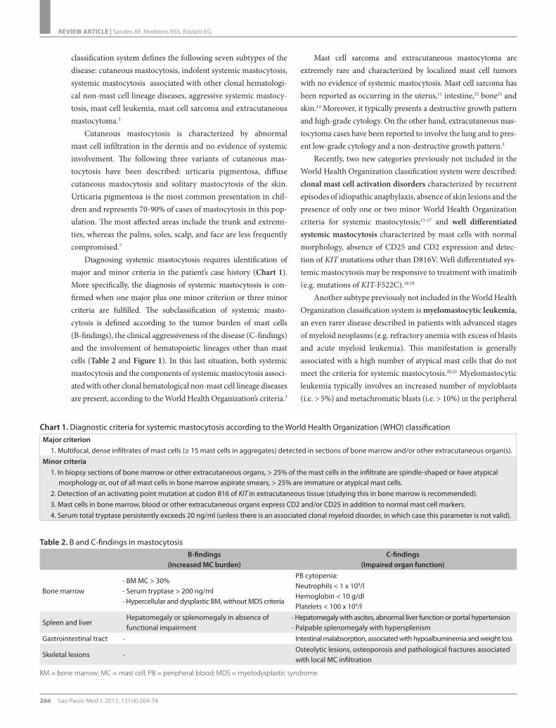
Diagnosing systemic mastocytosis requires identification of major and minor criteria in the patient’s case history (Chart 1). More specifically, the diagnosis of systemic mastocytosis is con-firmed when one major plus one minor criterion or three minor criteria are fulfilled. The subclassification of systemic masto-cytosis is defined according to the tumor burden of mast cells (B-findings), the clinical aggressiveness of the disease (C-findings) and the involvement of hematopoietic lineages other than mast cells (Table 2 and Figure 1). In this last situation, both systemic mastocytosis and the components of systemic mastocytosis associ-ated with other clonal hematological non-mast cell lineage diseases are present, according to the World Health Organization’s criteria.3
Mast cell sarcoma and extracutaneous mastocytoma are extremely rare and characterized by localized mast cell tumors with no evidence of systemic mastocytosis. Mast cell sarcoma has been reported as occurring in the uterus,11 intestine,12 bone13 and skin.14 Moreover, it typically presents a destructive growth pattern and high-grade cytology. On the other hand, extracutaneous mas-tocytoma cases have been reported to involve the lung and to pres-ent low-grade cytology and a non-destructive growth pattern.3
Recently, two new categories previously not included in the World Health Organization classification system were described: clonal mast cell activation disorders characterized by recurrent episodes of idiopathic anaphylaxis, absence of skin lesions and the presence of only one or two minor World Health Organization criteria for systemic mastocytosis;15-17 and well differentiated systemic mastocytosis characterized by mast cells with normal morphology, absence of CD25 and CD2 expression and detec-tion of KIT mutations other than D816V. Well differentiated sys-temic mastocytosis may be responsive to treatment with imatinib (e.g. mutations of KIT-F522C).18,19
Another subtype previously not included in the World Health Organization classification system is myelomastocytic leukemia, an even rarer disease described in patients with advanced stages of myeloid neoplasms (e.g. refractory anemia with excess of blasts and acute myeloid leukemia). This manifestation is generally associated with a high number of atypical mast cells that do not meet the criteria for systemic mastocytosis.20,21 Myelomastocytic leukemia typically involves an increased number of myeloblasts (i.e. > 5%) and metachromatic blasts (i.e. > 10%) in the peripheral
Chart 1. Diagnostic criteria for systemic mastocytosis according to the World Health Organization (WHO) classificationMajor criterion
1. Multifocal, dense infiltrates of mast cells (≥ 15 mast cells in aggregates) detected in sections of bone marrow and/or other extracutaneous organ(s).Minor criteria
1. In biopsy sections of bone marrow or other extracutaneous organs, > 25% of the mast cells in the infiltrate are spindle-shaped or have atypical morphology or, out of all mast cells in bone marrow aspirate smears, > 25% are immature or atypical mast cells.
2. Detection of an activating point mutation at codon 816 of KIT in extracutaneous tissue (studying this in bone marrow is recommended).3. Mast cells in bone marrow, blood or other extracutaneous organs express CD2 and/or CD25 in addition to normal mast cell markers.4. Serum total tryptase persistently exceeds 20 ng/ml (unless there is an associated clonal myeloid disorder, in which case this parameter is not valid).
Table 2. B and C-findings in mastocytosisB-findings
(Increased MC burden)C-findings
(Impaired organ function)
Bone marrow- BM MC > 30%- Serum tryptase > 200 ng/ml- Hypercellular and dysplastic BM, without MDS criteria
PB cytopenia:Neutrophils < 1 x 10⁹/lHemoglobin < 10 g/dlPlatelets < 100 x 109/l
Spleen and liverHepatomegaly or splenomegaly in absence of functional impairment
- Hepatomegaly with ascites, abnormal liver function or portal hypertension- Palpable splenomegaly with hypersplenism
Gastrointestinal tract - Intestinal malabsorption, associated with hypoalbuminemia and weight loss
Skeletal lesions -Osteolytic lesions, osteoporosis and pathological fractures associated with local MC infiltration
BM = bone marrow; MC = mast cell; PB = peripheral blood; MDS = myelodysplastic syndrome.

Diagnosis and treatment of mast cell disorders: practical recommendations | REVIEW ARTICLE
Sao Paulo Med J. 2013; 131(4):264-74 267
blood and/or bone marrow and lacks other features of mast cell disorders, such as mast cell infiltrates, expression of CD25 and CD2, or KIT-D816V mutation.
Bone marrow aspirateNormal and reactive mast cells are round or oval and small to medium sized cells that present a central round nucleus, con-densed chromatin without a nucleolus, abundant cytoplasm (i.e. low N/C ratio) and numerous metachromatic cytoplasmic gran-ules, as identified via Romanowsky-based staining methods.
The cytomorphology of neoplastic mast cells have been classi-fied into the following three subtypes:22 (1) metachromatic blasts with a high N/C ratio, fine nuclear chromatin, prominent nucleoli and few metachromatic granules; (2) atypical mast cells type I, which are spindle-shaped cells with oval nuclei in an eccentric position, elongated cytoplasmic projections and hypogranular cytoplasm; and (3) atypical mast cells type II (promastocytes), which exhibit bi- to multilobed nuclei associated with mature morphology (i.e. condensed chromatin and low N/C ratio) or immature morphology (i.e. fine chromatin and high N/C ratio) (Figure 2). Moreover, these morphological features of mast cells have been correlated with different clinical presentations of mas-tocytosis. Atypical mast cells type I are more commonly found
in systemic mastocytosis cases with an indolent course, whereas atypical mast cells type II and metachromatic blasts are more fre-quently found in mast cell leukemia cases, which are commonly associated with poor outcome and shorter survival time.
The percentage of mast cells in the bone marrow is deci-sive for the final diagnosis of systemic mastocytosis at advanced stages, and an increase in the number of mast cells to ≥ 5% sug-gests an unfavorable prognosis. In patients with mast cell leu-kemia, the number of mast cells in the bone marrow smear is ≥ 20%. The differential count should be performed in areas far from bone spicules, since mast cells tend to be present in higher numbers near the spicules. It should be noted that the limit of 20% applies only to bone marrow smears but not to histologi-cal sections.22 In addition, a cytomorphological analysis contrib-utes towards detection of eosinophilia, myelodysplasia and addi-tional morphological features that might suggest coexistence with another hematological malignancy.10
Bone marrow histopathologyPresence of multifocal, dense infiltrates of mast cells (i.e. > 15 mast cells in aggregates) in bone marrow trephine biopsy sec-tions and/or other extracutaneous organs is a major crite-rion for systemic mastocytosis, according to the World Health
Figure 1. Algorithm for classification of systemic mastocytosis (SM).
Systemicmastocytosis (SM)
BM aspirate < 20%MC
BM aspirate > 20%MC
PB <10% MC
PB >10% MC
No B and C-�ndings
B-�ndings
C-�ndings
Clonalhematologicalnon-mast cell
lineage disease(WHO)
Clonalhematologicalnon-mast cell
lineage disease(WHO)
Indolent SM
Smoldering SM
Aggressive SM
SM-AHDNM MCL-AHDNM
“aleukemic” mast cell leukemia (MCL)
Mast cellleukemia
BM = bone marrow; PB = peripheral blood; MC = mast cell; WHO = World Health Organization

REVIEW ARTICLE | Sandes AF, Medeiros RSS, Rizzatti EG
268 Sao Paulo Med J. 2013; 131(4):264-74
Organization classifi cation.1,3 Documentation of bone marrow involvement accompanying systemic mastocytosis is oft en estab-lished by examination of a bone marrow trephine biopsy speci-men. Indeed, histological sections demonstrate multifocal clus-ters or cohesive aggregates/infi ltrates of mast cells mainly in the form of peritrabecular and intertrabecular distribution. Th e pres-ence of mast cells can be confi rmed by Giemsa staining, which enables observation of their main morphological features, i.e. a blue to purple-colored pattern and the typical cytoplasmic gran-ules. However, these features can also be observed under reactive mast cell conditions and in cases of myelomastocytic leukemia.
Cytomorphological features can help to diff erentiate nor-mal/reactive mast cells from neoplastic mast cells. In tissue sec-tions stained with hematoxylin and eosin, normal/reactive mast cells are usually loosely scattered throughout the sample and display round to oval nuclei with clumped chromatin, a low N/C ratio and an absent or indistinct nucleolus. Th e cytoplasm is abundant and usually fi lled with small, faintly visible gran-ules that are best highlighted by Giemsa staining. Dense aggre-gates of mast cells are only exceptionally detected in reactive states or in patients treated with stem cell factor. For the diag-nosis of systemic mastocytosis, atypical features of mast cell must be observed, such as a compact cluster of both spindle-shaped and round mast cells in varying numbers intermingled
with lymphocytes, eosinophils, histiocytes and fi broblasts, all of which are more frequently seen in indolent systemic mastocy-tosis (Figure 3). Less oft en, the clusters are more monomorphic and mainly composed of spindle-shaped mast cells that abut or stream along the bony trabeculae. Signifi cant reticulin fi bro-sis and thickening of the adjacent bone trabeculae are also fre-quently observed.
A diff use pattern of infi ltration is the predominant pat-tern in aggressive systemic mastocytosis and mast cell leuke-mia.23 In these patients, systemic mastocytosis can be diagnosed without additional tests, since the presence of one major crite-rion (i.e. aggregates of mast cells) and one minor criterion (i.e. abnormal morphology) match the World Health Organization’s diagnostic criteria.
Th is is not the case in patients with tryptase-positive round cell infi ltrates of mast cells (TROCI), in which further tests are necessary for diagnosis (e.g. staining for CD117, CD25 and CD34 by means of fl ow cytometry), given that basophils and myelo-blasts can also express low levels of tryptase.24 Coexpression of CD25 and CD117 in tryptase-positive round cell infi ltrate cells is highly suggestive of systemic mastocytosis.
In addition, detection of a single infi ltrate of a mast cell or presence of mast cell aggregates with less than 15 cells should be considered to be a minor diagnostic criterion.1 It should be
A
A
B
B
C
C
D
D
Figure 2. Bone marrow aspirates from a healthy individual (panel A) and from systemic mastocytosis cases (panels B to D). Panel B demonstrates an atypical mast cell type II, with a bilobed nucleus; panels C and D show spindle-shaped atypical mast cells type I, with eccentric oval nuclei and cytoplasmic projections.
Figure 3. (A) Hematoxylin-eosin-stained photomicrograph of bone marrow showing a cluster of packed fusiform cells with eosinophilic cytoplasm and some degree of atypia, in which there are large nuclei with clumped chromatin and nucleoli. These cells are permeated by small reactive lymphocytes. The mast cell nature is highlighted on a Giemsa-stained slide (in inset detail), showing as bluish hypogranulated cytoplasm. (B) Immunolabeled stain for CD 117 confi rming the mast cell nature of the cells, which also aberrantly express tryptase (C) and CD 25 (D). These fi ndings confi rm the diagnosis of bone marrow mastocytosis.
Diagnosis and treatment of mast cell disorders: practical recommendations | REVIEW ARTICLE
Sao Paulo Med J. 2013; 131(4):264-74 269
noted that absence of mast cell infiltrates in bone marrow biop-sies has been reported in around 20-30% of indolent systemic mastocytosis cases,17,25 thereby suggesting that lower sensitiv-ity towards the World Health Organization major criterion is needed in diagnosing systemic mastocytosis at the initial stages of the disease.10
The most specific methods for identifying immature or atypi-cal mast cells in tissue sections are based on immunohistochem-ical staining for tryptase and CD117.23,26 These neoplastic mast cells aberrantly express CD2 and CD25, which are the best mark-ers for the definitive diagnosis of systemic mastocytosis.10
Finally, careful inspection of the hematopoietic character-istics other than the mast cells in the bone marrow is of crucial importance. Often, the unaffected bone marrow is seemingly typical with normal distribution of fat cells and hematopoi-etic precursors. Such cases usually either belong to indo-lent systemic mastocytosis with involvement of the skin and bone marrow or represent scenarios of isolated mastocyto-sis of the bone marrow. In other cases, the bone marrow may be extremely hypercellular due to proliferation of cells of non-mast cell lineage. These findings may be reactive (i.e. myeloid hyperplasia) or may indicate a coexisting hematopoietic neo-plasm. Lymphoproliferative diseases are less frequently iden-tified in this setting. Clinicians must pay special attention to increased cellularity of the bone marrow and disturbed matura-tion of hematopoietic cells, because these patterns may be asso-ciated with an unfavorable outcome or with a smoldering vari-ant of systemic mastocytosis, even if the criteria for a coexisting myeloid neoplasm are not fulfilled.
Multiparameter flow cytometryImmunophenotyping by means of flow cytometry provides rel-evant information for diagnosis, classification and monitoring of hematological malignancies.27-31 Normal and reactive mast cells in the bone marrow present high forward and sideward light scatter characteristics and are promptly detected through strong expression of CD117.32 The normal mast cell phenotype is characterized by expression of CD45, CD63, CD203c, FcRIe and cytoplasmic total tryptase (CyB12); and by absence of CD2, CD25, CD123 and CD34 antigens.33,34 HLA-DR is usually nega-tive, but may also be partially expressed in a small fraction of normal individuals.
Mast cells in systemic mastocytosis patients frequently pres-ent aberrant antigenic expression, thereby allowing a means of clear differentiation from normal mast cells with diagnostic sen-sitivity higher than 98% and specificity of 100%.32 Thus, multipa-rameter flow cytometry is highly informative in this setting and is currently considered to be the gold standard for identification of aberrant mast cells.10
Neoplastic mast cells usually demonstrate aberrant expression of CD25, with or without CD2, and abnormal expression of these markers is considered to be a minor diagnostic criterion in the World Health Organization’s classification system10,32-37 (Chart 1, Figure 4). Other abnormal phenotypic profiles that have been described include aberrant expression of CD123; overexpression of CD203c, CD63, CD69 or CD45; and low expression of CyB12.34,38
It was recently shown that systemic mastocytosis is pheno-typically heterogeneous and presents three different maturation-associated immunophenotypic profiles that correlate with molec-ular subtypes of the disease and have prognostic relevance.33,36 Patients with indolent systemic mastocytosis and clonal mast cell activation disorders have an immunophenotypic profile simi-lar to that observed in activated mast cells (i.e. overexpression of CD63, CD69 and CD203c), in addition to high forward and sideward light scatter characteristics, positivity to CD25 and high expression of CD2. In contrast, mast cells from well differenti-ated systemic mastocytosis cases show normal expression of acti-vation markers, lack of both CD25 and CD2, a phenotype simi-lar to that of mature resting mast cells and high expression of CD117 and FcRIe, as the main features. Finally, in cases of mas-tocytosis that are typically associated with unfavorable outcomes (i.e. aggressive systemic mastocytosis and mast cell leukemia), the mast cells exhibit aberrant expression of CD25 and usually do not express CD2. This phenotype has been associated with low light scatter values (i.e. forward and sideward light scatter, but especially low sideward light scatter) and low expression of CD117 and FcRIe, thereby reflecting an immature phenotype. Expression of CD30 is also related to distinct subtypes of masto-cytosis, i.e. it is strongly positive in cases of aggressive systemic mastocytosis and negative or dimly expressed in cases of indolent systemic mastocytosis.37,39
Serum tryptaseSerum tryptase levels are elevated in most patients with systemic mastocytosis, and detection of serum tryptase levels higher than 20 ng/ml is considered to be a minor diagnostic criterion in the World Health Organization’s classification system.1,5,40 Tryptase levels tend to be higher in patients with a high mast cell burden; patients with aggressive systemic mastocytosis generally present tryptase levels > 200 ng/ml, especially as the disease progresses.41
However, tryptase levels are also elevated in a significant propor-tion of cases of acute myeloid leukemia, chronic myeloid leukemia and myelodysplastic syndromes, and should not be considered to be a diagnostic criterion for patients with suspected myeloid neo-plasms associated with mastocytosis.3 Serum tryptase concen-trations generally decrease after treatment of aggressive systemic mastocytosis and mast cell leukemia, thus making this a suitable marker for evaluating the response to cytoreductive drugs.1

REVIEW ARTICLE | Sandes AF, Medeiros RSS, Rizzatti EG
270 Sao Paulo Med J. 2013; 131(4):264-74
c-KIT mutation and molecular studiesThe KIT-D816V mutation is considered to be a minor diagnos-tic criterion according to the World Health Organization’s classi-fication system. Indeed, it is detected in more than 90% of cases of mastocytosis and, as the number of pathological cells in the sample increases, the likelihood of detecting the KIT mutation also increases.2,42 The sensitivity of KIT mutation detection can be increased by enriching the mast cells in the sample by fluo-rescence-activated cell sorting and use of highly sensitive poly-merase chain reaction techniques.1,10
In suspected systemic mastocytosis cases, the analysis should be performed using bone marrow samples collected in ethylenedi-aminetetraacetic acid, and both non-fractionated and mononuclear bone marrow cells can be examined for the KIT mutation. Peripheral
blood should not be used as an alternative to bone marrow, since the mutation is not found in the peripheral blood of most patients with indolent systemic mastocytosis.1 Detection of the KIT mutation in the dermis is indicative of mastocytosis in the skin, but it is not diag-nostically indicative of systemic involvement.
In rare cases of systemic mastocytosis, no KIT-D816V mutation is detected. In patients with small-sized mast cell infiltrates, a negative result must be interpreted with caution due to the small number of mast cells analyzed. Nevertheless, in aggressive systemic mastocytosis and suspected well differ-entiated systemic mastocytosis cases, demonstration of absence of D816V is clinically important since cases with wild type-KIT and certain types of KIT mutations other than D816V may respond to imatinib therapy.19 If no mutation at codon 816 is
250000
200000
150000
100000
50000
0
250000
200000
150000
100000
50000
0Side
sca
tter
CD117 APC
CD11
7 A
PC
CD11
7 A
PC
CD45 PerCP-Cy5.5
Side
sca
tter
-1E2 1E2 1E3 1E4 1E50
-1E2
1E2
1E3
1E4
1E5
0
-1E2
1E2
1E3
1E4
1E5
0
1E3 1E4 1E50
CD25 PECD2 FITC 1E3 1E4 1E501E31E2 1E4 1E50
Figure 4. Representative bivariate dot plots illustrating mast cells (black) identified by means of flow cytometry based on CD117 and CD45 expression, with anomalous expression of CD2 and CD25, in a case of systemic mastocytosis.

Diagnosis and treatment of mast cell disorders: practical recommendations | REVIEW ARTICLE
Sao Paulo Med J. 2013; 131(4):264-74 271
detected, sequencing of KIT should be considered.1 Moreover, in female patients without the KIT mutation, the pattern of chromosome X inactivation can be assessed by means of the human-androgen receptor-a gene (HUMARA) assay, in order to evaluate mast cell clonality.25
In the presence of peripheral blood eosinophilia (> 1500 cells/ml), investigation of the CHIC-2 deletion and FIP1L1-PDGFRA rearrangement by means of fluorescence in situ hybridiza-tion or the reverse transcriptase polymerase chain reaction is indicated.1,5,40,43 Moreover, detection of translocations in regions containing chromosome bands 5q31-5q33 via conventional cyto-genetic analysis generally enables identification of cases associ-ated with the PDGFRB rearrangement.43,44 Cases that have either a PDGFRA or PDGFRB mutation associated with mast cell hyper-plasia should be properly classified as “myeloid neoplasms with PDGFRA or PDGFRB rearrangements,” according to the World Health Organization’s classification system.3
Treatment Treatment of mastocytosis is based on the specific clinical presen-tation and should take the following principles into account:1,4,40,45 (i) patients should receive detailed information about the disease and specific information concerning avoidance of agents and sit-uations that trigger mast cell degranulation (Chart 2); (ii) treat-ment of symptoms associated with acute and chronic release of mast cell mediators should be part of the therapeutic regimen; and (iii) cytoreductive treatment should be restricted to cases involving advanced forms of the disease.
Patients with cutaneous mastocytosis and indolent forms of mastocytosis (i.e. indolent systemic mastocytosis and smoldering mastocytosis) must be treated symptomatically, i.e. with topical and systemic drugs that act on the release of mast cell mediators, such as histamine receptor antagonists (H1 and H2), sodium cro-moglycate, and glucocorticoids (Oxford evidence level 1b). It is important to emphasize that most patients should not receive treatment to decrease the mast cell burden.1,4,7,40,43,45
Aggressive forms of mastocytosis (C-findings) should be treated with cytoreductive drugs; more specifically, interferon-alpha (i.e. 3 to 5 million units/m2/day) and cladribine (i.e. 5 mg/m2/day x 5 days q 4-6 weeks; 3 cycles) are the most widely used agents (Oxford evidence level 2b).46-50 Cases with the KIT-D816V mutation are resistant to treatment with imatinib, and this drug should be reserved for patients with KIT mutations other than the D816V mutation (Oxford evidence level 2b).51,52 Despite the fact that second-generation tyrosine kinase inhibitors (e.g. dasat-inib) have shown in vitro efficacy against mast cells with the D816V mutation,53 recent clinical studies have reported only modest activity in D816V-positive systemic mastocytosis cases (Oxford evidence level 2b).54 New tyrosine kinase inhibitors,
such as midostaurin (PKC412), are currently being evaluated and appear promising as potential therapeutic agents for sys-temic mastocytosis cases.51,55 Bisphosphonates should be used in cases with marked osteopenia and osteoporosis (Oxford evi-dence level 2b).56
Despite the limited evidence supporting hematopoietic stem cell transplantation, this treatment is an alternative approach and may induce remission in selected cases with advanced systemic mastocytosis (i.e. aggressive systemic mastocytosis and mast cell leukemia) (Oxford evidence level 3b).57,58 Valent et al. recom-mend that a debulking phase consisting of polychemotherapy or repeated cycles of cladribine should be included before perform-ing the hematopoietic stem cell transplantation.45
In contrast to C-findings, B-findings are not indicative of the presence of aggressive forms of the disease and should not lead to treatment decisions unless the symptoms progress (i.e. convert to C-findings). The standard message is “B: Borderline, Benign, Be careful — wait and watch whether progression occurs; C: Consider cytoreduction.”1
Recently, hypomethylating agents (i.e. decitabine and 5-aza-cytidine) were tested in vitro and showed proapoptotic and growth-inhibitory effects on cultured neoplastic mast cells (Oxford evidence level 5).59 However, clinical trials are required to confirm the clinical efficacy of these compounds.
Chart 2. Triggering agents in mastocytosisPhysical agents
Heat and coldSunlight Sudden changes of temperatureRubbing/pressure of skin lesions
Emotional factorsStress/anxietySleep deprivation
DrugsAspirin and other NSAIDsMorphine, codeine and derivativesCough medicationsAlcoholLocal anestheticsBeta-blockersVancomycin
VenomsHymenopteraSnakes
Infectious diseases with feverViral and bacterial
OthersDental and endoscopic proceduresVaccinesSurgeryContrast media
REVIEW ARTICLE | Sandes AF, Medeiros RSS, Rizzatti EG
272 Sao Paulo Med J. 2013; 131(4):264-74
CONCLUSIONSMastocytosis refers to a heterogeneous group of disorders that are characterized by accumulation of neoplastic mast cells in tis-sues. These disorders are diagnosed and classified based on clini-cal (i.e. B and C-findings), morphological, immunophenotypic (i.e. aberrant expression of CD25 and CD2) and molecular (i.e. KITD816V mutation) criteria. Correct subtype identification is essential for treatment and management of these disorders.
REFERENCES1. Valent P, Akin C, Escribano L, et al. Standards and standardization
in mastocytosis: consensus statements on diagnostics, treatment
recommendations and response criteria. Eur J Clin Invest.
2007;37(6):435-53.
2. Orfao A, Garcia-Montero AC, Sanchez L, Escribano L; REMA. Recent
advances in the understanding of mastocytosis: the role of KIT
mutations. Br J Haematol. 2007;138(1):12-30.
3. Horny HP, Metcalfe DD, Bennett JM, et al. Mastocytosis. In: Swerdlow
SH, Campo E, Harris NL, et al., editors. WHO classification of tumours
of haematopoietic and lymphoid tissues. 4th ed. Lyon: World Health
Organization; 2008. p. 54-63.
4. De la Hoz B, González de Olano D, Alvarez I, et al. Guías clínicas
para el diagnóstico, tratamiento y seguimiento de las mastocitosis
[Guidelines for the diagnosis, treatment and management of
mastocytosis]. An Sist Sanit Navar. 2008;31(1):11-32.
5. Pardanani A, Akin C, Valent P. Pathogenesis, clinical features, and
treatment advances in mastocytosis. Best Pract Res Clin Haematol.
2006;19(3):595-615.
6. Metcalfe DD. Mast cells and mastocytosis. Blood. 2008;112(4):946-56.
7. Castells M, Metcalfe DD, Escribano L. Diagnosis and treatment of
cutaneous mastocytosis in children: practical recommendations. Am
J Clin Dermatol. 2011;12(4):259-70.
8. Wolff K, Komar M, Petzelbauer P. Clinical and histopathological
aspects of cutaneous mastocytosis. Leuk Res. 2001;25(7):519-28.
9. Valent P, Horny HP, Escribano L, et al. Diagnostic criteria and
classification of mastocytosis: a consensus proposal. Leuk Res.
2001;25(7):603-25.
10. Alvarez-Twose I, Morgado JM, Sánchez-Muñoz L, et al. Current state
of biology and diagnosis of clonal mast cell diseases in adults. Int J
Lab Hematol. 2012;34(5):445-60.
11. Ma HB, Xu X, Liu WP, et al. Successful treatment of mast cell sarcoma
of the uterus with imatinib. Int J Hematol. 2011;94(5):491-4.
12. Bugalia A, Abraham A, Balasubramanian P, Srivastava A, Nair S.
Mast cell sarcoma of the small intestine: a case report. J Clin Pathol.
2011;64(11):1035-7.
13. Brcić L, Vuletić LB, Stepan J, et al. Mast-cell sarcoma of the tibia. J Clin
Pathol. 2007;60(4):424-5.
14. Auquit-Auckbur I, Lazar C, Deneuve S, et al. Malignant transformation
of mastocytoma developed on skin mastocytosis into cutaneous
mast cell sarcoma. Am J Surg Pathol. 2012;36(5):779-82.
15. Akin C, Scott LM, Kocabas CN, et al. Demonstration of an aberrant
mast-cell population with clonal markers in a subset of patients with
“idiopathic” anaphylaxis. Blood. 2007;110(7):2331-3.
16. Valent P, Akin C, Arock M, et al. Definitions, criteria and global
classification of mast cell disorders with special reference to mast
cell activation syndromes: a consensus proposal. Int Arch Allergy
Immunol. 2012;157(3):215-25.
17. Alvarez-Twose I, González de Olano D, Sánchez-Muñoz L, et al.
Clinical, biological, and molecular characteristics of clonal mast cell
disorders presenting with systemic mast cell activation symptoms.
J Allergy Clin Immunol. 2010;125(6):1269-78.e2.
18. Akin C, Fumo G, Yavuz AS, et al. A novel form of mastocytosis
associated with a transmembrane c-kit mutation and response to
imatinib. Blood. 2004;103(8):3222-5.
19. Alvarez-Twose I, González P, Morgado JM, et al. Complete response
after imatinib mesylate therapy in a patient with well-differentiated
systemic mastocytosis. J Clin Oncol. 2012;30(12):e126-9.
20. Valent P, Samorapoompichit P, Sperr WR, Horny HP, Lechner K.
Myelomastocytic leukemia: myeloid neoplasm characterized
by partial differentiation of mast cell-lineage cells. Hematol J.
2002;3(2):90-4.
21. Arredondo AR, Gotlib J, Shier L, et al. Myelomastocytic leukemia
versus mast cell leukemia versus systemic mastocytosis associated
with acute myeloid leukemia: a diagnostic challenge. Am J Hematol.
2010;85(8):600-6.
22. Sperr WR, Escribano L, Jordan JH, et al. Morphologic properties
of neoplastic mast cells: delineation of stages of maturation and
implication for cytological grading of mastocytosis. Leuk Res.
2001;25(7):529-36.
23. Horny HP, Valent P. Diagnosis of mastocytosis: general histopathological
aspects, morphological criteria, and immunohistochemical findings.
Leuk Res. 2001;25(7):543-51.
24. Horny HP, Sotlar K, Stellmacher F, et al. The tryptase positive
compact round cell infiltrate of the bone marrow (TROCI-BM): a
novel histopathological finding requiring the application of lineage
specific markers. J Clin Pathol. 2006;59(3):298-302.
25. Sánchez-Muñoz L, Alvarez-Twose I, García-Montero AC, et al.
Evaluation of the WHO criteria for the classification of patients with
mastocytosis. Mod Pathol. 2011;24(9):1157-68.
26. Horny HP, Sotlar K, Valent P. Differential diagnoses of systemic
mastocytosis in routinely processed bone marrow biopsy specimens:
a review. Pathobiology. 2010;77(4):169-80.
27. Falcão RP, Rizzatti EG, Saggioro FP, et al. Flow cytometry characterization
of leukemic phase of nasal NK/T-cell lymphoma in tumor biopsies and
peripheral blood. Haematologica. 2007;92(2):e24-5.
28. Sandes AF, Castro IN, Miura TE, et al. Bone marrow infiltration by
cells resembling plasmablasts in a patient with blastic plasmacytoid
dendritic cell neoplasm. Journal of Hematopathology. 2011;4(2):
123-6. Available from: http://link.springer.com/article/10.1007/
s12308-011-0097-5#page-1. Accessed in 2013 (Mar 13).

Diagnosis and treatment of mast cell disorders: practical recommendations | REVIEW ARTICLE
Sao Paulo Med J. 2013; 131(4):264-74 273
29. Sandes AF, Yamamoto M, Matarraz S, et al. Altered immunophenotypic
features of peripheral blood platelets in myelodysplastic syndromes.
Haematologica. 2012;97(6):895-902.
30. Rizzatti EG, Portieres FL, Martins SL, et al. Microgranular and t(11;17)/PLZF-
RARalpha variants of acute promyelocytic leukemia also present the flow
cytometric pattern of CD13, CD34, and CD15 expression characteristic of
PML-RARalpha gene rearrangement. Am J Hematol. 2004;76(1):44-51.
31. Matos DM, Rizzatti EG, Fernandes M, Buccheri V, Falcão RP.
Gammadelta and alphabeta T-cell acute lymphoblastic leukemia:
comparison of their clinical and immunophenotypic features.
Haematologica. 2005;90(2):264-6.
32. Escribano L, Diaz-Agustin B, López A, et al. Immunophenotypic
analysis of mast cells in mastocytosis: When and how to do it.
Proposals of the Spanish Network on Mastocytosis (REMA). Cytometry
B Clin Cytom. 2004;58(1):1-8.
33. Teodosio C, García-Montero AC, Jara-Acevedo M, et al. Mast cells
from different molecular and prognostic subtypes of systemic
mastocytosis display distinct immunophenotypes. J Allergy Clin
Immunol. 2010;125(3):719-26, 726.e1-726.e4.
34. Sánchez-Muñoz L, Teodósio C, Morgado JM, Escribano L.
Immunophenotypic characterization of bone marrow mast cells
in mastocytosis and other mast cell disorders. Methods Cell Biol.
2011;103:333-59.
35. Teodosio C, García-Montero AC, Jara-Acevedo M, et al. An immature
immunophenotype of bone marrow mast cells predicts for
multilineage D816V KIT mutation in systemic mastocytosis. Leukemia.
2012;26(5):951-8.
36. Morgado JM, Sánchez-Muñoz L, Teodósio CG, et al.
Immunophenotyping in systemic mastocytosis diagnosis: ‘CD25
positive’ alone is more informative than the ‘CD25 and/or CD2’ WHO
criterion. Mod Pathol. 2012;25(4):516-21.
37. Valent P, Cerny-Reiterer S, Herrmann H, et al. Phenotypic
heterogeneity, novel diagnostic markers, and target expression
profiles in normal and neoplastic human mast cells. Best Pract Res
Clin Haematol. 2010;23(3):369-78.
38. Hauswirth AW, Escribano L, Prados A, et al. CD203c is overexpressed
on neoplastic mast cells in systemic mastocytosis and is upregulated
upon IgE receptor cross-linking. Int J Immunopathol Pharmacol.
2008;21(4):797-806.
39. Valent P, Sotlar K, Horny HP. Aberrant expression of CD30 in aggressive
systemic mastocytosis and mast cell leukemia: a differential diagnosis
to consider in aggressive hematopoietic CD30-positive neoplasms.
Leuk Lymphoma. 2011;52(5):740-4.
40. Pardanani A. Systemic mastocytosis in adults: 2012 Update on
diagnosis, risk stratification, and management. Am J Hematol.
2012;87(4):401-11.
41. Sperr WR, Jordan JH, Fiegl M, et al. Serum tryptase levels in patients
with mastocytosis: correlation with mast cell burden and implication
for defining the category of disease. Int Arch Allergy Immunol.
2002;128(2):136-41.
42. Garcia-Montero AC, Jara-Acevedo M, Teodosio C, et al. KIT mutation
in mast cells and other bone marrow hematopoietic cell lineages
in systemic mast cell disorders: a prospective study of the Spanish
Network on Mastocytosis (REMA) in a series of 113 patients. Blood.
2006;108(7):2366-72.
43. Pardanani A. Systemic mastocytosis in adults: 2011 update on
diagnosis, risk stratification, and management. Am J Hematol.
2011;86(4):362-71.
44. Arefi M, García JL, Peñarrubia MJ, et al. Incidence and clinical
characteristics of myeloproliferative neoplasms displaying a PDGFRB
rearrangement. Eur J Haematol. 2012;89(1):37-41.
45. Valent P, Sperr WR, Akin C. How I treat patients with advanced
systemic mastocytosis. Blood. 2010;116(26):5812-7.
46. Lim KH, Pardanani A, Butterfield JH, Li CY, Tefferi A. Cytoreductive
therapy in 108 adults with systemic mastocytosis: Outcome analysis
and response prediction during treatment with interferon-alpha,
hydroxyurea, imatinib mesylate or 2-chlorodeoxyadenosine. Am J
Hematol. 2009;84(12):790-4.
47. Kluin-Nelemans HC, Oldhoff JM, Van Doormaal JJ, et al. Cladribine
therapy for systemic mastocytosis. Blood. 2003;102(13):4270-6.
48. Hauswirth AW, Simonitsch-Klupp I, Uffmann M, et al. Response to
therapy with interferon alpha-2b and prednisolone in aggressive
systemic mastocytosis: report of five cases and review of the
literature. Leuk Res. 2004;28(3):249-57.
49. Pardanani A, Tefferi A. Systemic mastocytosis in adults: a review on
prognosis and treatment based on 342 Mayo Clinic patients and
current literature. Curr Opin Hematol. 2010;17(2):125-32.
50. Bjerrum OW. Interferon-α treatment in systemic mastocytosis. Curr
Drug Targets. 2011;12(3):433-6.
51. Ustun C, DeRemer DL, Akin C. Tyrosine kinase inhibitors in the
treatment of systemic mastocytosis. Leuk Res. 2011;35(9): 1143-52.
52. Vega-Ruiz A, Cortes JE, Sever M, et al. Phase II study of imatinib
mesylate as therapy for patients with systemic mastocytosis. Leuk
Res. 2009;33(11):1481-4.
53. Shah NP, Lee FY, Luo R, et al. Dasatinib (BMS-354825) inhibits
KITD816V, an imatinib-resistant activating mutation that triggers
neoplastic growth in most patients with systemic mastocytosis.
Blood. 2006;108(1):286-91.
54. Verstovsek S, Tefferi A, Cortes J, et al. Phase II study of dasatinib in
Philadelphia chromosome-negative acute and chronic myeloid
diseases, including systemic mastocytosis. Clin Cancer Res.
2008;14(12):3906-15.
55. Gleixner KV, Mayerhofer M, Aichberger KJ, et al. PKC412 inhibits in
vitro growth of neoplastic human mast cells expressing the D816V-
mutated variant of KIT: comparison with AMN107, imatinib, and
cladribine (2CdA) and evaluation of cooperative drug effects. Blood.
2006;107(2):752-9.
56. Barete S, Assous N, de Gennes C, et al. Systemic mastocytosis and
bone involvement in a cohort of 75 patients. Ann Rheum Dis.
2010;69(10):1838-41.

REVIEW ARTICLE | Sandes AF, Medeiros RSS, Rizzatti EG
274 Sao Paulo Med J. 2013; 131(4):264-74
57. Nakamura R, Chakrabarti S, Akin C, et al. A pilot study of
nonmyeloablative allogeneic hematopoietic stem cell transplant
for advanced systemic mastocytosis. Bone Marrow Transplant. 2006;
37(4):353-8.
58. Spyridonidis A, Thomas AK, Bertz H, et al. Evidence for a graft-versus-
mast-cell effect after allogeneic bone marrow transplantation. Bone
Marrow Transplant. 2004;34(6):515-9.
59. Ghanim V, Herrmann H, Heller G, et al. 5-azacytidine and
decitabine exert proapoptotic effects on neoplastic mast cells:
role of FAS-demethylation and FAS re-expression, and synergism
with FAS-ligand. Blood. 2012;119(18):4242-52.
Sources of funding: None
Conflict of interest: None
Date of first submission: August 28, 2012
Last received: December 28, 2012
Accepted: March 20, 2013
Address for correspondence:
Alex Freire Sandes
Grupo Fleury
Av. General Valdomiro de Lima, 508
São Paulo (SP) — Brasil
CEP 04344-903
Tel. (+55 11) 5014-4058
Fax. (+55 11) 5014-7223
E-mail: [email protected]