Review Article Chylous Ascites: Evaluation and...
11
Review Article Chylous Ascites: Evaluation and Management Said A. Al-Busafi, 1,2 Peter Ghali, 1 Marc Deschênes, 1 and Philip Wong 1 1 Hepatology Unit, Department of Gastroenterology, Royal Victoria Hospital, McGill University Health Center, Montreal, QC, Canada 2 Department of Medicine, College of Medicine and Health Science, Sultan Qaboos University, P.O. Box 35, 123 Muscat, Oman Correspondence should be addressed to Said A. Al-Busafi; busafi[email protected] Received 28 September 2013; Accepted 19 December 2013; Published 3 February 2014 Academic Editors: D. Morioka, O. Topcu, and M. Watanabe Copyright © 2014 Said A. Al-Busafi et al. is is an open access article distributed under the Creative Commons Attribution License, which permits unrestricted use, distribution, and reproduction in any medium, provided the original work is properly cited. Chylous ascites refers to the accumulation of lipid-rich lymph in the peritoneal cavity due to disruption of the lymphatic system secondary to traumatic injury or obstruction. Worldwide, abdominal malignancy, cirrhosis, and tuberculosis are the commonest causes of CA in adults, the latter being most prevalent in developing countries, whereas congenital abnormalities of the lymphatic system and trauma are commonest in children. e presence of a milky, creamy appearing ascitic fluid with triglyceride content above 200 mg/dL is diagnostic, and, in the majority of cases, unless there is a strong suspicion of malignancy, further investigations are not required in patients with cirrhosis. If an underlying cause is identified, targeted therapy is possible, but most cases will be treated conservatively, with dietary support including high-protein and low-fat diets supplemented with medium-chain triglycerides, therapeutic paracentesis, total parenteral nutrition, and somatostatins. Rarely, resistant cases have been treated by transjugular intrahepatic portosystemic shunt, surgical exploration, or peritoneovenous shunt. 1. Introduction Chylous ascites (CA) is an uncommon form of ascites, defined as the leakage of the lipid-rich lymph into the peritoneal cavity [1]. Damage or obstruction to the lymphatic system or one of its tributaries produces ascites with a turbid or milky appearance from the high triglyceride content [1]. Asellius, in 1622, first described the lymphatic system in a dog aſter observing vessels in the mesentery containing a white milky fluid [2] and, in 1694, Morton reported the first case of CA in a 2-year-old boy who died with tuberculosis [2]. e reported incidence of CA is approximately 1 in 20,000 admissions at a large university-based hospital over 20-year period [3]. However, it is believed that the incidence has increased, probably because of prolonged survival of patients with cancer and more aggressive cardiothoracic and abdominal interventions as well as laparoscopic surgery and transplantation [2]. is trend is supported by the finding of a 1 per 11,589 incidence in the last years of the study [3]. e reported incidence would also probably greatly increase if paracentesis and an appropriate analysis of the ascitic fluid were performed with all patients with ascites [2]. e prognosis basically varies based on the underlying cause. In the same study, the 1-year mortality rate was 71%, which increased to 90% when a malignancy was the underlying cause. Other study that included a greater proportion of congenital or traumatic cases has reported a lower mortality rate (43% in adults and 24% in children) [4]. e mortality becomes even lower in selected groups, such as those with postoperative CA [2]. e aim of this review is to outline the causes of chylous ascites, present a paradigm for investigations, and describe the various management options. 2. Anatomy of the Lymphatic System e lymphatic system includes lymph, lymphatic vessels, lymphatic tissues, and red bone marrow (Figure 1)[5]. It is a one-way drainage system which allows the return of excess interstitial fluids and proteins to the vascular system [2]. Lymph passes from lymphatic capillaries into lymphatic vessels and then through lymph nodes into lymph trunks (Figure 1). e thoracic duct, the main duct for the return of lymph to blood, is about 38–45 cm long and begins as dilation called the cisterna chyli anterior to the second lumbar Hindawi Publishing Corporation ISRN Hepatology Volume 2014, Article ID 240473, 10 pages http://dx.doi.org/10.1155/2014/240473
Transcript of Review Article Chylous Ascites: Evaluation and...
Review ArticleChylous Ascites: Evaluation and Management
Said A. Al-Busafi,1,2 Peter Ghali,1 Marc Deschênes,1 and Philip Wong1
1 Hepatology Unit, Department of Gastroenterology, Royal Victoria Hospital, McGill University Health Center, Montreal, QC, Canada2Department of Medicine, College of Medicine and Health Science, Sultan Qaboos University, P.O. Box 35, 123 Muscat, Oman
Correspondence should be addressed to Said A. Al-Busafi; [email protected]
Received 28 September 2013; Accepted 19 December 2013; Published 3 February 2014
Academic Editors: D. Morioka, O. Topcu, and M. Watanabe
Copyright © 2014 Said A. Al-Busafi et al. This is an open access article distributed under the Creative Commons AttributionLicense, which permits unrestricted use, distribution, and reproduction in any medium, provided the original work is properlycited.
Chylous ascites refers to the accumulation of lipid-rich lymph in the peritoneal cavity due to disruption of the lymphatic systemsecondary to traumatic injury or obstruction. Worldwide, abdominal malignancy, cirrhosis, and tuberculosis are the commonestcauses of CA in adults, the latter being most prevalent in developing countries, whereas congenital abnormalities of the lymphaticsystem and trauma are commonest in children. The presence of a milky, creamy appearing ascitic fluid with triglyceride contentabove 200mg/dL is diagnostic, and, in the majority of cases, unless there is a strong suspicion of malignancy, further investigationsare not required in patients with cirrhosis. If an underlying cause is identified, targeted therapy is possible, but most caseswill be treated conservatively, with dietary support including high-protein and low-fat diets supplemented with medium-chaintriglycerides, therapeutic paracentesis, total parenteral nutrition, and somatostatins. Rarely, resistant cases have been treated bytransjugular intrahepatic portosystemic shunt, surgical exploration, or peritoneovenous shunt.
1. Introduction
Chylous ascites (CA) is an uncommon form of ascites,defined as the leakage of the lipid-rich lymph into theperitoneal cavity [1]. Damage or obstruction to the lymphaticsystem or one of its tributaries produces ascites with a turbidor milky appearance from the high triglyceride content [1].Asellius, in 1622, first described the lymphatic system in a dogafter observing vessels in the mesentery containing a whitemilky fluid [2] and, in 1694, Morton reported the first case ofCA in a 2-year-old boy who died with tuberculosis [2].
The reported incidence of CA is approximately 1 in20,000 admissions at a large university-based hospital over20-year period [3]. However, it is believed that the incidencehas increased, probably because of prolonged survival ofpatients with cancer and more aggressive cardiothoracic andabdominal interventions as well as laparoscopic surgery andtransplantation [2]. This trend is supported by the findingof a 1 per 11,589 incidence in the last years of the study [3].The reported incidence would also probably greatly increaseif paracentesis and an appropriate analysis of the asciticfluid were performed with all patients with ascites [2]. Theprognosis basically varies based on the underlying cause. In
the same study, the 1-year mortality rate was 71%, whichincreased to 90% when a malignancy was the underlyingcause. Other study that included a greater proportion ofcongenital or traumatic cases has reported a lower mortalityrate (43% in adults and 24% in children) [4]. The mortalitybecomes even lower in selected groups, such as those withpostoperative CA [2].
The aim of this review is to outline the causes of chylousascites, present a paradigm for investigations, and describethe various management options.
2. Anatomy of the Lymphatic System
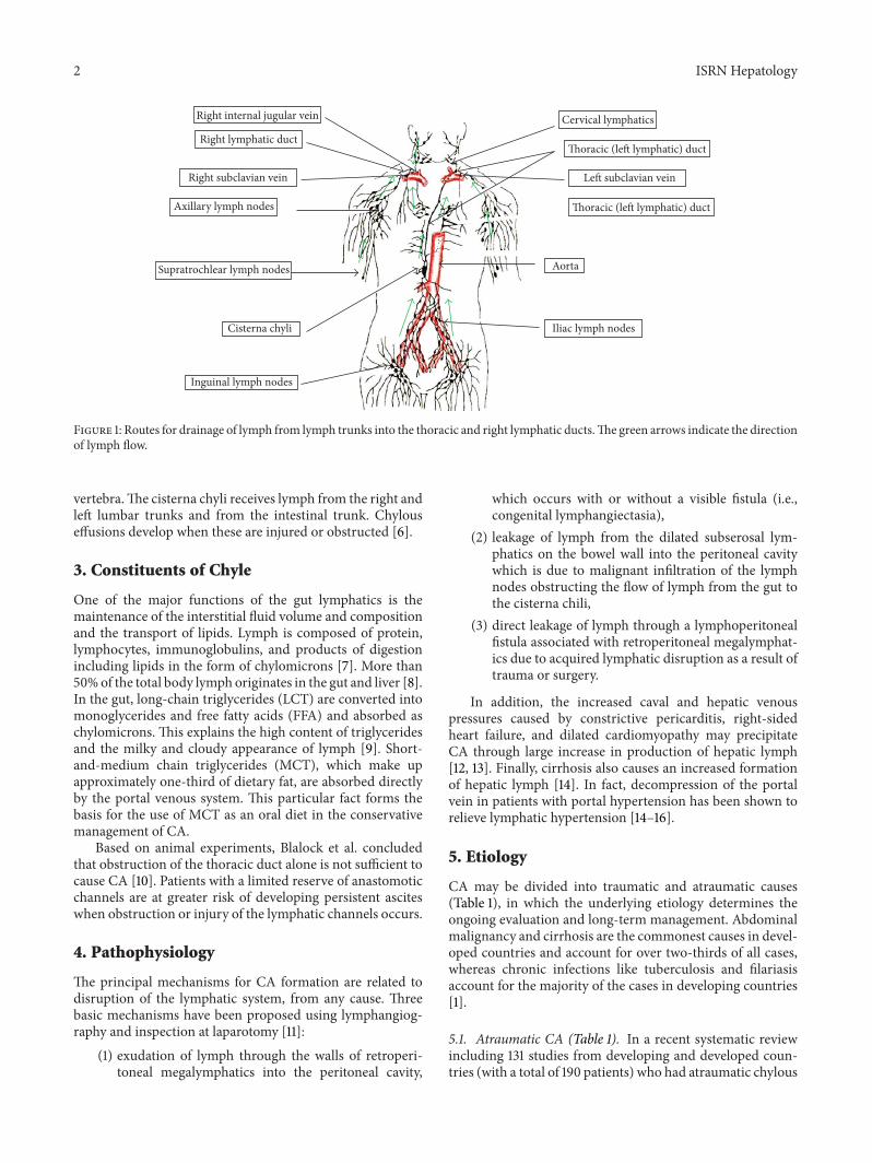
The lymphatic system includes lymph, lymphatic vessels,lymphatic tissues, and red bone marrow (Figure 1) [5]. Itis a one-way drainage system which allows the return ofexcess interstitial fluids and proteins to the vascular system[2]. Lymph passes from lymphatic capillaries into lymphaticvessels and then through lymph nodes into lymph trunks(Figure 1). The thoracic duct, the main duct for the returnof lymph to blood, is about 38–45 cm long and begins asdilation called the cisterna chyli anterior to the second lumbar
Hindawi Publishing CorporationISRN HepatologyVolume 2014, Article ID 240473, 10 pageshttp://dx.doi.org/10.1155/2014/240473
2 ISRN Hepatology
Right internal jugular vein
Right subclavian vein
Axillary lymph nodes
Supratrochlear lymph nodes
Cervical lymphatics
Cisterna chyli
Inguinal lymph nodes
Aorta
Iliac lymph nodes
Right lymphatic ductThoracic (left lymphatic) duct
Left subclavian vein
Thoracic (left lymphatic) duct
Figure 1: Routes for drainage of lymph from lymph trunks into the thoracic and right lymphatic ducts.The green arrows indicate the directionof lymph flow.
vertebra.The cisterna chyli receives lymph from the right andleft lumbar trunks and from the intestinal trunk. Chylouseffusions develop when these are injured or obstructed [6].
3. Constituents of Chyle
One of the major functions of the gut lymphatics is themaintenance of the interstitial fluid volume and compositionand the transport of lipids. Lymph is composed of protein,lymphocytes, immunoglobulins, and products of digestionincluding lipids in the form of chylomicrons [7]. More than50%of the total body lymph originates in the gut and liver [8].In the gut, long-chain triglycerides (LCT) are converted intomonoglycerides and free fatty acids (FFA) and absorbed aschylomicrons. This explains the high content of triglyceridesand the milky and cloudy appearance of lymph [9]. Short-and-medium chain triglycerides (MCT), which make upapproximately one-third of dietary fat, are absorbed directlyby the portal venous system. This particular fact forms thebasis for the use of MCT as an oral diet in the conservativemanagement of CA.
Based on animal experiments, Blalock et al. concludedthat obstruction of the thoracic duct alone is not sufficient tocause CA [10]. Patients with a limited reserve of anastomoticchannels are at greater risk of developing persistent asciteswhen obstruction or injury of the lymphatic channels occurs.
4. Pathophysiology
The principal mechanisms for CA formation are related todisruption of the lymphatic system, from any cause. Threebasic mechanisms have been proposed using lymphangiog-raphy and inspection at laparotomy [11]:
(1) exudation of lymph through the walls of retroperi-toneal megalymphatics into the peritoneal cavity,
which occurs with or without a visible fistula (i.e.,congenital lymphangiectasia),
(2) leakage of lymph from the dilated subserosal lym-phatics on the bowel wall into the peritoneal cavitywhich is due to malignant infiltration of the lymphnodes obstructing the flow of lymph from the gut tothe cisterna chili,
(3) direct leakage of lymph through a lymphoperitonealfistula associated with retroperitoneal megalymphat-ics due to acquired lymphatic disruption as a result oftrauma or surgery.
In addition, the increased caval and hepatic venouspressures caused by constrictive pericarditis, right-sidedheart failure, and dilated cardiomyopathy may precipitateCA through large increase in production of hepatic lymph[12, 13]. Finally, cirrhosis also causes an increased formationof hepatic lymph [14]. In fact, decompression of the portalvein in patients with portal hypertension has been shown torelieve lymphatic hypertension [14–16].
5. Etiology
CA may be divided into traumatic and atraumatic causes(Table 1), in which the underlying etiology determines theongoing evaluation and long-term management. Abdominalmalignancy and cirrhosis are the commonest causes in devel-oped countries and account for over two-thirds of all cases,whereas chronic infections like tuberculosis and filariasisaccount for the majority of the cases in developing countries[1].
5.1. Atraumatic CA (Table 1). In a recent systematic reviewincluding 131 studies from developing and developed coun-tries (with a total of 190 patients) who had atraumatic chylous

ISRN Hepatology 3
Table 1: Etiological classification of chylous ascites.
Atraumatic [2] Traumatic(I) Neoplastic Cardiac (I) IatrogenicSolid organ cancers Constrictive pericarditis (A) SurgicalLymphoma Congestive heart failure Abdominal aneurysm repairSarcoma Gastrointestinal Retroperitoneal lymphadenectomyCarcinoid tumors Celiac sprue Placement of peritoneal dialysis catheterLymphangioleiomyomatosis Whipple’s disease Inferior vena cava resectionChronic lymphatic leukemia Intestinal malrotation Pancreaticoduodenectomy(II) Diseases Small bowel volvulus Vagotomy(A) Congenital Menetrier disease Radical and laparoscopic nephrectomyPrimary lymphatic hypoplasia Inflammatory Nissen fundoplicationKlippel-Trenaunay syndrome Pancreatitis Distal splenorenal shuntsYellow nail syndrome Fibrosing mesenteritis Laparoscopic adrenalectomyPrimary lymphatic hyperplasia Retroperitoneal fibrosis Gynecological surgeryLymphangioma Sarcoidosis (B) NonsurgicalFamilial visceral myopathy Systemic lupus erythematosus Radiotherapy(B) Acquired Behcet’s disease (II) NoniatrogenicCirrhosis Peritoneal dialysis Blunt abdominal traumaInfectious Hyperthyroidism Battered child syndromeTuberculosis Nephrotic syndrome Penetrating abdominal traumaFilariasis Drugs Shear forces to the root of the mesenteryMycobacterium avium in AIDS Calcium channel blockers (III) IdiopathicAscariasis Sirolimus Rule out lymphoma
ascites, the most common causes in adults were malignancy(25%), cirrhosis (16%), mycobacterium infection (15%), and avariety of uncommon causes (23%) [17]. In children, themostcommon causes were lymphatic anomalies (84%) followedby a variety of uncommon causes (15%). Other causes ofCA include trauma, including surgical and radiotherapy, andother atraumatic, including congenital, inflammatory, andsystemic disorders.
5.1.1. Neoplastic Causes. The most common cause of CAin adults is malignancy. Among the group of malignancies,lymphoma accounts for at least one-third of the cases [17].Tumors through direct invasion or extrinsic compressionlead to disruption of normal lymphatic flow [18]. In addi-tion to lymphomas, other tumors that cause CA may arisefrom the intra-abdominal solid organ malignancies such asstomach, esophagus, pancreas, endometrial, and prostate,which account for 40% of all malignant causes [17]. Carcinoidtumours andKaposi sarcoma account for 15% and 9%, respec-tively, of the malignancy-related cases. Lymphangioleiomy-omatosis is a rare benign tumor of lymphatic channelsand lymph nodes, clinically manifested by chylous effusionsincluding CA [19].
5.1.2. Congenital. Congenital lymphatic anormalities are pre-dominant cause in the pediatric population, wich accountsfor 84% of all causes. In contrast, lymphatic anomalies
account only for 9% of atraumatic CA in adults [17]. Pri-mary lymphatic hyperplasia has been recognized as causeof chylous ascites [20]. It consists of two principal patterns:“bilateral hyperplasia” inwhich the lymphatics are not grosslydilated and contain valves and lymphangiectasia in whichlymphatics are grossly dilated in the wall of small bowel andhave no valves. The primary intestinal lymphangiectasia isresponsible for the majority of the cases in children [17].
Primary lymphatic hypoplasia is another conditionseen most commonly in children and presents with lym-phedema, chylothorax, CA, or combination [21].TheKlippel-Trenaunay syndrome is an autosomal dominant inheriteddisorder characterized by venous and lymphatic hypoplas-tic malformations which can manifest as lower limb lym-phedema and it is often associated with chylous ascites[22]. The yellow-nail syndrome is a childhood disorder ofunknown etiology. The patients have hypoplastic or aplasticlymphatics leading to the characteristic features of lowerlimb lymphedema, pleural effusion, and/or CA and a yellowdiscoloration with dystrophy of the nails [23].
5.1.3. Cirrhosis. Although ascites is a commonmanifestationof hepatic cirrhosis, CA presents in 0.5–1% of patients withcirrhosis [15, 16, 24]. Recently, a systematic review showedthat cirrhosis was responsible for 11% of atraumatic chylousascites [17]. This discrepancy was explained by the authors tobe related to the under diagnosis.

4 ISRN Hepatology
Cirrhotic patients may present with CA as an initialpresentation or might present at a later stage of the diseasedue to complications such as shunt surgery, sclerotherapy-related thoracic duct injury, or hepatocellular carcinoma[24–26]. However, unless clinically indicated, an aggressiveapproach to exclude malignancy is not warranted. Othercause of CA in cirrhotic patients that should be consideredis portal vein thrombosis [27].
5.1.4. Infectious. Lymphatic filariasis and peritoneal tuber-culosis are the most common infectious causes of CA andaccount for the majority of the cases in the developing coun-tries [1]. Low socioeconomic status, malnutrition, cirrhosis,HIV infection, diabetes mellitus, underlyingmalignancy, andambulatory peritoneal dialysis are risk factors for tuberculousCA [28–30]. Lymphatic filariasis causes severe inflammatoryreaction in lymphatic vessels leading to lymphedema and CA[31]. Infection with Mycobacterium avium-intracellulare hasbeen reported to cause CA in AIDS patients [32]. A recentsystematic review showed that infections with mycobacterialspecies, as MAI infection and tuberculosis, contribute to 10%of all cases of atraumatic CA [17].
5.1.5. Inflammatory. A variety of inflammatory causes havebeen reported to be associated with CA. Both acute pancre-atitis and chronic pancreatitis have been associated with CA[33]. Two mechanisms have been proposed to play a role inthe development of CA by pancreatitis, which are compres-sion of lymphatic channels or direct damage by pancreaticenzymes [33]. Fibrosing mesenteritis is rare benign processthat involves inflammation, fat necrosis, and fibrosis of themesentery [34] has been also reported to causeCA [17].Otherrare inflammatory causes include idiopathic retroperitonealfibrosis, [35] sarcoidosis, [36] systemic lupus erythematous,[37] peritoneal dialysis, [38] and hyperthyroidism [39].
5.1.6. Other Causes. Constrictive pericarditis has been alsoreported to cause CA [40, 41]. It causes impaired lymphdrainage with thoracic duct dilatation and hypertensionleading to an increase in hepatic venous pressure, therebyincreasing lymph production [10]. Congestive heart failurecan also cause CA by increasing formation of and impairedlymphatic drainage [12, 42, 43]. CA may develop as a resultof heart failure secondary to thyrotoxic cardiomyopathy andresolve promptly if treated appropriately [44]. The nephroticsyndrome has been reported for unknown mechanism tocause chylous effusions including CA [45, 46]. A studyincluding 90 patients with nephritic syndrome and ascites,showed that 45% of those who underwent paracentesis(16/35) had CA. However, the diagnosis was based ondetection of opalescent effusion rather than by checking thetriglyceride level of peritoneal fluid [47].
Celiac disease andWhipple’s disease can cause CA due tomesenteric node hyperplasia [48]. Calcium channel blockershave also been implicated as a cause of CA in patients under-going peritoneal dialysis [49–51]. In addition, sirolimus, inrenal transplant setting, has also been reported to cause CA[52].
5.2. Traumatic CA (Table 1)
5.2.1. Postoperative. Surgical interventions are well-knowncauses of CA secondary to direct lymphatic vessels injury.CA can occur as early as 1 week after abdominal surgerybecause of disruption of the lymphatic vessels or as late asseveral weeks to months because of adhesions or extrinsiccompression of lymphatic vessels [1]. A retrospective studyover a 2-year period of a cohort including 1,103 oncologicalpatients undergoing abdominal surgical procedures revealed1.1% incidence of postoperative chylous ascites. 7.4% ofpatients who underwent retroperitoneal, esophageal, gastric,or cytoreductive surgeries developed CA [53]. Other sur-gical procedures that can result in CA include aortic andabdominal aneurysm repair [54], retroperitoneal lymphnodedissection [55], inferior vena cava resection [56], catheterimplantation for peritoneal dialysis [38], distal splenorenalshunts [57], small bowel transplantation [58], liver transplan-tation [59], choledochal cyst excision [60], pancreaticoduo-denectomy [61], anterior spinal surgery [62], laparoscopicsurgeries including Nissen fundoplication [63], Roux-en-Y gastric bypass [64], adrenalectomy [65], cholecystectomy[66], and donor nephrectomy [67].
5.2.2. Radiotherapy. Abdomen and pelvic radiation is a com-mon cause of CA [2]. In a review done at Mayo clinic involv-ing 207 patients who received whole abdomen irradiationfor gynecologic malignancies, a 3% incidence of CA wasreported [68]. Irradiation to abdomen can cause fibrosis oflymphatic vessels within small bowel and mesentery leadingto obstruction and extravasation of lymph [69] which istypically observed after a mean of 12 months after radiationtherapy [70].
5.2.3. Noniatrogenic Causes. In contrast to direct injury oflymphatic vessels during surgery, blunt abdominal traumacauses CA through hyperextension and hyperflexion leadingto rupture of lymphatic vessels and lymph leakage [71].Penetrating abdominal trauma has also been reported tocause CA [72]. The battered child syndrome, which can leadto blunt abdominal trauma, accounts for approximately 10%of cases of CA in children [73].Therefore, it is very importantto exclude this diagnosis in any child presenting with CA.
6. Complications of Chylous Ascites
Loss of chyle into peritoneal cavity can lead to seriousconsequences because of the loss of essential proteins, lipids,immunoglobulins, vitamins, electrolytes, and water. Whilerepeated therapeutic paracentesis provides relief from symp-toms, the nutritional deficiency will continue to persistor deteriorate unless definitive therapeutic measures areinstituted to stop leakage of chyle into the peritoneal space.In fact, in postoperative settings, this may cause increasedmortality [74]. Therefore, it is very important to provideadequate nutritional support replenishing fluid loss, vitamindeficiencies, and electrolyte loss while specific therapeuticmeasures are planned.

ISRN Hepatology 5
In addition, continued loss of lymphocyte-rich lymphinto the peritoneal space and enormous loss of protein ingastrointestinal tract lead to hypogammaglobulinemia andtherefore increased susceptibility to infection [75]. Prolongedthoracic duct drainage has been used previously to induceimmunosuppression in several diseases including rheuma-toid arthritis and myasthenia gravis [75].
The bioavailability of certain drugs could be drasticallyimpaired in the presence of significant chyle leak. There arereports of this phenomenon in patients with chylothorax-causing subtherapeutic digoxin [76], amiodarone [77], andcyclosporine [78] levels in the serum. Sequestration of drugsin chyle should be recognized early, to prevent subtherapeuticplasma levels in patients undergoing drainage of CA.
7. Evaluation and Diagnosis
The diagnostic approach of CA consists of first suspectingthe diagnosis, then confirming the presence of chyle in theperitoneal cavity, and finally determining the underlyingabnormality. A careful history, physical examination, anddiagnostic paracentesis are the key in the initial evaluationof any patient presenting with ascites.
7.1. Clinical Findings. Progressive and painless abdominaldistention (81%) and nonspecific pain (14%) are the mostcommon presenting symptoms in CA, occurring over acourse of weeks to months depending on the underlyingcause [17]. Patients who have undergone abdominal orthoracic surgery may present with an acute onset of CA [2].Patients may also present weight gain and dyspnea resultingfrom increased abdominal girth [1]. Other features includeweight loss, anorexia, malaise, steatorrhea, malnutrition,enlarged lymph nodes, fevers, and night sweats [2, 3, 11].However, most often the diagnosis of CA is not suspectedbefore performing a diagnostic paracentesis [1].
Physical signs that may be present on examinationinclude ascites, pleural effusions, lower extremity edema,lymphadenopathy, cachexia, temporal wasting, and hernias[1]. Other findings depend on the underlying cause.
7.2. Laboratory Findings. Abdominal paracentesis is themost important diagnostic tool in evaluating and managingpatients with ascites. In contrast to the yellow and transparentappearance of ascites due to cirrhosis and portal hyperten-sion, chyle typically has a cloudy and turbid appearance(Table 2). This should be distinguished from pseudochylousascites, in which the turbid appearance is due to cellulardegeneration from infection or malignancy without actuallycontaining high levels of triglycerides [79]. Depending on theclinical suspicion, ascitic fluid should be sent for cell count,culture,Gram stain, total protein, albumin, triglyceride levels,glucose, lactate dehydrogenase, amylase, and cytology [80].The serum to ascites albumin gradient (SAAG) should becalculated to determine if the ascites is related to portalhypertension or other causes [80]. The triglyceride levels inascitic fluid are very important in defining CA. Triglyceridevalues are typically above 200mg/dL, although some authors
Table 2: Characteristics of ascitic fluids in chylous ascites (adaptedfrom Cardenas and Chopra) [1].
Color Milky and cloudyTriglyceride level Above 200mg/dLCell count Above 500 (lymphocytic predominance)Total protein Between 2.5 and 7.0 g/dLSAAG Below 1.1 g/dL∗
Cholesterol Low (ascites/serum ratio < 1)Lactate dehydrogenase Between 110 and 200 IU/LCulture Positive in selected cases of tuberculosisCytology Positive in malignancyAmylase Elevated in cases of pancreatitisGlucose Below 100mg/dLIU: international units; SAAG: serum-ascites albumin gradient.∗Is elevated above 1.1 g/dL in CA secondary to cirrhosis.
use a cutoff value of 110mg/dL [1, 3]. A tuberculosis smearand culture and adenosine deaminase activity (ADA) shouldbe performed in selected cases when tuberculosis is suspected[1]. ADA has high sensitivity and specificity in the diagnosisof tuberculous peritonitis [81]. In contrast, its utility in pop-ulations with high prevalence of cirrhosis such as the UnitedStates is limited [82].The diagnosis of tuberculous peritonitisusually requires a peritoneal biopsy via laparoscopy [83].
Standard blood tests, including a complete blood count,electrolytes, liver tests, total protein, albumin, lactate dehy-drogenase, triglycerides, cholesterol, amylase, and lipaseshould be performed. Additional testing should be basedupon the clinical setting [84].
7.3. Imaging Studies
7.3.1. Computed Tomography (CT). Chyle has a water densityappearance on CT which can be readily distinguished fromacute hemorrhage in the setting of trauma [2]. CT of theabdomen is useful in identifying pathologic intra-abdominallymph nodes and masses. In the setting of postoperative ortraumatic causes of CA, it also helps in determining the extentand localization of fluid, particularly, if there is a suspicion ofthoracic duct injury [1, 2]. Other finding on CTmight suggestthat CA is formation of fluid-fluid level [85]. Another CTtechnique reported is the direct opacification of the thoracicduct with oral fat emulsions [86].
7.3.2. Lymphoscintigraphy. Lymphoscintigraphy allows func-tional assessment of lymphatic transport and so can be usedto detect abnormal lymphatic drainage in CA. It is usefulfor detecting patients for surgery and assessing the effect oftreatment [87–89]. It can be used when lymphangiography iscontraindicated [2]. Its advantages include no adverse effects,no contraindications, and the ability to perform repetitivestudies. The technical challenges of this technique and itsrare implementation may make it an unfavorable diagnosticmodality [2].

6 ISRN Hepatology
7.3.3. Lymphangiography. Lymphangiography use has beendeclining with the availability of noninvasive imaging, butit remains the gold standard in defining cases of lymphaticobstruction. It has been successfully used to detect abnormalretroperitoneal nodes, leakage from dilated lymphatics, fis-tulization, and patency of the thoracic duct [11, 90, 91].
In addition, lymphangiography is also used in treatingpatients with chyle leakages who are resistant to conservativeapproach [92]. However, it is associated with complica-tions including contrast hypersensitivity, tissue necrosis, fatembolism, and even transient lymphedema and CA [87, 93].
7.4. Laparoscopy. Ascites of unknown etiology is a commonindication for laparoscopy in patients with ascites especiallywhen tuberculosis or malignancy is suspected [2]. It wasreported to be perhaps the best and most definitive methodto diagnose intestinal lymphangiectasia [20].
7.5. Laparotomy. Early reoperation has been recommendedfor postoperative CA to address the underlying cause aswell as for providing treatment [94]. It is advocated todo combined pre- and intraoperative lymphangiography tofacilitate successful treatment of postoperative CA [95].
8. Management of CA
Few studies have addressed the best treatment regimens forCA [2]. Nutritional regimens and pharmacological and sur-gical therapies exist but there is still a lack of a clear consensuson the optimal management of CA [96]. Treatment of theunderlying cause is an important initial step in managingthese patients. In most cases, particularly, in patients withinfectious, inflammatory, or hemodynamic cause, this willresult in resolution of symptoms and of the ascites [1].
8.1. Medical Treatment. Medical management of CA is basedon the theory that decreasing chyle flow will allow forspontaneous closure of the chyle leak [96]. However, there isno precise, functional method formonitoring the response totherapy [2].
8.1.1. Dietary Therapy. Based on the limited studies and noclear consensus, it is a reasonable approach for patients inwhom the cause was not found or for those who did notrespond to treatment of the underlying cause to recommendthe nutritional therapy. Although it is common in practiceto recommend bowel rest and dietary modification, enteralfeedings, or the use of total parenteral nutrition (TPN),definitive evidence supporting one nutrition therapy overanother does not exist [96]. Goal of nutrition therapy is todecrease production of chyle, replace fluid and electrolytes,and maintain or improve nutrition status [97].
A reasonable approach is to recommend a high-proteinand low-fat diet with MCT. Dietary restriction of LCTprevents their conversion into monoglycerides and FFA,which are transported as chylomicrons to the intestinallymph ducts. In contrast, MCT are absorbed directly intointestinal cells and transported as FFA and glycerol directly
to the liver via the portal vein. Thus, a low-fat diet with MCTsupplementation reduces the production and flow of chyle[98]. Patients with advanced cirrhosis,MCT oil should not beused as narcosis and coma may occur. Such patients shouldbe managed with a low-sodium diet and diuretics such asspironolactone [99].
Patients who do not respond to the above measuresshould have bowel rest to reduce lymphflow and be started onTPN [2]. TPN is theoretically superior to any enteral feedingsbecause the bowel is bypassed. The presence of intraluminalwater alone has been shown to increase thoracic duct lymphflow [2]. TPN along with somatostatin or octreotide canrelieve the symptoms and rapidly close the fistula in patientswith CA [100]. This approach appears to be an effectivetherapy for the treatment of CA caused by various disorders[101].
8.1.2. Pharmacology. There are other medical measureswhich have been described in literature as either case reportsor small observational studies. Orlistat, a reversible inhibitorof gastric and pancreatic lipases, was reported to minimizeascites and triglyceride levels in ascitic fluid in a patientwith CA due to cirrhosis [102]. Case reports have suggestedthat both somatostatin and octreotide either alone or incombination with TPN are effective in the management ofCA due to different causes [67, 101, 103–106].Themechanismmay involve inhibition of lymph fluid excretion throughspecific receptors found in the normal intestinal wall of lym-phatic vessels [107]. In case reports, a promising treatment,etilefrine, a sympathomimetic drug, was shown to causeresolution of postesophagectomy chylous effusions [108].
8.2. Abdominal Paracentesis. In patients with symptomaticascites, a therapeutic paracentesis should be performed torelieve symptoms and could be repeated as needed [1].Unless the patient has cirrhosis, the replacement of albu-min to prevent postparacentesis circulatory dysfunction isnot recommended. Repeated large-volume paracentesis is areasonable option for patients who have end-stage disease notamenable to medical or surgical treatment.
8.3. Transjugular Intrahepatic Portosystemic Shunt (TIPS).The use of TIPS to successfully treat CA has been reportedin patients with cirrhosis and CA resistant to conservativetherapy and who have reasonable liver function [109–111].However, the placement of TIPS in a patient with cirrhosisis associated with significant problems so patients must beselected carefully.
8.4. Peritoneovenous Shunting. In the past, peritoneovenousshunts (LeVeen or Denver shunts) were considered optionsfor patients who were refractory to medical therapy and poorcandidates for surgery. However, these shunts were associatedwith a high rate of serious complications, such as sepsis,disseminated intravascular coagulation, hypokalemia, smallbowel obstruction, and risk for air embolism, and are thusseldom used [112]. In addition, the high viscosity of the chyle

ISRN Hepatology 7
results in a high rate of shunt occlusion in majority of thecases [3, 94, 113].
8.5. Angiography. In addition, to make diagnosis, lymphan-giography with or without embolization is another promisingtechnique which has been described for the literature in thetreatment of postoperative CA when conservative therapyfails [90, 92, 114].
8.6. Surgical Treatment. If the above conservative manage-ment is not successful in treating CA, surgical interventionmay be beneficial especially in patients with postoperative,neoplastic, and congenital causes [2]. Preoperative lymphan-giography or lymphoscintigraphy is helpful in identifyingthe anatomical location of the leakage or the presence ofa fistula [3]. Laparotomy is also essential in the diagnosisand management of acute chylous peritonitis. In a reviewwhere all patients were initially treated conservatively withdietary therapy, surgery (fistula closure, bowel resection, orinsertion of a peritoneovenous shunt) was performed inpatients who failed conservative therapy (66%). Closure of aretroperitoneal fistula, when present, was the most success-ful operation [11]. However, in postoperative CA, surgicalreinterventions are associated with significant incidence ofmorbidity and mortality [115]. In addition, surgery mayoccasionally fail to identify the leak. Some promising newtechniques (e.g., use of octreotide, etilefrine, or angiography),which—alone or in combination with well-established con-servative measures—may have the potential to avoid surgicalreinterventions [115].
In addition, to prevent postoperative CA, it was foundthat the milk test is a safe and effective method followingpancreatectomy [116]. In children, fibrin glue application forcontrol of lymph leakage is also effective in prevention andmanagement of postoperative CA [117] as well as manage-ment of congenital CA [118].
9. Conclusion
In summary, CA is a relatively uncommon disorder. Malig-nancy and cirrhosis are the leading causes of this condition inadults. In contrast, congenital abnormalities of the lymphaticsystem and trauma are common causes in children. Paracen-tesis with confirmation of elevated triglyceride is consideredto be the gold standard for diagnosis of CA. In a cirrhoticpatient, unless there is a strong suspicion of malignancy,there is no need for unnecessary and invasive diagnostictests to rule out a malignant cause. Treatment of the under-lying cause is an important initial step in managing thesepatients. Conservative approach includes the use of a low-fat diet, MCT intake, paracentesis, TPN, and somatostatins.Other treatment options for resistant cases include TIPS,surgical exploration, and peritoneovenous shunt. However,some promising new techniques such as use of etilefrineor percutaneous embolisation of cisterna chyli are waitingfurther evaluation.
Conflict of Interests
The authors declare that there is no conflict of interestsregarding the publication of this paper.
References
[1] A. Cardenas and S. Chopra, “Chylous ascites,”American Journalof Gastroenterology, vol. 97, no. 8, pp. 1896–1900, 2002.
[2] O. O. Aalami, D. B. Allen, and C. H. Organ Jr., “Chylous ascites:a collective review,” Surgery, vol. 128, no. 5, pp. 761–778, 2000.
[3] O. W. Press, N. O. Press, and S. D. Kaufman, “Evaluation andmanagement of chylous ascites,” Annals of Internal Medicine,vol. 96, no. 3, pp. 358–364, 1982.
[4] J. S. Vasko and R. I. Tapper, “The surgical significance of chylousascites,” Archives of Surgery, vol. 95, no. 3, pp. 355–368, 1967.
[5] G. J. Tortora and M. Nielsen, Principles of Human Anatomy,John Wiley & Sons, Hoboken, NJ, USA, 12th edition, 2012.
[6] J. R.Malagelada, F. L. Iber, andW. G. Linscheer, “Origin of fat inchylous ascites of patientswith liver cirrhosis,”Gastroenterology,vol. 67, no. 5, pp. 878–886, 1974.
[7] D. B. Zilversmit, “The composition and structure of lymphchylomicrons in dog, rat, and man,” The Journal of ClinicalInvestigation, vol. 44, no. 10, pp. 1610–1622, 1965.
[8] G. T. Lesser, M. S. Bruno, and K. Enselberg, “Chylous ascites.Newer insights and many remaining enigmas,” Archives ofInternal Medicine, vol. 125, no. 6, pp. 1073–1077, 1970.
[9] M. L. Paes and H. Powell, “Chylothorax: an update,” BritishJournal of Hospital Medicine, vol. 51, no. 9, pp. 482–490, 1994.
[10] A. Blalock, C. S. Robinson, R. S. Cunningham, and M. E.Gray, “Experimental studies on lymphatic blockage,” Archivesof Surgery, vol. 34, pp. 1049–1071, 1937.
[11] N. L. Browse, N. M. Wilson, F. Russo, H. Al-Hassan, and D.R. Allen, “Aetiology and treatment of chylous ascites,” BritishJournal of Surgery, vol. 79, no. 11, pp. 1145–1150, 1992.
[12] M. K. Hurley, V. J. Emiliani, G. M. Comer, A. Patel, C. Navarro,and C. O. Maiki, “Dilated cardiomyopathy associated withchylous ascites,” American Journal of Gastroenterology, vol. 84,no. 12, pp. 1567–1569, 1989.
[13] S. Guneri, C. Nazli, O. Kinay, O. Kirimli, C. Mermut, andE. Hazan, “Chylous ascites due to constrictive pericarditis,”International Journal of Cardiac Imaging, vol. 16, no. 1, pp. 49–54, 2000.
[14] B. T. Maywood, L. Goldstein, and R. W. Busuttil, “Chylousascites after a Warren shunt,” American Journal of Surgery, vol.135, no. 5, pp. 700–702, 1978.
[15] W. S. C. Cheng, I. R. Gough, M. Ward, J. Croese, and L. W.Powell, “Chylous ascites in cirrhosis: a case report and reviewof literature,” Journal of Gastroenterology and Hepatology, vol. 4,no. 1, pp. 95–99, 1989.
[16] W. G. Rector Jr., “Spontaneous chylous ascites of cirrhosis,”Journal of Clinical Gastroenterology, vol. 6, no. 4, pp. 369–372,1984.
[17] D. C. Steinemann, D. Dindo, P.-A. Clavien, and A. Nocito,“Atraumatic chylous ascites: systematic review on symptomsand causes,” Journal of the AmericanCollege of Surgeons, vol. 212,no. 5, pp. 899–905, 2011.
[18] T. Almakdisi, S. Massoud, and G. Makdisi, “Lymphomas andchylous ascites: review of the literature,” Oncologist, vol. 10, no.8, pp. 632–635, 2005.
8 ISRN Hepatology
[19] V. J. Ferrans, Z. X. Yu, W. K. Nelson et al., “Lymphangi-oleiomyomatosis (LAM): a reviewof clinical andmorphologicalfeatures,” Journal of NipponMedical School, vol. 67, no. 5, pp. 311–329, 2000.
[20] U. Fox and G. Lucani, “Disorders of the intestinal mesentericlymphatic system,” Lymphology, vol. 26, no. 2, pp. 61–66, 1993.
[21] S. W. Unger and J. G. Chandler, “Chylous ascites in infants andchildren,” Surgery, vol. 93, no. 3, pp. 455–461, 1983.
[22] M. Cohen Jr., “Klippel-Trenaunay syndrome,”American Journalof Medical Genetics, vol. 93, no. 3, pp. 171–175, 2000.
[23] P. M. Duhra, E. M. M. Quigley, and M. N. Marsh, “Chylousascites, intestinal lymphangiectasia and the “yellow-nail” syn-drome,” Gut, vol. 26, no. 11, pp. 1266–1269, 1985.
[24] S. Sultan, A. Pauwels, R. Poupon, and V. G. Levy, “Chylousascites in cirrhosis. A retrospective study of 20 cases,”Gastroen-terologie Clinique et Biologique, vol. 14, no. 11, pp. 842–847, 1990.
[25] L. Vargas-Tank, R. Estay, L. Ovalle, J. R. Soto, and M. E.Villanueva, “Esophageal sclerotherapy and chylous ascites,”Gastrointestinal Endoscopy, vol. 40, no. 3, p. 396, 1994.
[26] F. M. Gomez Soto, F. Marcos Sanchez, A. I. Franco Moreno,A. Viana Alonso, A. I. Munoz Ruiz, and A. D. Perez-Navarro,“Chylous ascites chylosus as a manifestation of hepatocarci-noma,” Gastroenterologia y Hepatologia, vol. 26, no. 4, pp. 276–277, 2003.
[27] R. W. L. Leong, A. K. House, and G. P. Jeffrey, “Chylous ascitescaused by portal vein thrombosis treated with octreotide,”Journal of Gastroenterology and Hepatology, vol. 18, no. 10, pp.1211–1213, 2003.
[28] J. B. Mehta, A. Dutt, L. Harvill, and K. M. Mathews, “Epidemi-ology of extrapulmonary tuberculosis: a comparative analysiswith pre-AIDS era,” Chest, vol. 99, no. 5, pp. 1134–1138, 1991.
[29] M. M. Braun, R. H. Byers, W. L. Heyward et al., “Acquiredimmunodeficiency syndrome and extrapulmonary tuberculosisin the United States,” Archives of Internal Medicine, vol. 150, no.9, pp. 1913–1916, 1990.
[30] J. M. Aguado, F. Pons, F. Casafont, G. San Miguel, and R.Valle, “Tuberculous peritonitis: a study comparing cirrhotic andnoncirrhotic patients,” Journal of Clinical Gastroenterology, vol.12, no. 5, pp. 550–554, 1990.
[31] R. M. Patel and E. Purow, “Chylous ascites and chylothorax.Presenting manifestation of pancreatic carcinoma,” New YorkState Journal of Medicine, vol. 82, no. 3, pp. 349–351, 1982.
[32] P. Phillips, J. K. Lee, C. Wang, E. Yoshida, V. D. Lima, andJ. Montaner, “Chylous ascites: a late complication of intra-abdominal Mycobacterium avium complex immune recon-stitution syndrome in HIV-infected patients,” InternationalJournal of STD and AIDS, vol. 20, no. 4, pp. 285–287, 2009.
[33] J. P. Goldfarb, “Chylous effusions secondary to pancreatitis:case report and review of the literature,” American Journal ofGastroenterology, vol. 79, no. 2, pp. 133–135, 1984.
[34] B. White, A. Kong, and A.-L. Chang, “Sclerosing mesenteritis,”Australasian Radiology, vol. 49, no. 2, pp. 185–188, 2005.
[35] Y. Sakai and T. Nasu, “Idiopathic retroperitoneal fibrosis,” ActaPathologica Japonica, vol. 26, no. 5, pp. 637–647, 1976.
[36] J. M. Provenza and B. R. Bacon, “Chyloperitoneum associatedwith sarcoidosis,”American Journal of Gastroenterology, vol. 88,no. 9, pp. 1462–1463, 1993.
[37] S. B’Chir Hamzaoui, M. Abdallah, K. Bouslama et al., “Chylousascites revealing a systemic lupus erythematosus,” Gastroen-terologie Clinique et Biologique, vol. 31, no. 1, pp. 100–101, 2007.
[38] C. K. Cheung and A. Khwaja, “Chylous ascites: an unsualcomplication of peritoneal dialysis. A case report and literaturereview,” Peritoneal Dialysis International, vol. 28, no. 3, pp. 229–231, 2008.
[39] N. Hiroi, Y. Sakamoto, Y. Urita, M. Higa, K. Kuboki, and G.Yoshino, “Graves’ disease with intractable diarrhea, chylousascites, and chylothorax: a case report,” Thyroid, vol. 17, no. 12,pp. 1299–1303, 2007.
[40] R.W. England, K.W.Grathwohl, andG. E. Powell, “Constrictivepericarditis presenting as chylous ascites,” Journal of ClinicalGastroenterology, vol. 35, no. 1, pp. 104–105, 2002.
[41] B. Amasyali, G. Heper, O. Akkoc, U. C. Yuksel, A. Kilic,and E. Isik, “Chylous ascites and pleural effusion secondaryto constrictive pericarditis presenting with signs of lymphaticobstruction,” Japanese Heart Journal, vol. 45, no. 3, pp. 535–540,2004.
[42] H. A. Cakmak, G. Yenidunya, B. Karadag, and Z. Ongen,“Development of chylothorax and chylous ascites in a patientwith congestive heart failure,” Turk Kardiyoloji Dernegi Arsivi,vol. 39, no. 6, pp. 495–498, 2011.
[43] E. Ridruejo and O. G. Mando, “Chylous ascites as the mainmanifestation of left ventricular dysfunction: a case report,”BMC Gastroenterology, vol. 5, article 25, 2005.
[44] M.-H. Hsieh, C.-C. Chen, T.-Y. Wang, and C.-T. Chang, “Chy-lous ascites as a manifestation of thyrotoxic cardiomyopathy ina patient with untreated Graves’ disease,”Thyroid, vol. 20, no. 6,pp. 653–655, 2010.
[45] A. Kato, S. Kohno, T. Ohtake, T. Takita, and A. Hirshida,“Chylous ascites in an adult patient with nephrotic syndromedue to membranous nephropathy,” Nephron, vol. 89, no. 3, pp.361–362, 2001.
[46] S. Lewsuwan, T. Kanjanabuch, Y. Avihingsanon, K. Praditporn-silpa, and S. Eiam-Ong, “A rare case of chylous ascites andchyluria in an adult nephrotic syndrome with focal segmentalglomerulosclerosis,” Journal of the Medical Association of Thai-land, vol. 89, supplement 2, pp. S253–S256, 2006.
[47] J. Lindenbaum and S. S. Scheidt, “Chylous ascites and thenephrotic syndrome. Report of a case, associatedwith renal veinthrombosis,” The American Journal of Medicine, vol. 44, no. 5,pp. 830–836, 1968.
[48] H. J. Kaufmann, “Chylous ascites and intestinal muscularhypertrophy occurring in the course of celiac sprue,” AmericanJournal of Digestive Diseases, vol. 20, no. 5, pp. 494–497, 1975.
[49] Y.-T. Tsao and W.-L. Chen, “Calcium channel blocker-inducedchylous ascites in peritoneal dialysis,” Kidney International, vol.75, no. 8, p. 868, 2009.
[50] K. Yoshimoto, S. Saima, Y. Nakamura et al., “Dihydropyri-dine type calcium channel blocker-induced turbid dialysate inpatients undergoing peritoneal dialysis,” Clinical Nephrology,vol. 50, no. 2, pp. 90–93, 1998.
[51] W.-S. Yang, J.-W. Huang, H.-W. Chen, T.-J. Tsai, and K.-D.Wu, “Lercanidipine-induced chyloperitoneum in patients onperitoneal dialysis,” Peritoneal Dialysis International, vol. 28, no.6, pp. 632–636, 2008.
[52] G. Castro, C. Freitas, I. Beirao, G. Rocha, A. C. Henriques, andA.Cabrita, “Chylous ascites in a renal transplant recipient undersirolimus (rapamycin) treatment,” Transplantation Proceedings,vol. 40, no. 5, pp. 1756–1758, 2008.
[53] R. Kaas, L. D. Rustman, and F. A. N. Zoetmulder, “Chylousascites after oncological abdominal surgery: incidence andtreatment,” European Journal of Surgical Oncology, vol. 27, no.2, pp. 187–189, 2001.

ISRN Hepatology 9
[54] E. Olthof, J. D. Blankensteijin, and G. J. M. Akkersdijk, “Chy-loperitoneum following abdominal aortic surgery,” Vascular,vol. 16, no. 5, pp. 258–262, 2008.
[55] J. Baniel, R. S. Foster, R. G. Rowland, R. Bihrle, and J. P.Donohue, “Management of chylous ascites after retroperitoneallymph node dissection for testicular cancer,” Journal of Urology,vol. 150, no. 5, part 1, pp. 1422–1424, 1993.
[56] T. C. Bower, D.M.Nagorney, K. J. Cherry Jr. et al., “Replacementof the inferior vena cava for malignancy: an update,” Journal ofVascular Surgery, vol. 31, no. 2, pp. 270–281, 2000.
[57] Y. Edoute, P. Nagachandran, A. Assalia, and H. Ben-Ami,“Transient chylous ascites following a distal splenorenal shunt,”Hepato-Gastroenterology, vol. 47, no. 32, pp. 531–532, 2000.
[58] R. A. Weseman, “Review of incidence and management ofchylous ascites after small bowel transplantation,” Nutrition inClinical Practice, vol. 22, no. 5, pp. 482–484, 2007.
[59] S. Asfar, R. Lowndes, andW. J. Wall, “Chylous ascites after livertransplantation,” Transplantation, vol. 58, no. 3, pp. 368–369,1994.
[60] P.H.Y.Chung,K.K. Y.Wong, andP.K.H. Tam, “Chylous ascitesfollowing choledochal cyst excision and Ladd’s procedure,”Journal of Paediatrics and Child Health, vol. 44, no. 9, pp. 526–527, 2008.
[61] N. A. van der Gaag, A. C. Verhaar, E. B. Haverkort, O. R. C.Busch, T. M. van Gulik, and D. J. Gouma, “Chylous ascites afterpancreaticoduodenectomy: introduction of a grading system,”Journal of the American College of Surgeons, vol. 207, no. 5, pp.751–757, 2008.
[62] I.-C. Su and C.-M. Chen, “Spontaneous healing of retroperi-toneal chylous leakage following anterior lumbar spinalsurgery: a case report and literature review,” European SpineJournal, vol. 16, supplement 3, pp. S332–S337, 2007.
[63] T. S. Bacelar, A. C. de Albuquerque, P. C. de Arruda, A. A.Ferraz, and E. M. Ferraz, “Postoperative chylous ascites: a rarecomplication of laparoscopic Nissen fundoplication,” JSLS, vol.7, no. 3, pp. 269–271, 2003.
[64] J. E. Hidalgo, A. Ramirez, S. Patel et al., “Chyloperitoneumafter laparoscopic Roux-en-Y gastric bypass (LRYGB),” ObesitySurgery, vol. 20, no. 2, pp. 257–260, 2010.
[65] P. de Sousa, L. Viart, J. Petit, and F. Saint, “Chylous ascitesafter laparoscopic adrenalectomy trans-peritoneal: anatomicaldistribution of lymph nodes and management,” Progres enUrologie, vol. 20, no. 5, pp. 385–388, 2010.
[66] E.H. Jensen andC.A.Weiss III, “Management of chylous ascitesafter laparoscopic cholecystectomy using minimally invasivetechniques: a case report and literature review,” AmericanSurgeon, vol. 72, no. 1, pp. 60–63, 2006.
[67] J. Aerts, A.Matas, D. Sutherland, and R. Kandaswamy, “Chylousascites requiring surgical intervention after donor nephrec-tomy: case series and single center experience,” AmericanJournal of Transplantation, vol. 10, no. 1, pp. 124–128, 2010.
[68] S. S. Lentz, M. F. Schray, and T. O. Wilson, “Chylous ascitesafter whole-abdomen irradiation for gynecologic malignancy,”International Journal of Radiation Oncology, Biology, Physics,vol. 19, no. 2, pp. 435–438, 1990.
[69] P. A. Hurst and J. M. Edwards, “Chylous ascites and obstructivelymphoedema of the small bowel following abdominal radio-therapy,” British Journal of Surgery, vol. 66, no. 11, pp. 780–781,1979.
[70] Y.-K. Keung, R. P. Whitehead, and E. Cobos, “Chemotherapytreatment of chyloperitoneum and peritoneal carcinomatosis
due to cervical cancer—review of literature,”Gynecologic Oncol-ogy, vol. 61, no. 3, pp. 448–450, 1996.
[71] J. M. Haan, S. Montgomery, T. J. Novosel, D. M. Stein, andT. M. Scalea, “Chyloperitoneum after blunt abdominal injury,”American Surgeon, vol. 73, no. 8, pp. 811–813, 2007.
[72] J. M. Plummer, M. E. McFarlane, and A. H. McDonald,“Chylous ascites associated with chylothorax; a rare sequelaof penetrating abdominal trauma: a case report,” Journal ofMedical Case Reports, vol. 1, article 49, 2007.
[73] A. L. Beal, C. M. Gormley, D. L. Gordon, and C. M. C. Ellis,“Chylous ascites: a manifestation of blunt abdominal trauma inan infant,” Journal of Pediatric Surgery, vol. 33, no. 4, pp. 650–652, 1998.
[74] P. J. Gaglio, C. B. Leevy, and B. Koneru, “Peri-operative chylousascites,” Journal of Medicine, vol. 27, no. 5-6, pp. 369–376, 1996.
[75] M. R. Camiel, D. L. Benninghoff, and L. L. Alexander, “Chylouseffusions, extravasation of lymphographic contrast material,hypoplasia of lymph nodes and lymphocytopenia,” Chest, vol.59, no. 1, pp. 107–110, 1971.
[76] M. D. Taylor, S. S. Kim, and L. J. Vaias, “Therapeutic digoxinlevel in chylous drainage with no detectable plasma digoxinlevel,” Chest, vol. 114, no. 5, pp. 1482–1484, 1998.
[77] C. Strange, D. P. Nicolau, and S. R. Dryzer, “Chylous transportof amiodarone,” Chest, vol. 101, no. 2, pp. 573–574, 1992.
[78] R. Repp, H. H. Scheld, J. Bauer, H. Becker, J. Kreuder, and H.Netz, “Cyclosporine losses by a chylothorax,” Journal of Heartand Lung Transplantation, vol. 11, no. 2, pp. 397–398, 1992.
[79] B. A. Runyon, E. A. Akriviadis, and A. J. Keyser, “The opacityof portal hypertension-related ascites correlates with the fluid’striglyceride concentration,”American Journal of Clinical Pathol-ogy, vol. 96, no. 1, pp. 142–143, 1991.
[80] B. A. Runyon, “Management of adult patients with ascites due tocirrhosis: an update,” Hepatology, vol. 49, no. 6, pp. 2087–2107,2009.
[81] A. Riquelme, M. Calvo, F. Salech et al., “Value of adeno-sine deaminase (ADA) in ascitic fluid for the diagnosis oftuberculous peritonitis: a meta-analysis,” Journal of ClinicalGastroenterology, vol. 40, no. 8, pp. 705–710, 2006.
[82] D. J. Hillebrand, B. A. Runyon, W. G. Yasmineh, and G. P.Rynders, “Ascitic fluid adenosine deaminase insensitivity indetecting tuberculous peritonitis in the United States,”Hepatol-ogy, vol. 24, no. 6, pp. 1408–1412, 1996.
[83] J. M. Martinez-Vazquez, I. Ocana, E. Ribera, R. M. Segura, andC. Pascual, “Adenosine deaminase activity in the diagnosis oftuberculous peritonitis,” Gut, vol. 27, no. 9, pp. 1049–1053, 1986.
[84] B. A. Runyon, “Current concepts: care of patients with ascites,”The New England Journal of Medicine, vol. 330, no. 5, pp. 337–342, 1994.
[85] J. F. Hibbeln, M. D. Wehmueller, and A. C. Wilbur, “Chylousascites: CT and ultrasound appearance,” Abdominal Imaging,vol. 20, no. 2, pp. 138–140, 1995.
[86] D. L. Day and W. J. Warwick, “Thoracic duct opacification forCT scanning,” American Journal of Roentgenology, vol. 144, no.2, pp. 403–404, 1985.
[87] M. H. Pui and T.-C. Yueh, “Lymphoscintigraphy in chy-luria, chyloperitoneum and chylothorax,” Journal of NuclearMedicine, vol. 39, no. 7, pp. 1292–1296, 1998.
[88] J. T. Andrews and L. J. Binder, “Lymphoscintigraphy pre- andpost-surgical lymphatic leak repair,” Australasian Radiology,vol. 40, no. 1, pp. 19–21, 1996.

10 ISRN Hepatology
[89] P. B. Sachs, M. G. Zelch, T. W. Rice, M. A. Geisinger, B. Risius,and G. K. Lammert, “Diagnosis and localization of lacerationof the thoracic duct: usefulness of lymphangiography and CT,”American Journal of Roentgenology, vol. 157, no. 4, pp. 703–705,1991.
[90] C. Cope, “Diagnosis and treatment of postoperative chyleleakage via percutaneous transabdominal catheterization of thecisterna chyli: a preliminary study,” Journal of Vascular andInterventional Radiology, vol. 9, no. 5, pp. 727–734, 1998.
[91] S. Kohnoe, I. Takahashi, H. Kawanaka, M. Mori, K. Okadome,and K. Sugimachi, “Combination of preoperative lymphan-giography using lipiodol and intraoperative lymphangiographyusing Evans Blue facilitates the accurate identification of post-operative chylous fistulas,” Surgery Today, vol. 23, no. 10, pp.929–931, 1993.
[92] T. Matsumoto, T. Yamagami, T. Kato et al., “The effectivenessof lymphangiography as a treatment method for various chyleleakages,” British Journal of Radiology, vol. 82, no. 976, pp. 286–290, 2009.
[93] M. J. Nube, P. H. J. Slee, G. H. Ooms, and K. J. Heering,“Lymphoedema and chylo ascites; an unusual complication oflymphography,” Netherlands Journal of Medicine, vol. 20, no. 1,pp. 18–22, 1977.
[94] C. J. Ablan, F. N. Littooy, and R. J. Freeark, “Postoperativechylous ascites: diagnosis and treatment. A series report andliterature review,” Archives of Surgery, vol. 125, no. 2, pp. 270–273, 1990.
[95] J. B. Kinmonth, “Disorders of the circulation of chyle,” Journalof Cardiovascular Surgery, vol. 17, no. 4, pp. 329–339, 1976.
[96] A. Smoke and M. H. Delegge, “Chyle leaks: consensus onmanagement?” Nutrition in Clinical Practice, vol. 23, no. 5, pp.529–532, 2008.
[97] C. R. Parrish and S. McCray, “When chyle leaks: nutritionmanagement options,” Practical Gastroenterology, vol. 28, no. 5,pp. 60–76, 2004.
[98] S. K. Ohri, T. Patel, L. A. Desa, and J. Spencer, “Themanagementof postoperative chylous ascites. A case report and literaturereview,” Journal of Clinical Gastroenterology, vol. 12, no. 6, pp.693–697, 1990.
[99] J. Uriz, A. Cardenas, andV.Arroyo, “Pathophysiology, diagnosisand treatment of ascites in cirrhosis,” Bailliere’s Best Practice &Research. Clinical Gastroenterology, vol. 14, no. 6, pp. 927–943,2000.
[100] S. Akbulut, D. Yilmaz, S. Bakir, E. Cucuk, and M. Tas, “Acuteappendicitis together with chylous ascites: is it a coincidence?”Case Reports in Medicine, vol. 2010, Article ID 206860, 4 pages,2010.
[101] B. S. Karagol, A. Zenciroglu, S. Gokce, A. A. Kundak, and M.S. Ipek, “Therapeutic management of neonatal chylous ascites:report of a case and review of the literature,” Acta Paediatrica,vol. 99, no. 9, pp. 1307–1310, 2010.
[102] J. Chen, R. K. Lin, and T. Hassanein, “Use of orlistat (xenical) totreat chylous ascites,” Journal of Clinical Gastroenterology, vol.39, no. 9, pp. 831–833, 2005.
[103] A. E. Yildirim, R. Altun, S. Can et al., “Idiopathic chylousascites treated with total parenteral nutrition and octreotide. Acase report and review of the literature,” European Journal ofGastroenterology and Hepatology, vol. 23, no. 10, pp. 961–963,2011.
[104] G. Joanny, A. Celia, G. Zeccolini, D. Del Biondo, and G. Breda,“Chylous ascites following laparoscopic adrenalectomy: case
report and literature review,” Archivio Italiano di Urologia eAndrologia, vol. 82, no. 3, pp. 186–188, 2010.
[105] D. X. Zhou, H. B. Zhou, Q. Wang, S. S. Zou, H. Wang, and H. P.Hu, “The effectiveness of the treatment of octreotide on chylousascites after liver cirrhosis,” Digestive Diseases and Sciences, vol.54, no. 8, pp. 1783–1788, 2009.
[106] G. Baiocchi, C. C. Faloppa, R. L. C. Araujo et al., “Chylousascites in gynecologic malignancies: cases report and literaturereview,”Archives of Gynecology andObstetrics, vol. 281, no. 4, pp.677–681, 2010.
[107] A. Widjaja, K. F. Gratz, J. Ockenga, S. Wagner, and M. P.Manns, “Octreotide for therapy of chylous ascites in yellow nailsyndrome,” Gastroenterology, vol. 116, no. 4, pp. 1017–1018, 1999.
[108] P. Guillem, V. Billeret, M. L. Houcke, and J. P. Triboulet,“Successful management of post-esophagectomy chylotho-rax/chyloperitoneum by etilefrine,” Diseases of the Esophagus,vol. 12, no. 2, pp. 155–156, 1999.
[109] G. J. de Vries, B. M. Ryan, M. De Bievre, A. Driessen, R.W. Stockbrugger, and G. H. Koek, “Cirrhosis related chylousascites successfully treated with TIPS,” European Journal ofGastroenterology and Hepatology, vol. 17, no. 4, pp. 463–466,2005.
[110] T. B. Kinney, S. L. Ferrara, F. J. Miller, A. C. Roberts, andT. Hassanein, “Transjugular intrahepatic portosystemic shuntcreation as treatment for refractory chylous ascites and chy-lothorax in a patient with cirrhosis,” Journal of Vascular andInterventional Radiology, vol. 15, no. 1, part 1, pp. 85–89, 2004.
[111] B. G. Rosser, J. J. Poterucha, M. A. McKusick, and P. S. Kamath,“Thoracic duct-cutaneous fistula in a patient with cirrhosis ofthe liver: successful treatment with a transjugular intrahepaticportosystemic shunt,”Mayo Clinic Proceedings, vol. 71, no. 8, pp.793–796, 1996.
[112] D. Rubinstein, I. McInnes, and F. Dudley, “Morbidity andmortality after peritoneovenous shunt surgery for refractoryascites,” Gut, vol. 26, no. 10, pp. 1070–1073, 1985.
[113] D. Voros and S. Hadziyannis, “Successful management ofpostoperative chylous ascites with a peritoneojugular shunt,”Journal of Hepatology, vol. 22, no. 3, p. 380, 1995.
[114] D. Mittleider, T. A. Dykes, K. P. Cicuto, S. M. Amberson, and C.R. Leusner, “Retrograde cannulation of the thoracic duct andembolization of the cisterna chyli in the treatment of chylousascites,” Journal of Vascular and Interventional Radiology, vol.19, no. 2, part 1, pp. 285–290, 2008.
[115] F. Benedix, H. Lippert, and F. Meyer, “Post-surgical lympho-cutaneous fistula, chylous ascites and chylothorax—infrequentbut serious complications: etiology, diagnosis and therapeuticoptions review,” Zentralblatt fur Chirurgie, vol. 132, no. 6, pp.529–538, 2007.
[116] H. Aoki, N. Takakura, S. Shiozaki, and H. Matsukawa, “Milk-based test as a preventive method for chylous ascites followingpancreatic resection,” Digestive Surgery, vol. 27, no. 5, pp. 427–432, 2010.
[117] S. Zeidan, A. Delarue, A. Rome, and B. Roquelaure, “Fibrin glueapplication in the management of refractory chylous ascites inchildren,” Journal of Pediatric Gastroenterology and Nutrition,vol. 46, no. 4, pp. 478–481, 2008.
[118] B. Antao, D. Croaker, and R. Squire, “Successful managementof congenital chyloperitoneum with fibrin glue,” Journal ofPediatric Surgery, vol. 38, no. 11, pp. E7–E8, 2003.

Submit your manuscripts athttp://www.hindawi.com
Stem CellsInternational
Hindawi Publishing Corporationhttp://www.hindawi.com Volume 2014
Hindawi Publishing Corporationhttp://www.hindawi.com Volume 2014
MEDIATORSINFLAMMATION
of
Hindawi Publishing Corporationhttp://www.hindawi.com Volume 2014
Behavioural Neurology
EndocrinologyInternational Journal of
Hindawi Publishing Corporationhttp://www.hindawi.com Volume 2014
Hindawi Publishing Corporationhttp://www.hindawi.com Volume 2014
Disease Markers
Hindawi Publishing Corporationhttp://www.hindawi.com Volume 2014
BioMed Research International
OncologyJournal of
Hindawi Publishing Corporationhttp://www.hindawi.com Volume 2014
Hindawi Publishing Corporationhttp://www.hindawi.com Volume 2014
Oxidative Medicine and Cellular Longevity
Hindawi Publishing Corporationhttp://www.hindawi.com Volume 2014
PPAR Research
The Scientific World JournalHindawi Publishing Corporation http://www.hindawi.com Volume 2014
Immunology ResearchHindawi Publishing Corporationhttp://www.hindawi.com Volume 2014
Journal of
ObesityJournal of
Hindawi Publishing Corporationhttp://www.hindawi.com Volume 2014
Hindawi Publishing Corporationhttp://www.hindawi.com Volume 2014
Computational and Mathematical Methods in Medicine
OphthalmologyJournal of
Hindawi Publishing Corporationhttp://www.hindawi.com Volume 2014
Diabetes ResearchJournal of
Hindawi Publishing Corporationhttp://www.hindawi.com Volume 2014
Hindawi Publishing Corporationhttp://www.hindawi.com Volume 2014
Research and TreatmentAIDS
Hindawi Publishing Corporationhttp://www.hindawi.com Volume 2014
Gastroenterology Research and Practice
Hindawi Publishing Corporationhttp://www.hindawi.com Volume 2014
Parkinson’s Disease
Evidence-Based Complementary and Alternative Medicine
Volume 2014Hindawi Publishing Corporationhttp://www.hindawi.com