Review Article Chitosan and Its Potential Use as a...
16
Review Article Chitosan and Its Potential Use as a Scaffold for Tissue Engineering in Regenerative Medicine Martin Rodríguez-Vázquez, Brenda Vega-Ruiz, Rodrigo Ramos-Zúñiga, Daniel Alexander Saldaña-Koppel, and Luis Fernando Quiñones-Olvera Translational Neurosciences Institute, Department of Neurosciences, University Center of Health Sciences CUCS, Universidad de Guadalajara, 44340 Guadalajara, JAL, Mexico Correspondence should be addressed to Rodrigo Ramos-Z´ u˜ niga; [email protected] Received 2 March 2015; Revised 26 April 2015; Accepted 13 May 2015 Academic Editor: Francesco Piraino Copyright © 2015 Martin Rodr´ ıguez-V´ azquez et al. is is an open access article distributed under the Creative Commons Attribution License, which permits unrestricted use, distribution, and reproduction in any medium, provided the original work is properly cited. Tissue engineering is an important therapeutic strategy to be used in regenerative medicine in the present and in the future. Functional biomaterials research is focused on the development and improvement of scaffolding, which can be used to repair or regenerate an organ or tissue. Scaffolds are one of the crucial factors for tissue engineering. Scaffolds consisting of natural polymers have recently been developed more quickly and have gained more popularity. ese include chitosan, a copolymer derived from the alkaline deacetylation of chitin. Expectations for use of these scaffolds are increasing as the knowledge regarding their chemical and biological properties expands, and new biomedical applications are investigated. Due to their different biological properties such as being biocompatible, biodegradable, and bioactive, they have given the pattern for use in tissue engineering for repair and/or regeneration of different tissues including skin, bone, cartilage, nerves, liver, and muscle. In this review, we focus on the intrinsic properties offered by chitosan and its use in tissue engineering, considering it as a promising alternative for regenerative medicine as a bioactive polymer. 1. Introduction Currently, regenerative medicine is one of the most popular scientific fields and the future of life sciences, where new technology and today’s public health challenges converge. Regenerative medicine is defined as the association of tissue engineering, stem cells research, gene therapy, and therapeutic cloning, as important strategies for regenerative medicine in the present and future. Recently, research on biomaterials has pointed to the design, development, and improvement of scaffolds as new drug release systems and bioactive molecules for regenerative medicine [1]. All of this has been possible because of dramatic advances in the field of tissue engineering during the last 10 years, offering potential regeneration of almost all tissues and organs of the human body. Tissue engineering is an important therapeutic strategy for present and future medicine. erefore, the goal in tissue engineering is to restore, regenerate, maintain, or improve function in defective tissue or lost tissue due to different disease conditions. is may be possible by using the development of biologic substitutes or by rebuilding structural scaffolds that induce tissue regeneration. Tissue engineering is defined as the use of isolated cells or cell substitutes, tissue inducers, and cells placed on or in a matrix to repair and regenerate tissue [2]. Strategies can be classified into three groups: (1) isolated cell implants or cell substitutes in the body, (2) tissue inducer substances (such as growth factors), and (3) cells placed on or in different matrices or substrates that work as a vehicle or scaffold that induce tissue regeneration [3]. us, the focus on tissue engineering points to the use of structures used to repair injured tissue or tissue with structural malformations and also reinforce and, in certain cases, organize regenerating tissue that would work as a scaffolding to restore or regenerate the damaged tissue [4]. Hindawi Publishing Corporation BioMed Research International Volume 2015, Article ID 821279, 15 pages http://dx.doi.org/10.1155/2015/821279
Transcript of Review Article Chitosan and Its Potential Use as a...
Review ArticleChitosan and Its Potential Use as a Scaffold forTissue Engineering in Regenerative Medicine
Martin Rodríguez-Vázquez, Brenda Vega-Ruiz, Rodrigo Ramos-Zúñiga,Daniel Alexander Saldaña-Koppel, and Luis Fernando Quiñones-Olvera
Translational Neurosciences Institute, Department of Neurosciences, University Center of Health Sciences CUCS,Universidad de Guadalajara, 44340 Guadalajara, JAL, Mexico
Correspondence should be addressed to Rodrigo Ramos-Zuniga; [email protected]
Received 2 March 2015; Revised 26 April 2015; Accepted 13 May 2015
Academic Editor: Francesco Piraino
Copyright © 2015 Martin Rodrıguez-Vazquez et al. This is an open access article distributed under the Creative CommonsAttribution License, which permits unrestricted use, distribution, and reproduction in any medium, provided the original work isproperly cited.
Tissue engineering is an important therapeutic strategy to be used in regenerative medicine in the present and in the future.Functional biomaterials research is focused on the development and improvement of scaffolding, which can be used to repair orregenerate an organ or tissue. Scaffolds are one of the crucial factors for tissue engineering. Scaffolds consisting of natural polymershave recently been developedmore quickly and have gainedmore popularity.These include chitosan, a copolymer derived from thealkaline deacetylation of chitin. Expectations for use of these scaffolds are increasing as the knowledge regarding their chemical andbiological properties expands, and new biomedical applications are investigated. Due to their different biological properties suchas being biocompatible, biodegradable, and bioactive, they have given the pattern for use in tissue engineering for repair and/orregeneration of different tissues including skin, bone, cartilage, nerves, liver, and muscle. In this review, we focus on the intrinsicproperties offered by chitosan and its use in tissue engineering, considering it as a promising alternative for regenerative medicineas a bioactive polymer.
1. Introduction
Currently, regenerative medicine is one of the most popularscientific fields and the future of life sciences, where newtechnology and today’s public health challenges converge.
Regenerative medicine is defined as the association oftissue engineering, stem cells research, gene therapy, andtherapeutic cloning, as important strategies for regenerativemedicine in the present and future.
Recently, research on biomaterials has pointed to thedesign, development, and improvement of scaffolds as newdrug release systems and bioactivemolecules for regenerativemedicine [1].
All of this has been possible because of dramatic advancesin the field of tissue engineering during the last 10 years,offering potential regeneration of almost all tissues andorgans of the human body.
Tissue engineering is an important therapeutic strategyfor present and future medicine. Therefore, the goal in
tissue engineering is to restore, regenerate, maintain, orimprove function in defective tissue or lost tissue due todifferent disease conditions. This may be possible by usingthe development of biologic substitutes or by rebuildingstructural scaffolds that induce tissue regeneration. Tissueengineering is defined as the use of isolated cells or cellsubstitutes, tissue inducers, and cells placed on or in a matrixto repair and regenerate tissue [2]. Strategies can be classifiedinto three groups: (1) isolated cell implants or cell substitutesin the body, (2) tissue inducer substances (such as growthfactors), and (3) cells placed on or in different matrices orsubstrates that work as a vehicle or scaffold that induce tissueregeneration [3].
Thus, the focus on tissue engineering points to the useof structures used to repair injured tissue or tissue withstructural malformations and also reinforce and, in certaincases, organize regenerating tissue that would work as ascaffolding to restore or regenerate the damaged tissue [4].
Hindawi Publishing CorporationBioMed Research InternationalVolume 2015, Article ID 821279, 15 pageshttp://dx.doi.org/10.1155/2015/821279
2 BioMed Research International
Table 1: Characteristics that must contain a biomaterial for use in tissue engineering and regenerative medicine.
Characteristics Description of the characteristic
Biocompatibility They must be accepted by the receptor and must not lead to rejectionmechanisms because of its presence
Absorbability and degradability Absorbable, with controllable degradation and resorption rate to be the sameas the in vitro and in vivo cell/tissue growth
Not to be toxic or carcinogenic Its degradation products cannot cause local or systemic adverse effect on abiological system
Chemically stableChemical modifications not being present in a biological system implant orbiodegradable in nontoxic products, at least during the scheduled time toregenerate tissue
Chemically adequate surface To have a chemically adequate surface for cell access, proliferation and celldifferentiation
Adequate resistance and mechanical properties Resistance and mechanical properties, superficial characteristics, fatigue time,and weight, according to the receptor tissue needs, as well
The proper design, size, and shape of the scaffolding Which allows having a structure with properties according to the needs of thereceiving tissue to regenerate or repair.
2. The Scaffolding
Scaffolding is defined as 3D porous solid biomaterials,designed for the following functions: (1) promoting cell-biomaterial interactions, cell adhesion, and extracellularmatrix deposits (ECMD), (2) allowing for sufficient transportof gases, nutrients, and regulatory factors to allow for cell sur-vival, proliferation, and differentiation, (3) breaking down ata controllable rate that is close to the regeneration rate of thetissue of interest, and (4) creating minimal inflammation [5].
Biomaterials used as scaffolds in tissue engineering mustmeet certain requirements or characteristics in order thatthey can perform the above functions (Table 1) [23]. Thisscaffolding composed of natural or synthetic materials iscommonly used as scaffolds to interact with biological sys-tems to accomplish desirable medical outcomes in modernhealthcare, providing alternatives to overcome the limitationsand restrictions imposed by the use of autograft and allografttissues [23].
Polymers are biocompatible biomaterials for applicationin regenerative medicine and tissue engineering. They canbe natural, synthetic biodegradable, and synthetic non-biodegradable. Among them, the polymers that are mostlyused as biomaterials are the natural and synthetic biodegrad-able ones, which have attracted significant interest because oftheir flexibility in terms of chemical manipulation and theability to break down into low molecular weight fragmentsthat can be eliminated or resorbed by the human body [24,25].
Natural polymers can be considered as the first biode-gradable biomaterials used in human clinical conditions [24].These natural materials, due to their bioactive properties,tend to have greater biological interaction with the cells,which allow them to have better performance in the biolog-ical system. Natural polymers can be classified as pro-teins (silk, collagen, fibrinogen, elastin, cheratin, actin, andmyosin) and polysaccharides (cellulose, amylose, dextran,chitin, and glycosaminoglycans) or polynucleotides (DNA,RNA) [26].
CH2OH CH2OH CH2OH
NH2NH2
NH2
OH
OH OH
OH
OH
O
O
O
O
O
n
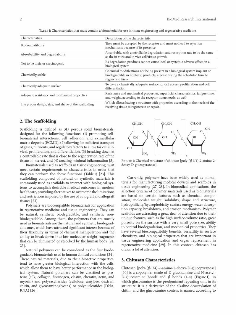
Figure 1: Chemical structure of chitosan [poly-(𝛽-1/4)-2-amino-2-deoxy-D-glucopyranose].
Currently, polymers have been widely used as bioma-terials for manufacturing medical devices and scaffolds intissue engineering [27, 28]. In biomedical applications, theselection criteria of polymer materials used as biomaterialsare based on certain features such as chemical compo-sition, molecular weight, solubility, shape and structure,hydrophilicity/hydrophobicity, surface energy, water absorp-tion capacity, breakdown, and erosion mechanism. Polymerscaffolds are attracting a great deal of attention due to theirunique features, such as the high surface-volume ratio, greatporosity on the surface with a very small pore size, abilityto control biodegradation, and mechanical properties. Theyhave several biocompatibility benefits, versatility in surfacechemistry, and biological properties that are important intissue engineering application and organ replacement inregenerative medicine [29]. In this context, chitosan hasdrawn a lot of attention.
3. Chitosan Characteristics
Chitosan [poly-(𝛽-1/4)-2-amino-2-deoxy-D-glucopyranose][30] is a copolymer made of D-glucosamine and N-acetyl-D-glucosamine bonds and 𝛽 bonds (1–4) (Figure 1), inwhich glucosamine is the predominant repeating unit in itsstructure; it is a derivative of the alkaline deacetylation ofchitin, and the glucosamine content is named according to

BioMed Research International 3
the degree of deacetylation (DD). Depending on the proce-dure of origin and preparation, molecular weight may varyfrom 300 kD to over 1,000 kD, with a deacetylation between30% and 95% in the available commercial preparations.Chitosan has been the best version of the chitin polymerbecause it is readily soluble in diluted organic acids, therebyhaving greater availability to be used in chemical reactions[27, 28, 31–33].
Chitosan properties are very much affected by the con-ditions in which the material is processed because the man-ufacturing process conditions are the ones that control theresulting amount of deacetylation.TheDD in chitosan is a keyfeature that determines its physical, chemical, and biologicalcharacteristics. The DD is determined by the amount of freeamino groups in the polymer chain, and this free aminogroup confers a positive charge to chitosan.The amino groupand the hydroxyl group provide functionality, so chitosanturns out to be a highly reactive polysaccharide. The positivecharge in chitosan allows for many electrostatic interactionswith negatively charged molecules. The processing condi-tions, as well as the amount of functional groups createdby deacetylation, allow for coupling of the groups, havingan impact on the crystallinity of chitosan which in turn isdirectly associated with the ability of chitosan to be soluble inaqueous acid solutions, resulting in one of its main featuresfor processing [34].
Chitosan has many physical and chemical propertiesconferred by its functional groups (amino NH2 and hydroxylOH), as well as biological properties coming from its chem-ical composition. Solubility, biodegradability, reactivity, andabsorption of many of its substrates depend on the amountof protonated amino groups in the polymer chain andthus in the rate of acetylated or nonacetylated glucosamine[35, 36]. All of these features make it an attractive optionfor several applications in science such as food/nutrition,medicine, microbiology, immunology, agriculture, and vet-erinary medicine [37].
4. Physicochemical Properties
4.1. pH Dependence and Solubility. Chitosan solubilitydepends on the distribution of free amino and N-acetylgroups. In diluted acid solutions (pH ≤ 6) the free aminogroups are protonated and confer a polycationic behavior andthe molecule becomes soluble [4]. From the pka standpoint,similarly, the amino groups (pka 6.2–7.0) are completelyprotonated in acids with a pka less than 6.2, making chitosansoluble, remaining so until reaching a pH near 6.2, when, ata higher pH (>de 6.5) the amines in chitosan deprotonateand chitosan become insoluble, after which precipitates, suchas hydrated gels, are formed. Chitosan is insoluble in water,aqueous solutions, concentrated acids, and common organicsolvents, but it is totally soluble when stirred into aqueoussolutions such as acetic acid, nitric acid, hydrochloric acid,perchloric acid, lactic acid, and phosphoric acid [36, 38, 39].
4.2. Degree of Deacetylation (DD). The degree of deacetyla-tion (DD) represents the rate of D-glucosamine units with
respect to the total amount of N-acetyl-D-glucosamine thatmakes the chitosan molecule, since this unit is found in theamino group created from the elimination of the acetyl group.A deacetylated chitin over 60 or 70% is already considered tobe chitosan.TheDD is a structural parameter that determinessome physical and chemical properties such as solubilitylimit in acid solutions (pH 2–6), molecular weight, andmechanical properties (elasticity and traction resistance).The deacetylation process turning chitin into chitosan willtransform the acetyl group into a primary amino group,which is more hydrophilic than the preceding molecule;thereby, the DD in chitosan increases the water content insamples taken from chitosan samples, tending to have animpact on the ability to absorb water and limiting the abilityto have maximum swelling [40].
The DD also has an impact on biological properties,such as the in vitro and in vivo biodegradation. It has beenproven that, at a greater DD (between 84 and 90%), thedegradation process is delayed. Highly deacetylated chitosan(over 85%) shows a low degradation index in the aqueousenvironment andwill degrade after a fewmonths, and a lowerDD (between 82 and 65%) would lead to a faster degradation.The commercially available preparations have a DD between60 and 90%. This feature has an impact on some biologicalproperties in chitosan, such as healing capacity, increasein osteogenesis, and a breakdown process by lysozymes inbiological systems [41, 42].
The DD plays a key role in cell adhesion and proliferationbut does not change the cytocompatibility of chitosan. “Invitro” studies have shown that the lower the DD in chitosan,the lower the cell adhesion in the films. It has been found thatkeratinocytes attached to the chitosan film may change theiradhesion and cell growth depending on their deacetylationdegree (DD), but proliferation is not promoted. Thus, DDaffects growth of cells in the same way as cell adhesion.
4.3. Molecular Weight. Depending on where and how thepreparation procedure is done, the molecular weight maychange from 300 to over 1000 kD [4]; viscosity andmolecularweight are inversely proportional to the degree of acety-lation. Therefore, the greater the molecular weight is, thechitosan membranes tend to be more viscous, thus allowingfor controlling fluidity in them, an important feature intissue interaction. Due to its high molecular weight and itslineal nonbranched structure, chitosan is a strong viscosity-building agent in acid mediums and behaves as a pseu-doplastic material, where viscosity depends on agitation[43].
It has also been proven that the molecular weight hasan indirect effect or is inversely proportional to the swellingcapacity and/or hydration of chitosan membranes, and whenthe molecular weight is greater and the DD is higher,the swelling or permeability is less in chitosan membranes[44].
There is a direct association between molecular weightand DD. These two parameters have a direct effect onthe biodegradation process of chitosan, since at a greatermolecular weight the degradation process is delayed in “invitro” as well as “in vivo” systems [45].
4 BioMed Research International
(a)
(b) (c)
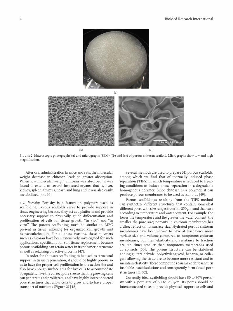
Figure 2: Macroscopic photographs (a) and micrographs (SEM) ((b) and (c)) of porous chitosan scaffold. Micrographs show low and highmagnification.
After oral administration in mice and rats, the molecularweight decrease in chitosan leads to greater absorption.When low molecular weight chitosan was absorbed, it wasfound to extend to several inspected organs, that is, liver,kidney, spleen, thymus, heart, and lung and it was also easilymetabolized [44, 46].
4.4. Porosity. Porosity is a feature in polymers used asscaffolding. Porous scaffolds serve to provide support intissue engineering because they act as a platform and providenecessary support to physically guide differentiation andproliferation of cells for tissue growth “in vivo” and “invitro.” The porous scaffolding must be similar to MECpresent in tissue, allowing for organized cell growth andneovascularization. For all these reasons, these polymerssuch as chitosan have been extensively investigated for suchapplications, specifically for soft tissue replacement becauseporous scaffolding can retain water in its polymeric structureas well as retaining bioactive proteins [47].
In order for chitosan scaffolding to be used as structuralsupport in tissue regeneration, it should be highly porous soas to have the proper cell proliferation in the action site andalso have enough surface area for live cells to accommodateadequately, have the correct pore size so that the growing cellscan penetrate and proliferate, and have highly interconnectedpore structures that allow cells to grow and to have propertransport of nutrients (Figure 2) [48].
Several methods are used to prepare 3D porous scaffolds,among which we find that of thermally induced phaseseparation (TIPS) in which temperature is reduced to freez-ing conditions to induce phase separation in a degradablehomogenous polymer. Since chitosan is a polymer, it canproduce porous membranes to be used as scaffolds [49].
Porous scaffoldings resulting from the TIPS methodcan synthetize different structures that contain somewhatdifferent poreswith size ranges from 1 to 250𝜇mand that varyaccording to temperature andwater content. For example, thelower the temperature and the greater the water content, thesmaller the pore size; porosity in chitosan membranes hasa direct effect on its surface size. Hydrated porous chitosanmembranes have been shown to have at least twice moresurface size and volume compared to nonporous chitosanmembranes, but their elasticity and resistance to tractionare ten times smaller than nonporous membranes usedas controls [50]. The porous structure can be stabilizedadding glutaraldehyde, polyethylenglycol, heparin, or colla-gen, allowing the structure to become more resistant and tomaintain elasticity.These compounds canmake chitosan turninsoluble in acid solutions and consequently form closed porestructures [51, 52].
Currently, ideal scaffolding should have 80 to 90% poros-ity with a pore size of 50 to 250𝜇m. Its pores should beinterconnected so as to provide physical support to cells and

BioMed Research International 5
guide their proliferation and differentiation, also facilitatingneovascularization. The pore sizes recommended for skinscaffolding should be greater than 160 𝜇m, varying between15–100𝜇m and 100–200𝜇m, with a desired 90% porosity toprovide the necessary space and enough surface to growcells and create priority temporary scaffolds for implantationallowing for regeneration or damaged tissue repair [53].
4.5. Water Absorption Capacity. When placed in liquidmedia, chitosan membranes can swell and retain a givenwater volume absorbed from the medium in their three-dimensional network. In order to be used for biomedicalpurposes, they should absorb fluid from the body for celltransference, plus they should allow for an adequate distri-bution of nutrients, metabolites, and growth factors, throughextracellular media [47].
4.6. Mechanical Properties. Considering the appropriatemechanical properties we can state that chitosan membraneshave a disadvantage when used for support in tissue engi-neering because these membranes are very stiff and brittle;that is, they have low mechanical resistance [23]. So then,in order to optimize resistance and elasticity, crosslinkingagents are used with at least two functional reactive groupsthat allow for making bridges between polymeric chains withformaldehyde, epoxides reacting with polyethyleneglycol,dialdehydes (glutaraldehyde and glyoxal), and starch [54].In cross-linked hydrogels the polymeric chains are boundby the crosslinking agent, building a 3D network. Theirnature will mainly depend on the density or crosslinkingdegree, named according to the ratio of moles in the agentwith the moles in the repeat units of the polymer. Amongthe reactions to the crosslinks in chitosan with some othermaterials we can find the aldehyde-amine reaction withpolyethylene glycol, which is used because it is hydrophilic,with low toxicity and good biocompatibility. Studies havebeen conducted showing the effectiveness of the chitosan-DiepoxyPEG (Diepoxy-polyethylene glycol) crosslinking,resulting in an improvement of the mechanical properties ofthe crosslinked compound [54, 55].
4.7. Biological Properties. Chitosan has many beneficialbiomedical properties, such as biocompatibility, biodegrad-ability, andno toxicity. Biological activity of chitosan is closelyrelated to its solubility and therefore molecular weight andDD [56].
4.8. Biodegradability. The process of biodegradation of chi-tosan can be through various media both physical (thermaldegradation) and chemical (enzymatic degradation); the rateof degradation of the chitosan is inversely proportional to thedegree of crystallinity of the polymer and therefore the DDand, thus, can manipulate the degradation rate by controllingthe DD which occurs during processing [41, 57].
There is a broad range of hydrolytic enzymes such aslysozyme, which is the primary enzyme responsible forchitosan degradation in “in vivo systems” and that is found inlymphoid human and animal tissue and that can be used tonaturally degrade chitosan [58, 59]. Inside the body it leads
to the release of amino sugars that can be processed andreleased by the metabolic system. Chitosan degradation isan important property assuming that the end processes andapplications it will ultimately be given can agree with theresulting design [35].
Some of the specific enzymes that degrade chitin andchitosan have a clearly identified structure but their actionmechanisms are still unknown. In mammals, these enzymesseem to be completely absent; however, when chitosan isimplanted, it will eventually disappear completely after sometime and the degradation speed seems to depend onDD [35].
Through enzymatic hydrolysis mechanisms chitosanmaybe easily depolymerized due to susceptibility 𝛽 bonds (1–4) mediated by different hydrolases including lysozymes,pectinase, cellulases, hemicellulases, lipases, and amylasesamong others, which means that chitosan has a pecu-liar vulnerability to other enzymes that are different fromchitinases. Its degradation products are oligosaccharides ormonosaccharides, naturalmetabolites of glycosaminoglycansor glycoaminoproteins. Lysozyme is an unspecific proteolyticenzyme common in mammals. It can hydrolyze chitosan, butthis action quickly disappears when chitosan has a degree ofacetylation (DA) below 30% [41].
When chitosan is fully acetylated, it is totally insensitive tothis enzyme.Moreover, it seems that at least three consecutiveN-acetylated groups are necessary to be recognized by thisenzyme and, in spite of depending on the amine content inchitin and chitosan the unspecific enzymes that degrade thesepolymers are inactivated, and such degradation can be foundin in vivo implants. It has been proven that whatever thecircumstancesmay be, biodegradation of chitosan takes placedepending onmany and diverse factors, especially the degreeof acetylation, molecular weight, degree of crystallinity, watercontent, and also the shape and condition of the surface onthe material, aside from its microstructure [41].
4.9. Biocompatibility. Biomaterials should be biocompatible;that is, contact with the body should not result in adversereactions, so they must be capable of recognizing and coop-erate harmoniously with structures and cells of the humanbody, without producing unspecific reactions [60]. Clinicaltests conducted so far have not reported any inflammatoryor allergic reactions after implantation, injection, topicalapplication, or ingestion of chitosan in the human body [40].This is due to the fact that chitosan is made of GlcN andGlcNac that are natural components of mammalian tissues[61].
There are studies that support the biocompatibility of thematerial and the direct relationship of this property withits DD material used. In the first reports that determinedtoxicity, none of the materials were chitosan films using astandard “in vivo” toxicity tests to assess their safety [62].The biocompatibility of chitosan films has also been shownwith different DD; studies where a model of subcutaneousimplantation in rats is applied showed that the films ofchitosan with DD between 69 and 74% induced a relativelyacute inflammatory reaction by rapid biodegradation, withalmost complete resorption after 4 weeks of implantation;DD films with high (between 74 to 90%) resulted in
6 BioMed Research International
a mild inflammatory reaction in tissue degradation rate anda slower rate. This is in agreement with the well-knownfact that rapidly biodegradable biomaterials elicit an acuteinflammation reaction due to a significantly large productionof low-molecular-weight compounds within a short time.Therefore it was determined that films with ≥ 84% DDshowed reaction to softer tissue because these were degradedmore slowly [45].
4.10. Non Toxic. Several studies have proven that, so far,clinical tests conducted with chitosan have not reportedadverse inflammatory or allergic reactions when used intissue engineering or as a vehicle in drugs, nor after implan-tation, injection, or topical application in the human bodyor for oral application. This property is due to the fact thatchitosan is made of GlcN and GlcNAc, natural componentsof mammalian tissue [41, 42, 63].
5. Cytocompatibility
A wide number of cells have been successfully cultured on2D and 3D chitosan matrices envisaging cell-based regen-erative therapies, among them keratinocytes, chondrocytes,osteoblasts, hepatocytes, and Schwann cells [40, 64–68].Some studies found that the DDwas an important parameteraffecting cell adhesion, by promoting high adhesion. Thiseffect was reported for a number of anchorage-dependentcells, such as keratinocytes, fibroblasts, dorsal root ganglionneurons, and Schwann cells [69–71].
In other studies the effect of DD concerning behavior ofthe osteogenic cells and chitosan films was investigated inporous matrices, using DD in the range of 96–51%. Thesestudies revealed a trend that determines that there is anincrease of cell adhesion related to an increase in the DD,and the differences showed that a DD greater than 91% can becritical in terms of osteogenic response fromchitosan [72, 73].
6. Nonimmunogenic
Chitosan and its oligomers stimulate macrophage activityincreasing nitric oxide, reactive oxygen species, TNF-𝛼,interferon, and IL-1, as well as TGF-𝛽1 and PDGF. However,since there are no proteins and lipids in its structure, it isnot possible to develop specific antibodies against it, unlessit is coupled with other substances such as albumin. Theconclusion from all the studies on the subject indicates thatchitosan is hypoallergenic and only transiently stimulates theimmune system because when impregnated with tissue fluidsin the receptor body it ultimately becomes biotolerated andmetabolized [41, 63, 74].
7. Antimicrobial and Antifungal
Chitosan inhibits growth of many types of fungi, yeasts, andbacteria. In solutions made of acid dilutions the positiveloads in them interact with the negatively charged residues ofmacromolecules on the cell surface of microorganisms, sup-posedly competing against the Ca+2 for the electronegative
sites in the membrane, but without conferring dimensionalstability, compromising the membrane integrity and makingit weak [75]. Among the antimicrobial effects of chitosanare those related to Candida albicans, Enterobacter cloacae,Enterococcus faecalis, Escherichia coli, Klebsiella pneumoniae,Pseudomonas aeruginosa, Staphylococcus aureus, and Strepto-coccus pyogenes.
This property is of special significance because it has beenproven that the antimicrobial agents such as bandagingmate-rials and dressings generally lead to cytotoxicity, delaying thehealing process, or leading to pathogen resistance. In the caseof chitosan, since the antimicrobial effects come directly fromthe membrane, there is almost no need to use antibacterialsubstances or change bandages, the implants themselves, ordressings when they are applied [63].
8. Tissue Repair and Regenerative Medicine
In regenerative medicine applications of biomaterials fortissue repair and regeneration include their use as orthopedicimplants and, as bone fillers, adhesives for tissue repair andthe use of scaffolds for tissue engineering; the latter areused for repair and/or regeneration of skin, bone, cartilageand nerve tissues, since these tissues have been the focus ofgreater research in regenerative medicine. This involves theuse of chitosan as a scaffolding material or as an analog orextracellular matrix (ECMD), which works as support for theregeneration of damaged tissue (Table 2) [6–22, 76].
The popularity of chitosan for tissue repair and regen-eration is due to the fact that it can be easily processedand manufactured in a variety of forms including fibers,films, sponges, and hydrogels. This provides the ability tomimic the shape of the receiving tissue or biomaterial tissueinterface. Moreover, the similarity of its chemical structureto some polysaccharides and ECM constituents offers thepossibility of being chemically modified to adapt structurallyand functionally to the host tissue, due to its previouslydescribed properties that allow for the ability to regenerateprimary tissue cells and even stem cells. Thus its potential isto be used in regenerative medicine [4, 33, 77, 78].
9. Chitosan and Tissue Engineering
9.1. Skin, Nerves, and Soft Tissues. The generation of scaffoldswith porous structures is important in the engineering ofepithelial and soft tissues. Chitosan can be manufacturedin a porous structure to allow for cell seeding. This spacecreated by the porous structure allows for cell proliferation,migration, and the exchange of nutrients. In addition, thecontrollable porosity of chitosan scaffolds is beneficial toangiogenesis, which is fundamental in supporting the sur-vival and function of the regenerated soft tissues [50, 79].Chitosan scaffolds have shown both cytocompatibility invitro and biocompatibility in vivo. Generally, chitosan evokesonly a minimal foreign body reaction in vivo, and implantedchitosan scaffolds seldom induce chitosan-specific reactions[32].

BioMed Research International 7
Table2:Ap
plications
ofchito
san-basedscaffolds
fortissue
engineering.
Chito
sancombinatio
nScaffoldob
tained
Experim
entalm
odel
Tissue
application
Reference
Chito
san+hyaluron
anHybrid
polymer
fiber
Fibrob
lasts
from
patellartendo
nof
Japanese
white
rabbit
Ligament
[6]
Collagen-chito
san+fib
ringlue
Asymmetric
porous
scaffold
Hum
anderm
alfib
roblastsandkeratin
ocytes
Skin
[7]
Chito
san+alginate
Polyele
ctrolyte
multilayer
film
C2C1
2myoblasts
Muscle
[8]
Chito
san+aloe
vera
Blendedmem
brane
Bovine
artic
ular
chon
drocytes
andmesenchym
alste
mcells
Skin
[9]
Chito
sanalon
eMem
brane
Embryonalsub
mandibu
larg
land
cells
Salivarygland
[10]
Chito
san+layero
fchitosan/gelatin
Sand
wichtubu
lar
scaffold
Vascular
smoo
thmuscle
cells
from
rabbitaorta
Bloo
dvessel
[11]
Genipin-crosslin
kedchito
san,
chito
san-nano
hydroxyapatite
Fram
ework
Hum
anperio
dontalligam
enttissue,periodo
ntal
ligam
entstem
cells
Bone
[12]
Chito
san+collagen
Hydrogel
Epididym
alfatp
adsc
ells,
andsubcutaneous
pocketof
maleL
ewis
rat
Adiposetissue
[13]
Chito
san+po
lyester
Com
pressedpo
rous
disc
Bovine
artic
ular
chon
drocytes
Cartilage
[14]
Chito
san+collagen+genipin
Crosslink
edpo
rous
mem
brane
Rabb
itartic
ular
chon
drocytes
Cartilage
[15]
Chito
san+chon
droitin
sulphate
Bidimensio
nalglass
surfa
ceso
r3Dpacketof
paraffin
Bovine
artic
ular
chon
drocytes
andhu
man
mesenchym
alste
mcells
cultu
reCa
rtilage
[16]
Chito
san+adipose-deriv
edste
mcells
Tube
nervec
ondu
itMale,Sprague-Daw
leyratssciatic
nervetransectio
nNerve
[17]
Chito
sanalon
eTu
beMale,beagledo
gsph
renicn
erve
resection
Nerve
[18]
Chito
sanalon
eViscou
ssolutionanda
mon
olayer
rigid
physical
hydrogel
Femalem
inipigsthird-degreeb
urns
Skin
[19]
Chito
san+silkfib
roin
Thin
blendedfilm
Femaleg
uineap
igsv
entralhernia
Muscle
[20]
Chito
san+𝛽-sod
ium
glycerop
hosphate+hydroxyethyl
cellu
lose
Hydrogel
Malea
ndfemales
heep
artic
ular
defect
Cartilage
[21]
Chito
san+calcium
phosph
atec
ement
Chito
sanmicrospheres
insid
ecem
entp
aste
Maler
abbitfem
oraldefect
Bone
[22]
8 BioMed Research International
Due to the fact that some chitin-based biomaterials donot provide a friendly interface for cell adhesion of somespecific tissue types, other biomaterials, such as collagen orfibronectin with tissue-specific binding sequence, should beblended with chitosan to produce scaffolds with higher cellaffinity. Also, chitosan is blended with other biomaterials tocreate scaffolds that are more appropriate for directing thedesired cell behaviors and to mechanically strengthen thetissues engineering of tissues such as the skeletal system [80,81]. The biological activity beneficial to tissue regenerationcan be introduced through the entrapment of bioactive agentsin the scaffolds through physical adsorption [6]. For example,trimethylated chitosan has been reported to be efficient ingene transfection without increasing cytotoxicity [82].
Specifically speaking about the difference in tissues, thereis evidence that chitin-based materials support neuronalgrowth. In addition, many different substrates and bioactivemolecules have been added into chitin-based scaffold toincrease their affinity with nerve cells [83]. A chitosan tubeimmobilized with laminin peptides can facilitate proximalnerve sprouting and regenerate axon bridging [84]. In 2004,a study showed that chitosan fibers supported the adhesion,migration, and proliferation of Schwann cells, which allowedfor axonal regeneration in the peripheral nervous system[68]. Whenever there is a peripheral nerve lesion, the currentstandard treatment is to use an autologous nerve graftto bridge the neural gap and facilitate nerve regenerationand reconnection; however, since there have been frequentand severe complications, several attempts have been madeover the last decades to overcome this problem by usingdifferent biomaterials but functional recovery is still far frombeing acceptable [85]. Nevertheless, different scaffolds werefabricated in a study by cross-linking chitosan with aceticacid and chitosan with 𝛾-glycidoxypropyltrimethoxysilane,which would later be cultivated with N1E-115 cells, derivedfrom mouse neuroblastoma C-1300. The resulting hybridmembranes presented good cytocompatibility besides thefact that, when cultured in the presence of dimethylsulfoxide(DMSO) or cyclic AMP (cAMP), they show characteristicsfrom neural cells, such as ceased multiplication, extensiveneurite outgrowth, and polarization of cellular membranes,being able to locally produce and deliver nerve growthfactors, essential in the reconstruction of peripheral nervelesions. In vivo studies suggest that these chitosan-basedmembranes show promising results regarding its applicationsin peripheral nerve engineering due to their porous structure,their chemical modifications, and high affinity to cellular sys-tems [85]. These modifications make chitin-based materialsmore diverse and functional for soft tissue regeneration. Inthe same manner, it was found that in the regeneration ofligaments, chitosan-hyaluronic hybrid polymers can provideappropriate environments for cellular adhesion, proliferation,and extracellular membrane (ECM) production, as well asfacilitating the biological effects of seeded cells [6].
For vascular tissues, in order to mimic the morphologicaland mechanical properties of blood vessels and improvelong-term patency rates, collagen has been crosslinked withchitosan to generate a tubular scaffold. This biocompatiblescaffold proved to have desirable porosity and pliability,
enhanced cell adhesion, proliferation, and ECM produc-tion [86, 87]. In addition to vascular applications, chi-tosan/collagen blended scaffolds have also been employed inadipose tissue regeneration. When adipocytes were seeded,the in vitro cytocompatibility and in vivo biocompatibility ofscaffolds were confirmed experimentally [88].
Chitosan also has a potential use in skin repair andregeneration subsequent to injuries or burns. A study wasperformed in which chitosan was cross-linked with silicaparticles (SiO2), used as a porogen agent and the extractionsfrom the developed membranes demonstrated no cytotoxi-city against L-929 cells 24 hours after the culture. In addi-tion, the macroporous membrane exhibited excellent cellularadhesion and proliferation after 24 and 48 hours of culturing,which is why the developed scaffold might be adequate forskin tissue engineering [89]. Chitin-based materials havealso demonstrated their potential in maintaining and induc-ing cell phenotypes used in culturing melanocytes, cornealkeratinocytes, and skin keratinocytes [90, 91]. Even in thesalivary gland, the morphogenetic efficacy of mesenchyme-derived growth factors is dramatically augmented with theassistance of chitosan.The effects of epithelial morphogeneticfactors, such as fibroblast growth factors 7 (FGF7), fibroblastgrowth factor 10 (FGF10), and hepatocyte growth factor(HGF), have been upregulated in the presence of chitosan[88].
Furthermore, chitosan use in the design of new tissueadhesives was motivated by the fact that it can bind to colla-gen due to hydrogen bonding and polyanionic–polycationicinteractions [88].There is evidence that hydrogels andmeshesof chitosan cross-linked with other biomaterials are usefulin the prevention of postoperative abdominal adhesions. Astudy was developed in which they used thermosensitivehydroxybutyl chitosan (HBC) in a rat side-wall defect-cecumabrasion model for prevention of postoperative abdominaladhesions. HBC is a new derivative of chitosan whose maincharacter is the intelligent response to changing temperature.HBC demonstrated antiadhesive activity as well as being easyto handle during the operation. Therefore, it may be effectivein prevention of postoperative adhesions [92]. Another pro-tocol was performed where they compared 3 different typesof meshes: Dynamesh-IPoM mesh, a simple polypropylenemesh, and a polypropylene/chitosan mesh. The results werethat the polypropylene/chitosan mesh proved to be the leastirritating for the recipient’s tissue as well as surroundingtissues, as evidenced by the lowest rate of inflammatoryreaction within the connective tissue, which guarantees theimplant acceptance and the least extensive adhesion to inter-nal organs, and thus the lowest rate of complications [93].Finally, there was another protocol in which they determinedthat a chitosan-gelatin modified film modified chitosan filmis effective on preventing peritoneal adhesions induced bywound, ischemia, and infection, but the effect is not apparentin foreign body-induced adhesion [94].
Due to the properties that chitosan has shown in reduc-tion and prevention of postoperative intraperitoneal adhe-sions, it is also widely being studied for its use in repairand regeneration of the abdominal wall in ventral hernias. Astudy was conducted to investigate the feasibility of using silk

BioMed Research International 9
fibroin and chitosan blend scaffolds for ventral hernia repairin guinea pigs [20]. This blended scaffold was comparedto a biodegradable human acellular dermal matrix and anonbiodegradable polypropylene mesh. The investigatorsconcluded that the silk fibroin and chitosan blend scaffold,unlike the mesh and the matrix, showed tissue remodelingin all 3 dimensions, with seamless integration at the interfacewith adjacent native tissue, the repair sites remained intact,and their mechanical strength was similar to that of thenative abdominal wall. Additionally, the scaffold promotedthe deposition of new extracellular matrix, uniform vascu-larization, and cellular infiltration in the repair sites, whichcontributed to the increase in mechanical strength of theregenerated tissue. Thus, this scaffold is potentially usefulin reconstruction and regeneration of the abdominal wall[20]. Furthermore, due to its utility in ventral hernia repair,it might also have a potential use in inguinal hernias andother types of herniation. It might even be useful in therepair of certain congenital defects such as omphalocele orgastroschisis, although to date there are still no apparentmodels that prove its effectiveness for application in humansfor these types of defects.
Specifically speaking about intestinal tissue, its engineer-ing is an emerging field due to a growing demand for intesti-nal lengthening and replacement procedures secondary tomassive bowel resections [95, 96]. Intestinal transplantationis a common treatment but its limitation resides in the highincidence of rejection, availability of donor organs, and thesize of the donor graft. The biocompatibility of chitosanwas investigated by growing rabbit colonic circular smoothmuscle cells on chitosan-coated plates [95]. The cells main-tained their spindle-like morphology and preserved theirsmooth muscle phenotypic markers. Tubular scaffolds weremanufactured with central openings composed of chitosanand collagen in a 1 : 1 ratio. Concentrically aligned 3D circularmuscle constructs were bioengineered using fibrin-basedhydrogel seededwith the colonic circular smoothmuscle cellsfrom the rabbit.Themuscle constructs contracted in responseto acetylcholine (Ach) and potassium chloride (KCl) and theyrelaxed in response to vasoactive intestinal peptide (VIP).These results demonstrate that chitosan is a biomaterial possi-bly suitable for intestinal tissue engineering applications [95].
In conclusion, it is safe to say that chitosan has greatpotential in applications for soft tissue engineering, whetherit is used for wound closure or for its potential use ingenerating specific tissue grafts. Nonetheless, there is stillmuch research to be done in terms of their properties andformation of scaffolds. Next generation scaffolds should beable to carrymany different bioactive factors and release themin specific order. To this end, decisions on how to control theseparate loading capacity, kinetics of drug release, and rateof substrate degradation are the major challenges to be faced[88].
10. Potential Applications Supported byIts Biological Activity
10.1. Hemostatic Properties. Chitosan is capable of promotingplatelet adhesion by initializing a cascade of intracellular
signaling which activates glycoproteins IIb/IIa as well asthromboxane A2/ADP, increasing platelet spreading andstrengthening the stability of adhesion [97]. Chitosan is avail-able in different presentations, including films, fibers, andhydrogels, and each one of them offers specific advantages interms of absorption depending in its therapeutic use.
Several studies have proven efficacy of chitosan as ahemostatic agent. However, it has been reported that thehemostatic mechanisms of chitosan are separate from theclassic coagulation cascade [62]. The hemostatic effect inchitosan is achieved from the direct interactionwith platelets,mainly in alpha granules. The intracellular signaling inducesPDGF-AB and TGF-𝛽1 release. An increased rate in thePDGF-AB has been found, as much as up to 130%, with theuse of chitosan compared to a control group [98]. Chitosancan be used in medical and surgical procedures by directapplication on bleeding surfaces, using several presentationssuch as powder, solutions, coatings, films, hydrogels, com-pounded filaments, and more. Nevertheless, its clinical usewill depend on the application technique used as well as thetype of wound that it is applied on. Many investigators havedescribed the hemostatic applications for chitosan; however,different presentations have been used in each study; thatis, why results should be analyzed according to the differentgroups, depending on the physical form of the material [99].
10.2. Liver Repair Application. There are three crucial factorsfor successful use of chitosan in surgery: it must be placed inthe intra-abdominal cavity, it must have good bonding to thesurface of the lesion, and it must be able to maintain properhemostasis.
A study compared the effectiveness of a freeze-dry chi-tosan graft versus the use of sponge gauze in a venousbleeding due to a severe liver lesion in a pig model. Theanimal model involves extensive vascular damage, as wellas damage to the liver parenchyma. Several vascular lesionsof approximately 1 cm in diameter were made. The outcomereportedwith the chitosan graft had less blood loss (𝑝 < 0.01)compared to the group of sponges (264mL and 2,879mL,resp.) [100].
In further investigations the hemostatic effects of chitosanas a solution have been previously analyzed. The results of alingual incision in a heparinized animal model according tothe evaluations with electron microscopy concluded that theincisions treated with chitosan showed a disturbance in themorphology of red cells, as well as an unusual affinity amongthe red cells. It was reported that the red cell fractions thatinteracted with chitosan reduced bleeding in 60%, reachinghemostasis at 800 𝜇g/mL [99].
10.3. Healing Properties. In another study it was found that,by means of chitosan hydrogel application on skin woundsin diabetic mice, the wound shrinking speed improved andwound closure was significantly faster.The chitosan hydrogelcombined with fibroblast growth factor type 2 was seento accelerate the closing process even further. Histologyexamination showed that the combination of chitosan andfibroblast growth factor type 2 fostered the formation of gran-ulation tissue, capillary network, and epithelialization [101].
10 BioMed Research International
The regenerative properties of chitosan are based on amatrix building capacity that is adequate for growth andactivation of macrophages and proliferative cells in three-dimensional tissue. A comparative study between woundstreated with chitosan and a control group treated only withsaline solution was conducted in a dog animal model. Thewounds were clinically assessed throughout the study andinspected histologically once the animal was euthanized.Clinically, complete healing was achieved in the chitosan-treated group after three weeks, while in the control groupit took four weeks. A complete repair of epidermal cells witha keratin layer associated with connective tissue proliferationwas seen. In the chitosan group a collagen network of fibersproduced by fibroblasts was found, which surrounded theneovasculature of the wound, while in the control grouphyalinosis of subcutaneous tissue occurred [102].
10.4. Chitosan Composites for Bone and Cartilage Regenera-tion. Chitosan composites have been synthesized for hardtissue regeneration, as in the case of bone and cartilage.
Evaluation of chitosan composites for bone tissue regen-eration is based on physicochemical and biological char-acterizations. Physicochemical characterization comprisesthe study of homogeneity, purity, percentage composition,chemical bonding, thermic stability, mechanical tests, andincubation on simulated body fluid [103, 104]. On the otherhand biological evaluation includes the test in in vitro culturecells (MC-3T3, hFOB, MG63, and bone marrow stem cells)[105–108].These assays evaluate composite cytocompatibilityand cytotoxicity, in addition to its intinsic capacity of induc-tion of cellular differentiation [109]. Finally the efficacy in thetreatment of surgical defects on animal models is evaluated[21, 107, 110].
In the first place, chitosan as a matrix allows for bio-compatibility of an implantable material, and in the second,it allows for the interaction or combination with inducermaterials for tissue regeneration. In this sense, the use ofchitosan composites for tissue regeneration in experimentalmodels is a promising strategy for treatment of skeletal andjoint diseases [76, 111, 112].
In the case of bone tissue engineering, chitosan matriceshave been combined with osteogenic materials, like hydrox-yapatite [103, 108, 109, 111], calciumphosphate and sulfate [113,114], and others [115–117]. The purpose of the combinationof those biomaterials is to obtain organic and inorganiccomposites that simulate the bone structure [118].
The evaluation of chitosan composites in osteoblast cul-ture is an important factor to identify its biocompatibility.In in vitro assays with the MC3T3 cell line, which comesfrom calvarial murine osteoblasts, cross-linked membraneof chitosan with tripolyphosphate showed the same valuesin MTT assay of cell viability compared to controls; resultswere observed in composites compound of chitosan withcalcium phosphate, and chitosan with the release of bonemorphogenic protein type-2, concluding that those mem-branes are biocompatible with osteoblasts. Additionally ithas mechanical properties that make it a good implantablecomposite in bone defects [113, 119].
When bone tissue is damaged by trauma, cancer, orinfection, a source of autogenous bone tissue or replace-ment materials is needed to regenerate the compromisedtissue. Experimental animal models are a good resourceto understand how biological and pathological conditionsparticipate in the healing of bone tissue. In this sense,chitosan composites can be used to induce bone healing andregeneration.
Chitosan composites have been tested in bone defectsin experimental models successfully for bone regeneration.Chitosan hydrogel, gelifiable by blue light, was used for BMP-2 release and showed good bone regeneration in a femoraldefect in rat [120]. Similar results were observed with the useof a lyophilized porous membrane, a compound of chitosanand hydroxyapatite, in a calvarial defect in rat, the compositemembrane filled up the defect as compared to a control,in addition, the presence of osteogenic markers was moreabundant in the experimental group [121].
Osteogenesis is the processwhere osteoblast cells prolifer-ate frommesenchymal cells and deposit extracellular matrix;at the end of the bone defect these cells differentiate intomature osteocytes. All of this tissue process includes theexpression of multiple bone markers and enzymes involvedin cell maturation and bone calcification.
Chitosan/nanohydroxyapatite composites have beenmore relevant for tissue engineering, because of its ability toinduce a good proliferative response in osteoblasts, and in atibial defect in a rabbit it showed good bone regeneration at8 weeks seen by microcomputarized tomography [122].
Joint defects are common in elderly people, caused byrheumatoid arthritis and dehydration of cartilage tissue inthe entire body or by the lifting of heavy loads with thecorresponding joint wear, leading to the total dependence ofjoint replacement therapy.
In the case of cartilage tissue regeneration, chitosancomposites have been designed to form hydrogels. Thesecomposites allow for the inclusion of cells and molecules forcartilage regeneration. The most combined inducer agent ofcartilage regeneration is collagen type II, this is the mainprotein in cartilage tissue, and it enhances the adhesion andformation of clusters of chondrocytes in vitro [15, 123, 124],a requirement for cartilage regeneration. Collagen II andchondroitin sulfate in chitosan hydrogels stimulate chondro-cytes attachment in a blue light gelification composite, italso induces the mesenchymal stem cells differentiation tochondrocytes in vitro [125], and similar results have beenobserved with chitosan hydrogel with alginate and fibroin. Inorder to follow the seeding cells on chitosan composites, thenext step in regeneration is the formation of functional tissue;collagen II expression by chondroblasts and chondrocytes isa determinant factor in cartilage formation, and it has beenobserved in the glycerophosphate-chitosan hydrogel with silkfibrils, where chondrocyte phenotype is maintained for theexpression of glycosaminoglycans and type II collagen invitro [126], as obtained with alginate and fibroin in chitosanhydrogels [127]. These findings suggest that chitosan cansupport the addition of an inducer material and also isa biocompatible material for cartilage tissue engineering.Extracellular matrix deposition is a key factor to recognize

BioMed Research International 11
biocompatibility and normal cell function, in addition a 3Dmatrix for growth tissue by proliferation and differentiationof precursor cells. In the case of chondroblast and chondro-cytes, production of collagen II is a functional tissue determi-nant, and this has been found in glycerophosphate-chitosanhydrogel and silk fibrils, which stimulates the production ofcollagen II and glycosaminoglycans by chondrocytes in vitro[126].
The polylactide acid-chitosan membranes with collagenprovide a laminatematrix withmechanical properties similarto cartilage, but in addition they have worked as a supportfor chondrocytes from rabbit cartilage [128], and this opensthe possibility to use chitosan composites in the regenerationof cartilage defects [21], as in the case of arthritis or jointcartilage damage from aging.
A biomaterial combination of chitosan with polycapro-lactone [112, 124, 129], silk fibrils [126], genipin [15], chon-droitin sulfate [130], and polyester [14] has been designedwith the purpose of obtaining composites with propertiesthat resemble those of cartilage, and it also provides a properenvironment for extracellularmatrix deposition, cell viability,and differentiation.
This perspective allows for understanding the potentialproperties of chitosan composites in hard tissue regeneration.In summary, chitosan composites provide physical and chem-ical and mechanical support, cell attachment, proliferation,and differentiation, with the corresponding biocompatibilityto induce the bone and cartilage tissue regeneration.
11. Conclusions
Regenerative medicine is facing new challenges in the wayto induce tissue repair in live tissue. Advances have led tothe availability of bioactive compounds for damaged tissue.Such compounds must have a regenerative effect and fosterwound repair, with the least possible morbidity and with highbiocompatibility conditions. Chitosan polymers have beenproven to serve as scaffolds that induce tissue regeneration,but beyond that, they are considered to be an ideal polymerfor making bioactive compounds [131]. This is possiblebecause there is a potential synergy in their byproducts,when combined with growth factors and stem cells, eitherof mesenchymal origin or neural origin. Additional studiesshould try to confirm the translational issues on the roleof bioactive polymers and their real impact on regenerativemedicine.
Abbreviations
DD: Degree of deacetylationTIPS: Thermally induced phase separationECMD: Extracellular matrix depositsECM: Extracellular membrane.
Conflict of Interests
The authors report no conflict of interests.
References
[1] C. Shi, Y. Zhu, X. Ran, M. Wang, Y. Su, and T. Cheng, “Ther-apeutic potential of chitosan and its derivatives in regenerativemedicine,” Journal of Surgical Research, vol. 133, no. 2, pp. 185–192, 2006.
[2] Y. Fung, “A proposal to the National science Foundation for anEngineering Research Centre at USCD,” UCSD 865023, Centerfor the Engineering of Living Tissues, 2001.
[3] R. Langer and J. P. Vacanti, “Tissue engineering,” Science, vol.260, no. 5110, pp. 920–926, 1993.
[4] I.-Y. Kim, S.-J. Seo, H.-S. Moon et al., “Chitosan and itsderivatives for tissue engineering applications,” BiotechnologyAdvances, vol. 26, no. 1, pp. 1–21, 2008.
[5] R. Langer and D. A. Tirrell, “Designing materials for biologyand medicine,” Nature, vol. 428, no. 6982, pp. 487–492, 2004.
[6] T. Funakoshi, T. Majima, N. Iwasaki et al., “Novel chitosan-based hyaluronan hybrid polymer fibers as a scaffold inligament tissue engineering,” Journal of Biomedical MaterialsResearch Part A, vol. 74, no. 3, pp. 338–346, 2005.
[7] C.-M. Han, L.-P. Zhang, J.-Z. Sun, H.-F. Shi, J. Zhou, and C.-Y.Gao, “Application of collagen-chitosan/fibrin glue asymmetricscaffolds in skin tissue engineering,” Journal of Zhejiang Univer-sity Science B, vol. 11, no. 7, pp. 524–530, 2010.
[8] S. G. Caridade, C. Monge, F. Gilde, T. Boudou, J. F. Mano, andC. Picart, “Free-standing polyelectrolyte membranes made ofchitosan and alginate,” Biomacromolecules, vol. 14, no. 5, pp.1653–1660, 2013.
[9] S. S. Silva, S. G. Caridade, J. F. Mano, and R. L. Reis, “Effectof crosslinking in chitosan/aloe vera-based membranes forbiomedical applications,” Carbohydrate Polymers, vol. 98, no. 1,pp. 581–588, 2013.
[10] T.-L. Yang andT.-H. Young, “The enhancement of submandibu-lar gland branch formation on chitosan membranes,” Biomate-rials, vol. 29, no. 16, pp. 2501–2508, 2008.
[11] L. Zhang, Q. Ao, A. Wang et al., “A sandwich tubular scaffoldderived from chitosan for blood vessel tissue engineering,”Journal of Biomedical Materials Research Part A, vol. 77, no. 2,pp. 277–284, 2006.
[12] S. Ge, N. Zhao, L. Wang et al., “Bone repair by periodontal liga-ment stem cell-seeded nanohydroxyapatite-chitosan scaffold,”International Journal of Nanomedicine, vol. 7, pp. 5405–5414,2012.
[13] X. Wu, L. Black, G. Santacana-Laffitte, and C. W. PatrickJr., “Preparation and assessment of glutaraldehyde-crosslinkedcollagen–chitosan hydrogels for adipose tissue engineering,”Journal of Biomedical Materials Research Part A, vol. 81, no. 1,pp. 59–65, 2007.
[14] M. L. A. da Silva, A. Crawford, J. M. Mundy et al.,“Chitosan/polyester-based scaffolds for cartilage tissue engi-neering: assessment of extracellular matrix formation,” ActaBiomaterialia, vol. 6, no. 3, pp. 1149–1157, 2010.
[15] L.-P. Yan, Y.-J. Wang, L. Ren et al., “Genipin-cross-linkedcollagen/chitosan biomimetic scaffolds for articular cartilagetissue engineering applications,” Journal of BiomedicalMaterialsResearch, Part A, vol. 95, no. 2, pp. 465–475, 2010.
[16] J. M. Silva, N. Georgi, R. Costa et al., “Nanostructured 3D con-structs based on chitosan and chondroitin sulphate multilayersfor cartilage tissue engineering,” PLoS ONE, vol. 8, no. 2, ArticleID e55451, 2013.
12 BioMed Research International
[17] Y.-Y. Hsueh, Y.-J. Chang, T.-C. Huang et al., “Functionalrecoveries of sciatic nerve regeneration by combining chitosan-coated conduit and neurosphere cells induced from adipose-derived stem cells,” Biomaterials, vol. 35, no. 7, pp. 2234–2244,2014.
[18] N. Tanaka, I. Matsumoto, M. Suzuki et al., “Chitosan tubescan restore the function of resected phrenic nerves,” InteractiveCardioVascular andThoracic Surgery, 2015.
[19] N. Boucard, C. Viton, D. Agay et al., “The use of physicalhydrogels of chitosan for skin regeneration following third-degree burns,”Biomaterials, vol. 28, no. 24, pp. 3478–3488, 2007.
[20] A. S. Gobin, C. E. Butler, and A. B. Mathur, “Repair andregeneration of the abdominal wall musculofascial defect usingsilk fibroin-chitosan blend,” Tissue Engineering, vol. 12, no. 12,pp. 3383–3394, 2006.
[21] T. Hao, N. Wen, J.-K. Cao et al., “The support of matrixaccumulation and the promotion of sheep articular cartilagedefects repair in vivo by chitosan hydrogels,” Osteoarthritis andCartilage, vol. 18, no. 2, pp. 257–265, 2010.
[22] D. Meng, L. Dong, Y. Wen, and Q. Xie, “Effects of addingresorbable chitosan microspheres to calcium phosphatecements for bone regeneration,” Materials Science andEngineering: C, vol. 47, pp. 266–272, 2015.
[23] M. A. Lizarbe, “Sustitutivos de tejidos: de los biomateriales ala ingenierıa tisular,” Revista de la Real Academia de CienciasExactas, Fısicas y Naturales, vol. 101, no. 1, pp. 227–249, 2007.
[24] L. S. Nair and C. T. Laurencin, “Biodegradable polymers asbiomaterials,” Progress in Polymer Science, vol. 32, no. 8-9, pp.762–798, 2007.
[25] S. Ramakrishna, J. Mayer, E. Wintermantel, and K. W. Leong,“Biomedical applications of polymer-composite materials: areview,” Composites Science and Technology, vol. 61, no. 9, pp.1189–1224, 2001.
[26] B. D. Ratner, Biomaterials Science: An Introduction to Materialsin Medicine, Academic Press, 2004.
[27] E. Khor and L. Y. Lim, “Implantable applications of chitin andchitosan,” Biomaterials, vol. 24, no. 13, pp. 2339–2349, 2003.
[28] R. A. Muzzarelli, C. Jeuniaux, and G. W. Gooday, Chitin inNature and Technology, Springer, 1986.
[29] B. Dhandayuthapani, Y. Yoshida, T. Maekawa, and D. S. Kumar,“Polymeric scaffolds in tissue engineering application: a review,”International Journal of Polymer Science, vol. 2011, Article ID290602, 19 pages, 2011.
[30] G. Lodhi, Y.-S. Kim, J.-W. Hwang et al., “Chitooligosaccharideand its derivatives: preparation and biological applications,”BioMed Research International, vol. 2014, Article ID 654913, 13pages, 2014.
[31] K. Rudall, “Chitin and its association with other molecules,”Journal of Polymer Science C: Polymer Symposia, vol. 28, no. 1,pp. 83–102, 1969.
[32] P. J. VandeVord, H. W. T. Matthew, S. P. Desilva, L. Mayton,B. Wu, and P. H. Wooley, “Evaluation of the biocompatibilityof a chitosan scaffold in mice,” Journal of Biomedical MaterialsResearch, vol. 59, no. 3, pp. 585–590, 2002.
[33] M. N. V. R. Kumar, “A review of chitin and chitosan applica-tions,” Reactive and Functional Polymers, vol. 46, no. 1, pp. 1–27,2000.
[34] D. W. Lee, H. Lim, H. N. Chong, and W. S. Shim, “Advances inchitosanmaterial and its hybrid derivatives: a review,”TheOpenBiomaterials Journal, vol. 1, no. 1, pp. 10–20, 2009.
[35] N. H. Foda, H. M. El-Laithy, and M. I. Tadros, “Implantablebiodegradable sponges: effect of interpolymer complex forma-tion of chitosan with gelatin on the release behavior of tramadolhydrochloride,” Drug Development and Industrial Pharmacy,vol. 33, no. 1, pp. 7–17, 2007.
[36] M. Rinaudo, “Chitin and chitosan: properties and applications,”Progress in Polymer Science, vol. 31, no. 7, pp. 603–632, 2006.
[37] K. V. Harish Prashanth and R. N. Tharanathan,“Chitin/chitosan:modifications and their unlimited applicationpotential—an overview,” Trends in Food Science & Technology,vol. 18, no. 3, pp. 117–131, 2007.
[38] A. Chenite, C. Chaput, D. Wang et al., “Novel injectableneutral solutions of chitosan form biodegradable gels in situ,”Biomaterials, vol. 21, no. 21, pp. 2155–2161, 2000.
[39] H. Yi, L.-Q. Wu, W. E. Bentley et al., “Biofabrication withchitosan,” Biomacromolecules, vol. 6, no. 6, pp. 2881–2894, 2005.
[40] C. Chatelet, O. Damour, and A. Domard, “Influence of thedegree of acetylation on some biological properties of chitosanfilms,” Biomaterials, vol. 22, no. 3, pp. 261–268, 2001.
[41] R. A. A. Muzzarelli, “Human enzymatic activities related to thetherapeutic administration of chitin derivatives,” Cellular andMolecular Life Sciences, vol. 53, no. 2, pp. 131–140, 1997.
[42] K. Tomihata and Y. Ikada, “In vitro and in vivo degradationof films of chitin and its deacetylated derivatives,” Biomaterials,vol. 18, no. 7, pp. 567–575, 1997.
[43] D. K. Singh and A. R. Ray, “Biomedical applications of chitin,chitosan, and their derivatives,” Journal of MacromolecularScience C: Polymer Reviews, vol. 40, no. 1, pp. 69–83, 2000.
[44] L. Zeng, C. Qin, W. Wang, W. Chi, and W. Li, “Absorptionand distribution of chitosan in mice after oral administration,”Carbohydrate Polymers, vol. 71, no. 3, pp. 435–440, 2008.
[45] X.-G. Chen, L. Zheng, Z. Wang, C.-Y. Lee, and H.-J. Park,“Molecular affinity and permeability of different molecularweight chitosan membranes,” Journal of Agricultural and FoodChemistry, vol. 50, no. 21, pp. 5915–5918, 2002.
[46] S. Y. Chae, M.-K. Jang, and J.-W. Nah, “Influence of molecularweight on oral absorption of water soluble chitosans,” Journal ofControlled Release, vol. 102, no. 2, pp. 383–394, 2005.
[47] A. Sanchez, M. R. S. Ballesteros, J. R. Vega-Baudrit, andM. Rojas, “Utilizacion de soportes de hidrogel de quitosanoobtenidos a partir de desechos del camaron langostino (Pleu-roncodes planipes) para el crecimiento ‘in vitro’ de fibroblastoshumanos,”Revista Iberoamericana de Polımeros, vol. 8, no. 5, pp.347–362, 2007.
[48] M.-H. Ho, P.-Y. Kuo, H.-J. Hsieh et al., “Preparation of porousscaffolds by using freeze-extraction and freeze-gelation meth-ods,” Biomaterials, vol. 25, no. 1, pp. 129–138, 2004.
[49] D. W. Hutmacher, “Scaffolds in tissue engineering bone andcartilage,” Biomaterials, vol. 21, no. 24, pp. 2529–2543, 2000.
[50] S. V. Madihally and H. W. T. Matthew, “Porous chitosanscaffolds for tissue engineering,”Biomaterials, vol. 20, no. 12, pp.1133–1142, 1999.
[51] L. Ma, C. Gao, Z. Mao et al., “Collagen/chitosan porousscaffoldswith improved biostability for skin tissue engineering,”Biomaterials, vol. 24, no. 26, pp. 4833–4841, 2003.
[52] I. J. Roh and I.-C. Kwon, “Fabrication of a pure porous chitosanbead matrix: influences of phase separation on the microstruc-ture,” Journal of Biomaterials Science, Polymer Edition, vol. 13,no. 7, pp. 769–782, 2002.
[53] H. Liu, F. Yao, Y. Zhou et al., “Porous poly (DL-lactic acid)modified chitosan-gelatin scaffolds for tissue engineering,”
BioMed Research International 13
Journal of Biomaterials Applications, vol. 19, no. 4, pp. 303–322,2005.
[54] H. Kiuchi, W. Kai, and Y. Inoue, “Preparation and characteriza-tion of poly(ethylene glycol) crosslinked chitosan films,” Journalof Applied Polymer Science, vol. 107, no. 6, pp. 3823–3830, 2008.
[55] I. Adekogbe and A. Ghanem, “Fabrication and characterizationofDTBP-crosslinked chitosan scaffolds for skin tissue engineer-ing,” Biomaterials, vol. 26, no. 35, pp. 7241–7250, 2005.
[56] J. Stefan, B. Lorkowska-Zawicka, K. Kaminski, K. Szczubialka,M. Nowakowska, and R. Korbut, “The current view on bio-logical potency of cationically modified chitosan,” Journal ofPhysiology and Pharmacology, vol. 65, no. 3, pp. 341–347, 2014.
[57] T. Dvir, O. Tsur-Gang, and S. Cohen, “‘Designer’ scaffolds fortissue engineering and regeneration,” Israel Journal of Chem-istry, vol. 45, no. 4, pp. 487–494, 2005.
[58] S. Senel and S. J. McClure, “Potential applications of chitosan inveterinary medicine,” Advanced Drug Delivery Reviews, vol. 56,no. 10, pp. 1467–1480, 2004.
[59] A. Di Martino, M. Sittinger, and M. V. Risbud, “Chitosan:a versatile biopolymer for orthopaedic tissue-engineering,”Biomaterials, vol. 26, no. 30, pp. 5983–5990, 2005.
[60] J. Jagur-Grodzinski, “Biomedical application of functional poly-mers,” Reactive and Functional Polymers, vol. 39, no. 2, pp. 99–138, 1999.
[61] T.A.Khan,K.K. Peh, andH. S. Ch’ng, “Mechanical, bioadhesivestrength and biological evaluations of chitosan films for wounddressing,” Journal of Pharmacy & Pharmaceutical Sciences, vol.3, no. 3, pp. 303–311, 2000.
[62] S. B. Rao and C. P. Sharma, “Use of chitosan as a biomaterial:studies on its safety and hemostatic potential,” Journal ofBiomedical Materials Research, vol. 34, no. 1, pp. 21–28, 1997.
[63] H. Ueno, T. Mori, and T. Fujinaga, “Topical formulationsand wound healing applications of chitosan,” Advanced DrugDelivery Reviews, vol. 52, no. 2, pp. 105–115, 2001.
[64] S. M. Oliveira, D. Q. Mijares, G. Turner, I. F. Amaral, M. A.Barbosa, and C. C. Teixeira, “Engineering endochondral bone:in vivo studies,”Tissue Engineering Part A, vol. 15, no. 3, pp. 635–643, 2009.
[65] I. F. Amaral, P. L. Granja, and M. A. Barbosa, “Chemicalmodification of chitosan by phosphorylation: an XPS, FT-IR and SEM study,” Journal of Biomaterials Science, PolymerEdition, vol. 16, no. 12, pp. 1575–1593, 2005.
[66] I. F. Amaral, M. Lamghari, S. R. Sousa, P. Sampaio, and M.A. Barbosa, “Rat bone marrow stromal cell osteogenic differ-entiation and fibronectin adsorption on chitosan membranes:the effect of the degree of acetylation,” Journal of BiomedicalMaterials Research Part A, vol. 75, no. 2, pp. 387–397, 2005.
[67] A. Lahiji, A. Sohrabi, D. S. Hungerford, and C. G. Fron-doza, “Chitosan supports the expression of extracellular matrixproteins in human osteoblasts and chondrocytes,” Journal ofBiomedical Materials Research, vol. 51, no. 4, pp. 586–595, 2000.
[68] Y. Yuan, P. Zhang, Y. Yang, X.Wang, andX.Gu, “The interactionof Schwann cells with chitosan membranes and fibers in vitro,”Biomaterials, vol. 25, no. 18, pp. 4273–4278, 2004.
[69] M. Prasitsilp, R. Jenwithisuk, K. Kongsuwan, N. Damrongchai,and P. Watts, “Cellular responses to chitosan in vitro: theimportance of deacetylation,” Journal of Materials Science:Materials in Medicine, vol. 11, no. 12, pp. 773–778, 2000.
[70] T. Freier, H. S. Koh, K. Kazazian, and M. S. Shoichet, “Con-trolling cell adhesion and degradation of chitosan films by N-acetylation,” Biomaterials, vol. 26, no. 29, pp. 5872–5878, 2005.
[71] T. Freier, R.Montenegro, H. S. Koh, andM. S. Shoichet, “Chitin-based tubes for tissue engineering in the nervous system,”Biomaterials, vol. 26, no. 22, pp. 4624–4632, 2005.
[72] I. F. Amaral, A. L. Cordeiro, P. Sampaio, and M. A. Barbosa,“Attachment, spreading and short-term proliferation of humanosteoblastic cells cultured on chitosan films with differentdegrees of acetylation,” Journal of Biomaterials Science, PolymerEdition, vol. 18, no. 4, pp. 469–485, 2007.
[73] I. F. Amaral, P. Sampaio, and M. A. Barbosa, “Three-dimensional culture of human osteoblastic cells in chitosansponges: the effect of the degree of acetylation,” Journal ofBiomedical Materials Research, Part A, vol. 76, no. 2, pp. 335–346, 2006.
[74] K. Nishimura, S. Nishimura, N. Nishi, S. Tokura, and I. Azuma,“Immunological activity of chitin derivatives,” in Chitin inNature and Technology, R. Muzzarelli, C. Jeuniaux, and G.Gooday, Eds., pp. 477–483, Springer, New York, NY, USA, 1986.
[75] T.Mori,M.Murakami,M.Okumura, T. Kadosawa, T.Uede, andT. Fujinaga, “Mechanism of macrophage activation by chitinderivatives,” Journal of Veterinary Medical Science, vol. 67, no.1, pp. 51–56, 2005.
[76] S. Ge, N. Zhao, L. Wang et al., “Bone repair by periodontal liga-ment stem cell-seeded nanohydroxyapatite-chitosan scaffold,”International Journal of Nanomedicine, vol. 7, pp. 5405–5414,2012.
[77] J. M. Dang and K. W. Leong, “Natural polymers for gene deliv-ery and tissue engineering,” Advanced Drug Delivery Reviews,vol. 58, no. 4, pp. 487–499, 2006.
[78] R. A. A. Muzzarelli, “Chitins and chitosans for the repairof wounded skin, nerve, cartilage and bone,” CarbohydratePolymers, vol. 76, no. 2, pp. 167–182, 2009.
[79] Y.-G. Ko, N. Kawazoe, T. Tateishi, and G. Chen, “Preparation ofchitosan scaffolds with a hierarchical porous structure,” Journalof Biomedical Materials Research Part B: Applied Biomaterials,vol. 93, no. 2, pp. 341–350, 2010.
[80] S.-J. Lin, S.-H. Jee, W.-C. Hsaio, S.-J. Lee, and T.-H. Young,“Formation of melanocyte spheroids on the chitosan-coatedsurface,” Biomaterials, vol. 26, no. 12, pp. 1413–1422, 2005.
[81] T.-W. Huang, Y.-H. Young, P.-W. Cheng, Y.-H. Chan, and T.-H.Young, “Culture of nasal epithelial cells using chitosan-basedmembranes,”The Laryngoscope, vol. 119, no. 10, pp. 2066–2070,2009.
[82] T. Kean, S. Roth, and M. Thanou, “Trimethylated chitosans asnon-viral gene delivery vectors: cytotoxicity and transfectionefficiency,” Journal of Controlled Release, vol. 103, no. 3, pp. 643–653, 2005.
[83] A. Matsuda, H. Kobayashi, S. Itoh, K. Kataoka, and J. Tanaka,“Immobilization of laminin peptide in molecularly alignedchitosan by covalent bonding,” Biomaterials, vol. 26, no. 15, pp.2273–2279, 2005.
[84] S. Itoh, A. Matsuda, H. Kobayashi, S. Ichinose, K. Shinomiya,and J. Tanaka, “Effects of a laminin peptide (YIGSR) immo-bilized on crab-tendon chitosan tubes on nerve regeneration,”Journal of Biomedical Materials Research, Part B: Applied Bio-materials, vol. 73, no. 2, pp. 375–382, 2005.
[85] M. J. Simoes, A. Gartner, Y. Shirosaki et al., “In vitro and in vivochitosan membranes testing for peripheral nerve reconstruc-tion,” Acta Medica Portuguesa, vol. 24, no. 1, pp. 43–52, 2011.
[86] C. Zhu, D. Fan, Z. Duan et al., “Initial investigation of novelhuman-like collagen/chitosan scaffold for vascular tissue engi-neering,” Journal of Biomedical Materials Research Part A, vol.89, no. 3, pp. 829–840, 2009.

14 BioMed Research International
[87] H. Shi, C. Han, Z. Mao, L. Ma, and C. Gao, “Enhancedangiogenesis in porous collagen-chitosan scaffolds loaded withangiogenin,” Tissue Engineering Part A, vol. 14, no. 11, pp. 1775–1785, 2008.
[88] M. A. Barbosa, A. P. Pego, and I. F. Amaral, “2.213—Chitosan,”in Comprehensive Biomaterials, P. Ducheyne, Ed., pp. 221–237,Elsevier, Oxford, UK, 2011.
[89] L. Mei, D. Hu, J. Ma, X. Wang, Y. Yang, and J. Liu, “Preparation,characterization and evaluation of chitosan macroporous forpotential application in skin tissue engineering,” InternationalJournal of Biological Macromolecules, vol. 51, no. 5, pp. 992–997,2012.
[90] J. S. Choi and H. S. Yoo, “Pluronic/chitosan hydrogels con-taining epidermal growth factor with wound-adhesive andphoto-crosslinkable properties,” Journal of BiomedicalMaterialsResearch, Part A, vol. 95, no. 2, pp. 564–573, 2010.
[91] Y.-H. Chen, I.-J. Wang, and T.-H. Young, “Formation ofkeratocyte spheroids on chitosan-coated surface can maintainkeratocyte phenotypes,” Tissue Engineering Part A, vol. 15, no. 8,pp. 2001–2013, 2009.
[92] C. Wei, C. Hou, Q. Gu, L. Jiang, B. Zhu, and A. Sheng, “Efficacyof thermosensitive hydroxybutyl chitosan in prevention of post-operative abdominal adhesions in a rat model,” Iranian PolymerJournal, vol. 18, no. 5, pp. 355–364, 2009.
[93] P. Ławniczak, B. Grobelski, and Z. Pasieka, “Properties compar-ison of intraperitoneal hernia meshes in reconstruction of theabdominalwall—animalmodel study,”Polish Journal of Surgery,vol. 83, no. 1, pp. 19–26, 2011.
[94] X.-L. Zhou, S.-W. Chen, G.-D. Liao et al., “Preventive effect ofgelatinizedly-modified chitosan film on peritoneal adhesion ofdifferent types,”World Journal of Gastroenterology, vol. 13, no. 8,pp. 1262–1267, 2007.
[95] E. Zakhem, S. Raghavan, R. R. Gilmont, and K. N. Bitar,“Chitosan-based scaffolds for the support of smooth muscleconstructs in intestinal tissue engineering,”Biomaterials, vol. 33,no. 19, pp. 4810–4817, 2012.
[96] T.-L. Yang, “Chitin-basedmaterials in tissue engineering: appli-cations in soft tissue and epithelial organ,” International Journalof Molecular Sciences, vol. 12, no. 3, pp. 1936–1963, 2011.
[97] C.-C. Wu, F.-N. Ko, T.-F. Huang, and C.-M. Teng,“Mechanisms-regulated platelet spreading after initial plateletcontact with collagen,” Biochemical and Biophysical ResearchCommunications, vol. 220, no. 2, pp. 388–393, 1996.
[98] H. Mercy, A. Halim, and A. Hussein, “Chitosan-derivatives ashemostatic agents: their role in tissue regeneration,” Regenera-tive Research, vol. 1, no. 1, pp. 38–46, 2012.
[99] H. S. Whang, W. Kirsch, Y. H. Zhu, C. Z. Yang, and S. M.Hudson, “Hemostatic agents derived from chitin and chitosan,”Journal ofMacromolecular Science, Part C: Polymer Reviews, vol.45, no. 4, pp. 309–323, 2005.
[100] A. E. Pusateri, S. J. McCarthy, K. W. Gregory et al., “Effect of achitosan-based hemostatic dressing on blood loss and survivalin a model of severe venous hemorrhage and hepatic injury inswine,” The Journal of Trauma-Injury Infection & Critical Care,vol. 54, no. 1, pp. 177–182, 2003.
[101] K. Obara, M. Ishihara, T. Ishizuka et al., “Photocrosslink-able chitosan hydrogel containing fibroblast growth factor-2stimulates wound healing in healing-impaired db/db mice,”Biomaterials, vol. 24, no. 20, pp. 3437–3444, 2003.
[102] N. E.-H. Inas and A. A. Kawkab, “Application of chitosan forwound repair in dogs,” Life Science Journal, vol. 1, no. 9, article2201, 2012.
[103] K. R. Mohamed, Z. M. El-Rashidy, and A. A. Salama, “Invitro properties of nano-hydroxyapatite/chitosan biocompos-ites,” Ceramics International, vol. 37, no. 8, pp. 3265–3271, 2011.
[104] L. Kong, Y. Gao, W. Cao, Y. Gong, N. Zhao, and X. Zhang,“Preparation and characterization of nano-hydroxyapatite/chi-tosan composite scaffolds,” Journal of Biomedical MaterialsResearch Part A, vol. 75, no. 2, pp. 275–282, 2005.
[105] Y. Chen, Z. Huang, X. Li et al., “In vitro biocompatibilityand osteoblast differentiation of an injectable chitosan/nano-hydroxyapatite/collagen scaffold,” Journal ofNanomaterials, vol.2012, Article ID 401084, 6 pages, 2012.
[106] K. R. Mohamed, H. H. Beherei, and Z. M. El-Rashidy, “In vitrostudy of nano-hydroxyapatite/chitosan-gelatin composites forbio-applications,” Journal of Advanced Research, vol. 5, no. 2, pp.201–208, 2014.
[107] S. W. Whu, K.-C. Hung, K.-H. Hsieh, C.-H. Chen, C.-L. Tsai,and S.-H. Hsu, “In vitro and in vivo evaluation of chitosan-gelatin scaffolds for cartilage tissue engineering,” MaterialsScience & Engineering C, vol. 33, no. 5, pp. 2855–2863, 2013.
[108] C. Xianmiao, L. Yubao, Z. Yi, Z. Li, L. Jidong, and W. Hua-nan, “Properties and in vitro biological evaluation of nano-hydroxyapatite/chitosan membranes for bone guided regener-ation,” Materials Science and Engineering C, vol. 29, no. 1, pp.29–35, 2009.
[109] S. M. Zo, D. Singh, A. Kumar, Y. W. Cho, T. H. Oh, and S. S.Han, “Chitosan-hydroxyapatite macroporous matrix for bonetissue engineering,” Current Science, vol. 103, no. 12, pp. 1438–1446, 2012.
[110] J. G. Saltarrelli Junior and D. P. Mukherjee, “In vivo testing of abone graft containing chitosan, calcium sulfate and osteoblastsin a paste form in a critical size defect model in rats,” Journal ofBiomedical Science and Engineering, vol. 02, pp. 24–29, 2009.
[111] J. Zhang, G. Liu, Q. Wu, J. Zuo, Y. Qin, and J. Wang,“Novel mesoporous hydroxyapatite/chitosan composite forbone repair,” Journal of Bionic Engineering, vol. 9, no. 2, pp. 243–251, 2012.
[112] S. C. Neves, L. S. Moreira Teixeira, L. Moroni et al.,“Chitosan/Poly(𝜀-caprolactone) blend scaffolds for cartilagerepair,” Biomaterials, vol. 32, no. 4, pp. 1068–1079, 2011.
[113] M. D. Weir and H. H. K. Xu, “Osteoblastic induction oncalcium phosphate cement-chitosan constructs for bone tissueengineering,” Journal of Biomedical Materials Research, Part A,vol. 94, no. 1, pp. 223–233, 2010.
[114] K. Dahlan, S. U. Dewi, A. Nurlaila, and D. Soejoko, “Synthesisand characterization of calcium phosphate/chitosan compos-ites,” International Journal of Basic & Applied Sciences, vol. 12,no. 1, pp. 50–57, 2012.
[115] G.-J. Lai, K. T. Shalumon, S.-H. Chen, and J.-P. Chen, “Compos-ite chitosan/silk fibroin nanofibers formodulation of osteogenicdifferentiation and proliferation of human mesenchymal stemcells,” Carbohydrate Polymers, vol. 111, pp. 288–297, 2014.
[116] J. H. Lee and B. O. Jeong, “The effect of hyaluronate-carboxymethyl cellulose on bone graft substitute healing in a ratspinal fusion model,” Journal of Korean Neurosurgical Society,vol. 50, no. 5, pp. 409–414, 2011.
[117] M. Lee, W. Li, R. K. Siu et al., “Biomimetic apatite-coatedalginate/chitosanmicroparticles as osteogenic protein carriers,”Biomaterials, vol. 30, no. 30, pp. 6094–6101, 2009.
[118] A. Anitha, S. Sowmya, P. T. S. Kumar et al., “Chitin and chi-tosan in selected biomedical applications,” Progress in PolymerScience, vol. 39, no. 9, pp. 1644–1667, 2014.

BioMed Research International 15
[119] S. Ma, Z. Chen, F. Qiao et al., “Guided bone regenerationwith tripolyphosphate cross-linked asymmetric chitosan mem-brane,” Journal of Dentistry, vol. 42, no. 12, pp. 1603–1612, 2014.
[120] S. Kim, K. Bedigrew, T. Guda et al., “Novel osteoinduc-tive photo-cross-linkable chitosan-lactide-fibrinogen hydrogelsenhance bone regeneration in critical size segmental bonedefects,” Acta Biomaterialia, vol. 10, no. 12, pp. 5021–5033, 2014.
[121] Y.H.Kang, S.H. Shin, andK.C.Kim, “Osteoconductive effect ofchitosan/hydroxyapatite composite matrix on rat skull defect,”Tissue Engineering and Regenerative Medicine, vol. 8, no. 1, pp.23–31, 2011.
[122] J. S. Lee, S. D. Baek, J. Venkatesan et al., “In vivo studyof chitosan-natural nano hydroxyapatite scaffolds for bonetissue regeneration,” International Journal of Biological Macro-molecules, vol. 67, pp. 360–366, 2014.
[123] B. Choi, S. Kim, B. Lin et al., “Visible-light-initiated hydrogelspreserving cartilage extracellular signaling for inducing chon-drogenesis of mesenchymal stem cells,” Acta Biomaterialia, vol.12, pp. 30–41, 2015.
[124] Y. Zhu, H. Wu, S. Sun, T. Zhou, J. Wu, and Y. Wan, “Designedcomposites for mimicking compressive mechanical propertiesof articular cartilagematrix,” Journal of theMechanical Behaviorof Biomedical Materials, vol. 36, pp. 32–46, 2014.
[125] B. Choi, S. Kim, B. Lin, B. M. Wu, and M. Lee, “Cartilaginousextracellular matrix-modified chitosan hydrogels for cartilagetissue engineering,” ACS Applied Materials & Interfaces, vol. 6,no. 22, pp. 20110–20121, 2014.
[126] F. Mirahmadi, M. Tafazzoli-Shadpour, M. A. Shokrgozar, andS. Bonakdar, “Enhanced mechanical properties of thermosen-sitive chitosan hydrogel by silk fibers for cartilage tissue engi-neering,”Materials Science and Engineering C, vol. 33, no. 8, pp.4786–4794, 2013.
[127] E. J. Sheehy, T.Mesallati, T. Vinardell, andD. J. Kelly, “Engineer-ing cartilage or endochondral bone: a comparison of differentnaturally derived hydrogels,” Acta Biomaterialia, vol. 13, pp.245–253, 2015.
[128] D. Yin, H. Wu, C. Liu et al., “Fabrication of composition-graded collagen/chitosan–polylactide scaffolds with gradientarchitecture and properties,” Reactive and Functional Polymers,vol. 83, pp. 98–106, 2014.
[129] C. Li, L. Wang, Z. Yang, G. Kim, H. Chen, and Z. Ge,“A viscoelastic chitosan-modified three-dimensional porouspoly (L-lactide-co-𝜀-caprolactone) scaffold for cartilage tissueengineering,” Journal of Biomaterials Science, Polymer Edition,vol. 23, no. 1–4, pp. 405–424, 2012.
[130] C. Kuo, C. Chen, C. Hsiao, and J. Chen, “Incorporation of chi-tosan in biomimetic gelatin/chondroitin-6-sulfate/hyaluronancryogel for cartilage tissue engineering,” Carbohydrate Poly-mers, vol. 117, pp. 722–730, 2015.
[131] J. H. Sandoval-Sanchez, R. Ramos-Zuniga, S. L. de Anda etal., “A new bilayer chitosan scaffolding as a dural substitute:experimental evaluation,” World Neurosurgery, vol. 77, no. 3-4,pp. 577–582, 2012.

Submit your manuscripts athttp://www.hindawi.com
ScientificaHindawi Publishing Corporationhttp://www.hindawi.com Volume 2014
CorrosionInternational Journal of
Hindawi Publishing Corporationhttp://www.hindawi.com Volume 2014
Polymer ScienceInternational Journal of
Hindawi Publishing Corporationhttp://www.hindawi.com Volume 2014
Hindawi Publishing Corporationhttp://www.hindawi.com Volume 2014
CeramicsJournal of
Hindawi Publishing Corporationhttp://www.hindawi.com Volume 2014
CompositesJournal of
NanoparticlesJournal of
Hindawi Publishing Corporationhttp://www.hindawi.com Volume 2014
Hindawi Publishing Corporationhttp://www.hindawi.com Volume 2014
International Journal of
Biomaterials
Hindawi Publishing Corporationhttp://www.hindawi.com Volume 2014
NanoscienceJournal of
TextilesHindawi Publishing Corporation http://www.hindawi.com Volume 2014
Journal of
NanotechnologyHindawi Publishing Corporationhttp://www.hindawi.com Volume 2014
Journal of
CrystallographyJournal of
Hindawi Publishing Corporationhttp://www.hindawi.com Volume 2014
The Scientific World JournalHindawi Publishing Corporation http://www.hindawi.com Volume 2014
Hindawi Publishing Corporationhttp://www.hindawi.com Volume 2014
CoatingsJournal of
Advances in
Materials Science and EngineeringHindawi Publishing Corporationhttp://www.hindawi.com Volume 2014
Smart Materials Research
Hindawi Publishing Corporationhttp://www.hindawi.com Volume 2014
Hindawi Publishing Corporationhttp://www.hindawi.com Volume 2014
MetallurgyJournal of
Hindawi Publishing Corporationhttp://www.hindawi.com Volume 2014
BioMed Research International
MaterialsJournal of
Hindawi Publishing Corporationhttp://www.hindawi.com Volume 2014
Nano
materials
Hindawi Publishing Corporationhttp://www.hindawi.com Volume 2014
Journal ofNanomaterials