Review Article Achieving the Balance between ROS and ...
12
Hindawi Publishing Corporation Oxidative Medicine and Cellular Longevity Volume 2013, Article ID 956792, 11 pages http://dx.doi.org/10.1155/2013/956792 Review Article Achieving the Balance between ROS and Antioxidants: When to Use the Synthetic Antioxidants Borut Poljsak, 1 Dušan Šuput, 2 and Irina Milisav 1,2 1 University of Ljubljana, Laboratory of Oxidative Stress Research, Faculty of Health Sciences, Zdravstvena Pot 5, SI-1000 Ljubljana, Slovenia 2 University of Ljubljana, Faculty of Medicine, Institute of Pathophysiology, Zaloska 4, SI-1000 Ljubljana, Slovenia Correspondence should be addressed to Irina Milisav; [email protected] Received 4 February 2013; Accepted 7 April 2013 Academic Editor: Oren Tirosh Copyright © 2013 Borut Poljsak et al. is is an open access article distributed under the Creative Commons Attribution License, which permits unrestricted use, distribution, and reproduction in any medium, provided the original work is properly cited. Free radical damage is linked to formation of many degenerative diseases, including cancer, cardiovascular disease, cataracts, and aging. Excessive reactive oxygen species (ROS) formation can induce oxidative stress, leading to cell damage that can culminate in cell death. erefore, cells have antioxidant networks to scavenge excessively produced ROS. e balance between the production and scavenging of ROS leads to homeostasis in general; however, the balance is somehow shiſted towards the formation of free radicals, which results in accumulated cell damage in time. Antioxidants can attenuate the damaging effects of ROS in vitro and delay many events that contribute to cellular aging. e use of multivitamin/mineral supplements (MVMs) has grown rapidly over the past decades. Some recent studies demonstrated no effect of antioxidant therapy; sometimes the intake of antioxidants even increased mortality. Oxidative stress is damaging and beneficial for the organism, as some ROS are signaling molecules in cellular signaling pathways. Lowering the levels of oxidative stress by antioxidant supplements is not beneficial in such cases. e balance between ROS and antioxidants is optimal, as both extremes, oxidative and antioxidative stress, are damaging. erefore, there is a need for accurate determination of individual’s oxidative stress levels before prescribing the supplement antioxidants. 1. Introduction Free radicals are reactive chemicals with an unpaired electron in an outer orbit [1]. Reactive oxygen species (ROS) comprise both free radical and nonfree radical oxygen containing molecules such as hydrogen peroxide (H 2 O 2 ), superoxide (O 2 ∙− ), singlet oxygen (1/2O 2 ), and the hydroxyl radical ( ∙ OH). ere are also reactive nitrogen, iron, copper, and sul- fur species [1, 2] which could attribute to increased ROS for- mation and oxidative stress and impair the redox balance. No matter how careful we are, we cannot avoid endogenous and exogenous free radical formation due to normal metabolism and exposure to environmental oxidants [3]. Free radicals are produced when our cells create energy from food and oxygen and when we are exposed to microbial infections, extensive exercise, or pollutants/toxins such as cigarette smoke, alcohol, ionizing and UV radiations, pesticides, and ozone. e most important endogenous sources of oxidizing agents contribut- ing to aging are mitochondrial: electron transport chain and nitric oxide synthase reaction. Nonmitochondrial sources of free radicals are Fenton’s reaction, microsomal cytochrome P 450 enzymes, peroxisomal beta-oxidation, and respiratory burst of phagocytic cells [4]. It has been shown that oxidative stress is involved in over 100 diseases, as their cause or consequence [2, 5]. Oxidative stress was first defined by Sies [6] as “a disturbance in the prooxidant to antioxidant balance in favor of the former, leading to potential damage” (Figures 1 and 2). Oxidative stress can be defined as an excessive amount of ROS, which is the net result of an imbalance between production and destruction of ROS (the latter is regulated by antioxidant defences). Oxidative stress is a consequence of an increased generation of free radicals and/or reduced physiological activity of antioxidant defenses against free radicals. All forms of life maintain a reducing environment within their cells. is reducing environment is preserved by enzymes that maintain the reduced state through a constant input of metabolic energy. Disturbances in this normal redox state can cause toxic effects through the production of
Transcript of Review Article Achieving the Balance between ROS and ...

Hindawi Publishing CorporationOxidative Medicine and Cellular LongevityVolume 2013, Article ID 956792, 11 pageshttp://dx.doi.org/10.1155/2013/956792
Review ArticleAchieving the Balance between ROS and Antioxidants:When to Use the Synthetic Antioxidants
Borut Poljsak,1 Dušan Šuput,2 and Irina Milisav1,2
1 University of Ljubljana, Laboratory of Oxidative Stress Research, Faculty of Health Sciences, Zdravstvena Pot 5,SI-1000 Ljubljana, Slovenia
2University of Ljubljana, Faculty of Medicine, Institute of Pathophysiology, Zaloska 4, SI-1000 Ljubljana, Slovenia
Correspondence should be addressed to Irina Milisav; [email protected]
Received 4 February 2013; Accepted 7 April 2013
Academic Editor: Oren Tirosh
Copyright © 2013 Borut Poljsak et al. This is an open access article distributed under the Creative Commons Attribution License,which permits unrestricted use, distribution, and reproduction in any medium, provided the original work is properly cited.
Free radical damage is linked to formation of many degenerative diseases, including cancer, cardiovascular disease, cataracts, andaging. Excessive reactive oxygen species (ROS) formation can induce oxidative stress, leading to cell damage that can culminate incell death. Therefore, cells have antioxidant networks to scavenge excessively produced ROS. The balance between the productionand scavenging of ROS leads to homeostasis in general; however, the balance is somehow shifted towards the formation of freeradicals, which results in accumulated cell damage in time. Antioxidants can attenuate the damaging effects of ROS in vitro anddelay many events that contribute to cellular aging.The use of multivitamin/mineral supplements (MVMs) has grown rapidly overthe past decades. Some recent studies demonstrated no effect of antioxidant therapy; sometimes the intake of antioxidants evenincreased mortality. Oxidative stress is damaging and beneficial for the organism, as some ROS are signaling molecules in cellularsignaling pathways. Lowering the levels of oxidative stress by antioxidant supplements is not beneficial in such cases. The balancebetween ROS and antioxidants is optimal, as both extremes, oxidative and antioxidative stress, are damaging. Therefore, there is aneed for accurate determination of individual’s oxidative stress levels before prescribing the supplement antioxidants.
1. Introduction
Free radicals are reactive chemicals with an unpaired electronin an outer orbit [1]. Reactive oxygen species (ROS) compriseboth free radical and nonfree radical oxygen containingmolecules such as hydrogen peroxide (H
2O2), superoxide
(O2
∙−), singlet oxygen (1/2O2), and the hydroxyl radical
( ∙OH).There are also reactive nitrogen, iron, copper, and sul-fur species [1, 2] which could attribute to increased ROS for-mation and oxidative stress and impair the redox balance. Nomatter how careful we are, we cannot avoid endogenous andexogenous free radical formation due to normal metabolismand exposure to environmental oxidants [3]. Free radicals areproduced when our cells create energy from food and oxygenand when we are exposed to microbial infections, extensiveexercise, or pollutants/toxins such as cigarette smoke, alcohol,ionizing and UV radiations, pesticides, and ozone. The mostimportant endogenous sources of oxidizing agents contribut-ing to aging are mitochondrial: electron transport chain and
nitric oxide synthase reaction. Nonmitochondrial sources offree radicals are Fenton’s reaction, microsomal cytochromeP450
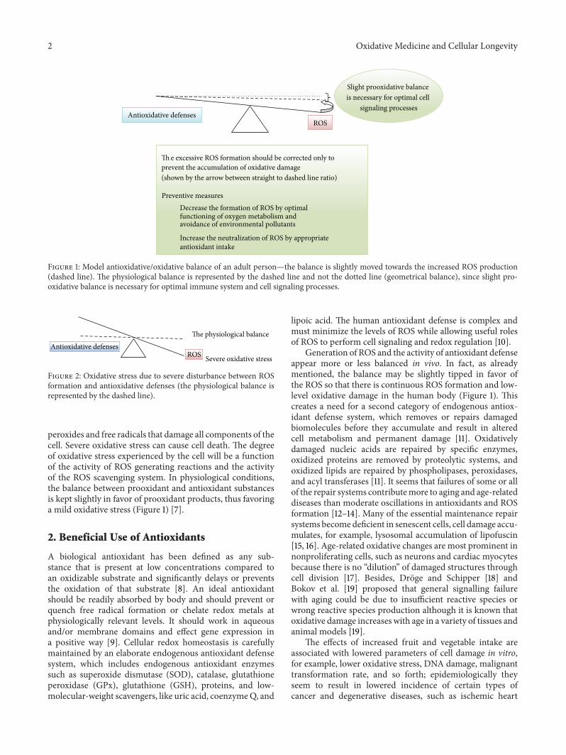
enzymes, peroxisomal beta-oxidation, and respiratoryburst of phagocytic cells [4]. It has been shown that oxidativestress is involved in over 100 diseases, as their cause orconsequence [2, 5]. Oxidative stress was first defined by Sies[6] as “a disturbance in the prooxidant to antioxidant balancein favor of the former, leading to potential damage” (Figures 1and 2). Oxidative stress can be defined as an excessive amountof ROS, which is the net result of an imbalance betweenproduction and destruction of ROS (the latter is regulatedby antioxidant defences). Oxidative stress is a consequenceof an increased generation of free radicals and/or reducedphysiological activity of antioxidant defenses against freeradicals. All forms of life maintain a reducing environmentwithin their cells. This reducing environment is preserved byenzymes that maintain the reduced state through a constantinput of metabolic energy. Disturbances in this normalredox state can cause toxic effects through the production of
2 Oxidative Medicine and Cellular Longevity
Slight prooxidative balance is necessary for optimal cell
signaling processesAntioxidative defenses
ROS
Th e excessive ROS formation should be corrected only toprevent the accumulation of oxidative damage(shown by the arrow between straight to dashed line ratio)
Preventive measuresDecrease the formation of ROS by optimalfunctioning of oxygen metabolism andavoidance of environmental pollutants
Increase the neutralization of ROS by appropriateantioxidant intake
Figure 1: Model antioxidative/oxidative balance of an adult person—the balance is slightly moved towards the increased ROS production(dashed line). The physiological balance is represented by the dashed line and not the dotted line (geometrical balance), since slight pro-oxidative balance is necessary for optimal immune system and cell signaling processes.
The physiological balance
Severe oxidative stressROSAntioxidative defenses
Figure 2: Oxidative stress due to severe disturbance between ROSformation and antioxidative defenses (the physiological balance isrepresented by the dashed line).
peroxides and free radicals that damage all components of thecell. Severe oxidative stress can cause cell death. The degreeof oxidative stress experienced by the cell will be a functionof the activity of ROS generating reactions and the activityof the ROS scavenging system. In physiological conditions,the balance between prooxidant and antioxidant substancesis kept slightly in favor of prooxidant products, thus favoringa mild oxidative stress (Figure 1) [7].
2. Beneficial Use of Antioxidants
A biological antioxidant has been defined as any sub-stance that is present at low concentrations compared toan oxidizable substrate and significantly delays or preventsthe oxidation of that substrate [8]. An ideal antioxidantshould be readily absorbed by body and should prevent orquench free radical formation or chelate redox metals atphysiologically relevant levels. It should work in aqueousand/or membrane domains and effect gene expression ina positive way [9]. Cellular redox homeostasis is carefullymaintained by an elaborate endogenous antioxidant defensesystem, which includes endogenous antioxidant enzymessuch as superoxide dismutase (SOD), catalase, glutathioneperoxidase (GPx), glutathione (GSH), proteins, and low-molecular-weight scavengers, like uric acid, coenzymeQ, and
lipoic acid. The human antioxidant defense is complex andmust minimize the levels of ROS while allowing useful rolesof ROS to perform cell signaling and redox regulation [10].
Generation of ROS and the activity of antioxidant defenseappear more or less balanced in vivo. In fact, as alreadymentioned, the balance may be slightly tipped in favor ofthe ROS so that there is continuous ROS formation and low-level oxidative damage in the human body (Figure 1). Thiscreates a need for a second category of endogenous antiox-idant defense system, which removes or repairs damagedbiomolecules before they accumulate and result in alteredcell metabolism and permanent damage [11]. Oxidativelydamaged nucleic acids are repaired by specific enzymes,oxidized proteins are removed by proteolytic systems, andoxidized lipids are repaired by phospholipases, peroxidases,and acyl transferases [11]. It seems that failures of some or allof the repair systems contributemore to aging and age-relateddiseases than moderate oscillations in antioxidants and ROSformation [12–14]. Many of the essential maintenance repairsystems becomedeficient in senescent cells, cell damage accu-mulates, for example, lysosomal accumulation of lipofuscin[15, 16]. Age-related oxidative changes are most prominent innonproliferating cells, such as neurons and cardiac myocytesbecause there is no “dilution” of damaged structures throughcell division [17]. Besides, Droge and Schipper [18] andBokov et al. [19] proposed that general signalling failurewith aging could be due to insufficient reactive species orwrong reactive species production although it is known thatoxidative damage increases with age in a variety of tissues andanimal models [19].
The effects of increased fruit and vegetable intake areassociated with lowered parameters of cell damage in vitro,for example, lower oxidative stress, DNA damage, malignanttransformation rate, and so forth; epidemiologically theyseem to result in lowered incidence of certain types ofcancer and degenerative diseases, such as ischemic heart

Oxidative Medicine and Cellular Longevity 3
disease and cataract [20–25]. On the other hand, increasedor prolonged free radical action can overwhelm ROS defensemechanisms, contributing to development of diseases andaging. Since oxidative damage of our cells increases with age,the increased intake of exogenous antioxidants from fruitand vegetables may support the endogenous antioxidativedefense. The antioxidants, like vitamin C and E, carotenoids,and polyphenols (e.g., flavonoids), are presently considered tobe the main exogenous antioxidants. Clinical studies implythat eating a diet rich in fruits, vegetables, whole grains,legumes, and omega-3 fatty acids can help humans in diseaseprevention [26].
3. Use of Synthetic Antioxidants:Their Control and Safety
A dietary supplement, also known as a food or nutritionalsupplement, is a preparation intended to provide nutrientssuch as vitamins, minerals, fibres, fatty, or amino acids thatare either missing or not consumed in sufficient amounts inperson’s diet. Surveys indicate that more than half of the USadult population uses food supplements, many of which con-tain antioxidants, such as vitamin A (retinoids, carotenes),vitamins C and E (tocopherols), lycopene, lutein, ubiquinone,glutathione, polyphenols (flavonoids), resveratrol, and N-acetylcysteine. In the USA, food supplements represent amarket of over $7 billion/year [27] and exceed $30 billionworldwide [28].
In the United States of America (and in many othercountries), the dietary supplement or dietary ingredientmanufacturer is responsible for ensuring that a dietarysupplement or ingredient is safe before it is marketedunder the Dietary Supplement Health and Education Act of1994 [29]. Generally, manufacturers neither need to registertheir products with Food and Drug Administration (FDA)nor get FDA approval before producing or selling dietarysupplements. FDA is responsible for taking action againstany unsafe dietary supplement product after it reaches themarket. Manufacturers must make sure that product labelinformation is truthful and not misleading and are requiredto submit to FDA all serious adverse event reports associatedwith use of the dietary supplement in the United States. Incontrast, the substances used as drugs must undergo clini-cal studies to determine their effectiveness, safety, possibleinteractions with other substances, and appropriate dosagesbefore entering the market [30]. FDA independently reviewscompany’s data and proposed labeling and, if health benefitsoutweigh its known risks, it approves the drug for sale.
The inappropriate use of dietary supplements may lead to“antioxidative stress.” This term was used for the first time byDundar and Aslan [31] for description of the negative effectsof antioxidants; it is discussed also by recent publication byPoljsak and Milisav [32]. Both “antioxidative” and oxida-tive stresses leading to the antioxidative imbalance can bedamaging for the organism and can result in cancerogenesis[33] and aging (Figures 2 and 3). There are a growingnumber of clinical trials in which individuals received oneor more synthetic antioxidants that fail to demonstrate
Severe “antioxidative stress”
ROS
The physiological balance
Antioxidative defenses
Figure 3: Severe disturbance between antioxidative defenses andROS leads to a state of increased “antioxidative stress.”
beneficial effects of antioxidant supplementation. Some evenimplied that antioxidant therapy had no effect and evencould increase mortality [34–46]. Ristow et al. [47] reportedthat nutritive antioxidants abolished the life extension byinhibiting a process called mitohormesis. Results of clinicaltrials on exogenous antioxidants intake are thus conflictingand contradictory.There seem to be homeostaticmechanismsin cells that govern the total antioxidant activity. Modifyingthe levels of one antioxidant causes compensatory changesin the levels of others, while the overall antioxidant capacityremains unaffected. The intake of only one antioxidant maythus alter the complex system of endogenous antioxidativedefence of cells or alter the cell apoptosis pathways [48].Dosing cells with exogenous antioxidants may decrease therate of synthesis or uptake of endogenous antioxidants, so thatthe total “cell antioxidant potential” remains unaltered. Cutler[49, 50] introduced “The oxidative stress compensationmodel” to explain why dietary supplements of antioxidantshave minimal effect on longevity. He explains that mosthumans are able to maintain their set point of oxidative stresseven if they consume additional antioxidant supplements; inother words, there is no further decrease in oxidative stress[49, 50].
4. Importance of the Balance
The production of free radicals increases with age [51],while some of the endogenous defense mechanisms decrease[52]. This imbalance leads to progressive damage of cellularstructures, presumably resulting in the aging phenotype [53,54].
The antioxidant defense system must thus minimizethe levels of most harmful ROS on one side while stillpermit enough ROS to remain for their useful purposes (e.g.,cell signaling and redox regulation). Cells usually toleratesuch mild oxidative stress; this stress can even upregulatecellular repair processes and other protective systems (e.g.,chaperones).
5. ‘‘Antioxidative Stress’’ Influences CellSignaling and Redox Regulation
The beneficial physiological cellular use of ROS is now beingdemonstrated in different fields, including intracellularsignaling and redox regulation. It is well documented thatlow levels of ROS are signaling molecules, modulating cellproliferation [55], apoptosis [56, 57], and gene expressionthrough activation of transcription factors [58], like NF-kappa-B and hypoxia-inducible-factor-1𝛼 (HIF) [59]. The

4 Oxidative Medicine and Cellular Longevity
inducers of NF-kappa-B include also tumor necrosis factoralpha (TNF𝛼) and interleukin 1-beta (IL-1𝛽) [60, 61]. ROScan act as signaling intermediates for cytokines, includingIL-1 and TNF𝛼 [62–64]. These proinflammatory cytokines,tumor necrosis factor (TNF)-𝛼, interleukin-1𝛽 (IL-1𝛽), andinterferon-𝛾 (IFN-𝛾), can additionally increase oxidativestress in humans [65], inducing production of ROS [64, 66].ROS also have a role in vascular cell signaling processesincluding activation of eNOS [67] and stimulation of cellgrowth and migration [68] through modulation of intra-cellular calcium [69] and activation of transcription factorssuch as NF-kappa-B [70] and protein kinases including ERK,p38MAPK, and Akt [71, 72]. ROS signaling is thus integratedinto many cellular pathways, including but not limited to (1)proliferation and survival pathways through MAP kinases,PI3 kinase, PTEN, and protein tyrosine phosphatases; (2)ROS homeostasis and antioxidant gene regulation throughRef-1, Nrf-2, thioredoxin, and so forth; (3) aging throughp66Shc; (4) DNAdamage response throughATMkinase; thismay lead to inhibition of mTORC1 resulting in suppressionof protein synthesis and activation of autophagy; (5) ironhomeostasis through iron-regulatory proteins (IRP) andiron-responsive elements (IRE), and so forth [73].
The production of O2
∙− and H2O2by activated phago-
cytes is the classic example of the deliberate metabolic gen-eration of ROS for useful purposes [74]. H
2O2is recognized
as an ubiquitous intracellular messenger [75–78]. Moderateamounts of mitochondrial superoxide and hydrogen per-oxide have important roles in a range of cellular signalingprocesses and can activate signaling pathways that promotecell survival and disease resistance due to hormesis [79–81]. Generation of O
2
∙−, HOCl, and H2O2by phagocytes is
important for defense against various bacterial and fungalstrains [82]. O
2
∙− is produced also by several cell types otherthan phagocytes, including lymphocytes and fibroblasts [82].As ROS are important in signal transduction, there seem tobe no great reserve of antioxidant defenses in mammals [83].
6. Imbalance between ROS and Antioxidants
6.1. Increased Oxidative Stress. The causes of increased ROSproduction include endogenous reasons (inflammation, ele-vation in O
2concentration, and increased mitochondrial
leakage) and exogenous (environmental pollution, strenuousexercise, smoking, nutrition, chronic inflammation, psycho-logical and emotional stress, and others) [3, 32, 79]. Causesof decreased antioxidant defenses include reduced activityof endogenous antioxidative enzymes and reduced intake orabsorption of antioxidants from food.
6.2. Increased “Antioxidative Stress”. Inappropriate antiox-idative intake may cause increased “antioxidative stress.”Antioxidants can neutralize ROS and decrease oxidativestress; however, this is not always beneficialwith respect to thedevelopment of a disease and its progression (e.g., cancer) orfor delaying aging [32] since antioxidants cannot distinguishamong the radicals with a beneficial physiological role andthose that cause oxidative damage to biomolecules.
Individuals who overdose antioxidant supplements couldenter the status of “antioxidative” stress (Figure 3). If admin-istration of antioxidant supplements decreases the level offree radicals, it may interfere with the immune system tofight bacteria and essential defensivemechanisms for removalof damaged cells, including those that are precancerousand cancerous [84]. Thus, antioxidant supplement overtakemay cause harm [35, 36, 56, 85, 86]. When large amountsof antioxidant nutrients are taken, they can also act asprooxidants by increasing oxidative stress [87, 88]. Pro- andantioxidant effects of antioxidants (e.g., vitamin C) are dosedependent, and thus, more is not necessarily better. Our dietstypically contain safe levels of vitamins; therefore, high-levelantioxidant supplementsmay upset this important physiolog-ical balance between the ROS formation and neutralization.
The amount of oxidized macromolecules in the cell is thesum of the rate of their formation subtracted by the rate ofrepair processes. The imposed oxidative damage potential isopposed by the antioxidant defense capacity of the system(Figure 4). In reality, the oxidative damage potential is greater,and thus there is a constant small amount of toxic free radicalformation, which escapes the defense of the cell. A certainamount of oxidized proteins and nucleic acids exists in cellsat all times; this reflects the oxidative events. Decreased com-pensation of oxidative stress and insufficient repair, in turn,accelerate aging, which consequently leads to further declineof cellular energy levels [89, 90]. Mechanisms that protectcells from oxidative stress (e.g., endogenous antioxidants,DNA repair processes) are consuming significant amounts ofenergy when being activated in all compartments of the cellfor prolonged time. It may require too much energy to pre-vent all oxidative damage throughout the life of an organism.Kowald and Kirkwood predicted that virtual immortalitymight be achieved if 55% of the total energy of the simulatedcell were devoted to repair and/or prevention of free radicaland oxidative damage on the quantitative MARS model(mitochondria, aberrant proteins, radicals, and scavengers)[91, 92]. It is the compromise to allocate suboptimal amountsof energy to cell repair systems, with a consequence of gradualdeterioration of the body structures with age [93]. Paradox-ically, the efficiency of defense and repair may be enhancedalso after the exposure to ROS, since the expression of manyDNA repair enzymes is upregulated during the oxidativestress [94–96]. Finkel and Holbrook [97] stated that thebest strategy to enhance endogenous antioxidant levels mayactually be oxidative stress itself, based on the classical phys-iological concept of hormesis [98]. This is in agreement witha recent Halliwell’s proposal that stimulating the increase inlevels of endogenous antioxidants by some prooxidants maybe more effective than consuming additional dietary antioxi-dants [10]. Many well-established components of the heart-healthy lifestyle are prooxidant, including the polyunsatu-rated fat, exercise, and moderate alcohol consumption [99].
7. The Importance of Determination of theOxidative/Antioxidative Status In Vivo
In order to determine the oxidative stress, both, the ROSformation as well as the antioxidative defense potential

Oxidative Medicine and Cellular Longevity 5
Physiological balanceGeometrical balance
Antioxidative defensesROS
Figure 4: The optimal situation: the physiological balance betweenthe ROS production and antioxidative defenses prevents the accu-mulation of damage by ROS and enables enough ROS for signaling.Enzymatic and nonenzymatic antioxidants can neutralize ROS andRNS and decrease oxidative stress and restore the balance.
should be measured; for example, low antioxidant amount isnot problematic when the ROS levels are low.
7.1. Determination of ROS. Free radicals have a very shorthalf-life, which makes them hard to measure in the lab-oratory. Nevertheless, multiple methods of oxidative stressmeasurement are available today, each with their own advan-tages and disadvantages (see review [101]). Many approachesare possible: identification of free radicals, either directly byparamagnetic electron resonance (electron spin resonance,ESR), or indirectly by identifying some more stable interme-diates: evaluation of the traces of radical attack on biologicalmolecules by high performance liquid chromatography, gas-liquid chromatography, colorimetric tests. The measurementof antioxidant status can be estimated by colorimetric,immune, or enzymatic methods [100] (Scheme 1). The directROS detection methods measure superoxide, H
2O2, ∙OH.
These are very reactive species and their quantitation isdifficult. In vivo ESR is relatively insensitive and requiressteady-state concentrations of free radicals in themicromolarrange, which limits its use formeasuringROS in patients. ESRcan be applied only through the technique of spin trapping forin vivo samples. Although it seems that toxicity is not a seriousproblem for most traps, there are no effective spin traps tobe administered to humans. Indirect methods are used inorder to overcome these problems. Indirect methods usuallymeasure the changes in endogenous antioxidant defensesystems or measure the ROS-induced damage of cellularcomponents [101]. Measuring the damage caused by ROSinstead of direct measuring of ROS seems logical, since itis the damage caused by ROS that is important rather thanthe total amount of generated ROS. Methods have beendeveloped to detect and quantify oxidative damage to pro-teins, lipids, and DNA. The principle behind fingerprintingmethods is to measure products of damage by ROS, that is, tomeasure not the species themselves but the damage that theycause. Of course, the end-products must be specific markersof oxidative damage [8]. According to Miwa et al. [102], agood marker of oxidative damage must increase by oxidativestress (i.e., upon the treatment with, e.g., paraquat, diquat,ionizing radiation, hyperoxia), and itmust remain unchangedin the absence of the oxidative event.
7.2. Determination of Antioxidant Status. There is a growinginterest to measure antioxidant status for clinical assessment[103]. Cellular protection against unwanted oxidation isachieved mainly by enzymes, such as superoxide dismutase(SOD), catalase, and glutathione peroxidase, whereas the
nonenzymatic antioxidants are playing the major role inthe plasma. Radical-scavenging antioxidants are consumedduring the reactions with ROS, and antioxidant statuscould be used indirectly to assess the free radical activity.One approach is to measure individual antioxidants (e.g.,ascorbate, 𝛼-tocopherol, urate) in blood, plasma, or tissuehomogenates. All of the individual molecules that are cur-rently recognized as antioxidants should be measured [103].However, this approach has several shortcomings: (1) it istime consuming, expensive, and technically demanding, (2)it may not detect the synergistic effects between the antioxi-dants, and (3) itmay not account for the influence of presentlyunknown antioxidant substances. The other approach is tomeasure the total antioxidant capacity or activity by sub-jecting the samples to controlled oxidative stress conditionsand measuring either the rate of oxidation or how long ittakes for oxidation to occur. Determination of antioxidativepotential per se is not sufficient, since it is difficult to establishhow the individual antioxidants work: by preventing theformation of ROS, by scavenging free radicals, by inducingthe signaling pathways, or by repairing the oxidative damage.Additionally, antioxidative status differs significantly betweenthe individuals and between the laboratory methods used inhumans [101]. Typical oxidative stress status of an individualis not established so far [104]. There are no reference valueson the optimal levels of antioxidants in urine, blood, or evenintracellularly. Additionally, several free radicals cannot crosscell membranes due to their charge, or they are so short-livedthat their diffusion is negligible. As such they cannot enter theblood from an affected region or organ. As there is no directcorrelation between the oxidative stress markers in bloodand their levels within the cells measuring the blood samplesmay be misleading. Besides, unknown are the amounts andcombinations of antioxidants needed for the beneficial effectin vivo. The improved methodology for determining theoxidative stress levels in humans may overcome at least someof these drawbacks.
Long-term effects of oxidative stress will occur if antiox-idant status is low and levels of free radicals are high. Nospecific clinical symptoms or clinical signs are associatedwithoxidative stress during the early stages of imbalance. There-fore, the oxidative stress is not diagnosed until there is anunavoidable damage and the consequences manifest as a signof a disease that could last for decades. Thus, oxidative stressshould be recognized and the oxidative imbalance should beameliorated in order to prevent or postpone the free radical-related disease development and premature aging [32]. Inpractice, it is difficult to determine all types of ROS withinthe cells or cellular compartments, as well as the overallantioxidative protection and repair of cells and organs at anyspecific time. Increased oxidative stress could result fromincreased ROS production or from decreased antioxidativedefences. Thus, increased ROS damage could be the resultof (a) increased ROS formation, (b) decreased antioxidativedefence, or/and (c) altered damage repair (Scheme 2). Sincenone of the biomarkers can predict the disease developmentas the consequence of the prolonged oxidative stress [105],it is important to use many methods for detection andquantification of oxidative stress whenever possible in order

6 Oxidative Medicine and Cellular Longevity
Direct methods
Indirect methods
EPR (ESR) and spin trapping
Oxidative DNA damage
Abasic (AP) sites
breaks
Comet assay (general DNA damage)
Others
Fingerprinting methods of:
Fluorescent probes
Lipid peroxidation
MDA
Others
Protein damage
PCC
AGE
AOPP
adducts Glycoxidation
aggregates, fragmentsLipid peroxidation and amino acid oxidation adducts Others
Superoxide dismutase
Glutathione reductase
Catalase
Glutathione peroxidase
Glutathione
Other antioxidative
Measurement of total antioxidant status
Measurement of endogenous enzymatic and
nonenzymatic antioxidant defense systems
Methods of free radicals and other reactive species detection
Double-strand DNA
8-OHdG4-HNE
Cross-links,
3-Nitrotyrosine8-isoprostaglandinF2𝛼 (8-isoprostane)
defense systems and repairprocesses
Scheme 1: Methods of oxidative stress determination. (8-OHG) = 8-hydroxyguanosine; 4-HNE = 4-Hydroxynonenal; MDA =malondialde-hyde; PCC = protein carbonyl content; AGE = advanced glycation end products; AOPP = advanced oxidation protein products.
to enhance their validity, as each method measures differentparameters and has inherent limitations. No single methodcan measure the oxidative stress or its subsequent damage invivo accurately at present. Additionally, the living organismsare complex and ever changing systems and therefore any
determination of the oxidative stress levels reflect the tem-porary state that may change considerably over time.
Presently, the use of supplemental antioxidants could beadvised only in cases of well-known conditions, where thedepletion of antioxidants is known and can be predicted.

Oxidative Medicine and Cellular Longevity 7
Damage
stress and/or damage and inplementation of strategies for balance restoration
Balance antioxidant levelAging
Disease
Damage to cells
Feedback loops between ROS,
antioxidants, and repair processes
Oxidative stress
Antioxidative stressFeedback loops between ROS,
antioxidants, and repair processes
Consequence
Consequence
Proteins
Lipids
DNA
Cause
Decreased ROS formation
Increased antioxidant defense
Defective immune and apoptosis systems
↓ Oxidative stress
↑ Free radical scavenging
↑ Damage repair
Approach
Early detection of (anti)oxidative
Increased ROS formationDecreased antioxidant defenseIneffective repair systems
Scheme 2: Oxidative and “antioxidative” stress: causes, consequences and methods for its control.
Daily use of synthetic supplements has not been proven asbeneficial, and excessive use may be harmful. Balanced foodstill seems to be the best option.
8. Discussion
A complex mix of substances in fruits and vegetables maycontribute to improved cardiovascular health and decreasedincidence of cancer in individuals who consume more ofthese foods [22, 23]. Even in elderly subjects a higher dailyintake of fruits and vegetables is associated with an improvedantioxidant status compared to subjects consuming dietspoor in fruits and vegetables [106]. Contrary, many clinicaltrials in which individuals received one or more syntheticantioxidants failed to prove their benefits. None of the
major clinical trials using mortality or morbidity as the endpoint has found positive effects of supplementation withantioxidants such as vitamin C, vitamin E, or 𝛽-carotene.Some recent studies showed that antioxidant therapy hadno effect and even increased the mortality [34–38, 107]. Theintake of only one antioxidant could alter the endogenousantioxidative defense of cells, modify cell death rates, ordecrease the synthesis of endogenous antioxidants. We haveto realize that the use of synthetic vitamin supplementsis not the alternative to the regular consumption of fruitsand vegetables. Cutler explains that most humans are ableto maintain their set points of oxidative stress regardlessof additional antioxidant supplementation through diet [49,50]. In contrast, antioxidant supplements do appear tobe effective in lowering an individual’s oxidative stress ifhis/her initial oxidative stress is above normal or above

8 Oxidative Medicine and Cellular Longevity
his/her set point of regulation [49, 50]. Thus, the antioxidantsupplements may help the organism to correct the elevatedlevels of oxidative stress that cannot be controlled by theendogenous antioxidants. There is also a problem of dosingthe synthetic antioxidants; for example, there are claims thatRDA (recommended daily allowance) levels of vitamin C andE are too low to prevent the oxidative stress. On the otherhand, many consumers ingest high amounts of supplementswith the antioxidant potential, which may lead to prooxidanteffects or to “antioxidative stress” [32]. Therefore, there isa need to determine the individual’s oxidative stress levelbefore administering the supplement therapy. However, thereference values for typical oxidative stress status of anindividual are not established so far and oxidative stress isdifficult and expensive to measure [104].
9. Conclusion
In vitro and in vivo studies imply that antioxidant nutrientsand related bioactive compounds from fruits and vegetablescan protect us from oxidative stress. Synthetic antioxidants asdietary supplements may prevent some ROS-induced dam-age in conditions of elevated oxidative stress during elevatedenvironmental oxidant exposure or at weaken endogenousoxidative stress responses of an aged organism. On theother hand, the presented evidence implies that syntheticantioxidant supplements cannot offer appropriate or totalprotection against oxidative stress and damage in “normal”situations and that the use of antioxidants to prevent diseaseor aging is controversial in situations of “normal” oxidativestress.
At the moment, it is difficult to evaluate the oxidativestress of the organism also because different criteria ofoxidative stress do not correlate with each other. Since there isno universal “scale” of oxidative stress, the future challenge(s)are in determination of total antioxidants and oxidativestress levels in different body fluids (urine, saliva, blood,and cytosol). Further, detection of the increased levels ofoxidative stress biomarker in the body fluid does not meannecessarily that the cells of the specific organ or tissue areunder oxidative stress. Besides, it is not possible for highlyreactive free radical produced within a tissue with a lifetimeofmicroseconds to diffuse into the blood to be detected at thedistant site. The researcher is thus limited to determinationof secondary products in human body fluids distant from thelocus of the ROS production [108]. With indirect oxidativestress markers, the person may be considered being underoxidative stress according to a given criterion but not toanother. Therefore, there is an urgent need to compare andstandardize the various methods for assessing the oxidativestate of biological systems, to establish the universal scaleof oxidative stress, and to provide age and gender specifictables of “normal values” for each body fluid. Until these areestablished, it is prudent to estimate the oxidative stress bycombining different methods and biomarkers.
The key to the future success of dietary antioxidantsupplementationmay be in the fine tuning of the suppressionof oxidative damagewithout disruption of the well-integrated
antioxidant defense networks. The selective enhancement ofthe defense system could be a major strategy for a successfulintervention by antioxidant administration [109].
References
[1] P. A. Riley, “Free radicals in biology: oxidative stress and theeffects of ionizing radiation,” International Journal of RadiationBiology, vol. 65, no. 1, pp. 27–33, 1994.
[2] B. Halliwell, J. M. C. Gutteridge, and C. E. Cross, “Free radicals,antioxidants, and human disease: where are we now?” Journalof Laboratory and Clinical Medicine, vol. 119, no. 6, pp. 598–620,1992.
[3] B. Poljsak, P. Jamnik, P. Raspor, and M. Pesti, “Oxidation-antioxidation-reduction processes in the cell: impacts of envi-ronmental pollution,” in Encyclopedia of Environmental Health,N. Jerome, Ed., pp. 300–306, Elsevier, 2011.
[4] M. Gilca, I. Stoian, V. Atanasiu, and B. Virgolici, “The oxidativehypothesis of senescence,” Journal of PostgraduateMedicine, vol.53, no. 3, pp. 207–213, 2007.
[5] J. M. C. Gutteridge, “Free radicals in disease processes: acompilation of cause and consequence,” Free Radical ResearchCommunications, vol. 19, no. 3, pp. 141–158, 1993.
[6] H. Sies, “Oxidative stress: from basic research to clinicalapplication,”American Journal of Medicine, vol. 91, no. 3, pp. 31–38, 1991.
[7] W. Droge, “Free radicals in the physiological control of cellfunction,” Physiological Reviews, vol. 82, no. 1, pp. 47–95, 2002.
[8] B. Halliwell and J.M. C. Gutteridge, Free Radicals in Biology andMedicine, Clarendon Press, Oxford, UK, 3nd edition, 1999.
[9] K. Rahman, “Studies on free radicals, antioxidants, and co-factors,”Clinical Interventions inAging, vol. 2, no. 2, pp. 219–236,2007.
[10] B. Halliwell, “Free radicals and antioxidants—quo vadis?”Trends in Pharmacological Sciences, vol. 32, no. 3, pp. 125–130,2011.
[11] K. H. Cheeseman and T. F. Slater, “An introduction to freeradical biochemistry,” British Medical Bulletin, vol. 49, no. 3, pp.481–493, 1993.
[12] D. Gems and R. Doonan, “Antioxidant defense and aging in C.elegans: is the oxidative damage theory of aging wrong?” CellCycle, vol. 8, no. 11, pp. 1681–1687, 2009.
[13] V. I. Perez, A. Bokov,H.V. Remmen et al., “Is the oxidative stresstheory of aging dead?” Biochimica et Biophysica Acta, vol. 1790,no. 10, pp. 1005–1014, 2009.
[14] Y. C. Jang and H. V. Remmen, “The mitochondrial theory ofaging: insight from transgenic and knockout mouse models,”Experimental Gerontology, vol. 44, no. 4, pp. 256–260, 2009.
[15] A. Terman and U. T. Brunk, “Oxidative stress, accumulationof biological “garbage”, and aging,” Antioxidants and RedoxSignaling, vol. 8, no. 1-2, pp. 197–204, 2006.
[16] U. T. Brunk, C. B. Jones, and R. S. Sohal, “A novel hypothesisof lipofuscinogenesis and cellular aging based on interac-tions between oxidative stress and autophagocytosis,”MutationResearch, vol. 275, no. 3–6, pp. 395–403, 1992.
[17] A. Terman, “Garbage catastrophe theory of aging: imperfectremoval of oxidative damage?” Redox Report, vol. 6, no. 1, pp.15–26, 2001.
[18] W. Droge and H. M. Schipper, “Oxidative stress and aberrantsignaling in aging and cognitive decline,” Aging Cell, vol. 6, no.3, pp. 361–370, 2007.

Oxidative Medicine and Cellular Longevity 9
[19] A. Bokov, A. Chaudhuri, and A. Richardson, “The role ofoxidative damage and stress in aging,” Mechanisms of Ageingand Development, vol. 125, no. 10-11, pp. 811–826, 2004.
[20] S. A. Stanner, J. Hughes, C. N.M. Kelly, and J. Buttriss, “A reviewof the epidemiological evidence for the ‘antioxidant hypothesis’,”Public Health Nutrition, vol. 7, no. 3, pp. 407–422, 2004.
[21] World Cancer Research Found, http://www.wcrf.org/.[22] A.Cherubini, G. B.Vigna,G. Zuliani, C. Ruggiero,U. Senin, and
R. Fellin, “Role of antioxidants in atherosclerosis: epidemiolog-ical and clinical update,” Current Pharmaceutical Design, vol. 11,no. 16, pp. 2017–2032, 2005.
[23] S. B. Lotito and B. Frei, “Consumption of flavonoid-rich foodsand increased plasma antioxidant capacity in humans: cause,consequence, or epiphenomenon?” Free Radical Biology andMedicine, vol. 41, no. 12, pp. 1727–1746, 2006.
[24] H. Boeing, A. Bechthold, A. Bub et al., “Critical review:vegetables and fruit in the prevention of chronic diseases,”European Journal of Nutrition, vol. 51, no. 6, pp. 637–663, 2012.
[25] F. L. Crowe, A. W. Roddam, T. J. Key et al., “Fruit and vegetableintake and mortality from ischaemic heart disease: resultsfrom the European Prospective Investigation into Cancer andNutrition (EPIC)-Heart study,” European Heart Journal, vol. 32,no. 10, pp. 1235–1243, 2011.
[26] W. C. Willett, “The Mediterranean diet: science and practice,”Public Health Nutrition A, vol. 9, no. 1, pp. 105–110, 2006.
[27] V. Glaser, “Billion-dollar market blossoms as botanicals takeroot,” Nature Biotechnology, vol. 17, no. 1, pp. 17–18, 1999.
[28] I. Raskin, D. M. Ribnicky, S. Komarnytsky et al., “Plants andhuman health in the twenty-first century,” Trends in Biotechnol-ogy, vol. 20, no. 12, pp. 522–531, 2002.
[29] DSHEA, http://www.fda.gov/Food/DietarySupplements/default.htm.
[30] FDA, http://www.fda.gov/Drugs/DevelopmentApprovalPro-cess/default.htm.
[31] Y. Dundar and R. Aslan, “Antioxidative stress,” Eastern Journalof Medicine, vol. 5, no. 2, pp. 45–47, 2000.
[32] B. Poljsak and I. Milisav, “The neglected significance of ‘Antiox-idative Stress’,” Oxidative Medicine and Cellular Longevity, vol.2012, Article ID 480895, 12 pages, 2012.
[33] B. Halliwell, “Oxidative stress and cancer: have we movedforward?” Biochemical Journal, vol. 401, no. 1, pp. 1–11, 2007.
[34] G. S. Omenn, G. E. Goodman, M. D. Thornquist et al., “Effectsof a combination of beta carotene and vitamin A on lungcancer and cardiovascular disease,”The New England Journal ofMedicine, vol. 334, no. 18, pp. 1150–1155, 1996.
[35] G. Bjelakovic, D. Nikolova, R. G. Simonetti, and C. Gluud,“Antioxidant supplements for prevention of gastrointestinalcancers: a systematic review andmeta-analysis,”TheLancet, vol.364, no. 9441, pp. 1219–1228, 2004.
[36] E. R. Miller, R. Pastor-Barriuso, D. Dalal, R. A. Riemersma, L.J. Appel, and E. Guallar, “Meta-analysis: high-dosage vitaminE supplementation may increase all-cause mortality,” Annals ofInternal Medicine, vol. 142, no. 1, pp. 37–46, 2005.
[37] Heart Protection Study Collaborative Group, “MRC/BHFHeartProtection Study of antioxidant vitamin supplementation in20 536 high-risk individuals: a randomised placebo-controlledtrial,”The Lancet, vol. 360, no. 9326, pp. 23–33, 2002.
[38] Age-Related Eye Disease Study Research Group, “A random-ized, placebo-controlled, clinical trial of high-dose supplemen-tation with vitamins C and E and beta carotine for age-relatedcataract and vision loss: AREDS report no. 9,” Archives ofOphthalmology, vol. 119, pp. 1439–1452, 200152.
[39] J. Mursu, K. Robien, L. J. Harnack, K. Park, and D. R. Jacobs Jr.,“Dietary supplements and mortality rate in older women: theIowaWomen’s Health Study,” Archives of Internal Medicine, vol.171, no. 18, pp. 1625–1633, 2011.
[40] E. A. Klein, I. M. Thompson Jr., C. M. Tangen et al., “VitaminE and the risk of prostate cancer: the Selenium and VitaminE Cancer Prevention Trial (SELECT),” Journal of the AmericanMedical Association, vol. 306, no. 14, pp. 1549–1556, 2011.
[41] G. Bjelakovic, D. Nikolova, L. L. Gluud, R. G. Simonetti, andC. Gluud, “Antioxidant supplements for prevention ofmortalityin healthy participants and patients with various diseases,”Cochrane Database of Systematic Reviews, vol. 16, no. 2, ArticleID CD007176, 2008.
[42] S. Hercberg, K. Ezzedine, C. Guinot et al., “Antioxidant supple-mentation increases the risk of skin cancers in women but notinmen,” Journal of Nutrition, vol. 137, no. 9, pp. 2098–2105, 2007.
[43] A. Bardia, I. M. Tleyjeh, J. R. Cerhan et al., “Efficacy of antiox-idant supplementation in reducing primary cancer incidenceand mortality: systematic review and meta-analysis,” MayoClinic Proceedings, vol. 83, no. 1, pp. 23–34, 2008.
[44] B. D. Lawenda, K. M. Kelly, E. J. Ladas, S. M. Sagar, A.Vickers, and J. B. Blumberg, “Should supplemental antioxidantadministration be avoided during chemotherapy and radiationtherapy?” Journal of the National Cancer Institute, vol. 100, no.11, pp. 773–783, 2008.
[45] S. K. Myung, Y. Kim, W. Ju, H. J. Choi, and W. K. Bae, “Effectsof antioxidant supplements on cancer prevention:meta-analysisof randomized controlled trials,”Annals of Oncology, vol. 21, no.1, pp. 166–179, 2010.
[46] G. Bjelakovic, D. Nikolova, L. L. Gluud, R. G. Simonetti, andC. Gluud, “Antioxidant supplements for prevention ofmortalityin healthy participants and patients with various diseases,”Cochrane Database of Systematic Reviews, no. 2, Article IDCD007176, 2008.
[47] M. Ristow, K. Zarse, A. Oberbach et al., “Antioxidants preventhealth Antioxidant supplements for prevention of mortalityin healthy participants and patients with various diseases.-promoting effects of physical exercise in humans,” Proceedingsof the National Academy of Sciences of the United States, vol. 106,pp. 8665–8670, 2009.
[48] S. Verhaegn, J. Adrain, J. McGovan, A. R. Brophy, R. S.Fernandes, and T. G. Gotler, “Inhibition of apoptosis by antiox-idants in the human IIL-60 leukemia cell line,” BiochemicalPharmacology, vol. 40, pp. 1021–1029, 1995.
[49] R. G. Cutler andM. P.Mattson, “Measuring oxidative stress andinterpreting its relevance in humans,” in Oxidative Stress andAging, R. G. Cutler and H. Rodriguez, Eds., World Scientific,New Jersey, NJ, USA, 2003.
[50] R. G. Cutler, “Genetic stability, dysdifferentiation, and longevitydeterminant genes,” in Critical Reviews of Oxidative Stress andDamage, R. G. Cutler and H. Rodriguez, Eds., World Scientific,New Jersey, NJ, USA, 2003.
[51] R. S. Sohal and R. Weindruch, “Oxidative stress, caloric restric-tion, and aging,” Science, vol. 273, no. 5271, pp. 59–63, 1996.
[52] I. Bogdan Allemann and L. Baumann, “Antioxidants used inskin care formulations,” Skin Therapy Letter, vol. 13, no. 7, pp.5–9, 2008.
[53] J. L. Hagen, D. J. Krause, D. J. Baker,M.Hua Fu,M. A. Tarnopol-sky, and R. T. Hepple, “Skeletal muscle aging in F344BN F1-hybrid rats: I.Mitochondrial dysfunction contributes to the age-associated reduction in VO2max,” Journals of Gerontology A,vol. 59, no. 11, pp. 1099–1110, 2004.

10 Oxidative Medicine and Cellular Longevity
[54] M. L. Hamilton, H. Van Remmen, J. A. Drake et al., “Doesoxidative damage to DNA increase with age?” Proceedings of theNational Academy of Sciences of theUnited States of America, vol.98, no. 18, pp. 10469–10474, 2001.
[55] G. A. C. Murrell, M. J. O. Francis, and L. Bromley, “Modulationof fibroblast proliferation by oxygen free radicals,” BiochemicalJournal, vol. 265, no. 3, pp. 659–665, 1990.
[56] K. M. Kim, P. K. Kim, Y. G. Kwon, S. K. Bai, W. D. Nam, and Y.M. Kim, “Regulation of apoptosis by nitrosative stress,” Journalof Biochemistry andMolecular Biology, vol. 35, pp. 127–133, 2002.
[57] G. Kroemer, N. Zamzami, and S. A. Susin, “Mitochondrialcontrol of apoptosis,” Immunology Today, vol. 18, no. 1, pp. 44–51, 1997.
[58] R. Schreck, P. Rieber, and P. A. Baeuerle, “Reactive oxygenintermediates as apparently widely used messengers in theactivation of the NF-𝜅B transcription factor and HIV-1,” TheEMBO Journal, vol. 10, no. 8, pp. 2247–2258, 1991.
[59] K. D. Kroncke, “Nitrosative stress and transcription,” TheJournal of Biological Chemistry, vol. 384, pp. 1365–1377, 2003.
[60] D. C. Fitzgerald, K. G. Meade, A. N. McEvoy et al., “Tumournecrosis factor-𝛼 (TNF-𝛼) increases nuclear factor 𝜅B (NF𝜅B)activity in and interleukin-8 (IL-8) release from bovinemammary epithelial cells,” Veterinary Immunology andImmunopathology, vol. 116, no. 1-2, pp. 59–68, 2007.
[61] P. Renard, M. D. Zachary, C. Bougelet et al., “Effects of antiox-idant enzyme modulations on interleukin-1-induced nuclearfactor kappa B activation,” Biochemical Pharmacology, vol. 53,no. 2, pp. 149–160, 1997.
[62] B.Meier, H.H. Radeke, S. Selle et al., “Human fibroblasts releasereactive oxygen species in response to interleukin-1 or tumournecrosis factor-𝛼,” Biochemical Journal, vol. 263, no. 2, pp. 539–545, 1989.
[63] M. L. Tiku, J. B. Liesch, and F. M. Robertson, “Production ofhydrogen peroxide by rabbit articular chondrocytes. Enhance-ment by cytokines,” Journal of Immunology, vol. 145, no. 2, pp.690–696, 1990.
[64] Y. Y. C. Lo and T. F. Cruz, “Involvement of reactive oxygenspecies in cytokine and growth factor induction of c-fosexpression in chondrocytes,” Journal of Biological Chemistry,vol. 270, no. 20, pp. 11727–11730, 1995.
[65] D. Yang, S. G. Elner, Z.M. Bian, G. O. Till, H. R. Petty, and V.M.Elner, “Pro-inflammatory cytokines increase reactive oxygenspecies throughmitochondria and NADPH oxidase in culturedRPE cells,” Experimental Eye Research, vol. 85, no. 4, pp. 462–472, 2007.
[66] Y. Y. C. Lo, J. M. S. Wong, and T. F. Cruz, “Reactive oxygenspecies mediate cytokine activation of c-Jun NH2- terminalkinases,” Journal of Biological Chemistry, vol. 271, no. 26, pp.15703–15707, 1996.
[67] H. Cai, Z. Li, S. Dikalov et al., “NAD(P)H oxidase-derivedhydrogen peroxide mediates endothelial nitric oxide produc-tion in response to angiotensin II,” Journal of Biological Chem-istry, vol. 277, no. 50, pp. 48311–48317, 2002.
[68] Q. Felty, W. C. Xiong, D. Sun et al., “Estrogen-inducedmitochondrial reactive oxygen species as signal-transducingmessengers,” Biochemistry, vol. 44, no. 18, pp. 6900–6909, 2005.
[69] G. B. Waypa, J. D. Marks, M. M. Mack, C. Boriboun, P. T.Mungai, and P. T. Schumacker, “Mitochondrial reactive oxygenspecies trigger calcium increases during hypoxia in pulmonaryarterial myocytes,” Circulation Research, vol. 91, no. 8, pp. 719–726, 2002.
[70] H. L. Pahl and P. A. Baeuerle, “Oxygen and the control of geneexpression,” BioEssays, vol. 16, no. 7, pp. 497–502, 1994.
[71] F. S. Wang, C. J. Wang, Y. J. Chen et al., “Ras inductionof superoxide activates ERK-dependent angiogenic transcrip-tion factor HIF-1𝛼 and VEGF-A expression in shock wave-stimulated osteoblasts,” Journal of Biological Chemistry, vol. 279,no. 11, pp. 10331–10337, 2004.
[72] K. M. Connor, S. Subbaram, K. J. Regan et al., “MitochondrialH2O2regulates the angiogenic phenotype via PTEN oxidation,”
Journal of Biological Chemistry, vol. 280, no. 17, pp. 16916–16924,2005.
[73] P. D. Ray, B.-W. Huang, and Y. Tsuji, “Reactive oxigen species(ROS) homeostasis and redox regulation in cellular signaling,”Cellular Signalling, vol. 24, no. 5, pp. 981–990, 2012.
[74] B. Halliwell and C. E. Cross, “Oxygen-derived species: theirrelation to human disease and environmental stress,” Environ-mental Health Perspectives, vol. 102, no. 10, pp. 5–12, 1994.
[75] S. G. Rhee, Y. S. Bae, S. R. Lee, and J. Kwon, “Hydrogen perox-ide: a key messenger that modulates protein phosphorylationthrough cysteine oxidation,” Science”s STKE, vol. 2000, no. 53,p. PE1, 2000.
[76] S. G. Rhee, “Redox signaling: hydrogen peroxide as intracellularmessenger,” Experimental &MolecularMedicine, vol. 31, pp. 53–59, 1999.
[77] T. Finkel, “Oxygen radicals and signaling,” Current Opinion inCell Biology, vol. 10, pp. 248–253, 1998.
[78] Y. J. Suzuki and G. D. Ford, “Redox regulation of signal trans-duction in cardiac and smooth muscle,” Journal of Molecularand Cellular Cardiology, vol. 31, pp. 345–353, 1999.
[79] B. Poljsak, “Strategies for reducing or preventing the generationof oxidative stress,” Oxidative Medicine and Cellular Longevity,vol. 2011, Article ID 194586, 15 pages, 2011.
[80] S. I. S. Rattan and D. Demirovic, “Hormesis can and does workin humans,” Dose-Response, vol. 8, no. 1, pp. 58–63, 2010.
[81] M. P.Mattson, T. G. Son, and S. Camandola, “Viewpoint: mech-anisms of action and therapeutic potential of neurohormeticphytochemicals,” Dose Response, vol. 5, no. 3, pp. 174–186, 2007.
[82] B. M. Babior and R. C. Woodman, “Chronic granulomatousdisease,” Seminars in Hematology, vol. 27, no. 3, pp. 247–259,1990.
[83] R. Stocker and B. Frei, “Endogenous antioxidant defenses inhuman blood plasma,” inOxidative Stress: Oxidants and Antiox-idants, H. Sies, Ed., pp. 213–242, Academic Press, London, UK,1991.
[84] R. I. Salganik, “The benefits and hazards of antioxidants: con-trolling apoptosis and other protective mechanisms in cancerpatients and the human population,” Journal of the AmericanCollege of Nutrition, vol. 20, no. 5, pp. 464S–72S, 2001.
[85] D. P. Vivekananthan, M. S. Penn, S. K. Sapp, A. Hsu, and E.J. Topol, “Use of antioxidant vitamins for the prevention ofcardiovascular disease: meta-analysis of randomised trials,”TheLancet, vol. 361, no. 9374, pp. 2017–2023, 2003.
[86] M. Caraballoso, M. Sacristan, C. Serra, and X. Bonfill, “Drugsfor preventing lung cancer in healthy people,” CochraneDatabase of Systematic Reviews, vol. 2, Article ID CD002141,2003.
[87] I. D. Podmore, H. R. Griffiths, K. E. Herbert, N. Mistry, P. Mis-try, and J. Lunec, “Vitamin C exhibits pro-oxidant properties,”Nature, vol. 392, no. 6676, p. 559, 1998.
[88] P. Palozza, “Pro-oxidant actions of carotenoids in biologicsystems,” Nutrition Reviews, vol. 56, no. 9, pp. 257–265, 1998.
[89] K. J. Davies, “Oxidative stress: the paradox of aerobic life,”Biochemical Society Symposium, vol. 61, pp. 1–31, 1995.

Oxidative Medicine and Cellular Longevity 11
[90] T. Blatt, H. Wenck, and K. P. Wittern, “Alterations of energymetabolism in cutaneous aging,” in Textbook of Aging Skin, M.A. Farage, K.W.Miller, andH. I.Maibach, Eds., Springer, Berlin,Germany, 2010.
[91] A. Kowald and T. B. L. Kirkwood, “Towards a network theoryof ageing: a model combining the free radical theory and theprotein error theory,” Journal ofTheoretical Biology, vol. 168, no.1, pp. 75–94, 1994.
[92] A. Kowald and T. B. L. Kirkwood, “A network theory of ageing:the interactions of defective mitochondria, aberrant proteins,free radicals and scavengers in the ageing process,” MutationResearch, vol. 316, no. 5-6, pp. 209–236, 1996.
[93] T. B. L. Kirkwood, “Evolution of ageing,” Nature, vol. 270, no.5635, pp. 301–304, 1977.
[94] T. J. Schulz, K. Zarse, A. Voigt, N. Urban, M. Birringer, andM. Ristow, “Glucose restriction extends caenorhabditis eleganslife span by inducing mitochondrial respiration and increasingoxidative stress,” Cell Metabolism, vol. 6, no. 4, pp. 280–293,2007.
[95] G. Wani, G. E. Milo, and S. M. D’Ambrosio, “Enhanced expres-sion of the 8-oxo-7,8-dihydrodeoxyguanosine triphosphatasegene in human breast tumor cells,” Cancer Letters, vol. 125, no.1-2, pp. 123–130, 1998.
[96] R. Bases, W. A. Franklin, T. Moy, and F. Mendez, “Enhancedexcision repair activity in mammalian cells after ionizingradiation,” International Journal of RadiationBiology, vol. 62, no.4, pp. 427–441, 1992.
[97] T. Finkel and N. J. Holbrook, “Oxidants, oxidative stress and thebiology of ageing,”Nature, vol. 408, no. 6809, pp. 239–247, 2000.
[98] I.Milisav, B. Poljsak, andD. Suput, “Adaptive response, evidenceof cross-resistance and its potential clinical use,” InternationalJournal of Molecular Sciences, vol. 13, no. 9, pp. 10771–10806,2012.
[99] K. J. Williams and E. A. Fisher, “Oxidation, lipoproteins, andatherosclerosis: which iswrong, the antioxidants or the theory?”Current Opinion in Clinical Nutrition andMetabolic Care, vol. 8,no. 2, pp. 139–146, 2005.
[100] Z. Sahnoun, K. Jamoussi, and K. M. Zeghal, “Free radicals:fundamental notions and methods of exploration,” Therapie,vol. 52, no. 4, pp. 251–270, 1997.
[101] B. Poljsak and P. Jamik, “Methodology for oxidative statedetection in biological systems,” in Handbook of Free Radicals:Formation, Types and Effects, D. Kozyrev and V. Slutsky, Eds.,Cell Biology Research Progress Series, Nova Science, New York,NY, USA, 2010.
[102] S. Miwa, F. L. Muller, and K. B. Beckman, “The basics ofoxidative biochemistry,” in Oxidative Stress in Aging. FromModel Systems to HumanDiseases, S. Miwa, K. B. Beckman, andF. L. Muller, Eds., Humana Press, Totowa, NJ, USA, 2008.
[103] J. Lunec andH. R.Griffits,Measuring in vivoOxidativeDamage,John Willey and Sons, New York, NY, USA, 2000.
[104] S. Arguelles, A. Gomez, A. Machado, and A. Ayala, “A pre-liminary analysis of within-subject variation in human serumoxidative stress parameters as a function of time,” RejuvenationResearch, vol. 10, no. 4, pp. 621–636, 2007.
[105] B. Halliwell, “The wanderings of a free radical,” Free RadicalBiology and Medicine, vol. 46, no. 5, pp. 531–542, 2009.
[106] T. Anlasik, H. Sies, H. R. Griffiths, P. Mecocci, W. Stahl, andM. C. Polidori, “Dietary habits are major determinants of theplasma antioxidant status in healthy elderly subjects,” BritishJournal of Nutrition, vol. 94, no. 5, pp. 639–642, 2005.
[107] “The effect of vitamin E and beta carotene on the incidenceof lung cancer and other cancers in male smokers,” The New
England Journal of Medicine, vol. 330, no. 15, pp. 1029–1035,1994.
[108] A. E. Holley and K. H. Cheeseman, “Measuring free radicalreactions in vivo,”BritishMedical Bulletin, vol. 49, no. 3, pp. 494–505, 1993.
[109] B. P. Yu, “Aging and oxidative stress: modulation by dietaryrestriction,” Free Radical Biology & Medicine, vol. 21, pp. 651–668, 1996.

Submit your manuscripts athttp://www.hindawi.com
Stem CellsInternational
Hindawi Publishing Corporationhttp://www.hindawi.com Volume 2014
Hindawi Publishing Corporationhttp://www.hindawi.com Volume 2014
MEDIATORSINFLAMMATION
of
Hindawi Publishing Corporationhttp://www.hindawi.com Volume 2014
Behavioural Neurology
EndocrinologyInternational Journal of
Hindawi Publishing Corporationhttp://www.hindawi.com Volume 2014
Hindawi Publishing Corporationhttp://www.hindawi.com Volume 2014
Disease Markers
Hindawi Publishing Corporationhttp://www.hindawi.com Volume 2014
BioMed Research International
OncologyJournal of
Hindawi Publishing Corporationhttp://www.hindawi.com Volume 2014
Hindawi Publishing Corporationhttp://www.hindawi.com Volume 2014
Oxidative Medicine and Cellular Longevity
Hindawi Publishing Corporationhttp://www.hindawi.com Volume 2014
PPAR Research
The Scientific World JournalHindawi Publishing Corporation http://www.hindawi.com Volume 2014
Immunology ResearchHindawi Publishing Corporationhttp://www.hindawi.com Volume 2014
Journal of
ObesityJournal of
Hindawi Publishing Corporationhttp://www.hindawi.com Volume 2014
Hindawi Publishing Corporationhttp://www.hindawi.com Volume 2014
Computational and Mathematical Methods in Medicine
OphthalmologyJournal of
Hindawi Publishing Corporationhttp://www.hindawi.com Volume 2014
Diabetes ResearchJournal of
Hindawi Publishing Corporationhttp://www.hindawi.com Volume 2014
Hindawi Publishing Corporationhttp://www.hindawi.com Volume 2014
Research and TreatmentAIDS
Hindawi Publishing Corporationhttp://www.hindawi.com Volume 2014
Gastroenterology Research and Practice
Hindawi Publishing Corporationhttp://www.hindawi.com Volume 2014
Parkinson’s Disease
Evidence-Based Complementary and Alternative Medicine
Volume 2014Hindawi Publishing Corporationhttp://www.hindawi.com