Review Article ABO Blood Groups and Cardiovascular Diseases - Hindawi
12
Hindawi Publishing Corporation International Journal of Vascular Medicine Volume 2012, Article ID 641917, 11 pages doi:10.1155/2012/641917 Review Article ABO Blood Groups and Cardiovascular Diseases Hanrui Zhang, 1, 2 Ciar´ an J. Mooney, 1, 2 and Muredach P. Reilly 1, 2 1 Cardiovascular Institute, Perelman School of Medicine, University of Pennsylvania, Philadelphia, PA 19104-6160, USA 2 Institute for Translational Medicine and Therapeutics, University of Pennsylvania School of Medicine, Philadelphia, PA 19104-5158, USA Correspondence should be addressed to Muredach P. Reilly, [email protected] Received 20 June 2012; Revised 25 August 2012; Accepted 1 September 2012 Academic Editor: Masaki Mogi Copyright © 2012 Hanrui Zhang et al. This is an open access article distributed under the Creative Commons Attribution License, which permits unrestricted use, distribution, and reproduction in any medium, provided the original work is properly cited. ABO blood groups have been associated with various disease phenotypes, particularly cardiovascular diseases. Cardiovascular diseases are the most common causes of death in developed countries and their prevalence rate is rapidly growing in developing countries. There have been substantial historical associations between non-O blood group status and an increase in some cardiovascular disorders. Recent GWASs have identified ABO as a locus for thrombosis, myocardial infarction, and multiple cardiovascular risk biomarkers, refocusing attention on mechanisms and potential for clinical advances. As we highlight in this paper, more recent work is beginning to probe the molecular basis of the disease associations observed in these observational studies. Advances in our understanding of the physiologic importance of various endothelial and platelet-derived circulating glycoproteins are elucidating the mechanisms through which the ABO blood group may determine overall cardiovascular disease risk. The role of blood group antigens in the pathogenesis of various cardiovascular disorders remains a fascinating subject with potential to lead to novel therapeutics and prognostics and to reduce the global burden of cardiovascular diseases. 1. Introduction In 1901, Landsteiner identified ABO blood groups as the first recognized human blood group system. The clinical significance of ABO blood type extends beyond transfusion medicine and solid organ/hematopoietic transplantation. To date, numerous reports have suggested important associa- tions between ABO blood groups and various diseases, for example, gastric cancer [1], periodontal diseases [2], and cardiometabolic diseases [3, 4]. According to World Health Organization (WHO) data, cardiovascular diseases (CVDs) are and will remain the leading causes of death globally: an estimated 17.3 million people died from CVD in 2008, representing 30% of all global deaths (WHO Media Centre (2011), cardiovascular diseases (fact sheet). retrieved from http://www.who.int/ mediacentre/factsheets/fs317/en/index.html). Studies on the associations between CVD and ABO blood groups have a long history. In 1955, Woolf proposed an odds ratio as a mea- sure to quantify the disease risk conferred by blood group type [5]. In 1969, Jick et al. reported a deficit of patients with blood group O among those who received anticoagulants for venous thromboembolism [6]. Prior to mutation detection in haemophilia carriership analysis, likelihood ratios of carriership of hemophilia A were based on Factor VIII levels conditional on blood group [7]. A number of later studies elucidated that ABO blood groups, particularly non-O blood groups, are associated with major cardiovascular risk factors and/or increased rate of cardiovascular events [8–13]. How- ever, there is limited consensus regarding the magnitude and significance of the ABO effects at the population level, and whether it relates to all disorders equally or predominantly modulates thrombotic pathways and disorders [14]. This paper summarizes the basic concepts of the biochemistry of ABO blood groups and recent findings of their relations to CVD.
Transcript of Review Article ABO Blood Groups and Cardiovascular Diseases - Hindawi

Hindawi Publishing CorporationInternational Journal of Vascular MedicineVolume 2012, Article ID 641917, 11 pagesdoi:10.1155/2012/641917
Review Article
ABO Blood Groups and Cardiovascular Diseases
Hanrui Zhang,1, 2 Ciaran J. Mooney,1, 2 and Muredach P. Reilly1, 2
1 Cardiovascular Institute, Perelman School of Medicine, University of Pennsylvania, Philadelphia, PA 19104-6160, USA2 Institute for Translational Medicine and Therapeutics, University of Pennsylvania School of Medicine, Philadelphia,PA 19104-5158, USA
Correspondence should be addressed to Muredach P. Reilly, [email protected]
Received 20 June 2012; Revised 25 August 2012; Accepted 1 September 2012
Academic Editor: Masaki Mogi
Copyright © 2012 Hanrui Zhang et al. This is an open access article distributed under the Creative Commons Attribution License,which permits unrestricted use, distribution, and reproduction in any medium, provided the original work is properly cited.
ABO blood groups have been associated with various disease phenotypes, particularly cardiovascular diseases. Cardiovasculardiseases are the most common causes of death in developed countries and their prevalence rate is rapidly growing in developingcountries. There have been substantial historical associations between non-O blood group status and an increase in somecardiovascular disorders. Recent GWASs have identified ABO as a locus for thrombosis, myocardial infarction, and multiplecardiovascular risk biomarkers, refocusing attention on mechanisms and potential for clinical advances. As we highlight in thispaper, more recent work is beginning to probe the molecular basis of the disease associations observed in these observationalstudies. Advances in our understanding of the physiologic importance of various endothelial and platelet-derived circulatingglycoproteins are elucidating the mechanisms through which the ABO blood group may determine overall cardiovascular diseaserisk. The role of blood group antigens in the pathogenesis of various cardiovascular disorders remains a fascinating subject withpotential to lead to novel therapeutics and prognostics and to reduce the global burden of cardiovascular diseases.
1. Introduction
In 1901, Landsteiner identified ABO blood groups as thefirst recognized human blood group system. The clinicalsignificance of ABO blood type extends beyond transfusionmedicine and solid organ/hematopoietic transplantation. Todate, numerous reports have suggested important associa-tions between ABO blood groups and various diseases, forexample, gastric cancer [1], periodontal diseases [2], andcardiometabolic diseases [3, 4].
According to World Health Organization (WHO) data,cardiovascular diseases (CVDs) are and will remain theleading causes of death globally: an estimated 17.3 millionpeople died from CVD in 2008, representing 30% of allglobal deaths (WHO Media Centre (2011), cardiovasculardiseases (fact sheet). retrieved from http://www.who.int/mediacentre/factsheets/fs317/en/index.html). Studies on theassociations between CVD and ABO blood groups have a
long history. In 1955, Woolf proposed an odds ratio as a mea-sure to quantify the disease risk conferred by blood grouptype [5]. In 1969, Jick et al. reported a deficit of patients withblood group O among those who received anticoagulants forvenous thromboembolism [6]. Prior to mutation detectionin haemophilia carriership analysis, likelihood ratios ofcarriership of hemophilia A were based on Factor VIII levelsconditional on blood group [7]. A number of later studieselucidated that ABO blood groups, particularly non-O bloodgroups, are associated with major cardiovascular risk factorsand/or increased rate of cardiovascular events [8–13]. How-ever, there is limited consensus regarding the magnitude andsignificance of the ABO effects at the population level, andwhether it relates to all disorders equally or predominantlymodulates thrombotic pathways and disorders [14]. Thispaper summarizes the basic concepts of the biochemistry ofABO blood groups and recent findings of their relations toCVD.

2 International Journal of Vascular Medicine
2. Biochemistry and PopulationDistribution of ABO Blood Groups
The ABO blood group is determined by the presence of Aand B antigens on the surface of the red blood cells (RBCs).In addition to RBCs, these antigens are widely expressed onthe membranes of a wide variety of cells, including platelets,vascular endothelium and epithelium [15] as well as in salivaand body fluids [16]. The biochemistry of the ABO bloodgroup system has been reviewed recently [16]. Briefly, theABH blood group antigens consist of terminal carbohydratemolecules which are synthesized by the sequential actionof the ABO glycosyltransferases. The ABO glycotransferase(transferase A, alpha 1,3-N-acetylgalactosaminyltransferase;transferase B, alpha 1,3-galactosyltransferase) gene encodesproteins related to the ABO blood group system [17, 18]. Theactive ABO glycotransferases catalyze the addition of specificmonosaccharides to a common core precursor antigen (H) toform distinct A and B antigens. Individuals with blood groupO express only the basic H antigen [19] due to a deletionof guanine-258 in the region of the gene encoding the N-terminus of the protein which results in a frameshift andtranslation of a protein lacking glycosyltransferase activity[17, 18].
The frequency of the common ABO phenotypes variesamong different populations. Populations with a high fre-quency of the A phenotype are found mainly in Northernand Central Europe [18]. The B phenotype is most frequentin Central Asia [18]. Blood group O is the most frequent phe-notype globally, with parts of Africa and Australia showinghighest frequencies [18]. The reasons for the observed differ-ences among populations are not well understood, althoughseveral theories have been proposed. Evolutionary selectionbased on pathogen-driven blood group antigen changes maybe one of the major contributors [18]. Under this theory,terminal carbohydrate modification on host proteins, lipids,and cells plays a significant role in modulating interactionswith pathogens. Thus, ambient pathogens are thought tohave driven the regional evolution and selection of hostblood group antigens that provide survival advantage todistinct geographic pathogen exposures.
3. Genome-Wide Association StudiesConfirmed ABO as a Locus forVenous Thromboembolism andMyocardial Infarction
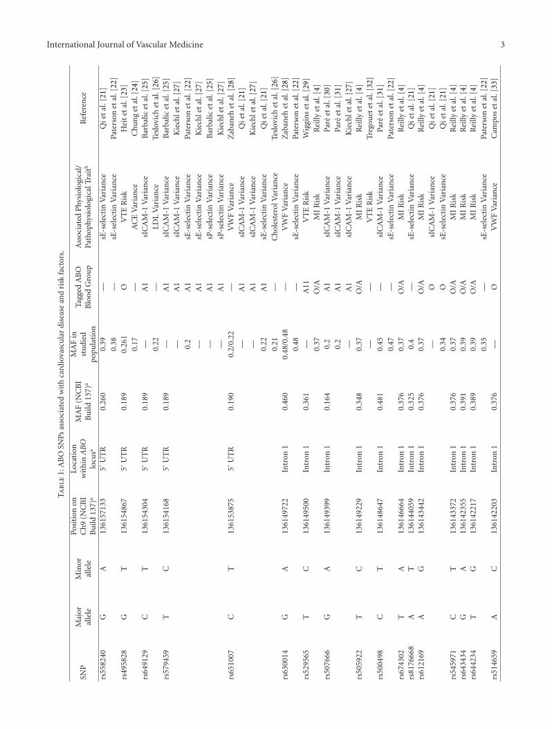
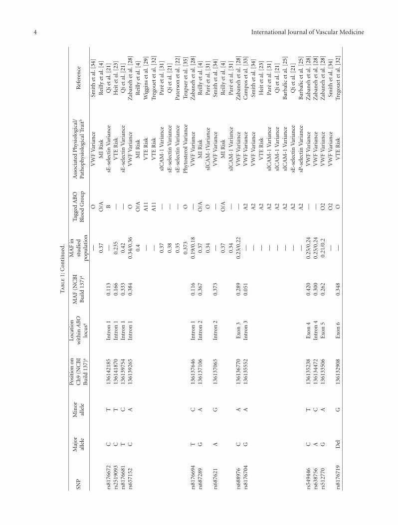
The widespread use of genome-wide association studies(GWASs) over the last 5 years has spurred an enormousacceleration in discoveries across the entire spectrum ofCVD [20]. Recent GWASs have confirmed ABO as a locusfor venous thromboembolism (VTE), myocardial infarction(MI), and multiple cardiovascular biomarkers (Table 1).
One of the most studied aspects of the ABO gene isits relationship with von Willebrand factor (VWF) [17, 36,37]. In 2003, a family-based linkage screen was carriedout to determine the loci involved in VWF variation in398 Spanish individuals. Markers at the chromosome 9q
ABO locus region harbored the highest LOD value of3.46 [38]. Subsequently, several GWASs have shown thatcarriers of single nucleotide polymorphisms (SNPs) thatmark non-O blood group types have higher levels of plasmaVWF when compared to O individuals. A recent GWASof 7856 European participants in the Atherosclerosis RiskIn Communities (ARIC) cohort study showed that ABOblood group O carriers had a 25% average reduction inplasma VWF levels when compared with non-O blood groupcarriers. SNP rs514659, which was used to tag the O bloodtype, contributed to 15.4% of circulating VWF variance[33]. Additionally, a genome-wide meta-analysis of 4 cohortsfound that rs657152, which also tags the blood group O,was strongly associated with circulating VWF levels andthat blood group O individuals had 22–30% lower plasmaVWF when compared to non-blood group O individuals[28]. A relationship between Factor VIII (FVIII) plasmaconcentrations and ABO blood groups has also been seen.However, in a Cohorts for Heart and Aging Research inGenome Epidemiology (CHARGE) Consortium GWAS, nounique genetic variants within the ABO locus were found toaffect FVIII independently of VWF [34]. As VWF binds andtransports FVIII, the correlation between the ABO gene andFVIII is most likely mediated via VWF [39].
As increased plasma levels of VWF and Factor VIII areassociated with greater risk of thrombosis [40, 41], manystudies have examined the connection between ABO bloodgroup and thrombotic risk. In a GWAS published in 2009,SNPs rs8176750, rs8176746 and rs8176719, which tag theA2, B, and O ABO blood groups, respectively, showed thatgenetically inferred blood type O had 67% lower risk ofVTE than non-O blood groups. Additionally, the A2 bloodgroup had 47% lower risk of VTE when compared to theother non-O blood group phenotypes [32]. Blood type A2was also shown to be associated with lower VTE risk ina recently published GWAS involving 1,503 VTE patientsin which rs8176704 was used to tag the A2 blood group[23]. These data suggest that the decreased risk of VTEis a result of reduced H antigen glycosylation, as the A2allele contains a 1061delC that results in the synthesis ofan enzyme that has 30–50 fold less A transferase activitythan the A1 allele product [42]. Blood group genotypesmay be more informative than blood group phenotypes instudying the association between blood groups and VTEsince genotypes can distinguish between heterozygous andhomozygous carriers of A, B, and O alleles and betweenA1 and A2 alleles [43, 44]. One GWAS found that the A11allele, tagged by rs529565 and rs657152, and the B allele,tagged by rs8176749, were associated with 56% and 16%increased risk of VTE, respectively, when compared to theO11 allele. Moreover, when compared with carriers of theO1O1 diplotype, VTE risk was increased by 79% for the A11diplotype, 82% for the B diplotype, and 170% for the ABdiplotype carriers. Overall, non-O categories combined wereassociated with a 77% increased risk of VTE when comparedto the O category [29]. The effect of ABO genotype onthrombosis risk was also investigated in a case-control studyof 471 patients and 471 controls of the Leiden ThrombophiliaStudy (LETS) which revealed that non-OO genotypes, except
International Journal of Vascular Medicine 3
Ta
ble
1:A
BO
SNPs
asso
ciat
edw
ith
card
iova
scu
lar
dise
ase
and
risk
fact
ors.
SNP
Maj
oral
lele
Min
oral
lele
Posi
tion
onC
h9
(NC
BI
Bu
ild13
7)a
Loca
tion
wit
hin
AB
Olo
cusa
MA
F(N
CB
IB
uild
137)
a
MA
Fin
stu
died
popu
lati
on
Tagg
edA
BO
Blo
odG
rou
pA
ssoc
iate
dP
hysi
olog
ical
/Pa
thop
hysi
olog
ical
Trai
tbR
efer
ence
rs55
8240
GA
1361
5713
35′
UT
R0.
260
0.39
—sE
-sel
ecti
nV
aria
nce
Qie
tal
.[21
]0.
38—
sE-s
elec
tin
Var
ian
cePa
ters
onet
al.[
22]
rs49
5828
GT
1361
5486
75′
UT
R0.
189
0.26
1O
VT
ER
isk
Hei
tet
al.[
23]
0.17
—A
CE
Var
ian
ceC
hun
get
al.[
24]
rs64
9129
CT
1361
5430
45′
UT
R0.
189
—A
1sI
CA
M-1
Var
ian
ceB
arba
licet
al.[
25]
0.22
—L
DL
Var
ian
ceTe
slov
ich
etal
.[26
]rs
5794
59T
C13
6154
168
5′U
TR
0.18
9—
A1
sIC
AM
-1V
aria
nce
Bar
balic
etal
.[25
]—
A1
sIC
AM
-1V
aria
nce
Kie
chle
tal
.[27
]
0.2
A1
sE-s
elec
tin
Var
ian
cePa
ters
onet
al.[
22]
—A
1sE
-sel
ecti
nV
aria
nce
Kie
chle
tal
.[27
]
—A
1sP
-sel
ecti
nV
aria
nce
Bar
balic
etal
.[25
]
—A
1sP
-sel
ecti
nV
aria
nce
Kie
chle
tal
.[27
]rs
6510
07C
T13
6153
875
5′U
TR
0.19
00.
2/0.
22—
VW
FV
aria
nce
Zab
aneh
etal
.[28
]—
A1
sIC
AM
-1V
aria
nce
Qie
tal
.[21
]
—A
1sI
CA
M-1
Var
ian
ceK
iech
let
al.[
27]
0.22
A1
sE-s
elec
tin
Var
ian
ceQ
iet
al.[
21]
0.21
—C
hol
este
rolV
aria
nce
Tesl
ovic
het
al.[
26]
rs63
0014
GA
1361
4972
2In
tron
10.
460
0.48
/0.4
8—
VW
FV
aria
nce
Zab
aneh
etal
.[28
]0.
48—
sE-s
elec
tin
Var
ian
cePa
ters
onet
al.[
22]
rs52
9565
TC
1361
4950
0In
tron
10.
361
—A
11V
TE
Ris
kW
iggi
ns
etal
.[29
]0.
37O
/AM
IR
isk
Rei
llyet
al.[
4]rs
5076
66G
A13
6149
399
Intr
on1
0.16
40.
2A
1sI
CA
M-1
Var
ian
cePa
reet
al.[
30]
0.2
A1
sIC
AM
-1V
aria
nce
Pare
etal
.[31
]
—A
1sI
CA
M-1
Var
ian
ceK
iech
let
al.[
27]
rs50
5922
TC
1361
4922
9In
tron
10.
348
0.37
O/A
MI
Ris
kR
eilly
etal
.[4]
——
VT
ER
isk
Treg
ouet
etal
.[32
]rs
5004
98C
T13
6148
647
Intr
on1
0.48
10.
45—
sIC
AM
-1V
aria
nce
Pare
etal
.[31
]0.
47—
sE-s
elec
tin
Var
ian
cePa
ters
onet
al.[
22]
rs67
4302
TA
1361
4666
4In
tron
10.
376
0.37
O/A
MI
Ris
kR
eilly
etal
.[4]
rs81
7666
8A
T13
6144
059
Intr
on1
0.32
50.
4—
sE-s
elec
tin
Var
ian
ceQ
iet
al.[
21]
rs61
2169
AG
1361
4344
2In
tron
10.
376
0.37
O/A
MI
Ris
kR
eilly
etal
.[4]
—O
sIC
AM
-1V
aria
nce
Qie
tal
.[21
]
0.34
OsE
-sel
ecti
nV
aria
nce
Qie
tal
.[21
]rs
5459
71C
T13
6143
372
Intr
on1
0.37
60.
37O
/AM
IR
isk
Rei
llyet
al.[
4]rs
6434
34G
A13
6142
355
Intr
on1
0.39
10.
39O
/AM
IR
isk
Rei
llyet
al.[
4]rs
6442
34T
G13
6142
217
Intr
on1
0.38
90.
39O
/AM
IR
isk
Rei
llyet
al.[
4]0.
35—
sE-s
elec
tin
Var
ian
cePa
ters
onet
al.[
22]
rs51
4659
AC
1361
4220
3In
tron
10.
376
—O
VW
FV
aria
nce
Cam
pos
etal
.[33
]
4 International Journal of Vascular Medicine
Ta
ble
1:C
onti
nu
ed.
SNP
Maj
oral
lele
Min
oral
lele
Posi
tion
onC
h9
(NC
BI
Bu
ild13
7)a
Loca
tion
wit
hin
AB
Olo
cusa
MA
F(N
CB
IB
uild
137)
a
MA
Fin
stu
died
popu
lati
on
Tagg
edA
BO
Blo
odG
rou
pA
ssoc
iate
dP
hysi
olog
ical
/Pa
thop
hysi
olog
ical
Trai
tbR
efer
ence
—O
VW
FV
aria
nce
Smit
het
al.[
34]
0.37
O/A
MI
Ris
kR
eilly
etal
.[4]
rs81
7667
2C
T13
6142
185
Intr
on1
0.11
3—
BsE
-sel
ecti
nV
aria
nce
Qie
tal
.[21
]rs
2519
093
CT
1361
4187
0In
tron
10.
166
0.23
5—
VT
ER
isk
Hei
tet
al.[
23]
rs81
7668
1T
C13
6139
754
Intr
on1
0.33
30.
42—
sE-s
elec
tin
Var
ian
ceQ
iet
al.[
21]
rs65
7152
CA
1361
3926
5In
tron
10.
384
0.34
/0.3
6O
VW
FV
aria
nce
Zab
aneh
etal
.[28
]0.
4O
/AM
IR
isk
Rei
llyet
al.[
4]
—A
11V
TE
Ris
kW
iggi
ns
etal
.[29
]
—A
11V
TE
Ris
kTr
egou
etet
al.[
32]
0.37
—sI
CA
M-1
Var
ian
cePa
reet
al.[
31]
0.38
—sE
-sel
ecti
nV
aria
nce
Qie
tal
.[21
]
0.35
—sE
-sel
ecti
nV
aria
nce
Pate
rson
etal
.[22
]
0.37
3O
Phy
tost
erol
Var
ian
ceTe
ups
eret
al.[
35]
rs81
7669
4T
C13
6137
646
Intr
on1
0.11
60.
19/0
.18
—V
WF
Var
ian
ceZ
aban
ehet
al.[
28]
rs68
7289
GA
1361
3710
6In
tron
20.
367
0.37
O/A
MI
Ris
kR
eilly
etal
.[4]
0.34
OsI
CA
M-1
Var
ian
cePa
reet
al.[
31]
rs68
7621
AG
1361
3706
5In
tron
20.
373
——
VW
FV
aria
nce
Smit
het
al.[
34]
0.37
O/A
MI
Ris
kR
eilly
etal
.[4]
0.34
—sI
CA
M-1
Var
ian
cePa
reet
al.[
31]
rs68
8976
CA
1361
3677
0E
xon
30.
289
0.23
/0.2
2—
VW
FV
aria
nce
Zab
aneh
etal
.[28
]rs
8176
704
GA
1361
3555
2In
tron
30.
051
—A
2V
WF
Var
ian
ceC
ampo
set
al.[
33]
—A
2V
WF
Var
ian
ceSm
ith
etal
.[34
]
—A
2V
TE
Ris
kH
eit
etal
.[23
]
—A
2sI
CA
M-1
Var
ian
cePa
reet
al.[
31]
—A
2sI
CA
M-1
Var
ian
ceQ
iet
al.[
21]
—A
2sI
CA
M-1
Var
ian
ceB
arba
licet
al.[
25]
—A
2sE
-sel
ecti
nV
aria
nce
Qie
tal
.[21
]
—A
2sP
-sel
ecti
nV
aria
nce
Bar
balic
etal
.[25
]rs
5494
46C
T13
6135
238
Exo
n4
0.42
00.
25/0
.24
—V
WF
Var
ian
ceZ
aban
ehet
al.[
28]
rs63
8756
AC
1361
3447
2In
tron
40.
300
0.25
/0.2
4—
VW
FV
aria
nce
Zab
aneh
etal
.[28
]rs
5127
70G
A13
6133
506
Exo
n5
0.26
20.
21/0
.2O
2V
WF
Var
ian
ceZ
aban
ehet
al.[
28]
—O
2V
WF
Var
ian
ceSm
ith
etal
.[34
]rs
8176
719
Del
G13
6132
908
Exo
n6
0.34
8—
OV
TE
Ris
kTr
egou
etet
al.[
32]

International Journal of Vascular Medicine 5
Ta
ble
1:C
onti
nu
ed.
SNP
Maj
oral
lele
Min
oral
lele
Posi
tion
onC
h9
(NC
BI
Bu
ild13
7)a
Loca
tion
wit
hin
AB
Olo
cusa
MA
F(N
CB
IB
uild
137)
a
MA
Fin
stu
died
popu
lati
on
Tagg
edA
BO
Blo
odG
rou
pA
ssoc
iate
dP
hysi
olog
ical
/Pa
thop
hysi
olog
ical
Trai
tbR
efer
ence
0.41
6O
VT
ER
isk
Hei
tet
al.[
23]
0.34
OsE
-sel
ecti
nV
aria
nce
Pate
rson
etal
.[22
]rs
8176
722
CA
1361
3275
4In
tron
60.
131
0.08
/0.0
8B
VW
FV
aria
nce
Zab
aneh
etal
.[28
]—
BV
WF
Var
ian
ceTr
egou
etet
al.[
32]
rs81
7673
4G
A13
6132
079
Intr
on6
——
O12
VT
ER
isk
Wig
gin
set
al.[
29]
rs81
7674
0A
T13
6131
472
Exo
n7
0.26
70.
25/0
.24
—V
WF
Var
ian
ceZ
aban
ehet
al.[
28]
rs81
7674
6G
T13
6131
322
Exo
n7
0.12
3—
BV
TE
Ris
kTr
egou
etet
al.[
32]
—B
sIC
AM
-1V
aria
nce
Pare
etal
.[31
]
0.16
—A
CE
Var
ian
ceC
hun
get
al.[
24]
rs81
7674
9C
T13
6131
188
Exo
n7
0.12
3—
BV
WF
Var
ian
ceSm
ith
etal
.[34
]—
BV
TE
Ris
kW
iggi
ns
etal
.[29
]rs
8176
750
CD
el13
6131
059
Exo
n7
——
A2
VT
ER
isk
Treg
ouet
etal
.[32
]rs
7857
390
GA
1361
2854
63′
UT
R0.
320
0.4
—sE
-sel
ecti
nV
aria
nce
Qie
tal
.[21
]
AC
E:a
ngi
oten
sin
-con
vert
ing
enzy
me;
LDL:
low
den
sity
lipop
rote
in;M
AF:
min
oral
lele
freq
uen
cy;M
I:m
yoca
rdia
lin
farc
tion
;sIC
AM
-1:s
olu
ble
inte
rcel
lula
rad
hes
ion
mol
ecu
le-1
;VT
E:v
enou
sth
rom
boem
bolis
m;
VW
F:vo
nW
illeb
ran
dfa
ctor
.a T
he
chro
mos
omal
loca
tion
and
MA
Ffo
rea
chSN
Pta
ble
entr
yw
asob
tain
edby
quer
yin
gea
chSN
Prs
nu
mbe
rin
the
NC
BI
Sin
gle
Nu
cleo
tide
Poly
mor
phis
mda
taba
se(d
bSN
P),
build
137
onth
e13
thof
Sept
embe
r,20
12(h
ttp:
//w
ww
.ncb
i.nlm
.nih
.gov
/pro
ject
s/SN
P/)
.bT
he
asso
ciat
edph
ysio
logi
cal/
path
ophy
siol
ogic
altr
aits
wer
eex
trac
ted
from
the
Nat
ion
alH
um
anG
enom
eR
esea
rch
Inst
itu
te(N
HG
RI)
GW
Aca
talo
gda
taba
se(h
ttp:
//w
ww
.gen
ome.
gov/
gwas
tudi
es/)
and
quer
ied
publ
icat
ion
sfr
omP
ubM
ed(h
ttp:
//w
ww
.ncb
i.nlm
.nih
.gov
/pu
bmed
).
6 International Journal of Vascular Medicine
homozygous A2 and A2-O combinations, were associatedwith increased thrombotic risk when compared to OOgenotypes. The relative thrombotic risk of AB genotypesand A1-combinations was increased by 90–110% whencompared to OO genotypes and the relative thrombotic riskof the homozygous B genotype and B-O combinations wasincreased by 60% [45].
The ABO locus has also been associated with arterialthrombosis in studies of MI. Our group reported that all11 SNPs that exceeded genome-wide significance for MI inpatients with established coronary atherosclerosis mapped tothe ABO locus. The risk alleles at rs514659 (odds ratio 1.21;P = 7.62× 10⁻⁹) and rs687289 (odds ratio 1.19; P = 7.75×10⁻⁹) were perfect tags for the loss of function ABO O bloodgroup demonstrating that functional ABO glycotransferasesconferred increased risk of MI. Further analysis foundthat rs514659 was associated with coronary artery diseases(CADs) when complicated by MI but not with CAD withoutMI, suggesting that the primary relationship of ABO toclinical CAD is through modulation of coronary thrombosisor plaque rupture in patients with established coronaryatherosclerosis rather than through primary promotion ofatherosclerosis per se.
The increase in MI risk for non-O blood type individualshas been suggested for some time through epidemiologicalstudies, although there has been debate as to which ABOblood group phenotypes confer the largest increase in risk[8, 11, 46–49]. Just recently, He et al. reported results of twolarge prospective studies of incident coronary heart disease(CHD) as well as a meta-analysis of all prospective data [50].The Nurses’ Health Study (including 62,073 women ages30 to 55 at baseline) and the Health Professionals Follow-up Study (including 27,428 men ages 40 to 75 at baseline)were followed up to 2006 and recorded 2,055 cases of CHDin the two cohorts. Individuals with self-reported non-Oblood type had an age-adjusted hazard ratio (HR) of 1.09(95% CI 1.03 to 1.17, P = 0.005) for risk of developingCHD. Associations between blood type and CHD risk werenot modified by age, physical activity, alcohol consumption,smoking status, or diabetes history. A meta-analysis of anadditional six prior cohorts, for a combined total of 114,648individuals and 5,741 CHD cases, also showed a significantpooled relative risk for CHD in patients with non-O bloodtype of 1.11 (95% CI 1.05 to 1.18, P = 0.001). Amongparticipants in the cohort, those with type O blood weresignificantly less likely to develop CHD when comparedagainst types B (HR 1.11, 95% CI 1.01 to 1.23) and AB (HR1.23, 95% CI 1.10 to 1.37), with a trend toward a higher riskfor patients with type A blood (HR 1.05, 95% CI 0.98 to1.13).
It is plausible that ABO modulation of VWF-relatedthrombosis accounts for the ABO association with MI.However, ABO antigens are expressed also on distinct plateletproteins, including GPIIb, a subunit of the fibrinogenreceptor heterodimer [51–54], and may therefore modulatespecific platelet functions in arterial thrombosis and MI(see below). In addition, ABO modulation of atheroscleroticplaque rupture and atherosclerosis itself cannot be discount-ed without further study.
4. Associations of the ABO Locus withMarkers of Endothelial Function andSerum Lipoproteins
The ABO glycotransferase may have broader impact onatherosclerotic CVD than simply through modulation ofthrombosis. A series of GWAS have linked the ABO locus tocirculating levels of soluble intercellular adhesion molecule-1 (sICAM-1), soluble P-selectin (sP-selectin), and soluble E-selectin (sE-selectin). Notably, mechanistic studies in rodentmodels have implicated these proteins in atherosclerosis [55–59] and their blood levels in humans correlate with increasedrisk of CVD events [60, 61]. Pare et al. showed that SNPrs507666, located in Intron 1 of the ABO gene and a perfecttag for ABO blood group A1, was associated with decreasedlevels of sICAM-1 (P = 5.1 × 10⁻²⁹) when compared tothe O allele and contributed to 1.5% of the total sICAM-1 concentration variance [31]. The same group confirmedthis relationship between rs507666 and circulating sICAM-1levels in a larger GWAS published in 2011 [30]. Blood levelsof sP-selectin and sE-selectin are also associated with SNPs inthe ABO region [21, 22, 25]. A recent meta-analysis showedthat, compared with major allele homozygotes, heterozygoteand minor allele homozygote individuals for the rs507666,rs579459, and rs651007 SNPs, which demonstrate a highdegree of linkage disequilibrium with each other (r2 ∼ 0.96)and tag the ABO A1 subtype, had lower plasma levels ofsICAM-1, sP-selectin, and sE-selectin [27]. A recent GWAShas also identified a potential relationship between ABOand circulating levels of angiotensin-converting enzyme(ACE); when compared to the ABO blood group O, meanACE activity in carriers of blood group B was significantlyincreased (P = 2.3×10⁻¹⁰) while ACE activity in blood groupA was decreased (P = 1.5× 10⁻⁸) [24].
Past epidemiological studies, some dating as far back as50 years ago, have suggested evidence for ABO associationwith circulating levels of cholesterol, with non-O groupsappearing to have higher levels [47, 62–65]. For example, in1976, Garrison et al. published an epidemiological analysis inthe Framingham Heart Study showing consistent elevationsof serum cholesterol levels in non-O blood groups whencompared to the O blood group [47]. Some recent GWASsand their meta-analyses support this potential role forABO genotypes in modulating circulating levels of totaland LDL cholesterol, as well as phytosterols, establishedcausal risk factors for atherosclerotic heart diseases [26, 35,66]. A meta-analysis of 46 lipid based GWAS reported anassociation between ABO SNPs and serum cholesterol levels.Total cholesterol was increased by 2.3 mg/dL in heterozygoteindividuals for the ABO rs651007 SNP when compared tomajor allele homozygotes (P = 8.66 × 10⁻²¹), while LDLcholesterol was increased by 2.05 mg/dL in heterozygoteindividuals for the ABO rs649129 SNP when comparedto major allele homozygotes (P = 7.85 × 10⁻²²) [26]. AGWAS published in 2010 found that the ABO locus showedgenome-wide-significance for association with phytosterollevels. Specifically, rs657152, which tightly tags the O1allele, was found to be associated with decreased levels of
International Journal of Vascular Medicine 7
circulating phytosterols. In a separate analysis, the studyreported that individuals with the O allele had decreasedcampesterol concentrations when compared to the A andB alleles. Furthermore, additional analyses revealed thatrs657152 was associated with reduced CAD risk (P =4.0 × 10⁻⁵) when compared to alleles that were associatedwith increased phytosterols [35]. These epidemiological andgenetic associations of ABO blood type with levels ofcirculating lipoproteins and sterols underscore the need forfunctional studies that define the mechanistic basis of theserelationships and the potential for therapeutic translation.
Overall, these pleiotropic associations with cardiovas-cular risk biomarkers suggest a complex role of ABO inatherosclerotic and vascular diseases with distinct ABO geno-types and ABO functions contributing to multiple causalpathways, for example, ABO genotypes related to bloodgroup O and loss of glycotransferase function protect againstVTE and MI while distinct genotypes relating to specificA and B blood subgroups and glycotransferase functionsmay have a more subtle and distinct impact on endothelialfunction, lipoproteins, and atherosclerosis (Table 1).
One paradox that has limited clinical interpretation ofGWAS discoveries, including that for the ABO locus, relatesto the highly significant P values reported for common dis-ease variants despite very small effect sizes (e.g., odds ratio).Unlike rare disease causing mutations, common variantsfor complex diseases have either minor functional effectsor are simply crude markers for rare functional variants;their strength of association with disease is often very smalldespite highly significant P values (which relate mostly tothe sample size of GWAS). However, the weak effect sizes ofthese variants should not be mistaken for a lack of clinicalimportance of a particular gene (or protein/biomarker) orits therapeutic manipulation in a complex disease process.For example, rare variants in 3-hydroxy-3-methyl-glutaryl-CoA (HMG-CoA) reductase have quite weak associationswith LDL cholesterol (LDL-C) levels and are well down thelist of top GWAS findings for LDL-C [26]. Thus, rather thanattempting to infer clinical relevance based on the strengthof associations, the key importance of GWAS is simply toidentify novel genes and proteins for human disease thatwarrant further study to establish their mechanistic roleand clinical importance. Over time, the totality of evidencederived from human, animal, and functional studies allowsan interpretation of the clinical and therapeutic importanceof the initial significance level for a locus in a GWAS, themagnitude of effect on a protein level, and the relativerisk of a given protein level for a disease phenotype. Forthe ABO locus, further mechanistic and functional studiesare required in order to define the clinical and therapeuticpossibilities.
5. Potential Mechanisms for theAssociation between ABO and CVD
Following recent GWAS discoveries, there is a resurgentinterest in identifying the mechanistic links underlying theassociation of the ABO locus and glycotransferase functions
with CVD. Most attention until recently was focused onthe role of ABO in regulating VWF bioactivity and relatedthrombotic pathways, but emerging work is defining morebroadly the role of ABO glycotransferase activity in mod-ulating multiple endothelial, platelet, and cardiometabolicpathways and exploring whether these effects are causallyinvolved in cardiometabolic diseases.
5.1. VWF and FVIII as Candidate Mechanisms. VWF andFVIII are glycoproteins (GPs) that circulate together innormal plasma as a noncovalent complex and both playimportant roles in normal hemostasis. VWF is a carrierfor FVIII and protects it from inactivation. VWF recruitsplatelets to the site of clot formation during primaryhemostasis. Factor VIII is released from VWF by the actionof thrombin and participates in the coagulation cascade.Thus, both VWF and FVIII are key proteins in the formationof occlusive thrombi in injured vessels [67]. As describedin preceding sections, relative to non-group O, carriers ofABO blood group O have significantly lower circulatingplasma VWF and FVIII levels [68]. Although this clinicallyimportant effect of ABO group on plasma VWF-FVIII levelsis well established, the mechanism through which it ismediated is not completely resolved. ABO appears to havedirect functional effects on circulating VWF and indirectly(via influence of VWF levels) modulates FVIII levels. Thepresence of abundant ABH carbohydrate molecules on theVWF oligosaccharide side chains provides the mechanisticbasis for ABO regulation of VWF levels. The active ABO Aand B glycotransferase enzymes, found in Golgi of endothe-lial cells, generate terminal carbohydrate modifications, Aand B antigens, on the existing VWF “H” oligisacharides,whereas the enzymatically inactive ABO O protein cannotmodify these VWF H antigens. The addition of A or Bterminal carbohydrate antigens to VWF in endothelial cellsmight influence circulating VWF levels and function byseveral mechanisms: altering the rate of VWF synthesisand/or secretion, regulating VWF proteolysis induced by itsmajor protease, ADAMTS13, modulating VWF clearance,or changing VWF biological activity: or perhaps somecombination of these events [17]. A number of studiessuggest that it is unlikely that ABO effects on VWF levelsare mediated by alterations in the biosynthesis and secretionof VWF [17]. In contrast, it has been demonstrated thatthe activity of ADAMTS13 differs against VWF of differentblood groups, with both the level of ADAMTS13 activity[69], and the rate of VWF proteolysis by ADAMTS13[70], being higher in blood group O as compared tonon-O individuals. Thus, the absence of VWF terminalcarbohydrate modifications in individuals with ABO bloodgroup O increases the susceptibility to and rate of proteolysisby ADAMTS13, although data available so far are not insupport of the role of proteolysis by ADAMTS13 in VWFclearance from the circulation [71]. Due to their physicalassociation in blood, lower circulating VWF results in lowerlevels of FVIII. Whether VWF in platelets (a relativelyabundant source) undergoes any modification by ABOremains controversial; such modification could alter platelet

8 International Journal of Vascular Medicine
production and subsequent turnover of VWF, particularlylocally during platelet-driven arterial thrombosis, althoughthis remains to be established. Indeed, current knowledgesuggests that ABO does not modify platelet VWF [72], butthe role of ABO in regulating platelet VWF and plateletfunction in thrombosis requires greater study (see below).
5.2. Other Endothelial Molecules and Platelet Proteins asPotential Mediators. Soluble levels of multiple adhesionmolecules, mostly derived from endothelial cells andplatelets, have been associated with coronary heart disease[61], and other cardiovascular conditions [73]. In large-scale genomic studies, plasma levels of sP-selectin andsICAM-1 were associated with ABO gene variants, but noassociation was found between platelet-bound P-selectinlevels and ABO blood group, suggesting that the ABOblood group may influence proteolysis and clearance fromthe circulation, rather than its production and cellularpresentation [25]. However, the specific mechanisms ofABO regulation of circulating levels of these endothelialGPs are largely unknown. Further, the implications fortheir function in atherosclerosis and clinical CVD requireelucidation.
Blood group ABO antigens are known to be carriedby several platelet GPs, for example, GPIb, GPIIb, GPIIIa,and platelet endothelial cell adhesion molecule (PECAM)[53], that play important roles in platelet function. PlateletABH expression is a stable, donor-specific characteristicwith 5% of A1 donors typing as either ABH high- or low-expressers [74], with high levels of A antigen on variousGPs from high-expresser platelets, especially GPIIb andPECAM (CD31) [51]. GPIIb is an integral componentof the GPIIb-GPIIIa fibrinogen receptor complex, whichrepresents the critical final common pathway for platelet-driven thrombosis in hemostasis and pathologic arterialthrombosis including acute MI. Genetic variation in GPIIbthat modulates fibrinogen binding has been associatedwith altered risk of thrombosis and MI [75–77], so itis conceivable that ABO-driven carbohydrate modificationof GPIIb might alter its functional interactions with fib-rinogen and thus platelet-mediated thrombosis. However,this hypothesis has not been adequately addressed to date.Besides GPIIb and PECAM, blood group A antigen is alsoexpressed on other uncharacterized platelet proteins (70–90 kDa) having electrophoretic mobilities closely resem-bling those of GPIV and GPV [52]. Thus these andother uncharacterized ABO-expressing platelet proteins mayalso act as potential functional modulators of the ABOassociations with arterial thrombosis and cardiovascularevents.
The list of GPs carrying blood type ABO antigens isgrowing. Some of the most abundantly expressed GPs, suchas PECAM and VWF, represent major proteins recognizedto carry ABO antigens. However, there is a great need toidentify all ABO target proteins and glycolipids in order tounderstand the nature and/or properties of GPs carryingABO antigens and to reveal novel, potentially cell-specific,mediators of CVD.
6. Perspectives
Despite the relative simplicity of the A and B antigens, espe-cially considering the modest biochemical difference betweenthem, the ABO blood group system appears to be one of themost interesting, both clinically and scientifically. It dividesthe world’s population into four major groups. The commonfrequency of each major blood group, especially blood groupO (the loss of glycotransferase function ABO type), suggestsimportant evolutionary selection pressures distinct fromheart disease which now have subtle but significant impactat the population level on complex diseases in modernsociety. Much larger epidemiological and genetic studies arerequired to determine risks of CVD with particular majorand minor ABO blood group. Such human data will informtranslational and mechanistic studies by pointing to specificABO glycan modifications that appear to be the driversof increased or decreased risk. However, such studies areinherently limited because they fail to illuminate the specificcell/tissue where glycan modifications are mediating theiractions and do not define the proteins or lipids carryingsuch modifications that are functionally altered and mediatedisease.
In future studies, it is of critical importance to establishclear mechanistic basis for the association between ABOblood groups and CVD. Advances in our understandingof the physiologic importance of the glycan structures ofVWF have provided a model and elucidated one mechanism,possibly the major one, through which ABO blood groupdetermines risk of thrombosis and acute cardiovascularevents. However, there are several other known endothelialand platelet GPs, discussed above, that are potentially func-tionally modified by ABO and candidate causal mediatorsin atherosclerosis and CVD. How and where these proteinsare modified and whether their carbohydrate alterations havefunctional impact on disease requires further study. Theincreased risks associated with non-O blood groups mightbe attributed to higher levels or functional modificationof specific endothelial-derived GPs, specific platelet GPs,both sources, and/or GPs from additional cells and tissuesalthough leukocytes are not likely primary sources as theylack the ABO enzyme [78].
The study of glycobiology in complex disease is inits relative infancy. Unbiased approaches are required todiscover and identify the population of proteins and lipidsthat are modified by ABO (and other glycotransferases), incells and tissues of specific relevance to atherothrombosis, sothat we can define all potential mediators of specific aspectsof risk and specific disease manifestations. Technologicaland bioinformatics advances, including mass-spectrometricapproaches, are beginning to emerge which will permit suchunbiased work. The tissue-specific expression and functionof ABO and many known glycotransferases [79] suggeststrongly that their actions and targets will be regulatedat a cell- and tissue- specific levels coincident with rolesin paracrine- and cell-specific events in atherothrombosis.A naıve impression might suggest that inhibition of ABOglycotransferase functions, akin to the naturally occurringABO O blood group, might provide a therapeutic strategy

International Journal of Vascular Medicine 9
for lowering the risk of thrombosis and CVD. However, sucha pharmacological approach would likely have untowardhematological and immunological consequences and/ornonspecific off target actions in cells and tissues not relevantto vascular diseases. Identification of the specific proteinand lipid targets of ABO which mediate the functionaleffects in thrombosis, atherosclerosis and CVD is likelyto provide greater opportunity for targeted therapeuticdevelopment and clinical translation. Whether targetingVWF carbohydrate modifications might provide clinicallymeaningful opportunities for treatment of thrombosis andMI is an open question.
There is much to be done to understand the role of ABOand glycobiology in CVD, and the next decade should seemany advances in the basic biology, mechanistic actions,and diagnostic, prognostic, and therapeutic possibilities inhumans. Thus, future studies to further define the associa-tion between ABO blood groups and cardiovascular eventsand risks, and to elucidate biochemical mechanisms respon-sible for these associations, are not only of basic scientificinterest but also of translational clinical importance.
Acknowledgments
This work was supported by Award from the NationalInstitutes of Health (NIH) including R01-HL-113147, R01-HL-111694, R01-DK-090505, U01-HL108636, and K24-HL107643 (to Muredach P. Reilly).
References
[1] I. I. El Hajj, J. G. Hashash, E. M. K. Baz, H. Abdul-Baki, andA. I. Sharara, “ABO blood group and gastric cancer: rekindlingan old fire?” Southern Medical Journal, vol. 100, no. 7, pp. 726–727, 2007.
[2] T. Demir, A. Tezel, R. Orbak, A. Eltas, C. Kara, and F.Kavrut, “The effect of ABO blood types on periodontal status,”European Journal of Dentistry, vol. 1, no. 3, pp. 139–143, 2007.
[3] M. A. Qureshi and R. Bhatti, “Frequency of abo blood groupsamong the diabetes mellitus type 2 patients,” Journal of theCollege of Physicians and Surgeons Pakistan, vol. 13, no. 8, pp.453–455, 2003.
[4] M. P. Reilly, M. Li, J. He et al., “Identification of ADAMTS7 asa novel locus for coronary atherosclerosis and association ofABO with myocardial infarction in the presence of coronaryatherosclerosis: two genome-wide association studies,” TheLancet, vol. 377, no. 9763, pp. 383–392, 2011.
[5] B. Woolf, “On estimating the relation between blood groupand disease,” Annals of Human Genetics, vol. 19, no. 4, pp. 251–253, 1955.
[6] H. Jick, D. Slone, B. Westerholm et al., “Venous thromboem-bolic disease and ABO blood type. A cooperative study,” TheLancet, vol. 1, no. 7594, pp. 539–542, 1969.
[7] P. P. Green, P. M. Mannucci, and E. Briet, “Carrier detectionin hemophilia A: a cooperative international study. II. Theefficacy of a universal discriminant,” Blood, vol. 67, no. 6, pp.1560–1567, 1986.
[8] J. H. Medalie, C. Levene, C. Papier et al., “Blood groups,myocardial infarction and angina pectoris among 10,000 adultmales,” New England Journal of Medicine, vol. 285, no. 24, pp.1348–1353, 1971.
[9] T. R. Ketch, S. J. Turner, M. T. Sacrinty et al., “ABO bloodtypes: influence on infarct size, procedural characteristics andprognosis,” Thrombosis Research, vol. 123, no. 2, pp. 200–205,2008.
[10] J. Erikssen, E. Thaulow, and H. Stormorken, “ABO bloodgroups and coronary heart disease (CHD). A study in subjectswith severe and latent CHD,” Thrombosis and Haemostasis, vol.43, no. 2, pp. 137–140, 1980.
[11] U. E. Nydegger, W. A. Wuillemin, F. Julmy, B. J. Meyer, and T.P. Carrel, “Association of ABO histo-blood group B allele withmyocardial infarction,” European Journal of Immunogenetics,vol. 30, no. 3, pp. 201–206, 2003.
[12] D. Platt, W. Muhlberg, L. Kiehl, and R. Schmitt-Ruth,“ABO blood group system, age, sex, risk factors and cardiacinfarction,” Archives of Gerontology and Geriatrics, vol. 4, no.3, pp. 241–249, 1985.
[13] I. Sari, O. Ozer, V. Davutoglu, S. Gorgulu, M. Eren, and M.Aksoy, “ABO blood group distribution and major cardiovas-cular risk factors in patients with acute myocardial infarction,”Blood Coagulation and Fibrinolysis, vol. 19, no. 3, pp. 231–234,2008.
[14] O. Wu, N. Bayoumi, M. A. Vickers, and P. Clark, “ABO(H)blood groups and vascular disease: a systematic review andmeta-analysis,” Journal of Thrombosis and Haemostasis, vol. 6,no. 1, pp. 62–69, 2008.
[15] R. Oriol, R. Mollicone, P. Coullin, A. M. Dalix, and J. J.Candelier, “Genetic regulation of the expression of ABH andLewis antigens in tissues,” APMIS, Supplement, vol. 100, no.27, pp. 28–38, 1992.
[16] E. Hosoi, “Biological and clinicel aspects of ABO blood groupsystem,” Journal of Medical Investigation, vol. 55, no. 3-4, pp.174–182, 2008.
[17] P. V. Jenkins and J. S. O’Donnell, “ABO blood group deter-mines plasma von Willebrand factor levels: a biologic functionafter all?” Transfusion, vol. 46, no. 10, pp. 1836–1844, 2006.
[18] J. R. Storry and M. L. Olsson, “The ABO blood group systemrevisited: a review and update,” Immunohematology, vol. 25,no. 2, pp. 48–59, 2009.
[19] J. B. Lowe, “The blood group-specific human glycosyltrans-ferases,” Bailliere’s Clinical Haematology, vol. 6, no. 2, pp. 465–492, 1993.
[20] T. Zeller, S. Blankenberg, and P. Diemert, “Genomewide asso-ciation studies in cardiovascular disease—an update 2011,”Clinical Chemistry, vol. 58, no. 1, pp. 92–103, 2012.
[21] L. Qi, M. C. Cornelis, P. Kraft et al., “Genetic variants in ABOblood group region, plasma soluble E-selectin levels and riskof type 2 diabetes,” Human Molecular Genetics, vol. 19, no. 9,Article ID ddq057, pp. 1856–1862, 2010.
[22] A. D. Paterson, M. F. Lopes-Virella, D. Waggott et al.,“Genome-wide association identifies the ABO blood groupas a major locus associated with serum levels of soluble E-selectin,” Arteriosclerosis, Thrombosis, and Vascular Biology,vol. 29, no. 11, pp. 1958–1967, 2009.
[23] J. A. Heit, S. M. Armasu, Y. W. Asmann et al., “A genome-wide association study of venous thromboembolism identifiesrisk variants in chromosomes 1q24.2 and 9q,” Journal ofThrombosis and Haemostasis, vol. 10, no. 8, pp. 1521–1531,2012.
[24] C. M. Chung, R. Y. Wang, J. W. Chen et al., “A genome-wide association study identifies new loci for ACE activity:potential implications for response to ACE inhibitor,” Phar-macogenomics Journal, vol. 10, no. 6, pp. 537–544, 2010.
[25] M. Barbalic, J. Dupuis, A. Dehghan et al., “Large-scalegenomic studies reveal central role of ABO in sP-selectin and
10 International Journal of Vascular Medicine
sICAM-1 levels,” Human Molecular Genetics, vol. 19, no. 9,Article ID ddq061, pp. 1863–1872, 2010.
[26] T. M. Teslovich, K. Musunuru, A. V. Smith et al., “Biological,clinical and population relevance of 95 loci for blood lipids,”Nature, vol. 466, no. 7307, pp. 707–713, 2010.
[27] S. Kiechl, G. Pare, M. Barbalic et al., “Association of variationat the ABO locus with circulating levels of soluble intercel-lular adhesion molecule-1, soluble P-selectin, and soluble E-selectin: a meta-analysis,” Circulation, vol. 4, no. 6, pp. 681–686, 2011.
[28] D. Zabaneh, T. R. Gaunt, M. Kumari et al., “Genetic variantsassociated with von Willebrand factor levels in healthy menand women identified using the humanCVD beadchip,”Annals of Human Genetics, vol. 75, no. 4, pp. 456–467, 2011.
[29] K. L. Wiggins, N. L. Smith, N. L. Glazer et al., “ABO genotypeand risk of thrombotic events and hemorrhagic stroke,”Journal of Thrombosis and Haemostasis, vol. 7, no. 2, pp. 263–269, 2009.
[30] G. Pare, P. M. Ridker, L. Rose et al., “Genome-wide associationanalysis of soluble ICAM-1 concentration reveals novel associ-ations at the NFKBIK, PNPLA3, RELA, and SH2B3 loci,” PLoSGenetics, vol. 7, no. 4, Article ID e1001374, 2011.
[31] G. Pare, D. I. Chasman, M. Kellogg et al., “Novel association ofABO histo-blood group antigen with soluble ICAM-1: resultsof a genome-wide association study of 6,578 women,” PLoSGenetics, vol. 4, no. 7, Article ID e1000118, 2008.
[32] D. A. Tregouet, S. Heath, N. Saut et al., “Common susceptibil-ity alleles are unlikely to contribute as strongly as the FV andABO loci to VTE risk: results from aGWAS approach,” Blood,vol. 113, no. 21, pp. 5298–5303, 2009.
[33] M. Campos, W. Sun, F. Yu et al., “Genetic determinants ofplasma von Willebrand factor antigen levels: a target gene SNPand haplotype analysis of ARIC cohort,” Blood, vol. 117, no.19, pp. 5224–5230, 2011.
[34] N. L. Smith, M. H. Chen, A. Dehghan et al., “Novel associa-tions of multiple genetic loci with plasma levels of factor VII,factor VIII, and von Willebrand factor: the CHARGE (Cohortsfor Heart and Aging Research in Genome Epidemiology)Consortium,” Circulation, vol. 121, no. 12, pp. 1382–1392,2010.
[35] D. Teupser, R. Baber, U. Ceglarek et al., “Genetic regulation ofserum phytosterol levels and risk of coronary artery disease,”Circulation, vol. 3, no. 4, pp. 331–339, 2010.
[36] J. C. Gill, J. Endres-Brooks, and P. J. Bauer, “The effect ofABO blood group on the diagnosis of von Willebrand disease,”Blood, vol. 69, no. 6, pp. 1691–1695, 1987.
[37] D. R. Rios, A. P. Fernandes, R. C. Figueiredo et al., “Rela-tionship between ABO blood groups and von Willebrandfactor, ADAMTS13 and factor VIII in patients undergoinghemodialysis,” Journal of Thrombosis and Thrombolysis, vol.33, no. 4, pp. 416–421, 2012.
[38] J. C. Souto, L. Almasy, J. M. Soria et al., “Genome-wide linkageanalysis of von Willebrand factor plasma levels: results fromthe GAIT project,” Thrombosis and Haemostasis, vol. 89, no. 3,pp. 468–474, 2003.
[39] V. Terraube, J. S. O’Donnell, and P. V. Jenkins, “Factor VIIIand von Willebrand factor interaction: biological, clinical andtherapeutic importance,” Haemophilia, vol. 16, no. 1, pp. 3–13,2010.
[40] R. A. Kraaijenhagen, P. S. In ’T Anker, M. M. W. Koopmanet al., “High plasma concentration of factor VIIIc is a majorrisk factor for venous thromboembolism,” Thrombosis andHaemostasis, vol. 83, no. 1, pp. 5–9, 2000.
[41] T. Koster, A. D. Blann, E. Briet, J. P. Vandenbroucke, and F.R. Rosendaal, “Role of clotting factor VIII in effect of vonWillebrand factor on occurrence of deep-vein thrombosis,”The Lancet, vol. 345, no. 8943, pp. 152–155, 1995.
[42] F. I. Yamamoto, P. D. McNeill, and S. I. Hakomori, “Humanhisto-blood group A2 transferase coded by A2 allele, one ofthe A subtypes, is characterized by a single base deletion inthe coding sequence, which results in an additional domain atthe carboxyl terminal,” Biochemical and Biophysical ResearchCommunications, vol. 187, no. 1, pp. 366–374, 1992.
[43] V. M. Morelli, M. C. H. de Visser, N. H. van Tilburg et al.,“ABO blood group genotypes, plasma von Willebrand factorlevels and loading of von Willebrand factor with A and Bantigens,” Thrombosis and Haemostasis, vol. 97, no. 4, pp. 534–541, 2007.
[44] S. le Cessie, J. Debeij, F. R. Rosendaal, S. C. Cannegieter, andJ. P. Vandenbroucke, “Quantification of bias in direct effectsestimates due to different types of measurement error in themediator,” Epidemiology, vol. 23, no. 4, pp. 551–560, 2012.
[45] V. M. Morelli, M. C. H. De Visser, H. L. Vos, R. M. Bertina,and F. R. Rosendaal, “ABO blood group genotypes and therisk of venous thrombosis: effect of factor V Leiden,” Journal ofThrombosis and Haemostasis, vol. 3, no. 1, pp. 183–185, 2005.
[46] T. W. Meade, J. A. Cooper, Y. Stirling, D. J. Howarth, V.Ruddock, and G. J. Miller, “Factor VIII, ABO blood groupand the incidence of ischaemic heart disease,” British Journalof Haematology, vol. 88, no. 3, pp. 601–607, 1994.
[47] R. J. Garrison, R. J. Havlik, R. B. Harris, M. Feinleib, W. B.Kannel, and S. J. Padgett, “ABO blood group and cardiovaculardisease the Framingham study,” Atherosclerosis, vol. 25, no. 2-3, pp. 311–318, 1976.
[48] N. Saha, C. C. S. Toh, and M. B. Ghosh, “Genetic associationin myocardial infarction. Ethnicity; ABO, Rh, Le(a), Xg(a)blood groups; G6PD deficiency; and abnormal hemoglobins,”Journal of Medical Genetics, vol. 10, no. 4, pp. 340–345, 1973.
[49] B. Bronte-Stewart, M. C. Botha, and L. H. Krut, “ABO bloodgroups in relation to ischaemic heart disease,” British MedicalJournal, vol. 1, no. 5293, pp. 1646–1650, 1962.
[50] M. He, B. Wolpin, K. Rexrode et al., “ABO blood groupand risk of coronary heart disease in two prospective cohortstudies,” Arteriosclerosis, Thrombosis, and Vascular Biology, vol.32, no. 9, pp. 2314–2320, 2012.
[51] B. R. Curtis, J. T. Edwards, M. J. Hessner, J. P. Klein, and R. H.Aster, “Blood group A and B antigens are strongly expressedon platelets of some individuals,” Blood, vol. 96, no. 4, pp.1574–1581, 2000.
[52] D. Stockelberg, M. Hou, L. Rydberg, J. Kutti, and H. Wadenvik,“Evidence for an expression of blood group A antigen onplatelet glycoproteins IV and V,” Transfusion Medicine, vol. 6,no. 3, pp. 243–248, 1996.
[53] M. Hou, D. Stockelberg, L. Rydberg, J. Kutti, and H. Wadenvik,“Blood group A antigen expression in platelets is prominentlyassociated with glycoprotein Ib and IIb. Evidence for an A1/A2difference,” Transfusion Medicine, vol. 6, no. 1, pp. 51–59,1996.
[54] K. Ogasawara, J. Ueki, M. Takenaka, and K. Furihata, “Studyon the expression of ABH antigens on platelets,” Blood, vol. 82,no. 3, pp. 993–999, 1993.
[55] N. Gaudreault, N. Kumar, J. M. Posada et al., “ApoE suppressesatherosclerosis by reducing lipid accumulation in circulatingmonocytes and the expression of inflammatory moleculeson monocytes and vascular endothelium,” Arteriosclerosis,Thrombosis, and Vascular Biology, vol. 32, no. 2, pp. 264–272,2012.

International Journal of Vascular Medicine 11
[56] T. Takeshi, N. Keisuke, I. Takaaki, Y. Makoto, and N. Tatsuji,“Involvement of adhesion molecule in in vitro plaque-likeformation of macrophages stimulated with Aggregatibacteractinomycetemcomitans lipopolysaccharide,” Journal of Peri-odontal Research, vol. 45, no. 4, pp. 550–556, 2010.
[57] W. Luo, H. Wang, M. K. Ohman et al., “P-selectin glycoproteinligand-1 deficiency leads to cytokine resistance and protectionagainst atherosclerosis in apolipoprotein E deficient mice,”Atherosclerosis, vol. 220, no. 1, pp. 110–117, 2012.
[58] J. Kisucka, A. K. Chauhan, B. Q. Zhao et al., “Elevated levels ofsoluble P-selectin in mice alter blood-brain barrier function,exacerbate stroke, and promote atherosclerosis,” Blood, vol.113, no. 23, pp. 6015–6022, 2009.
[59] T. P. Johnston, “Poloxamer 407 increases soluble adhesionmolecules, ICAM-1, VCAM-1 and E-selectin, in C57BL/6mice,” Journal of Pharmacy and Pharmacology, vol. 61, no. 12,pp. 1681–1688, 2009.
[60] P. M. Ridker, C. H. Hennekens, B. Roitman-Johnson, M. J.Stampfer, and J. Allen, “Plasma concentration of soluble inter-cellular adhesion molecule 1 and risks of future myocardialinfarction in apparently healthy men,” The Lancet, vol. 351,no. 9096, pp. 88–92, 1998.
[61] S. J. Hwang, C. M. Ballantyne, A. R. Sharrett et al., “Circulatingadhesion molecules VCAM-1, ICAM-1, and E-selectin incarotid atherosclerosis and incident coronary heart diseasecases: the Atherosclerosis Risk In Communities (ARIC) study,”Circulation, vol. 96, no. 12, pp. 4219–4225, 1997.
[62] M. F. Oliver, H. Geizerova, R. A. Cumming, and J. A. Heady,“Serum-cholesterol and ABO and rhesus blood-groups,” TheLancet, vol. 2, no. 7621, pp. 605–606, 1969.
[63] M. J. Langman, P. C. Elwood, J. Foote, and D. R. Ryrie, “ABOand Lewis blood-groups and serum-cholesterol,” The Lancet,vol. 2, no. 7621, pp. 607–609, 1969.
[64] C. Carpeggiani, M. Coceani, P. Landi, C. Michelassi, and A.L’Abbate, “ABO blood group alleles: a risk factor for coronaryartery disease. An angiographic study,” Atherosclerosis, vol.211, no. 2, pp. 461–466, 2010.
[65] E. Contiero, G. E. Chinello, and M. Folin, “Serum lipids andlipoproteins associations with ABO blood groups,” Anthropol-ogischer Anzeiger, vol. 52, no. 3, pp. 221–230, 1994.
[66] D. I. Chasman, G. Pare, S. Mora et al., “Forty-three lociassociated with plasma lipoprotein size, concentration, andcholesterol content in genome-wide analysis,” PLoS Genetics,vol. 5, no. 11, Article ID e1000730, 2009.
[67] A. K. Chauhan, J. Kisucka, C. B. Lamb, W. Bergmeier, andD. D. Wagner, “Von Willebrand factor and factor VIII areindependently required to form stable occlusive thrombi ininjured veins,” Blood, vol. 109, no. 6, pp. 2424–2429, 2007.
[68] I. Tirado, J. Mateo, J. M. Soria et al., “The ABO blood groupgenotype and factor VIII levels as independent risk factors forvenous thromboembolism,” Thrombosis and Haemostasis, vol.93, no. 3, pp. 468–474, 2005.
[69] P. M. Mannucci, C. Capoferri, and M. T. Canciani, “Plasmalevels of von Willebrand factor regulate ADAMTS-13, itsmajor cleaving protease,” British Journal of Haematology, vol.126, no. 2, pp. 213–218, 2004.
[70] D. J. Bowen, “An influence of ABO blood group on the rate ofproteolysis of von Willebrand factor by ADAMTS13,” Journalof Thrombosis and Haemostasis, vol. 1, no. 1, pp. 33–40, 2003.
[71] I. Badirou, M. Kurdi, J. Rayes et al., “von Willebrand factorclearance does not involve proteolysis by ADAMTS-13,” Jour-nal of Thrombosis and Haemostasis, vol. 8, no. 10, pp. 2338–2340, 2010.
[72] R. T. McGrath, E. McRae, O. P. Smith, and J. S. O’Donnell,“Platelet von Willebrand factor—structure, function andbiological importance,” British Journal of Haematology, vol.148, no. 6, pp. 834–843, 2010.
[73] S. C. Barbaux, S. Blankenberg, H. J. Rupprecht et al., “Asso-ciation between P-selectin gene polymorphisms and solubleP-selectin levels and their relation to coronary artery disease,”Arteriosclerosis, Thrombosis, and Vascular Biology, vol. 21, no.10, pp. 1668–1673, 2001.
[74] L. L. W. Cooling, K. Kelly, J. Barton, D. Hwang, T. A. W.Koerner, and J. D. Olson, “Determinants of ABH expressionon human blood platelets,” Blood, vol. 105, no. 8, pp. 3356–3364, 2005.
[75] Y. Cadroy, K. S. Sakariassen, J. P. Charlet, C. Thalamas, B.Boneu, and P. Sie, “Role of 4 platelet membrane glycoproteinpolymorphisms on experimental arterial thrombus formationin men,” Blood, vol. 98, no. 10, pp. 3159–3161, 2001.
[76] C. Meisel, J. A. Lopez, and K. Stangl, “Role of platelet glyco-protein polymorphisms in cardiovascular diseases,” Naunyn-Schmiedeberg’s Archives of Pharmacology, vol. 369, no. 1, pp.38–54, 2004.
[77] A. M. Carter, M. W. Mansfield, and P. J. Grant, “Polymor-phisms of platelet glycoproteins in relation to macrovasculardisease in type 2 diabetes mellitus,” Diabetic Medicine, vol. 15,no. 4, pp. 315–319, 1998.
[78] R. A. Dunstan, “Status of major red cell blood group antigenson neutrophils, lymphocytes and monocytes,” British Journalof Haematology, vol. 62, no. 2, pp. 301–309, 1986.
[79] A. Varki, “Evolutionary forces shaping the Golgi glycosylationmachinery: why cell surface glycans are universal to livingcells,” Cold Spring Harbor Perspectives in Biology, vol. 3, no. 6,p. a005462, 2011.

Submit your manuscripts athttp://www.hindawi.com
Stem CellsInternational
Hindawi Publishing Corporationhttp://www.hindawi.com Volume 2014
Hindawi Publishing Corporationhttp://www.hindawi.com Volume 2014
MEDIATORSINFLAMMATION
of
Hindawi Publishing Corporationhttp://www.hindawi.com Volume 2014
Behavioural Neurology
EndocrinologyInternational Journal of
Hindawi Publishing Corporationhttp://www.hindawi.com Volume 2014
Hindawi Publishing Corporationhttp://www.hindawi.com Volume 2014
Disease Markers
Hindawi Publishing Corporationhttp://www.hindawi.com Volume 2014
BioMed Research International
OncologyJournal of
Hindawi Publishing Corporationhttp://www.hindawi.com Volume 2014
Hindawi Publishing Corporationhttp://www.hindawi.com Volume 2014
Oxidative Medicine and Cellular Longevity
Hindawi Publishing Corporationhttp://www.hindawi.com Volume 2014
PPAR Research
The Scientific World JournalHindawi Publishing Corporation http://www.hindawi.com Volume 2014
Immunology ResearchHindawi Publishing Corporationhttp://www.hindawi.com Volume 2014
Journal of
ObesityJournal of
Hindawi Publishing Corporationhttp://www.hindawi.com Volume 2014
Hindawi Publishing Corporationhttp://www.hindawi.com Volume 2014
Computational and Mathematical Methods in Medicine
OphthalmologyJournal of
Hindawi Publishing Corporationhttp://www.hindawi.com Volume 2014
Diabetes ResearchJournal of
Hindawi Publishing Corporationhttp://www.hindawi.com Volume 2014
Hindawi Publishing Corporationhttp://www.hindawi.com Volume 2014
Research and TreatmentAIDS
Hindawi Publishing Corporationhttp://www.hindawi.com Volume 2014
Gastroenterology Research and Practice
Hindawi Publishing Corporationhttp://www.hindawi.com Volume 2014
Parkinson’s Disease
Evidence-Based Complementary and Alternative Medicine
Volume 2014Hindawi Publishing Corporationhttp://www.hindawi.com