Reverse Peroneus Brevis Muscle Flap for Dorsal Foot Wound ...
6
Reverse Peroneus Brevis Muscle Flap for Dorsal Foot Wound Coverage: A Case Study Mario Cala, DPM Hamed Jafary, DPM Alejandro Gonzalez, DPM Thomas Merrill, DPM INTRODUCTION Lower extremity wounds are among the most challenging wounds with respect to healing. Most patients with lower extremity wounds have comorbidities such as diabetes mellitus and peripheral vascular disease, both of which make the healing process even more difficult. In 1997, Mathes and Nahai first introduced the application principle and surgical techniques of the peroneus brevis muscle flap (1), and Eren et al described a distally pedicled peroneus brevis muscle flap in 2001 (2). Gosau et al (3) investigated the vascularity and use of peroneus brevis muscle using ultrasound, and concluded that peroneus brevis muscle tissue measuring up to 20 cm can be harvested and rotated by 180° to cover defects and still maintain its blood supply. These techniques require specialized surgical skills. A defect on the dorsum of the foot often results in exposed tendon and makes skin grafting more difficult. A small number of studies have been done using the peroneus brevis muscle flap to successfully cover ankle and distal leg wounds (4-7). However, to our knowledge there has not been a study on the use of a peroneus brevis muscle flap for foot wounds. In the following case study we demonstrate the use of a peroneus brevis muscle flap in coverage of a foot wound. CASE PRESENTATION A 63-year-old man was admitted into the hospital with a diagnosis of diabetic ketoacidosis and hyperkalemia. The patient’s past medical history was significant for uncontrolled type 2 diabetes mellitus and hypertension. After admission, podiatry service was consulted to evaluate the patient for cellulitis of the left foot. The physical examination revealed a blister on the dorsal aspect of the left foot with subcutaneous emphysema. The left lower extremity was red, hot, and swollen (Figure 1). Radiographs taken at the time of admission showed the presence of gas in the soft tissue (Figure 2). The laboratory results at the time of admission showed a glucose level of 629, white blood count of 23,800, and electrolytic decompensation. The patient was diagnosed with gas gangrene secondary to diabetic foot infection and was scheduled for surgery. The first surgery consisted of incision and drainage of the left foot and removal of all non-viable tissue on the dorsal aspect of the left foot down to the level of extensor tendons (Figure 3). Subsequently, the patient was scheduled for a second intervention 2 days later with a goal of stabilizing the patient clinically and metabolically. Infectious disease was also consulted and intravenous antibiotics were initiated. Since the soft tissue defect was very extensive with the extensor tendons exposed, a rotational muscle flap was thought to be the best option among few possibilities to cover the wound. A second surgery was performed 2 days after the first. At the time of surgery, the patient was more stable medically. The white blood count was 16,000, glucose level was 72, and the sodium and potassium levels were within normal ranges. During the surgery, the wound on the dorsal aspect of left foot was surgically prepared for application of the muscle flap. With the purpose of obtaining of peroneus muscle flap, a linear incision was performed over the anatomic projection of the fibula from the fibula CHAPTER 25 Figure 1. Clinical presentation of patient at the time of admission showing severe soft tissue emphysema.
Transcript of Reverse Peroneus Brevis Muscle Flap for Dorsal Foot Wound ...

Reverse Peroneus Brevis Muscle Flap for Dorsal Foot Wound Coverage: A Case Study
Mario Cala, DPMHamed Jafary, DPMAlejandro Gonzalez, DPMThomas Merrill, DPM
INTRODUCTION
Lower extremity wounds are among the most challenging wounds with respect to healing. Most patients with lower extremity wounds have comorbidities such as diabetes mellitus and peripheral vascular disease, both of which make the healing process even more difficult. In 1997, Mathes and Nahai first introduced the application principle and surgical techniques of the peroneus brevis muscle flap (1), and Eren et al described a distally pedicled peroneus brevis muscle flap in 2001 (2). Gosau et al (3) investigated the vascularity and use of peroneus brevis muscle using ultrasound, and concluded that peroneus brevis muscle tissue measuring up to 20 cm can be harvested and rotated by 180° to cover defects and still maintain its blood supply. These techniques require specialized surgical skills.
A defect on the dorsum of the foot often results in exposed tendon and makes skin grafting more difficult. A small number of studies have been done using the peroneus brevis muscle flap to successfully cover ankle and distal leg wounds (4-7). However, to our knowledge there has not been a study on the use of a peroneus brevis muscle flap for foot wounds. In the following case study we demonstrate the use of a peroneus brevis muscle flap in coverage of a foot wound.
CASE PRESENTATION
A 63-year-old man was admitted into the hospital with a diagnosis of diabetic ketoacidosis and hyperkalemia. The patient’s past medical history was significant for uncontrolled type 2 diabetes mellitus and hypertension. After admission, podiatry service was consulted to evaluate the patient for cellulitis of the left foot. The physical examination revealed a blister on the dorsal aspect of the left foot with subcutaneous emphysema. The left lower extremity was red, hot, and swollen (Figure 1). Radiographs taken at the time of admission showed the presence of gas in the soft tissue (Figure 2). The laboratory results at the time of admission showed a glucose level of 629, white blood count of 23,800, and electrolytic decompensation. The patient was diagnosed
with gas gangrene secondary to diabetic foot infection and was scheduled for surgery.
The first surgery consisted of incision and drainage of the left foot and removal of all non-viable tissue on the dorsal aspect of the left foot down to the level of extensor tendons (Figure 3). Subsequently, the patient was scheduled for a second intervention 2 days later with a goal of stabilizing the patient clinically and metabolically. Infectious disease was also consulted and intravenous antibiotics were initiated.
Since the soft tissue defect was very extensive with the extensor tendons exposed, a rotational muscle flap was thought to be the best option among few possibilities to cover the wound. A second surgery was performed 2 days after the first. At the time of surgery, the patient was more stable medically. The white blood count was 16,000, glucose level was 72, and the sodium and potassium levels were within normal ranges. During the surgery, the wound on the dorsal aspect of left foot was surgically prepared for application of the muscle flap. With the purpose of obtaining of peroneus muscle flap, a linear incision was performed over the anatomic projection of the fibula from the fibula
CHAPTER 25
Figure 1. Clinical presentation of patient at the time of admission showing severe soft tissue emphysema.

110
head down to 7 cm above the lateral malleoli. The incision was carried down through skin and fascia, and the lateral compartment of the leg was opened. The peroneal muscles were exposed, and the peroneus brevis was separated from the peroneus longus muscle. The superficial peroneal nerve was also identified and preserved (Figure 4). Next, the proximal three-quarter portion of the peroneus brevis muscle was detached from its osseous insertion (Figure 5). The peroneus brevis muscle belly was split in half in order to cover the entire open wound (Figure 6). The partially-detached proximal part of the peroneus brevis was rotated approximately 150 degrees over its long axis and transferred to the dorsum of the foot through the subcutaneous tunnel
created on the lateral aspect of the ankle (Figure 7), and was then fixated to the borders of the wound on the dorsum of foot with absorbable sutures (Figure 8). In order to expedite the healing process, split-thickness skin graft was harvested from the left thigh, and applied to cover the surgical site as a skin graft (Figure 9).
The patient was brought back to the operating room after 1 month (Figure 10), and 3 months (Figure 11) after the first application of split-thickness skin graft for re-application of skin graft and was followed in office on a weekly basis until the final recovery. After 6 months, the patient’s wound was healed completely and he was able to return to normal activities without any restrictions (Figure 12).
CHAPTER 25
Figure 2A. Radiograph of left foot showing extensive soft tissue emphysema.
Figure 2B. Lateral view.
Figure 3A.The soft tissue defect after the first debridement of all non-viable soft tissues.
Figure 3B. View after debridement.

111 CHAPTER 25
In conclusion, complicated lower extremity wounds in diabetic patients often result in below the knee or above the knee amputations. Patients with diabetes mellitus usually have co-morbidities such as peripheral vascular disease, peripheral neuropathy, or nutritional deficiency that make the healing process very challenging. There are not many
options to cover large defects on the distal leg and foot because of the muscular structure and blood flow to the distal extremities. In this study we showed that a peroneus muscle flap could be used to cover foot defects in medically-compromised patients.
Figure 4A. Identification of peroneus brevis muscle. Figure 4B. Retraction of peroneus longus muscle and peroneal nerve.
Figure 4C. Identification and retraction of peroneus longus muscle and peroneal nerve.
Figure 5. Detachment of proximal three-quarters of peroneus brevis muscle from its osseous insertion.
Figure 6A. Splitting of the peroneus brevis muscle belly. Figure 6B. Splitting of the peroneus brevis.

112 CHAPTER 25
Figure 7A. Rotating the peroneus brevis muscle through subcutaneous tunnel.
Figure 7B. Transferring the peroneus brevis muscle through the subcutaneous tunnel.
Figure 8A. Suturing muscle belly to wound borders.
Figure 9A. Application of split-thickness skin graft on muscle flap.
Figure 9B. Closure of lateral leg surgical incision.
Figure 8B. Appearance after suturing.

113 CHAPTER 25
Figure 10. Appearance 1 month after application of split-thickness skin graft.
Figure 11A. Appearance 3 months after the first application of split-thickness skin graft.
Figure 11B. Application of the third split-thickness skin graft.
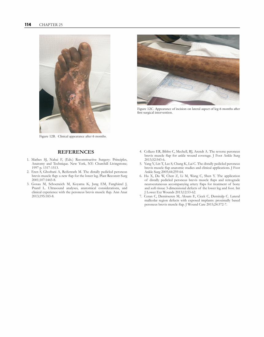
Figure 12A. Appearance 6 months after first surgical intervention.
114
REFERENCES 1. Mathes SJ, Nahai F, (Eds.) Reconstructive Surgery: Principles,
Anatomy and Technique. New York, NY: Churchill Livingstone; 1997 p. 1317-1513.
2. Eren S, Ghofrani A, Reifenrath M. The distally pedicled peroneus brevis muscle flap: a new flap for the lower leg. Plast Reconstr Surg 2001;107:1443-8.
3. Gosau M, Schoeneich M, Koyama K, Jung EM, Fanghänel J, Prantl L. Ultrasound analyses, anatomical considerations, and clinical experience with the peroneus brevis muscle flap. Ann Anat 2013;195:183-8.
CHAPTER 25
4. Collazo ER, Bibbo C, Mechell, RJ, Arendt A. The reverse peroneus brevis muscle flap for ankle wound coverage. J Foot Ankle Surg 2013;52:543-6.
5. Yang Y, Lin T, Lee S, Chang K, Lai C. The distally pedicled peroneus brevis muscle flap anatomic studies and clinical applications. J Foot Ankle Surg 2005;44:259-64.
6. Hu X, Du W, Chen Z, Li M, Wang C, Shen Y. The application of distally pedicled peroneus brevis muscle flaps and retrograde neurocutaneous accompanying artery flaps for treatment of bony and soft-tissue 3-dimensional defects of the lower leg and foot. Int J Lower Ext Wounds 2013;12:53-62.
7. Ceran C, Demirseren M, Aksam E, Cicek C, Demiralp C. Lateral malleolar region defects with exposed implants: proximally based peroneus brevis muscle flap. J Wound Care 2015;24:372-7.
Figure 12B. Clinical appearance after 6 months.
Figure 12C. Appearance of incision on lateral aspect of leg 6 months after first surgical intervention.


![Tachdjian's Pediatric Orthopaedics [Chapter 27] · diminished strength in the anterior tibialis and peroneus brevis. As the patient tries to actively dorsiflex the ankle, the metatarsophalangeal](https://static.fdocuments.in/doc/165x107/5b9ed0b509d3f2d0208c4506/tachdjians-pediatric-orthopaedics-chapter-27-diminished-strength-in-the-anterior.jpg)


![M34/1 M34 M34/175 M. fibularis tertius [M. peroneus tertius] 76 Tendo musculi fibularis brevis [Tendo musculi peronei brevis] 77 A. tarsalis lateralis 78 N. cutaneus dorsalis inter-medius](https://static.fdocuments.in/doc/165x107/60914ef95ae64858f1136fc7/m341-m34-m341-75-m-fibularis-tertius-m-peroneus-tertius-76-tendo-musculi-fibularis.jpg)


