
Retrospective analysis of adverse reactions to metal … · Retrospective analysis of adverse...
7
SA Orthopaedic Journal Summer 2014 | Vol 13 • No 4 Page 19 Retrospective analysis of adverse reactions to metal-on-metal lumbar disc arthroplasties in 378 consecutive patients S Berg MD, PhD Stockholm Spine Center, Löwenströmska Hospital, Stockholm, Sweden N Jansen M Eng North-West University (Potchefstroom campus), South Africa Correspondence: Dr Svante Berg Stockholm Spine Center Löwenströmska Hospital 19489 Upplands Väsby SE-195 89 Stockholm Sweden Email: [email protected] Introduction Neck or back pain associated with degenerative changes of the functional spinal unit is one of the leading causes of disability among adults. Chronic low back pain (CLBP) with a prevalence in excess of a one-year period is as high as 73%. 1 Disc degeneration is a frequent cause of CLBP, and conservative treatment including physio- therapy and exercise is the first step in attempting to reduce pain and improve function. Should conservative treatment fail, surgical intervention, which includes fusion or disc replacement procedure, is considered as a last resort. 2 The gold standard in surgical treatment of CLBP has been fusion. In the past decade, spinal disc arthroplasty and dynamic stabilisation devices have received growing acceptance as treatment for back and neck pain. 3,4 Finite element studies have confirmed that unnatural disc kinematics result in higher stress imparted to adjacent discs. 5,6 Unnatural kinematics may be caused by sclerotic, degenerated or fused intervertebral discs. Experience and knowledge gained from treatment of other joints with arthroplasty have led to the development of artificial discs (disc prostheses). The rationale for selecting total disc replacement (TDR) over fusion is a desire to preserve motion between vertebral bodies that might reduce adjacent segment disease. 6,7 Abstract Background: Spinal disc arthroplasty implants are primarily manufactured from metal/polymer materials. Biological reaction to wear debris ultimately requires clinical studies for assessment. Research into biological reaction of metal-on-polyethylene and metal-on-metal wear debris of knee and hip arthroplasties is well progressed as opposed to similar research on spinal arthroplasties. Materials and method: The Swedish Spine Register provides a resource for the evaluation of adverse events and clinical outcome to lumbar metal-on-metal total disc replacements. The resource will be used for a retrospective analysis of the cases in this study. The material reviewed consists of a total of 378 Swedish patients treated between October 2003 and May 2009 (181 male, 197 female); average age was 39.2 years. By means of a question- naire, 94% of the patients were followed up after two years and 88% after five years. Results: No reported cases were found of suspected or confirmed metal hypersensitivity or pseudotumors. This may be due to symptom-producing pseudotumors being extremely rare and the difficulty to form questions which would be able to indicate the presence of the adverse outcome. Conclusion: Based on the results from this study, it can be concluded that the results do not exclude the possi- bility that patients might have non-symptomatic pseudotumors, but being non-symptomatic, the authors doubt the importance and relevance of further investigating those isolated cases. Key words: spinal arthroplasty, metal-on-metal, pseudotumors, hypersensitivity, metallosis, SweSpine
Transcript of Retrospective analysis of adverse reactions to metal … · Retrospective analysis of adverse...
SA Orthopaedic Journal Summer 2014 | Vol 13 • No 4 Page 19
Retrospective analysis of adverse reactionsto metal-on-metal lumbar disc arthroplasties
in 378 consecutive patientsS Berg MD, PhD
Stockholm Spine Center, Löwenströmska Hospital, Stockholm, SwedenN Jansen M Eng
North-West University (Potchefstroom campus), South Africa
Correspondence: Dr Svante Berg
Stockholm Spine Center
Löwenströmska Hospital
19489 Upplands Väsby
SE-195 89 Stockholm
Sweden
Email: [email protected]
Introduction
Neck or back pain associated with degenerative changes
of the functional spinal unit is one of the leading causes
of disability among adults. Chronic low back pain
(CLBP) with a prevalence in excess of a one-year period
is as high as 73%.1 Disc degeneration is a frequent cause
of CLBP, and conservative treatment including physio-
therapy and exercise is the first step in attempting to
reduce pain and improve function. Should conservative
treatment fail, surgical intervention, which includes
fusion or disc replacement procedure, is considered as a
last resort.2
The gold standard in surgical treatment of CLBP has beenfusion. In the past decade, spinal disc arthroplasty anddynamic stabilisation devices have received growingacceptance as treatment for back and neck pain.3,4 Finiteelement studies have confirmed that unnatural disckinematics result in higher stress imparted to adjacentdiscs.5,6 Unnatural kinematics may be caused by sclerotic,degenerated or fused intervertebral discs. Experience andknowledge gained from treatment of other joints witharthroplasty have led to the development of artificial discs(disc prostheses). The rationale for selecting total discreplacement (TDR) over fusion is a desire to preserve motionbetween vertebral bodies that might reduce adjacentsegment disease.6,7
AbstractBackground: Spinal disc arthroplasty implants are primarily manufactured from metal/polymer materials.
Biological reaction to wear debris ultimately requires clinical studies for assessment. Research into biological
reaction of metal-on-polyethylene and metal-on-metal wear debris of knee and hip arthroplasties is well
progressed as opposed to similar research on spinal arthroplasties.
Materials and method: The Swedish Spine Register provides a resource for the evaluation of adverse events and
clinical outcome to lumbar metal-on-metal total disc replacements. The resource will be used for a retrospective
analysis of the cases in this study. The material reviewed consists of a total of 378 Swedish patients treated
between October 2003 and May 2009 (181 male, 197 female); average age was 39.2 years. By means of a question-
naire, 94% of the patients were followed up after two years and 88% after five years.
Results: No reported cases were found of suspected or confirmed metal hypersensitivity or pseudotumors. This
may be due to symptom-producing pseudotumors being extremely rare and the difficulty to form questions
which would be able to indicate the presence of the adverse outcome.
Conclusion: Based on the results from this study, it can be concluded that the results do not exclude the possi-
bility that patients might have non-symptomatic pseudotumors, but being non-symptomatic, the authors doubt
the importance and relevance of further investigating those isolated cases.
Key words: spinal arthroplasty, metal-on-metal, pseudotumors, hypersensitivity, metallosis, SweSpine
SAOJ Summer 2014_Orthopaedics Vol3 No4 2014/11/04 7:56 PM Page 1�
Page 20 SA Orthopaedic Journal Summer 2014 | Vol 13 • No 4
In all joint replacement surgery there are four main
questions to be answered: What is 1) the correct indication for
the procedure, 2) the best surgical technique to be used, 3) the
most favourable design of the implant and 4) the most appro-
priate materials to use? This study focuses on the fourth
questions, namely, the specific material frequently used.
Hip and knee replacements have been performed since the
1970s, providing knowledge that particulate wear debris
may be associated with post-operative complications such as
peri-implant osteolysis or pseudotumors characterised as an
enlargement of benign tissue.8
Selection of the wear couple material is for the most part a
compromise between the wear rate, robustness and toxicity.
There seems to be no single hip bearing material combi-
nation which reduces all clinical complications.9
In most cases, hip bearing design includes metal-on-
polymer (MoP), metal-on-metal (MoM), or ceramic-on-
ceramic (CoC) wear couples. Soft bearing wear, such as
polyethylene, in general is dominated by adhesive wear,
while hard-on-hard bearings such as MoM or CoC are
dominated by abrasive and surface fatigue wear.10
The use of MoM as a hip bearing has become popular in
the last years due to the possibility to reduce the risk of joint
luxation by larger femoral heads, but also perceived advan-
tages of being more robust than MoP or CoC, as well as
having a lower volumetric wear than MoP.11-13 Osteolysis
associated with MoM bearing implants is seldom reported,
and is considered as the prime motivation for a return to
MoM wear couples.14,15
Recently a particular MoM hip arthroplasty has been
found to have poor short-term results. Hexter et al. suggest
that a substantial portion of the blood ion levels are as a
result of corrosion at the taper interface between the hip
stem and ball and not the intended wear couple.16 Bernthal etal. suggest that the design flaw lies not with the specific
implant but rather with the ultra large monobloc cobalt-
chromium-molybdenum (CCM) acetabular shells.17
Over the medium to long term, the mechanical advantages
of MoM over MoP are potentially offset by a higher
frequency of foreign-body tissue reaction due to smaller
particle size of wear debris, as well as bioactive cobalt and
chrome ion release.15,18
The most common cause of total hip arthroplasty (THA)
failure is loosening of the acetabular cup or femoral part due
to osteolysis.19-22 Case studies indicate that failure due to
metallic debris infiltration have also been reported.23-25 It is
reported that reaction to polyethylene particles is that a
fibrotic granulation tissue typically develops.26
If the degree of metallosis (such as adjacent tissue staining
in black or grey) is significant and probably a certain size of
the particles is produced, a ‘foreign body reaction’ may take
place because submicron and micron-sized wear particles
are encapsulated by the host body.27 Fibroblast and inflam-
matory cells within the tissue are activated, and as a result
foreign body granulation tissue develops. The tissue tends
to be fibrotic, but may undergo additional changes such as
necrosis, and heterotopic ossification.
Local inflammation is mostly dependent on three factors;
particle load, shape and chemical reactivity. In general, a
higher inflammatory response will be produced with a
higher concentration of debris per tissue volume (particle
load), elongated fibres and more chemically reactive compo-
sition.20 Particle load is a function of the accuracy of device
placement, as misalignment may lead to fouling between
components near articulation extremes. The fixed centre of
rotation spinal disc replacements is likely to increase the
volume of wear debris if not accurately placed.28
The effects of pseudotumors have been found to be locally
destructive, requiring revision surgery in a high proportion
of patients.29 The extent of the encapsulation may be signif-
icant enough to appear on computed tomography scans.27
Potential risks associated with MoM hip implants are a
biological, foreign body reaction. Engh et al. suggest that the
process starts as an inflammatory response progressing to a
necrotic tissue involving soft and/or hard tissue.30 The most
appropriate blood fraction is controversial when studying
effects of MoM implants.29 The acceptable limit of blood
cobalt levels is therefore yet to be established.31,32
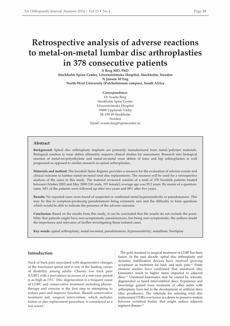
The potential for the release of metal ions was examined in
a subset of Kineflex MoM Cervical Disc subjects from a US
IDE clinical study, which included sample collection at all
investigational sites participating in the study that were
qualified and had the facilities to do the collection. These
sites collected samples from 32 subjects for analysis. Metal
ion analysis was evaluated by a core laboratory (Rush
University).
Figure 1. Serum metal ion levels (µg/L) for Kineflex disc subjects over time
Osteolysis associated with MoM bearing implants is seldom reported, and is considered as the prime motivation
for a return to MoM wear couples
SAOJ Summer 2014_Orthopaedics Vol3 No4 2014/11/04 7:56 PM Page 20
SA Orthopaedic Journal Summer 2014 | Vol 13 • No 4 Page 21
Mean serum cobalt and chromium serum levels ranged
from 0.37 to 1.24 µg/L and 0.27 to 1.11 µg/L, respectively,
at follow-up times ranging from 6 weeks to 72 months
after implantation. As shown in Figure 1, data through
extended follow-up with the metal ion cohort showed
metal ion levels were stable or decreasing over time after
12 months post-operative through the 72-month visit.
Metal ion data through extended follow-up with the metal
ion cohort showed metal ion levels levelling off and
decreasing over time after approximately 12 months post-
surgery. Maximum serum metal ion levels associated with
Kineflex implants were substantially lower than those found
with other metal-on-metal implants, and substantially lower
than threshold values that have been proposed as indicative
of metallosis in literature for orthopaedic implants. Long-
term follow-up data showed the metal ion levels associated
with the Kineflex disc to be approximately five to 26 times
lower than the 7 µg/L threshold of concern set by the British
MHRA Medical Alert for MoM hip replacements issued on
22 April 2010.33 Further, MoM hips and MoM lumbar discs
are substantially different in implant design, joint size and
environment (synovial vs non-synovial), loads and range of
motion, and in vitro and in vivo wear.
Hypersensitivity to metal ions may either occur within
minutes (initiated by anti-body or formation of antibody-
antigen complexes) or days (by a cell-mediated response).34,35
On the contrary, metallosis is a well-known phenomenon,
being the infiltration of metal, predominantly oxidised, in
the tissues in contact with the implant. The only known
effect of metallosis is that the periprosthetic tissue is stained
black, with microscopic necrosis.27
Materials commonly used for the manufacture oftotal disc replacementsSpinal disc arthroplasty devices mostly feature MoM and
MoP bearings in either fixed or mobile centre of rotation
configurations. The most popular metal and polymer used
by TDR device wear surfaces are cobalt-chromium-molyb-
denum (CCM), and ultra-high molecular weight polyeth-
ylene (UHMWPE). Polyether ether ketone (PEEK) is a
polymer which has a high degree of biocompatibility, and is
under investigation as a MoP wear couple.36
Materials presently used for lumbar and cervical spinal
disc replacement devices, which are US Food and Drug
Administration (FDA) approved, are listed in Tables I and IIrespectively. Although comprehensive laboratory tests as
well as animal studies are performed prior to clinical trials,
novel uses of biomaterials lead to difficulty in predicting invivo performance.26,37
Retrieval analyses of motion preserving spinal devices
have to a large extent validated the high mechanical strength
and low wear advantages of MoM over MoP. UHMWPE
wear surfaces have displayed the potential to crack or plasti-
cally deform in both hip and spinal applications.26,27,46 Ooij etal.2 report on high wear rates of the polyethylene component
of the SB Charité III lumbar disc replacement, suggesting
that the device would have a lifespan of less than 40 years.2
The nature of wear particles obtained by in vitro wear
simulation tests of hip, knee, and spinal wear are of a similar
morphology.20 Over the short term, the increase of ion levels
in blood after a single level MoM lumbar disc replacement
(Maverick™) was found to be of the same order of
magnitude as that of a well-functioning MoM hip arthro-
plasty.47 Subsequent to this publication it has been reported
that a specific MoM hip arthroplasty has been shown to
exhibit much higher wear or metal ion release, but in this
instance, it is believed to be related to the design of this
specific implant.48
Table I: List of presently FDA-approved lumbar disc arthroplasties
Manufacturer Device name PMA filed FDA approval Wear couple ReferenceDePuy Spine, Inc. Charite™ 2004.02.13 2004.10.26 CCM-polyethylene 38
S Synthes Spine, Inc. ProDisc®-L 2005.03.15 2006.08.14 CCM-polyethylene 39
Table II: List of presently FDA-approved cervical disc arthroplasties
Manufacturer Device name PMA filed FDA approval Wear couple Reference
Medtronic Sofamor
Danek
Bryan® 2006.06.29 2009.05.12 Ti-polyethylene 40
Prestige® 2006.05.19 2007.07.16 Stainless steel 316 41
S Synthes Spine, Inc. ProDisc™-C 2007.01.03 2007.12.17 CCM-polyethylene 42
Globus Medical, Inc. Secure®-C 2010.09.17 2012.09.28 CCM-polyethylene 43
NuVasive, Inc. PCM® 2010.04.01 2012.10.26 CCM-polyethylene 44
LDR Spine USA, Inc. Mobi-C® 2011.01.14 2013.08.07 CCM-polyethylene 45
Hypersensitivity to metal ions may either occur within minutes or days
SAOJ Summer 2014_Orthopaedics Vol3 No4 2014/11/04 7:56 PM Page 21
Page 22 SA Orthopaedic Journal Summer 2014 | Vol 13 • No 4
Hallab reviewed published data on concentrations of
metal in body fluids, reporting that Co ion levels in blood
serum increased from < 0.2–0.6 to 1.9–4.8 parts per billion
(ppb) for TDR and 0.6–7.9 ppb for MoM total hip arthro-
plasties.20 Cr-ion levels were found to be higher in THA
than TDR, 9.1 and 2.4 ppb respectively. It is therefore
noted that in lumbar TDR, Co and Cr ion levels are lower
than after THA.20,47
Complications such as peri-implant osteolysis and metal
hypersensitivity are rare and make for a challenging
diagnosis.30,49 Biological reaction to wear debris depends
on volume as well as morphology of wear debris
generated.50
Recently a few cases of suspected ‘pseudotumors’ after
cervical or lumbar TDR were reported.51,52 The purposes of
this study were to analyse in what patient material MoM
TDRs were used and to report on the clinical symptomatic
incidence of pseudotumors or metal hypersensitivity
brought on by MoM wear couples in lumbar spinal disc
arthroplasties.
Material and methods This is a retrospective study on consecutive patients who
had received MoM TDR between 8 October 2003 and 13
May 2009 due to chronic low back pain, where pain and
dysfunction were not reduced after prolonged conser-
vative treatment over at least one year. All surgeries were
carried out at Stockholm Spine Center in Sweden; 88 per
cent of the procedures were performed by one surgeon
(SB).
Follow-up on patients was performed two and five years
after surgery with The Swedish Spine Register, but for this
study, medical record data were also examined together
with a follow-up visit with clinical examination when
more than two years had passed since treatment. The
Swedish Spine Register (SweSpine) is a non-profit organi-
sation owned and administered by the Swedish Society of
Spinal Surgeons. SweSpine was founded in 1993, and is
currently a national registry.53
One of the aims of the registry is to provide outcome-
based feedback from patients after surgical intervention,
and thereby provide continuous improvement of spinal
treatments. Adverse medical conditions are recorded
within the register, which therefore serves as a rigorous
report of patient feedback for the recorded period.
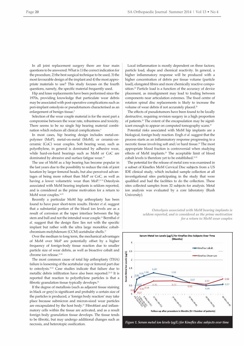
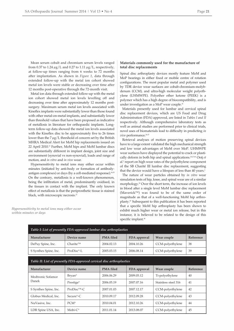
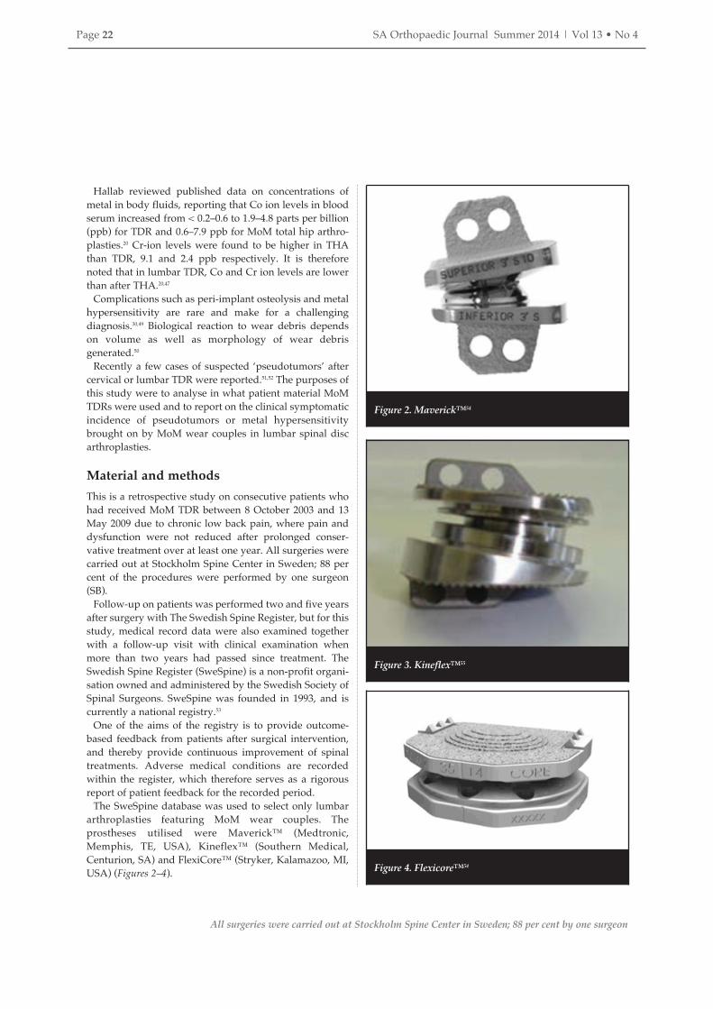
The SweSpine database was used to select only lumbar
arthroplasties featuring MoM wear couples. The
prostheses utilised were Maverick™ (Medtronic,
Memphis, TE, USA), Kineflex™ (Southern Medical,
Centurion, SA) and FlexiCore™ (Stryker, Kalamazoo, MI,
USA) (Figures 2–4).
Figure 2. Maverick™54
Figure 3. Kineflex™55
Figure 4. Flexicore™54
All surgeries were carried out at Stockholm Spine Center in Sweden; 88 per cent by one surgeon
SAOJ Summer 2014_Orthopaedics Vol3 No4 2014/11/04 7:56 PM Page 22
SA Orthopaedic Journal Summer 2014 | Vol 13 • No 4 Page 23
To evaluate the outcome and quality of spinal treatment,
patients answer questionnaires at one, two, five and ten
year intervals after treatment. The questionnaires are sent
to the patients with an attached pre-paid return envelope.
All returned questionnaires are registered at one place by
a single secretary. The questionnaires include present
work status and medication, but also re-operations,
EuroQol (EQ-5D), visual analogue scale (VAS) for back
and leg pain, SF-36, Global Assessment (GA) of back pain
and the original Oswestry Disability Index (ODI). At time
of surgery, the surgeon records diagnosis, type of surgery,
treated segments, implants, dates of in-hospital stay and
immediate complications.
Re-operations are recorded in the register, including
patients treated with MoM disc prostheses. Since more than
95% of TDR surgery in Sweden is performed in the clinic
from which these results are obtained, a re-operation would
most likely also be performed there. Thirty-seven per cent of
the patients in this study were examined by MRI after more
than two years had passed since surgery. The reason for the
examination was most frequently because the patients were
part of other studies, but in some cases they were performed
due to recurrent or persistent LBP. These MRI scans were
investigated for the occurrence of pseudotumors.
ResultsThe analysis of the treated patients showed that a total of
378 Swedish patients (181 male, 197 female), with average
age and weight of 39.2 years and 77.1 kg respectively were
treated. A total of 111 patients had previous spinal opera-
tions; 297 were active in sports, 45 were smokers. All
patients were treated with a total of 642 MoM total lumbar
disc replacements at one, two or three segments. Follow-
ups were performed with 94 per cent of the patients after
two years (356/378). In April 2013 317 of the patients had
answered their five-year questionnaires (the 88% that had
passed five years). The average time of follow-up was just
over six years (4–10). TDRs were performed utilising
Maverick™ in 227 cases, Kineflex™ in 140 cases and
FlexiCore™ in 11 cases.
No reported cases of pseudotumors were found in the
material, neither presented with clinical symptoms in
questionnaires or by words at clinical visit, nor identified
on MRI in the individual cases that had that examination
late post-operatively.
No case of suspected or verified metal hypersensitivity
was found.
Three patients were re-operated due to misplaced disc
prostheses, all within a week after index surgery. No
metallosis or inflammation was observed at this early
stage after initial surgical treatment. In one patient who
underwent a late decompression and fusion at a segment
that three years previously had been treated with a MoM
prosthesis, a slight metallosis was observed at one side
posteriorly in the annulus fibrosus, but without any local
reaction.
DiscussionInformation from other joint replacements (THA and TDR)
has been discouraging regarding the use of UHMWPE. This
is due to the concern of high wear rates, early plastic defor-
mation or component failure, and local reactions such as
osteolysis that may be caused by wear-particles. When TDR-
implants are compared with repeated movements under
loaded conditions, the volumetric wear of MoM is half of
that of MoP per million cycles.56 These factors have induced
the development of TDR-implants that do not use
UHMWPE. At this stage of disc development, other
materials now used for bearing couples in lumbar TDR are
CCM and PEEK.
Metallosis is the black or grey stain colouring of soft tissue
that is often seen when implants are removed. This is seen
even after fusion, when mobility-induced wear is not
present or limited. When discussing adverse events
resulting from the implantation of metal components into
humans, confusion prevails. In the authors’ opinion this is
because metallosis is not considered a complication, as it is
not linked to any symptoms, but rather as a normal occur-
rence due to surface corrosion or deposition of wear debris.
It is possible that there is an occurrence of true hypersensi-
tivity to any of the metals in the implant. This is however
almost unreported and it might occur whether or not the
implant also consists of polyethylene. The frequency of
hypersensitivity reactions is not well known.15 Due to the
rarity of metal hypersensitivity, as well as low accuracy of
allergy tests, it is debatable whether screening should be
performed prior to surgery.34
The formation of foreign body granulomas that can
develop into pseudotumors can be considered as a physio-
logical phenomenon. This is similar to what is observed if,
for example, a needle tip is left in human tissue and there is
no allergic reaction.
In recent reports there is a high frequency of foreign body
reactions leading to the formation of pseudotumors after
MoM total hip replacements. This is concerning as a large
portion of TDRs performed today in Sweden are MoM.
The proposed mechanism for the development of pseudo-
tumors is that metal wear particles start a ‘foreign-body
reaction’ in the host that encapsulates the wear particles.
This encapsulation is what forms the pseudotumor. It is also
reasonable to believe that the total size of the encapsulating
tissue is dependent on the amount of wear particles.
The total weight-strain on a hip prosthesis is assumed to be
within the same range as that on a TDR implant. The loading
pattern and total joint mobility is, however, far larger in a
hip than in an artificial disc. The authors hypothesise that
wear debris in a MoM artificial joint is, besides design and
placement differences, dependent on load and actual
‘travelled distance’ between joint surfaces per time unit. If
this hypothesis is relevant, fewer wear particles would be
expected in a TDR than in a THA and therefore there is a
lower likelihood of adverse reactions on wear products
emerging.
SAOJ Summer 2014_Orthopaedics Vol3 No4 2014/11/04 7:56 PM Page 23
Page 24 SA Orthopaedic Journal Summer 2014 | Vol 13 • No 4
In the case series of Swedish patients, no reports were seen
on ‘symptoms producing’ pseudotumors. This could be due
to the difficulty of forming questions that are directed to
finding patients who had developed pseudotumors.
Therefore the study has clear limitations. Despite this, the
result states that none of the patients had developed pseudo-
tumors that caused neurological symptoms or local pain.
The material is from Scandinavia’s largest spine clinic,
where until now more than 95% of lumbar TDRs in Sweden
were performed; therefore, these results can be considered
to be valid.
The Swedish Spine Register (SweSpine) covers not only
index surgery but also re-operations. Furthermore, the
patients treated at Stockholm Spine Center are all referred
from general practitioners or other orthopaedic clinics,
where the receiving surgeons are expected to deal with any
complications or need of re-operations that might occur. As
a consequence of this study there has been a continuous and
highly frequent use of MoM prostheses after April 2009 at
the clinic, still without any discovered cases of ‘pseudo-
tumors’.
ConclusionBased on the results from this study, it can be concluded that
symptom-producing pseudotumors after TDR with MoM
implants are extremely rare. The results do not exclude the
possibility that patients might have non-symptomatic
pseudotumors, but being non-symptomatic, the authors
doubt the importance and relevance of further investigating
those isolated cases.
References1. Rumboldt Z. Degenerative disorders of the spine. Semin.
Roentg. 2006; 41:327-62.
2. Ooij A van, Oner FC, Verbout AJ. Complications of artificial
disc replacement: a report of 27 patients with the SB Charité
disc. J Spinal Disord Tech. 2003; 16:369-83.
3. Billi F, Benya P, Ebramzadeh E, et al. Metal wear particles:
What we know, what we do not know, and why. SAS Journal2009; 3:133-42.
4. Schmoelz W, Huber JF, Nydegger T, et al. Dynamic stabi-
lization of the lumbar spine and its effects on adjacent
segments: an in vitro experiment. J Spinal Disord Tech. 2003;
16:418-23.
5. Ruberté LM, Natarajan RN, Andersson GB. Influence of
single-level lumbar degenerative disc disease on the behavior
of the adjacent segments - a finite element model study. JBiomech. 2009; 42:341-48.
6. Castellvi A, Huang H, Vestgaarden T, et al. Stress reduction in
adjacent level discs via dynamic instrumentation: a finite
element analysis. SAS Journal 2007; 1:74-81.
7. Huang RC, Tropiano P, Marnay T, et al. Range of motion and
adjacent level degeneration after lumbar total disc
replacement. Spine J. 2006; 6:242-47.
8. Dumbleton J, Manley M. Metal-on-metal total hip replacement:
What Does the literature say? J Arthroplasty. 2005; 20:174-88.
9. Passuti N, Philippeau J, Gouin F. Friction couples in total hip
replacement. Orthop Traumatol Surg Res. 2009; 95:S27-34.
10. Harper ML, Dooris A, Paré PE. The fundamentals of biotri-
bology and its application to spine arthroplasty. SAS Journal2009; 3:125-32.
11. Schiopu D, Girard J, Soenen M, et al. Metal ions levels measure-
ments for early total hip replacement malfunction diagnosis
with ‘plasma-sprayed ceramic’ bearings couple. OrthopTraumatol Surg Res. 2010; 96:75-79.
12. Beldame J, Carreras F, Oger P, et al. Cementless cups do not
increase osteolysis risk in metal-on-metal total hip arthroplasty.
Orthop Traumatol Surg Res. 2009; 95:478-90.
13. Crawford R, Ranawat CS, Rothman RH. Metal on metal: is it
worth the risk? J Arthroplasty. 2010; 25:1-2.
14. Macpherson GJ, Breusch SJ. Metal-on-metal hip resurfacing: a
critical review. Arch Orthop Trauma Surg. 2011; 131:101-10.
15. Delaunay C, Petit I, Learmonth ID, et al. Metal-on-metal
bearings total hip arthroplasty: The cobalt and chromium ions
release concern. Orthop Traumatol Surg Res. 2010; 96:894-904.
16. Adam Hexter, Anna Panagiotidou, et al. Wear and corrosion at
the taper interface in retrieved ASR XL metal-on-metal total hip
arthroplasty. Int J Surg. 2012; 12:S53-S109.
17. Nicholas M. Bernthal, Paul C. Celestre, et al. Disappointing
short-term results with the depuy ASR XL metal-on-metal total
hip arthroplasty. J Arthroplasty. 2012; 27:539-44.
18. Williams PA, Clarke IC. Understanding polyethylene wear
mechanisms by modeling of debris size distributions. Wear.2009; 267:646-52.
19. Murali R, Bonar SF, Kirsh G, et al. Osteolysis in third-generation
alumina ceramic-on-ceramic hip bearings with severe
impingement and titanium metallosis. J Arthroplasty. 2008;
23:1240.e13-1240.e19.
20. Hallab NJ. A review of the biologic effects of spine implant
debris: Fact from fiction. SAS Journal 2009; 3:143-60.
21. Catelas I. Semi-quantitative analysis of cytokines in MM THR
tissues and their relationship to metal particles. Biomaterials.2003; 24:4785-97.
22. Alhassan S, Goswami T. Wear rate model for UHMWPE in total
joint applications. Wear. 2008; 265:8-13.
23. Miloŝev L, Antoliĉ V, Minoviĉ A, et al. Extensive metallosis
and necrosis in failed prostheses with cemented titanium-
alloy stems and ceramic heads. J Bone Joint Surg Br. 2000;
82:352-57.
24. Chang J-D, Lee S-S, Hur M, et al. Revision total hip arthroplasty
in hip joints with metallosis: a single-center experience with 31
cases. J Arthroplasty. 2005; 20:568-73.
25. Oldenburg M, Wegner R, Baur X. Severe cobalt intoxication due
to prosthesis wear in repeated total hip arthroplasty. JArthroplasty. 2009; 24:825.e15-20.
26. Kurtz SM, Steinbeck M, Ianuzzi A, et al. Retrieval analysis of
motion preserving spinal devices and periprosthetic tissues.
SAS Journal 2009; 3:161-77.
27. Khan RJK, Wimhurst J, Foroughi S, et al. The natural history of
metallosis from catastrophic failure of a polyethylene liner in a
total hip. J Arthroplasty. 2009; 24:2-5.
28. Moumene M, Geisler FH. Comparison of biomechanical
function at ideal and varied surgical placement for two lumbar
artificial disc implant designs: mobile-core versus fixed-core.
Spine J. 2007; 32:1840-51.
29. Kwon Y-M, Ostlere SJ, McLardy-Smith P, et al. ‘Asymptomatic’
pseudotumors after metal-on-metal hip resurfacing arthro-
plasty: Prevalence and metal ion study. J Arthroplasty. 2011;
26:511-18.
30. Engh Jr CA, Ho H, Engh CA, et al. Metal-on-metal total hip
arthroplasty adverse local tissue reaction. Sem Arthroplasty.
2010; 21(1):19-23.
SAOJ Summer 2014_Orthopaedics Vol3 No4 2014/11/04 7:56 PM Page 24
SA Orthopaedic Journal Summer 2014 | Vol 13 • No 4 Page 25
31. Walter LR, Marel E, Harbury R, et al. Distribution of
chromium and cobalt ions in various blood fractions after
resurfacing hip arthroplasty. J Arthroplasty. 2008; 23:814-21.
32. Medical Device Alert, Ref: MDA/2012/036 Issued: 25 June
2012 at 11:00.
33. Macdonald S. Can a safe level for metal ions in patients with
metal-on-metal total hip arthroplasties be determined? JArthroplasty. 2004; 19:71-77.
34. Niki Y, Matsumoto H, Otani T, et al. Screening for sympto-
matic metal sensitivity: a prospective study of 92 patients
undergoing total knee arthroplasty. Biomaterials. 2005;
26:1019-26.
35. Hallab N, Jacobs JJ, Black J. Hypersensitivity to metallic
biomaterials: a review of leukocyte migration inhibition
assays. Biomaterials. 2000; 21:1301-14.
36. Austin H. 2008. Wear of PEEK all-polymer articulations for
cervical spinal disc arthroplasty. Master’s dissertation,
University of Waterloo. GB.
37. Chang B-S, Brown PR, Sieber A, et al. Evaluation of the
biological response of wear debris. Spine J. 2004; 4:239S-44S.
38. US Food and Drug Administration, Department of Health
and Human Services. P040006 CharitéTM Artificial Disc
approval letter, Oct 26, 2004. Retrieved August 30, 2013, from
http://www.accessdata.fda.gov/cdrh_docs/pdf4/p040006a.
39. US Food and Drug Administration, Department of Health
and Human Services. P050010 ProDics™-L Total Disc
Replacement approval letter, Aug 14, 2006. Retrieved August
30, 2013, from www.accessdata.fda.gov/cdrh_docs/pdf5/
p050010a.pdf
40. US Food and Drug Administration, Department of Health
and Human Services. P060023 BRYAN® Cervical Disc
approval letter, May 12, 2009. Retrieved August 27, 2013, from
www.accessdata.fda.gov/cdrh_docs/pdf6/ P060023a.pdf
41. US Food and Drug Administration, Department of Health
and Human Services. P060018 PRESTIGE® Cervical Disc
System approval letter, Jul 16, 2007. Retrieved August 27,
2013, from www.accessdata.fda.gov/cdrh_docs/pdf6/
P060018A.pdf
42. US Food and Drug Administration, Department of Health
and Human Services. P070001 ProDisc™-C Total Disc
Replacement approval letter, Dec 17, 2007. Retrieved August
27, 2013, from www.accessdata.fda.gov/cdrh_docs/pdf7/
P070001A.pdf
43. US Food and Drug Administration, Department of Health
and Human Services. P100003 Secure-C Artificial Cervical
Disc approval letter, Sep 28, 2012. Retrieved August 27, 2013,
from www.accessdata.fda.gov/cdrh_docs/pdf10/P100003a.
44. US Food and Drug Administration, Department of Health
and Human Services. P100012 PCM® Cervical Disc System
approval letter, Oct 26, 2012. Retrieved August 27, 2013, from
www.accessdata.fda.gov/cdrh_docs/pdf10/ P100012A.pdf
45. US Food and Drug Administration, Department of Health
and Human Services. P110002 Mobi-C® Cervical Disc
Prosthesis approval letter, Aug 07, 2013. Retrieved August 30,
2013, from http://www.accessdata.fda.gov/cdrh_docs/
pdf11/P110002a.pdf
46. Kurtz SM, Peloza J, Siskey R, et al. Analysis of a retrieved
polyethylene total disc replacement component. Spine J. 2005;
5:344-50.
47. Gornet MF, Burkus K, Skipor AK, et al. Prospective study of
serum metal ion levels in patients with cobalt-alloy metal-on-
metal lumbar disc replacements. Spine J. 2007; 7:74S-74S.
48. Hexter A, et al. Wear and corrosion at the taper interface in
retrieved asr xl metal-on-metal total hip arthroplasty. Int. J.Surg. 2012; 10:8;55.
49. Anand A, McGlynn F, Jiranek W. Metal Hypersensitivity: Can
it mimic infection? J Arthroplasty. 2009; 24:826.e25-826.e28.
50. Kowandy C, Mazouz H, Richard C. Isolation and analysis of
articular joints wear debris generated in vitro. Wear. 2006;
261:966-70.
51. Cavanaugh D, Nunley P, Kerr E, et al. Delayed hyper-
reactivity to metal ions after cervical disc arthroplasty. A case
report and literature review. Spine, 2009; 34:E262–5.
52. Guyer R, Shellock J, MacLennan B, et al. Early failure of metal-
on-metal artificial disc prostheses associated with lympho-
cytic reaction. Diagnosis and treatment experience in four
cases. Spine 2011;36:E492-97.
53. Strömqvist B, Jönsson B, Fritzell P, et al. The Swedish National
Register for Lumbar Spine Surgery. Swedish Society for
Spinal Surgery. Acta Orthop 2001; 72: 99-106.
54. Singh K, Vaccaro AR, et al. Assessing the potential impact of
total disc arthroplasty on surgeon practice patterns in North
America. Spine J., 2004; 4: S195-S201.
55. Hähnle UR, Weinberg IR, et al. Kineflex (Centurion) lumbar
disc prosthesis: insertion technique and 2-year clinical results
in 100 patients. SAS Journal 2007; 1:28-35.
56. Bushelow M, Walker J, Coppes J, et al. Comparison of wear
rates: metal/UHMWPE and metal-on-metal total disc arthro-
plasty. Spine J. 2007; 7:97S.
This article is also available online on the SAOA website(www.saoa.org.za) and the SciELO website (www.scielo.org.za).Follow the directions on the Contents page of this journal toaccess it.
• SAOJ
SAOJ Summer 2014_Orthopaedics Vol3 No4 2014/11/04 7:56 PM Page 25


![Optimal predictor for 6-mercaptopurine intolerance in ...ITPA deficiency and 6-MP -related adverse effects, sub-sequent studies, both retrospective [21] and prospective [22], have](https://static.fdocuments.in/doc/165x107/60c575d81829d47c701e4fc0/optimal-predictor-for-6-mercaptopurine-intolerance-in-itpa-deficiency-and-6-mp.jpg)