![Giant Retroperitoneal Myxoid Liposarcomas, Survival in ......[3,10]. The retroperitoneal tumors are an important surgical challenge. Total tumors surgical extirpation is not possible](https://static.fdocuments.in/doc/165x107/60526365bf6dbb585a170846/giant-retroperitoneal-myxoid-liposarcomas-survival-in-310-the-retroperitoneal.jpg)
Retroperitoneal Tumors-7-16
20
The gamut of primary retroperitoneal masses: multimodality evaluation with pathologic correlation Guillermo P. Sangster, 1 Matias Migliaro, 2 Maureen G. Heldmann, 3 Peeyush Bhargava, 3 Alireza Hamidian, 4 Jaiyeola Thomas-Ogunniyi 5 1 Departments of Radiology and Anesthesiology, LSU Health – Shreveport, 1501 Kings Highway, Shreveport, LA 71103, USA 2 Hospital Santa Isabel de Hungria, Mendoza 5521, Argentina 3 Department of Radiology, LSU Health – Shreveport, 1501 Kings Highway, Shreveport, LA 71103, USA 4 Department of Surgery, LSU Health – Shreveport, 1501 Kings Highway, Shreveport, LA 71103, USA 5 Department of Pathology, LSU Health – Shreveport, 1501 Kings Highway, Shreveport, LA 71103, USA Abstract The retroperitoneum is a large space where primary and metastatic tumors grow silently before clinical signs ap- pear. Neoplastic retroperitoneal diseases may be solid or cystic, primary or secondary and range from benign to aggressive in behavior. Retroperitoneal neoplasms are notable for their widely disparate histologies. The solid primary retroperitoneal neoplasms are extremely uncom- mon and can be classified based on their tissue of origin into three main categories: mesodermal tumors, neuro- genic tumors, and extragonadal germ cell tumors. These tumors can grow to a large size before clinical symptoms occur or become palpable. When symptoms do occur, they are nonspecific. The majority of these masses are malignant and imaging plays a pivotal role in the detec- tion, staging, and pre-operative planning. Benign and malignant masses should be distinguished whenever pos- sible to avoid unnecessary surgical procedures. Macro- scopic fat, calcification, necrosis, vascularity, and neural foraminal widening are common imaging features helping for tumor differentiation. Meticulous cross-sectional imaging can triage the patient to the most appropriate therapy. Tumor morphology dictates imaging character, and biologic activity is reflected by positron emission tomography (PET). Complete surgical excision with tu- mor free margins is essential for long-term survival. Biopsy should be performed in consultation with surgical oncology to avoid complicating curative surgery. This pictorial essay illustrates the spectrum of multidetector computed tomography (MDCT) imaging findings in common and uncommon primary retroperitoneal masses, with an emphasis on cross-sectional imaging features for an adequate tumor characterization and staging. Key words: Retroperitoneum—Primary—Sarcomas— Tumors—Masses Primary retroperitoneal neoplasms are rare, accounting for 0.1% to 0.2% of all malignancies [1], and approxi- mately 70% to 80% are malignant [2]. Tumors can arise from any retroperitoneal tissue outside of the major retroperitoneal organs, and neoplasms can be divided primarily into solid and cystic masses. The solid neo- plasms can be classified based on their tissue of origin into three main categories: mesodermal tumors, neuro- genic tumors, and extragonadal germ cell tumors [2]. The preferred treatment of retroperitoneal sarcomas (RS) is surgical resection. However, complete excision is only possible in 20% to 75% of cases due to invasion of vital organs or vascular structures. Even with complete exci- sion, recurrence rates are high for all subtypes of sar- coma [1]. Metastasis is common in high-grade sarcomas and spread occurs hematogenously to the liver, lung, bone, and brain [1]. CME activity This article has been selected as the CME activity for the current month. Please visit https://ce.mayo.edu/content/ abdominal-radiology-journal-gamut-primary-retroperitoneal-masses% C2%A0-multimodality-evaluation and follow the instructions to complete this CME activity. Electronic supplementary material The online version of this article (doi:10.1007/s00261-016-0735-6) contains supplementary material, which is available to authorized users. Correspondence to: Guillermo P. Sangster; email: [email protected] ª Springer Science+Business Media New York 2016 Published online: 6 June 2016 Abdominal Radiology Abdom Radiol (2016) 41:1411–1430 DOI: 10.1007/s00261-016-0735-6
-
Upload
peeyush-bhargava-md-mba -
Category
Documents
-
view
249 -
download
0
Transcript of Retroperitoneal Tumors-7-16

The gamut of primary retroperitoneal masses:multimodality evaluation with pathologiccorrelation
Guillermo P. Sangster,1 Matias Migliaro,2 Maureen G. Heldmann,3
Peeyush Bhargava,3 Alireza Hamidian,4 Jaiyeola Thomas-Ogunniyi5
1Departments of Radiology and Anesthesiology, LSU Health – Shreveport, 1501 Kings Highway, Shreveport, LA 71103, USA2Hospital Santa Isabel de Hungria, Mendoza 5521, Argentina3Department of Radiology, LSU Health – Shreveport, 1501 Kings Highway, Shreveport, LA 71103, USA4Department of Surgery, LSU Health – Shreveport, 1501 Kings Highway, Shreveport, LA 71103, USA5Department of Pathology, LSU Health – Shreveport, 1501 Kings Highway, Shreveport, LA 71103, USA
Abstract
The retroperitoneum is a large space where primary andmetastatic tumors grow silently before clinical signs ap-pear. Neoplastic retroperitoneal diseases may be solid orcystic, primary or secondary and range from benign toaggressive in behavior. Retroperitoneal neoplasms arenotable for their widely disparate histologies. The solidprimary retroperitoneal neoplasms are extremely uncom-mon and can be classified based on their tissue of origininto three main categories: mesodermal tumors, neuro-genic tumors, and extragonadal germ cell tumors. Thesetumors can grow to a large size before clinical symptomsoccur or become palpable. When symptoms do occur,they are nonspecific. The majority of these masses aremalignant and imaging plays a pivotal role in the detec-tion, staging, and pre-operative planning. Benign andmalignant masses should be distinguished whenever pos-sible to avoid unnecessary surgical procedures. Macro-scopic fat, calcification, necrosis, vascularity, and neuralforaminal widening are common imaging features helpingfor tumor differentiation. Meticulous cross-sectionalimaging can triage the patient to the most appropriate
therapy. Tumor morphology dictates imaging character,and biologic activity is reflected by positron emissiontomography (PET). Complete surgical excision with tu-mor free margins is essential for long-term survival.Biopsy should be performed in consultation with surgicaloncology to avoid complicating curative surgery. Thispictorial essay illustrates the spectrum of multidetectorcomputed tomography (MDCT) imaging findings incommon and uncommon primary retroperitoneal masses,with an emphasis on cross-sectional imaging features foran adequate tumor characterization and staging.
Key words: Retroperitoneum—Primary—Sarcomas—Tumors—Masses
Primary retroperitoneal neoplasms are rare, accountingfor 0.1% to 0.2% of all malignancies [1], and approxi-mately 70% to 80% are malignant [2]. Tumors can arisefrom any retroperitoneal tissue outside of the majorretroperitoneal organs, and neoplasms can be dividedprimarily into solid and cystic masses. The solid neo-plasms can be classified based on their tissue of origininto three main categories: mesodermal tumors, neuro-genic tumors, and extragonadal germ cell tumors [2]. Thepreferred treatment of retroperitoneal sarcomas (RS) issurgical resection. However, complete excision is onlypossible in 20% to 75% of cases due to invasion of vitalorgans or vascular structures. Even with complete exci-sion, recurrence rates are high for all subtypes of sar-coma [1]. Metastasis is common in high-grade sarcomasand spread occurs hematogenously to the liver, lung,bone, and brain [1].
CME activity This article has been selected as the CME activityfor the current month. Please visit https://ce.mayo.edu/content/abdominal-radiology-journal-gamut-primary-retroperitoneal-masses%C2%A0-multimodality-evaluation and follow the instructions to completethis CME activity.
Electronic supplementary material The online version of this article(doi:10.1007/s00261-016-0735-6) contains supplementary material,which is available to authorized users.
Correspondence to: Guillermo P. Sangster; email: [email protected]
ª Springer Science+Business Media New York 2016
Published online: 6 June 2016AbdominalRadiology
Abdom Radiol (2016) 41:1411–1430
DOI: 10.1007/s00261-016-0735-6

The American Joint Committee Staging System ofextremity soft-tissue sarcomas, which is based on theTNM classification, is commonly used for the staging ofthe retroperitoneal sarcomas [3]. The stage is determinedby the size of the tumor, the histologic grade, spread to
lymph nodes, and distant metastasis. We will briefly re-view the principles of the TNM classification used forsarcoma staging in Tables 1 and 2.
Mesodermal tumors
Mesodermal tumors constitute 47% to 57% of the primaryretroperitoneal tumors (Table 3) [1], the vast majority ofwhich (90%) are sarcomas [4]. Yet only 10% to 20% of allsarcomas arise from the retroperitoneum, with an overallincidence of 0.3% to 0.4% per 100,000 of the generalpopulation [3]. RS occur in all ages groups, with a peak inthe fifth decade of life. RS tend to enlarge in retroperi-toneal spaces without producing symptoms and aretherefore often large at the time of diagnosis and presentlater with nonspecific symptoms such as abdominal painand fullness. Liposarcomas, leiomyosarcomas, and malig-nant fibrous histiocytomas (MFH) constitute more than80% of these tumors [2]. Recently, the frequency of MFHin the retroperitoneum has been disputed. With the use ofimmunohistochemistry, many of these fibrous tumors havenow been shown to represent other sarcoma types such asleiomyosarcomas or dedifferentiated liposarcomas. Forthis reason, it is expected that the number of the reportedMFH will be significantly reduced in the future [3].
Liposarcoma
Liposarcoma is the most common (33%) primary RS [2].Liposarcoma can be sub-classified into three groups: well-differentiated liposarcoma with and without dedifferenti-ated components, myxoid and round-cell liposarcoma,and pleomorphic liposarcoma [5]. These subtypes haveradiologic, clinical, pathologic, and genetic distinctions;
Table 3. Mesodermal tumors: characteristic imaging findings and other diagnosis features
Mesodermal tumors Image finding Other diagnosis features
Well-differentiated liposarcoma Pure fat-containing massEnhancing nodular septa
FDG-PET/TC image shows in the majority of low-grade liposarcomas faint or weak FOG uptake
Dedifferentiated liposarcoma Heterogeneous mass with fat 30% calcifications The nonadipose solid component of dedifferentiatedliposarcoma is well demarcated
Intense FOG uptakeMyxoid liposarcoma Cystic appearance lesions with delayed progressive
enhancementMyxoid stromaVariable FOG uptake, although often weak
Pleomorphic liposarcoma NonspecificHeterogeneous soft-tissue mass with areas of necrosis
Do not contain fat on imagingFOG uptake
Leiomyosarcoma Large mass, with extensive necrosis, contiguousinvolvement of a vessel
Calcifications are uncommon
Malignant fibrous histiocytoma NonspecificHeterogeneously enhancing soft-tissue mass with
areas of necrosis and hemorrhage. 7% to 20%calcifications
Calcification and less extensive necrosis are tools todistinguish from leiomyosarcoma
Rhabdomyosarcoma NonspecificMass lesion with areas of calcification, necrosis, and
heterogeneous enhancement
Bimodal distribution, with peaks in occurrence at 7years and at adolescence
E-GIST Well-defined inhomogeneous mass with peripheralcalcification
Positive marker CD117 (KIT protein)
Lipoma Pure fat-containing mass without enhancement orsoft tissue component
Difficult to distinguish from well-differentiatedliposarcoma component
Leiomyoma Similar to leiomyosarcoma Almost exclusively in women
Table 1. TNM classification
TNM
T (primary site)T1 Tumor size £ 5 cmT2 Tumor size > 5 cma Superficial tumorb Deep tumor
N (lymph node involvement)Nx Inability to evaluate the regional lymph nodesN0 Absence of lymph nodes involvementN1 Histologically verified regional lymph node metastasis
M (distant metastasis)M0 Absence of distant metastasisM1 Presence of distant metastasis
G (histologic grade of malignancy)G1 Low, well differentiatedG2 Intermediate, moderately well differentiatedG3 Poorly or very poorly differentiated
Table 2. Tumor staging: American Joint Committee staging of soft-tissue sarcomas
Stage Classification
IA G1, T1, N0, M0IB G1, T2, N0, M0IIA G2, T1, N0, M0IIB G2, T2, N0, M0IIIA G3, T1, N0, M0IIIB G3, T2, N0, M0IIIC G1–3, T1, 2, N1, M0IVA G1–3, T3, N0, N1, M0IVB G1–3, T1–3, N0, N1, M1
1412 G. P. Sangster et al.: The gamut of primary retroperitoneal masses

however, they can coexist in the same patient (histologi-cally mixed subtypes) (Fig. 1). The well-differentiatedforms are low-grade malignancies, which usually containan appreciable amount of fat and rarely metastasize.However, the myxoid liposarcoma with round-cell com-ponents and the pleomorphic types show more aggressivebehavior, may have a lesser amount of adipose tissue andappear similar to other sarcomas, and generally showhigher rates of metastases [6–8].
Unfortunately, surgery is only curative in a minority ofpatients, and liposarcoma shows a high rate of localrecurrence even when surgical margins are negative fortumor [8]. Liposarcoma frequently recurs within severalyears, most frequently within 6 months to 2 years, after theinitial surgical resection. In routine follow-up computedtomography (CT) examinations, recurrent tumors werefound to occur not only along the resection site but alsoanywhere else in the abdominal cavity [7, 8]. CT examina-tions are useful for the diagnosis and post-operative follow-up of patients with liposarcoma. Recurrent liposarcomascanbe difficult to differentiate fromnormal retroperitonealfat on imaging. In cases where CT evaluation shows soft-tissue attenuation at the site of a previous resection, arecurrence may not be easily distinguished from post-op-erative scarring or fibrosis in the surgical bed [3]. Onesuggested follow-up scheme is to obtain imaging at regularintervals (i.e., CT or magnetic resonance imaging (MRI)every 3–4 months for 2 years, then every 4–6 months for 3–5 years, and every 12 months thereafter). Follow-up for
greater than 5 years is recommended. Although most sar-comas (including high-grade) recur within 2 years, markeddelay in appearance of recurrent disease is not unusual [3].
The well-differentiated (Fig. 2) subtype is the mostcommon type of retroperitoneal liposarcoma. It containsmature fatty tissue with imaging characteristics that maybe indistinguishable from those of a benign lipoma.Furthermore, lipoma is less common than liposarcoma inthe retroperitoneum. Well-differentiated retroperitonealliposarcomas often recur if a clear margin is not obtainedduring surgical excision; however, they do not metasta-size [4]. On imaging, liposarcomas demonstrate the CTattenuation and MRI signal intensity characteristics offat. They are usually lobulated, displacing, or sur-rounding normal structures. Unlike lipomas, liposar-coma has thicker, irregular, and nodular septa that showenhancement after contrast material administration.They also may contain solid-appearing regions that arepoorly defined, with no clear demarcation between thetumor and the surrounding fat. These areas usually en-hance after administration of contrast material. In amajority of low-grade liposarcomas, the 18F-fluo-rodeoxyglucose positron emission tomography (FDG-PET/TC) imaging shows faint or weak FDG uptake [9].
Dedifferentiated liposarcoma (also called well-differ-entiated liposarcoma with dedifferentiated component)represents a subgroup of well-differentiated liposarcoma,where a portion of the tumor histologically differentiatesinto a nonadipose cell type such as fibrous tissue,
Fig. 1. Three synchronous retroperitoneal liposarcomas ina single patient. A Axial contrast-enhanced computedtomography (CT) of the abdomen. B Axial 18F-fluo-rodeoxyglucose positron emission tomography (FDG-PET)/CT image. Extensive ill-defined fat-containing mass (as-terisk) causing anterior displacement of the small bowelloops. Minimal FDG activity is seen on the PET/CT fusionimage. Histology confirmed a well-differentiated liposarcoma.
A second soft-tissue lesion with intense metabolic activityand lack of perceptible fat is identified in the left perinephricspace consistent with dedifferentiated liposarcoma (arrow).The posterior left renal fossa lesion (arrowhead) appearsslightly hypodense compared with muscle density. On FDG-PET/CT fusion image, the lesion demonstrates a moderatemetabolic activity. The final diagnosis was myxoid liposar-coma.
G. P. Sangster et al.: The gamut of primary retroperitoneal masses 1413
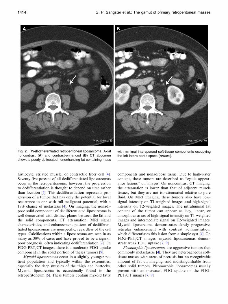
histiocyte, striated muscle, or contractile fiber cell [4].Seventy-five percent of all dedifferentiated liposarcomasoccur in the retroperitoneum; however, the progressionto dedifferentiation is thought to depend on time ratherthan location [5]. This dedifferentiation represents pro-gression of a tumor that has only the potential for localrecurrence to one with full malignant potential, with a17% chance of metastasis [4]. On imaging, the nonadi-pose solid component of dedifferentiated liposarcoma iswell demarcated with distinct planes between the fat andthe solid components. CT attenuation, MRI signalcharacteristics, and enhancement pattern of dedifferen-tiated liposarcomas are nonspecific, regardless of the celltypes. Calcifications within a liposarcoma are seen in asmany as 30% of cases and have proved to be a sign ofpoor prognosis, often indicating dedifferentiation [2]. OnFDG-PET/CT images, there is a moderate FDG uptakecomponent in the solid portion of theses tumors [9].
Myxoid liposarcomas occur in a slightly younger pa-tient population and typically within the extremities,especially the deep muscles of the thigh and buttocks.Myxoid liposarcoma is occasionally found in theretroperitoneum [5]. These tumors contain myxoid fatty
components and nonadipose tissue. Due to high-watercontent, these tumors are described as ‘‘cystic appear-ance lesions’’ on images. On noncontrast CT imaging,the attenuation is lower than that of adjacent muscletissues, but they are not iso-attenuated relative to purefluid. On MRI imaging, these tumors also have low-signal intensity on T1-weighted images and high-signalintensity on T2-weighted images. The intraluminal fatcontent of the tumor can appear as lacy, linear, oramorphous areas of high-signal intensity on T1-weightedimages and intermediate signal on T2-weighted images.Myxoid liposarcoma demonstrates slowly progressive,reticular enhancement with contrast administration,which differentiates this lesion from a simple cyst [4]. OnFDG-PET/CT images, myxoid liposarcomas demon-strate weak FDG uptake [7, 9].
Pleomorphic liposarcomas are aggressive tumors thatcommonly metastasize [4]. They are heterogeneous soft-tissue masses with areas of necrosis but no recognizableamount of fat on imaging, and indistinguishable fromother solid tumors. Pleomorphic liposarcomas usuallypresent with an increased FDG uptake on the FDG-PET/CT images [7, 9].
Fig. 2. Well-differentiated retroperitoneal liposarcoma. Axialnoncontrast (A) and contrast-enhanced (B) CT abdomenshows a poorly delineated nonenhancing fat-containing mass
with minimal interspersed soft-tissue components occupyingthe left latero-aortic space (arrows).
1414 G. P. Sangster et al.: The gamut of primary retroperitoneal masses

Leiomyosarcomas
Leiomyosarcomas are the second most frequent primaryretroperitoneal tumor (28%) and are most commonlyobserved in women between the fifth and sixth decades[2, 4]. They arise from the smooth muscle tissue withinthe retroperitoneum, walls of retroperitoneal veins, orfrom embryonic Wolffian remnants [7]. These tumorscan grow to a large size before compromising adjacentorgans and precipitating clinical symptoms such as ve-nous thrombosis [2]. Approximately, 40% of patientshave metastases to liver, lungs, or lymph nodes at thetime of diagnosis, which occur late in the course of thedisease [2, 4]. Three major growth patterns have been
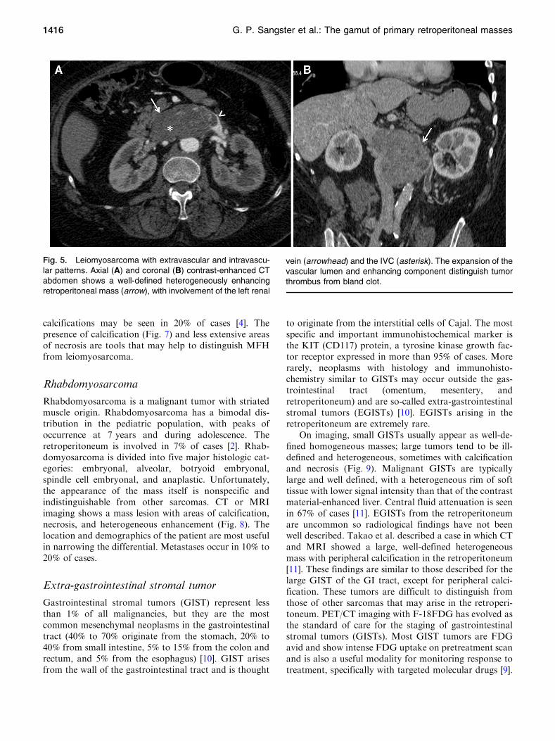
identified: 62% are completely extravascular and extra-luminal (Fig. 3), 33% are extravascular and intravascular(Figs. 4, 5), and only 5% are completely intravascularand intraluminal (Fig. 6) [4, 7]. These tumors can belarger than 10 cm and may contain extensive areas ofnecrosis with occasional hemorrhage. Calcifications areuncommon. [2]. The diagnosis of leiomyosarcoma shouldbe suspected when there is a large retroperitoneal masswith necrosis and contiguous involvement of a vessel [2].At CT, small tumors may be homogeneously solid, butlarge tumors are heterogeneous due to intralesionalnecrosis and hemorrhage. At MRI imaging, these tumorshave intermediate- to low-signal intensity on T1-weigh-ted images and intermediate- to high-signal intensity onT2-weighted images, depending on the amount ofnecrosis. Contrast enhancement is often heterogeneousand predominantly peripheral. Rarely, leiomyosarcomamay appear as mostly cystic.
Malignant fibrous histiocytoma (MFH)
MFH is the third most common primary retroperitonealtumor (19%) and overall the most common soft-tissuesarcoma in the body [2, 7]. After the extremities, theretroperitoneal space is the second most common site forthis tumor (15% of these tumors occur in the retroperi-toneum). MFH arises from undifferentiated mesenchy-mal cells, and it is more common in males in their fifthand sixth decades of life. Retroperitoneal MFHs aredifficult to diagnose preoperatively because CT and MRIimaging appearances are nonspecific. Images demon-strate a large, infiltrating, and heterogeneously enhanc-ing soft-tissue mass with areas of necrosis andhemorrhage and with invasion of adjacent organs [2].Contrast enhancement is typically heterogeneous, and
Fig. 4. Leiomyosarcoma with extravascular and intravascu-lar growth. A Axial contrast-enhanced CT abdomen shows awell-defined lobulated enhancing mass (arrow) arising fromthe inferior vena cava (IVC) (asterisk). Intralesional hetero-
geneity is secondary to tumor necrosis and hemorrhage. BPostsurgical contrast CT of the abdomen demonstratescomplete tumoral excision with IVC graft placement (arrow-head).
Fig. 3. Extravascular leiomyosarcoma. Axial contrast-en-hanced CT abdomen demonstrates a well-defined lobulatedsoft-tissue mass in the anterior pararenal space (arrow). Thislesion produces compression and displacement of the pan-creatic tail.
G. P. Sangster et al.: The gamut of primary retroperitoneal masses 1415
calcifications may be seen in 20% of cases [4]. Thepresence of calcification (Fig. 7) and less extensive areasof necrosis are tools that may help to distinguish MFHfrom leiomyosarcoma.
Rhabdomyosarcoma
Rhabdomyosarcoma is a malignant tumor with striatedmuscle origin. Rhabdomyosarcoma has a bimodal dis-tribution in the pediatric population, with peaks ofoccurrence at 7 years and during adolescence. Theretroperitoneum is involved in 7% of cases [2]. Rhab-domyosarcoma is divided into five major histologic cat-egories: embryonal, alveolar, botryoid embryonal,spindle cell embryonal, and anaplastic. Unfortunately,the appearance of the mass itself is nonspecific andindistinguishable from other sarcomas. CT or MRIimaging shows a mass lesion with areas of calcification,necrosis, and heterogeneous enhancement (Fig. 8). Thelocation and demographics of the patient are most usefulin narrowing the differential. Metastases occur in 10% to20% of cases.
Extra-gastrointestinal stromal tumor
Gastrointestinal stromal tumors (GIST) represent lessthan 1% of all malignancies, but they are the mostcommon mesenchymal neoplasms in the gastrointestinaltract (40% to 70% originate from the stomach, 20% to40% from small intestine, 5% to 15% from the colon andrectum, and 5% from the esophagus) [10]. GIST arisesfrom the wall of the gastrointestinal tract and is thought
to originate from the interstitial cells of Cajal. The mostspecific and important immunohistochemical marker isthe KIT (CD117) protein, a tyrosine kinase growth fac-tor receptor expressed in more than 95% of cases. Morerarely, neoplasms with histology and immunohisto-chemistry similar to GISTs may occur outside the gas-trointestinal tract (omentum, mesentery, andretroperitoneum) and are so-called extra-gastrointestinalstromal tumors (EGISTs) [10]. EGISTs arising in theretroperitoneum are extremely rare.
On imaging, small GISTs usually appear as well-de-fined homogeneous masses; large tumors tend to be ill-defined and heterogeneous, sometimes with calcificationand necrosis (Fig. 9). Malignant GISTs are typicallylarge and well defined, with a heterogeneous rim of softtissue with lower signal intensity than that of the contrastmaterial-enhanced liver. Central fluid attenuation is seenin 67% of cases [11]. EGISTs from the retroperitoneumare uncommon so radiological findings have not beenwell described. Takao et al. described a case in which CTand MRI showed a large, well-defined heterogeneousmass with peripheral calcification in the retroperitoneum[11]. These findings are similar to those described for thelarge GIST of the GI tract, except for peripheral calci-fication. These tumors are difficult to distinguish fromthose of other sarcomas that may arise in the retroperi-toneum. PET/CT imaging with F-18FDG has evolved asthe standard of care for the staging of gastrointestinalstromal tumors (GISTs). Most GIST tumors are FDGavid and show intense FDG uptake on pretreatment scanand is also a useful modality for monitoring response totreatment, specifically with targeted molecular drugs [9].
Fig. 5. Leiomyosarcoma with extravascular and intravascu-lar patterns. Axial (A) and coronal (B) contrast-enhanced CTabdomen shows a well-defined heterogeneously enhancingretroperitoneal mass (arrow), with involvement of the left renal
vein (arrowhead) and the IVC (asterisk). The expansion of thevascular lumen and enhancing component distinguish tumorthrombus from bland clot.
1416 G. P. Sangster et al.: The gamut of primary retroperitoneal masses
Lipoma
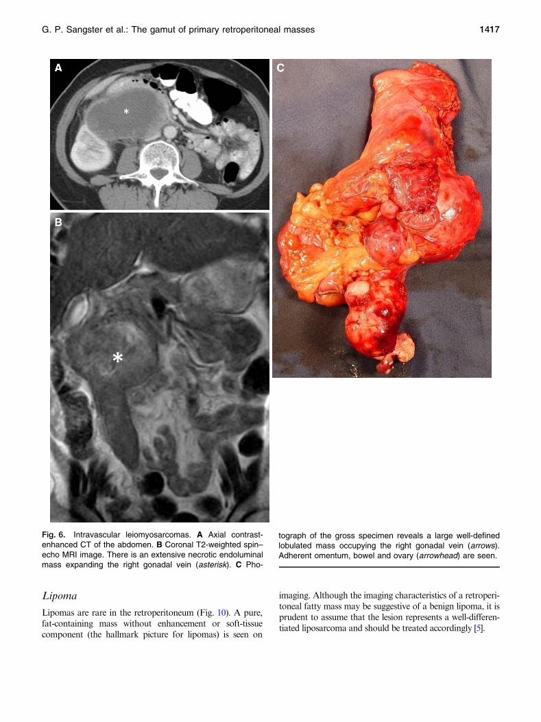
Lipomas are rare in the retroperitoneum (Fig. 10). A pure,fat-containing mass without enhancement or soft-tissuecomponent (the hallmark picture for lipomas) is seen on
imaging. Although the imaging characteristics of a retroperi-toneal fatty mass may be suggestive of a benign lipoma, it isprudent to assume that the lesion represents a well-differen-tiated liposarcoma and should be treated accordingly [5].
Fig. 6. Intravascular leiomyosarcomas. A Axial contrast-enhanced CT of the abdomen. B Coronal T2-weighted spin–echo MRI image. There is an extensive necrotic endoluminalmass expanding the right gonadal vein (asterisk). C Pho-
tograph of the gross specimen reveals a large well-definedlobulated mass occupying the right gonadal vein (arrows).Adherent omentum, bowel and ovary (arrowhead) are seen.
G. P. Sangster et al.: The gamut of primary retroperitoneal masses 1417

Leiomyoma
Commonly encountered in the uterus, leiomyomas rarelyarise primarily from the retroperitoneum, yet occur al-most exclusively in women. Many of these tumors his-
tologically resemble uterine leiomyomas, have a lowpotential for recurrence and a good prognosis. Imagingfindings are nonspecific and diagnosis is histopathologi-cal.
Angioleiomyoma (Fig. 11), also known as vascularleiomyoma, is an uncommon type of leiomyoma thatoriginates from smooth muscle cells and contains thick-walled vessels. There are only a few cases of retroperi-toneal angioleiomyoma reported in the literature. Sincethey are very uncommon, imaging characteristics ofretroperitoneal angioleiomyomas are not well known.Uterine angioleiomyoma shows the areas of cysticdegeneration and prominent tortuous vascular-likeenhancing structures. Thus, angioleiomyoma should beincluded in the differential diagnosis when a retroperi-toneal mass contains cystic components and prominentvascularization [12].
Neurogenic tumors
Neurogenic tumors constitute 10% to 20% of primaryretroperitoneal tumors (Table 4) [1], occur in a youngerage group than sarcomas, and are usually benign. Thesetumors can be classified as ganglion cell origin, para-ganglionic system origin (pheochromocytomas, para-gangliomas), or nerve sheath origin (neurilemmomas,neurofibromas, neurofibromatosis, and malignantnerve sheath tumors). Calcifications can occur in alltypes of neurogenic tumors [9]. Neurogenic tumors arisefrom sympathetic ganglia in the paraspinal region,
Fig. 7. Malignant fibrous histiocytoma. A Axial contrast-en-hanced CT of the abdomen shows a large lobulated hetero-geneous soft-tissue mass with areas of necrosis and coarseamorphous calcifications (arrow) located in the anterior para-renal space. This mass produces displacement of the bowelwithout mechanical obstruction. B Cut surface photograph ofthe gross specimen revealed fleshy areas mixed with necroticfoci. Focal areas of calcifications are noted (arrow).
Fig. 8. Alveolar rhabdomyosarcoma. Axial contrast-en-hanced CT of the abdomen demonstrates an anterior para-renal space lobulated solid mass with areas of necrosis andheterogeneous enhancement (asterisk). The mass com-presses the right kidney causing moderate hydronephrosis(arrow). The kidney shows delayed enhancement secondaryto ischemic vascular defect due to parenchymal compression.
1418 G. P. Sangster et al.: The gamut of primary retroperitoneal masses

adrenal medulla, or organ of Zuckerkandl, a body de-rived from neural crest located at the bifurcation of theaorta or at the origin of the inferior mesenteric artery.Occasionally neurogenic neoplasms originate from theurinary bladder, bowel wall, abdominal wall, and gall-bladder [2, 9].
Tumors of ganglion cell origin
Tumors of ganglion cell origin include ganglioneuroma(benign), neuroblastoma (malignant), and ganglioneu-roblastoma (intermediate). The adrenal gland is the mostcommon primary site of involvement for these tumors.Neuroblastoma and ganglioneuroblastoma most oftenoccur in infants and children, whereas ganglioneuromatends to occur in adolescents and young adults [13].
Ganglioneuroma is a rare benign tumor that arisesfrom the sympathetic ganglia. These tumors are com-posed entirely of ganglion cells and Schwannian stromaand do not contain neuroblasts, intermediate cells, ormitotic figures [13]. Malignant transformation is rare andthe prognosis is excellent [1]. However, there are rarereports of metastasis [14]. This tumor is commonly seenin the 20- to 40-year-old group, with no sex predilection[2]. The retroperitoneum (32% to 52% of cases) andmediastinum (39% to 43% of cases) are the most com-mon sites for ganglioneuroma, followed by the cervicalregion (8% to 9% of cases). In the retroperitoneum, thetumor is commonly seen along the paravertebral sym-pathetic ganglia (59% of cases) or, less commonly, in theadrenal medulla [2]. Ganglioneuromas range in size from5 to 15 cm at presentation. Discrete punctate calcifica-tions are seen in 20% to 30% of ganglioneuromas, unlikethe coarse amorphous calcification of neuroblastomas.Necrosis and hemorrhage are uncommon. In CT, gan-glioneuromas are well-circumscribed oval or lobular,often surrounding adjacent blood vessels without com-pressing the lumen. They are homogeneous and lowattenuation on unenhanced CT scans with delayedheterogeneous enhancement. These features are
bFig. 9. Retroperitoneal benign GIST. A Axial contrast-enhanced CT abdomen shows a well-defined roundedretroperitoneal mass with heterogeneous enhancement dueto central necrosis (arrow). B Photograph of the resectedspecimen of the GIST. There is a solid and partly hemorrhagiclesion in the duodenum wall, with intact and relatively normaloverlying mucosa. C IHC staining demonstrates cellularspindle cell tumor strongly positive with CD117 (209 magni-fication).
G. P. Sangster et al.: The gamut of primary retroperitoneal masses 1419

explained by an abundance of myxoid matrices in thetumors. On MR imaging, a ganglioneuroma is homo-geneously hypointense on T1-weighted images, withvarying signal intensity on T2-weighted images, depend-ing on the myxoid, cellular, and collagen components[2, 5, 13, 14].
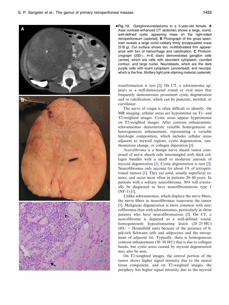
Ganglioneuroblastoma is an intermediate-grade tumorthat has elements of benign ganglioneuroma and malig-nant neuroblastoma. At histologic analysis, they aremalignant tumors that contain primitive neuroblasts andmature ganglion cells. The most common tumor site isthe abdomen, followed by the mediastinum, neck, andlower extremities [13]. Ganglioneuroblastoma is a pedi-atric tumor occurring in the 2- to 4-year-old group, withno sex predilection, and it is rare in adults [2]. Imagingappearances vary ranging from a predominantly solidmass to a predominantly cystic mass with a few thinstrands of solid tissue. These tumors may be partially ortotally encapsulated and frequently contain granularcalcification (Fig. 12) [13].
Neuroblastoma (Fig. 13) is a malignant tumor that ismore commonly seen in males in their first decade of life.Two-thirds of neuroblastomas are located in the adrenalgland, and the remaining occurs along the paravertebral
sympathetic chain [2]. On CT and MR imaging, a neu-roblastoma is irregular, lobulated, and heterogeneous,and demonstrates coarse amorphous calcifications andvariable contrast enhancement. Lesions may invadeadjacent organs and encase vessels with luminal com-pression. Neuroblastomas tend to metastasize to bone,bone marrow, liver, lymph nodes, and skin [12]. As manyas 70% of patients have metastatic disease at the time ofdiagnosis [2]. I-123 metaiodobenzylguanidine (MIBG) isthe radiopharmaceutical used for evaluation of patientsdiagnosed with neuroblastomas. I-123 MIBG uptake canbe seen in the primary tumor and the metastatic sites.
Tumors of the paraganglionic system
The paraganglionic system is composed of neural crestcells, which are found in the adrenal medulla, parasym-pathetic ganglia, and chemoreceptors. Tumors that arisefrom the chromaffin cells of the adrenal medulla arecalled pheochromocytomas, and those that arise in anextra-adrenal location (10%) are referred to as para-gangliomas [2].
The rate of malignancy in paragangliomas ismuch higher, 22% to 50%, compared to 10% for
Fig. 10. Lipoma. Coronal contrast-enhanced CT abdomen(A) FDG-PET images (B) shows a homogeneous, well-cir-cumscribed fat density mass in the right-sided retroperi-toneum (asterisk). Leftward displacement of the duodenum
and ascending colon is seen. FDG-PET image demonstratesa large area of photopenia (black arrow) corresponding to aretroperitoneal lipoma. Photopenia suggests minimal glucosemetabolism in this region.
1420 G. P. Sangster et al.: The gamut of primary retroperitoneal masses
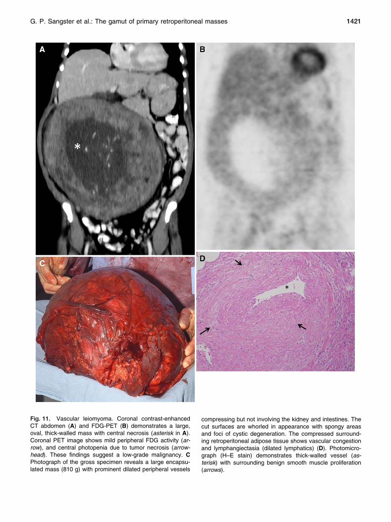
Fig. 11. Vascular leiomyoma. Coronal contrast-enhancedCT abdomen (A) and FDG-PET (B) demonstrates a large,oval, thick-walled mass with central necrosis (asterisk in A).Coronal PET image shows mild peripheral FDG activity (ar-row), and central photopenia due to tumor necrosis (arrow-head). These findings suggest a low-grade malignancy. CPhotograph of the gross specimen reveals a large encapsu-lated mass (810 g) with prominent dilated peripheral vessels
compressing but not involving the kidney and intestines. Thecut surfaces are whorled in appearance with spongy areasand foci of cystic degeneration. The compressed surround-ing retroperitoneal adipose tissue shows vascular congestionand lymphangiectasia (dilated lymphatics) (D). Photomicro-graph (H–E stain) demonstrates thick-walled vessel (as-terisk) with surrounding benign smooth muscle proliferation(arrows).
G. P. Sangster et al.: The gamut of primary retroperitoneal masses 1421

pheochromocytomas [1]. Pheochromocytomas have beencalled ‘‘ten percent tumors,’’ because approximately 10%are bilateral, 10% are extra-adrenal (paragangliomas ofthe retroperitoneum, mediastinum, or urinary bladder),10% occur in children, 10% are familial, 10% are malig-nant, and 10% are not associatedwithhypertension [13]. Inthe retroperitoneum, 40% of paragangliomas producehigh catecholamine levels that result in hypertension andelevated urinary metanephrine or vanillylmandelic acidlevels [2, 5]. Rarely, paraganglioma can manifest withacute abdomen caused by retroperitoneal hemorrhage [2].Paraganglioma can be associated with type 1 neurofibro-matosis, multiple endocrine neoplasia syndrome, and vonHippel–Lindau syndrome. They are commonly seen in thethird to fourth decades of life, with no sex predilection. Inthe retroperitoneum, the most common site for a para-ganglioma is the organ of Zuckerkandl (Fig. 14).
On CT, paragangliomas are usually 3 cm or larger,well-marginated, round, and lobular tumors in a para-aortic location. They are usually highly vascular tumorsdemonstrating intense early enhancement (Fig. 15).Central necrosis occurs in approximately 40%, and(Fig. 16) punctate calcification can be seen in approxi-mately 15% [1]. Malignant lesions tend to be larger withmore irregular margins and more extensive necrosis. Ifno lesion is found or identified on CT and laboratory testresults remain positive, radionuclide imaging withmetaiodobenzylguanidine is suggested to identify theoccult lesion [13]. On MR imaging, they are heteroge-neously bright on T2-weighted imaging, though less than
80% of paragangliomas show the characteristic uniformhigh signal on T2-weighted imaging (caused by thehemorrhage within the tumor). Hemorrhagic portionscan be bright on T1-weighted sequences, and fluid–fluidlevels may be seen [15].
Tumors of nerve sheath origin
Tumors of nerve sheath origin include schwannoma (alsoknown as neurilemoma), neurofibroma, neurofibro-matosis, and neurogenic sarcoma (malignant schwan-noma). More than 90% of these tumors are benign. Thebenign lesions are identified in young and middle-agedadults [13].
Schwannoma, or neurilemoma, is a benign tumor thatarises from the perineural sheath of Schwann (neur-ilemma) [2]. Schwannoma accounts for 6% of retroperi-toneal neoplasms, comprising approximately 3% of allschwannomas [2, 16]. They are usually asymptomaticand are more common in females (2:1), particularly inthe 20-to 50-year-old age group [2]. Retroperitonealschwannomas are usually solid encapsulated tumors thatare commonly located in the paravertebral region and,less commonly, adjacent to the kidney, pre-sacral space,and abdominal wall [2]. They are usually larger and havea higher tendency to undergo spontaneous degenerationand hemorrhage compared with their counterparts aris-ing in the head and neck or extremities, which are usuallysolid and small [16]. These tumors are usually asymp-tomatic and are thought to be slow growing. Malignant
Table 4. Neurogenic tumors: characteristic imaging findings and other diagnosis features
Neurogenic tumors Image finding Other diagnosis features
Ganglioneuroma Homogeneous paravertebral mass.Extension along normal structures.Delayed enhancement
20–40 years age group, with no sexpredilection
Ganglioneuroblastoma Vary ranging from a predominantly solidmass to a predominantly cystic mass.Encapsulated. May contain granularcalcification
2–4 years age group, with no sexpredilection
Neuroblastoma Irregular, lobulated, heterogeneous. 85%calcifications (coarse, amorphous,mottled). Variable contrast enhance-ment
Males, 1st decade of life
Paragangliomas Paravertebral mass, intense earlyenhancement. 40% central necrosis.15% punctate calcification. Fluid–fluidlevel
High catecholamine levels, hypertension
Schwannomas Oval mass that frequently demonstratesprominent cystic degeneration andcalcification
Female predilection. 20–50 years. Encap-sulated. Compression of nerve to oneside
Neurofibroma Well-circumscribed, round mass withhomogeneous contrast enhancement.Occasionally, myxoid degeneration
Male predilection. 20–40 years (younger inNF1). Usually unencapsulated. Diffuseexpansion of the entire nerve
Plexiform neurofibroma Bilaterally symmetric, elongated para-psoas masses with homogeneous lowattenuation
Is characteristic of NF-1
1422 G. P. Sangster et al.: The gamut of primary retroperitoneal masses
transformation is rare [2]. On CT, a schwannoma ap-pears as a well-demarcated round or oval mass thatfrequently demonstrates prominent cystic degenerationand or calcification, which can be punctate, mottled, orcurvilinear.
The nerve of origin is often difficult to identify. OnMR imaging, cellular areas are hypointense on T1- andT2-weighted images. Cystic areas appear hyperintenseon T2-weighted images. After contrast enhancement,schwannomas demonstrate variable homogeneous orheterogeneous enhancement, representing a variablehistologic composition, which includes cellular areasadjacent to myxoid regions, cystic degeneration, xan-thomatous change, or collagen deposition [1].
Neurofibroma is a benign nerve sheath tumor com-posed of nerve sheath cells intermingled with thick col-lagen bundles with a small to moderate amount ofmyxoid degeneration [1]. Cystic degeneration is rare [2].Neurofibromas only account for about 1% of retroperi-toneal tumors [1]. They are solid, usually superficial tu-mors, and occur most often in patients 20–40 years. Inpatients with a solitary neurofibroma, 30% will eventu-ally be diagnosed to have neurofibromatosis type 1(NF-1) [1].
Unlike schwannomas, which displace the nerve fibers,the nerve fibers in neurofibromas transverse the tumor[1]. Malignant degeneration is more common with neu-rofibromas than with schwannomas, particularly in thosepatients who have neurofibromatosis [2]. On CT, aneurofibroma is depicted as a well-defined round,homogeneously hypoattenuating lesion (20–25 HU)(HU = Hounsfield unit) because of the presence of li-pid-rich Schwann cells and adipocytes and the entrap-ment of adjacent fat. Typically, there is homogeneouscontrast enhancement (30–50 HU) that is due to collagenbands, but cystic areas caused by myxoid degenerationmay also be seen.
On T1-weighted images, the central portion of thetumor shows higher signal intensity due to the neuraltissue component, and on T2-weighted images, theperiphery has higher signal intensity due to the myxoid
bFig. 12. Ganglioneuroblastoma in a 3-year-old female. AAxial contrast-enhanced CT abdomen shows a large, round,well-defined cystic appearing mass on the right-sidedretroperitoneum (asterisk). B Photograph of the gross speci-men reveals a large ovoid rubbery thinly encapsulated mass(518 g). Cut surface shows tan, multilobulated firm appear-ance with foci of hemorrhage and calcification. C Photomi-crograph (2009; H–E stain) demonstrates ganglion cells(arrow), which are cells with abundant cytoplasm, roundedcontour, and large nuclei. Neuroblasts, which are the darkpurple cells with scant cytoplasm (arrowhead), and neuropil,which is the fine, fibrillary light pink-staining material (asterisk).
G. P. Sangster et al.: The gamut of primary retroperitoneal masses 1423

degeneration. Tumors involving the neural foramen havea dumbbell shape with expansion of the bone foraminaor vertebral body scalloping.
Plexiform neurofibroma (PN) is an interdigitatingnetwork of fronds of tumor occurring along a nerve andits branches. PN is characteristic of NF-1, and fulfills oneof the two criteria required for the diagnosis of NF-1(nearly all patients with PN will have NF-1) [18]. Al-though PN can occur in a variety of locations within thebody, the involvement of the retroperitoneal spaceand pelvis is uncommon [17]. Unlike most primary
retroperitoneal neoplasms, the CT features of most PNare sufficiently characteristic that a specific diagnosisusually can be made without invasive procedures [18].CT demonstration of bilaterally symmetric, elongatedpara-psoas masses with homogeneous low attenuationindicates a benign retroperitoneal PN. If not clinicallyknown, the diagnosis of NF-1 should be suggested [18].When bilateral lesions are present, asymmetry in axialdiameter and an asymmetric attenuation pattern shouldraise the suspicion of malignant transformation. If thebilateral para-psoas masses differ in size more than 2 cmor attenuation on CT, the larger lesion might represent amalignant change and should be biopsied [17]. F-18FDG-PET/CT has been shown to be very sensitive and
Fig. 14. Paraganglioma. A Axial unenhanced CT abdomenshows a well-marginated, low density tumor (arrow) withintralesional punctate calcifications in the left para-aorticspace consistent with a paraganglioma in the organ ofZuckerkandl. B Axial contrast-enhanced CT image showstypical intense early enhancement of the mass.
Fig. 13. Neuroblastoma in a 6-year-old male. Axial (A) andcoronal (B) contrast-enhanced CT of the abdomen demon-strates a lobulated heterogeneous soft-tissue mass with areasof necrosis in the right perinephric space (arrows). There isalso coarse amorphous calcifications (arrowhead) and variablecontrast enhancement. The lesion produces compression andinferior displacement of the ipsilateral kidney.
1424 G. P. Sangster et al.: The gamut of primary retroperitoneal masses
specific imaging modality for the diagnosis of malignantperipheral nerve sheath tumors in patients with neu-rofibromatosis type 1 [9].
On MRI, PN are seen as low-signal intensity lesionson T1-weighted images with a signal intensity slightlygreater than the surrounding muscles. On T2-weightedimages they are markedly hyperintense. Multiple hy-pointense septations can be seen within the tumor on T2-weighted images [17].
Malignant nerve sheath tumors (MNST) (Fig. 17) arerare tumors that account for approximately 3% to 10% ofall soft-tissue sarcomas [19]. They are aggressive high-grade neoplasms with a poor prognosis [4].MNST includemalignant schwannoma, neurogenic sarcoma, and neu-rofibrosarcoma. Fifty percent (50%) of these tumorsoriginate de novo, and the rest of them are derived fromneurofibroma or ganglioneuroma or occur after exposureto radiation [2]. They have a tendency to recur locally, anddistant metastases occur at a rate of 65% [4]. MNST aremore common in the 20- to 50-year-old group, with no sexpredilection [2]. In 25% to 50% of cases, these tumors areassociatedwithNF-1.Having anNF-1 is considered as themost important risk factor for developing malignantperipheral nerve sheath tumors [19]. Imaging criteria areunreliable in differentiating malignant from benign nervesheath tumors [13]. On imaging, malignant nerve sheathtumors are vascular masses closely associated with neu-rovascular bundles. Useful characteristics for distin-guishing between malignant peripheral nerve sheathtumors and neurofibromas include increased largestdimension of the mass, presence of peripheral enhancedpattern, presence of peri-lesional edema like zone, andpresence of intra-tumoral cystic lesions. The presence oftwo to four of these features is suggestive of malignancy(sensitivity, 61%; specificity, 90%) [19].
Germ cell, sex cord, and stromal celltumors
Extragonadal germ cell tumor
Although germ cell tumors are seen most commonly inthe testes or ovaries, 1% to 2.5% of germ cell tumorsoriginate in an extragonadal location (Table 5) [2]. Pri-mary extragonadal germ cell tumors (PEGCT) are rare,accounting for approximately 5% of all primaryretroperitoneal tumors. This is the second most commonsite for PEGCT after the mediastinum [4]. They almostexclusively occur in males with peak incidence in the fifth
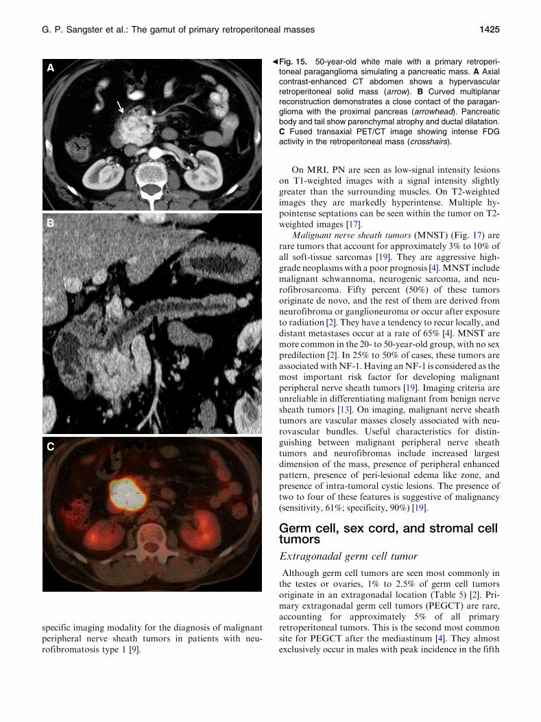
bFig. 15. 50-year-old white male with a primary retroperi-toneal paraganglioma simulating a pancreatic mass. A Axialcontrast-enhanced CT abdomen shows a hypervascularretroperitoneal solid mass (arrow). B Curved multiplanarreconstruction demonstrates a close contact of the paragan-glioma with the proximal pancreas (arrowhead). Pancreaticbody and tail show parenchymal atrophy and ductal dilatation.C Fused transaxial PET/CT image showing intense FDGactivity in the retroperitoneal mass (crosshairs).
G. P. Sangster et al.: The gamut of primary retroperitoneal masses 1425

decade of life [4]. They are believed to arise from pri-mordial germ cell rests that have failed to migrate duringthe fourth to sixth week of embryonic development [20].
The majority of retroperitoneal germ cell tumorsrepresents metastases from primary testicular neoplasms(retroperitoneal metastasis is seen in 30% of gonadaltumors, and therefore, careful examination of the testesis crucial). The basic histologic types are seminoma;embryonal carcinoma; choriocarcinoma; teratoma with
mature, immature, or malignant foci; and yolk sac tu-mor. Elevated levels of alpha-fetoprotein are seen inembryonal carcinoma and yolk sac tumor, whereas ele-vated levels of the beta subunit of human chorionic go-nadotropin are seen in choriocarcinoma [4].
On imaging, the findings for PEGCT are nonspecific.Retroperitoneal germ cell tumors typically appear as alarge midline lobular mass of heterogeneous CT attenu-ation or MR signal intensity. A midline mass is moresuggestive of a PEGCT than of metastasis [2]. Semino-mas and embryonal carcinomas may appear homoge-nous. Areas of hemorrhage and necrosis may also beseen, especially with nonseminomatous germ cell tumors[4]. Calcifications, tooth-like or well-defined, and fat canbe seen very frequently in teratomas [2]. A pathog-nomonic finding in teratomas is the presence of a fat-fluid level and chemical shift between fat and fluid [2].
Fig. 16. Atypical retroperitoneal paraganglioma. A Axial andB sagittal contrast-enhanced CT abdomen show a well-mar-ginated hypodense lesion with central necrosis (asterisk) lo-cated in the retroperitoneum between de aorta, superiormesenteric artery and the IVC.
Fig. 17. A 29-year-old African–American female withmalignant peripheral nerve sheath tumor (MPNST). Axialcontrast-enhanced CT abdomen shows (A) a large, oval, well-defined necrotic mass located in the left perinephric spaceconsistent with MPNST (asterisk). B Bilateral and symmetriclow-attenuation masses are noted in psoas regions consistentwith benign peripheral nerve sheath tumor (arrows).
1426 G. P. Sangster et al.: The gamut of primary retroperitoneal masses

Primary sex cord stromal tumors
These tumors are extremely rare and are seen in womenover a wide age range (30–76 years). Extra-ovarian pri-mary sex cord stromal tumors arise from ectopic sex cordstromal tissues or from the sex cord—like the differen-tiation of somatic cells. These tumors are more com-monly seen in the pelvis along the broad ligament orfallopian tubes and, less commonly, in the retroperi-toneum or adrenal glands. Most of these tumors aregranulosa cell tumors [2].
Adult granulosa cell tumors (Fig. 18) are estrogenproducing tumors that occur predominantly in peri- andpostmenopausal women. Granulosa cell tumors can arisein locations other than the ovary and may be derivedfrom the mesenchyme of the genital ridge. Women whohave undergone oophorectomy may have the potential todevelop granulosa cell tumors. The locations of thesetumors have been described as the broad ligament,retroperitoneum, and adrenal glands. The imaging find-ings are nonspecific. CT and MR images show hetero-geneous solid tumors, with heterogeneous enhancement[2, 21].
Lymphomas
Lymphomas constitute roughly one-third of all primaryretroperitoneal neoplasms. Hodgkin’s lymphoma (HD)has a bimodal age distribution and peaks in the early 20s.It usually presents with limited disease spread (stage I orII). Extranodal sites are usually not involved. Non-Hodgkin’s lymphoma (NHL) presents in patients 40–70 years of age, often already in an advanced diseasestage (stage III or IV) upon diagnosis. Extranodal dis-ease in NHL is much more common than in HD. Para-aortic lymph nodes are involved in 25% of patients withHD and 55% of patients with NHL. Bone marrowinvolvement at presentation occurs in more than 40% ofpatients with NHL. On imaging, a lymphoma typically
appears as a well-defined homogeneous and mildlyenhancing para-aortic or pelvic mass that extends be-tween structures without compressing them. Necrosisand calcification are very uncommon [1, 4]. Intenselyincreased FDG uptake on the PET-FDG scan is noted inthe areas of the pelvic and retroperitoneal lymphaticpathways, which correlates with areas of lym-phadenopathy evident on CT scan [9].
Primary non-neoplasticretroperitoneal lesions
Retroperitoneal fibrosis
Retroperitoneal fibrosis (RF) encompasses a range ofdiseases characterized by proliferation of aberrant fibro-inflammatory tissue, which usually surrounds the infra-renal portion of the abdominal aorta, inferior vena cava,and iliac vessels. It may extend to neighboring structures.In about 56% to 100% of patients with idiopathic RF,the fibro-inflammatory tissues entrap the ureters andcause obstructive uropathy and subsequent renal failure.
Ureteral involvement is most often bilateral and leadsto renal failure. RF is typically idiopathic (>70% ofcases). Although in the rest of the cases (<30%), RF isfound to be secondary to factors that include drug use(derivatives of ergot alkaloids), malignancies (lym-phoma, RS, carcinoid tumor, and metastatic diseasefrom primary cancers of the stomach, colon, breast, lung,genitourinary tract, or thyroid gland), infections (histo-plasmosis, tuberculosis, actinomycosis), radiation ther-apy, major trauma, major abdominal surgeries,retroperitoneal hemorrhage or hematoma, and prolifer-ative diseases (Erdheim–Chester disease and other histi-ocytoses). Although retroperitoneal fibrosis manifests asan isolated retroperitoneal disease, it can also be asso-ciated with other fibrosing conditions, such as sclerosingcholangitis, Riedel thyroiditis, fibrotic pseudotumor ofthe orbit, and autoimmune pancreatitis, all of which
Table 5. Extragonadal germ cell tumors: characteristic imaging findings and other diagnosis features
Extragonadal germ cell tumors
Image finding Other diagnosis features
� Large midline lobular masses of heterogeneous CT attenuation orMR signal intensity
� Elevated levels of alpha-fetoprotein are seen in embryonal carcinomaand yolk sac tumor
� Seminomas and embryonal carcinomas may appear homogenous � Elevated levels of the beta subunit of human chorionic gonadotropinare seen in choriocarcinoma
� Areas of hemorrhage and necrosis may be seen, especially withnonseminomatous germ cell tumors
� The majority of retroperitoneal germ cell tumors represent metas-tases from primary testicular neoplasms
� Calcification (tooth-like or well defined) and fat can be seen in 56%and 93% of teratomas
� Elevated levels of alpha-fetoprotein are seen in embryonal carcinomaand yolk sac tumor
� A fat-fluid (sebum) level and chemical shift between fat and fluid arepathognomonic for teratomas
� Elevated levels of the beta subunit of human chorionic gonadotropinare seen in choriocarcinoma
� Large midline lobular masses of heterogeneous CT attenuation orMR signal intensity
� Seminomas and embryonal carcinomas may appear homogenous
G. P. Sangster et al.: The gamut of primary retroperitoneal masses 1427

form part of the group labeled ‘‘multifocal fibrosclero-sis.’’ If promptly diagnosed and treated, idiopathic andmost other benign forms of RF have a good prognosis.In contrast, malignant RF, which accounts for up to 10%of cases, has a poor prognosis. Idiopathic RF is more
common in males (3:1), particularly in the 40- to 60-year-old group [2, 22].
RF consist of a well-delimited but irregular soft-tissueperi-aortic mass that extends from the level of the renalarteries to the iliac vessels and often progresses through the
Fig. 18. Adult granulosa cell tumor. A Coronal contrast-en-hanced CT abdomen shows a round, well-defined mass withheterogeneous enhancement, located in the left perinephricspace (asterisk). This lesion produces compression of the leftureter causing proximal hydronephrosis (arrow). B Pho-tograph of the gross specimen reveals a circumscribed thinlyencapsulated mass with fleshy solid areas, foci of cystic
degeneration, and yellow areas of necrosis. C Photomicro-graph (H&E, 2009) shows sheets of round to oval tumor cellswith Call–Exner bodies (this is formed by a discrete array ofgranulosa cells with angular grooved nuclei and centralinspissated eosinophilic material). D Tumor cells are stronglypositive for estrogen receptor.
1428 G. P. Sangster et al.: The gamut of primary retroperitoneal masses
retroperitoneum to envelop the ureters and inferior venacava (Fig. 19). The mass usually lies anterior and lateral tothe aorta, so it does not displace the aorta and IVCanteriorly, as lymphoma or metastatic nodes often do.
Avid enhancement is seen in the active stages ofretroperitoneal fibrosis, with little or no enhancement inthe chronic phase. MR imaging shows high-signal intensityon T2-weighted images in the acute phase of the disease,
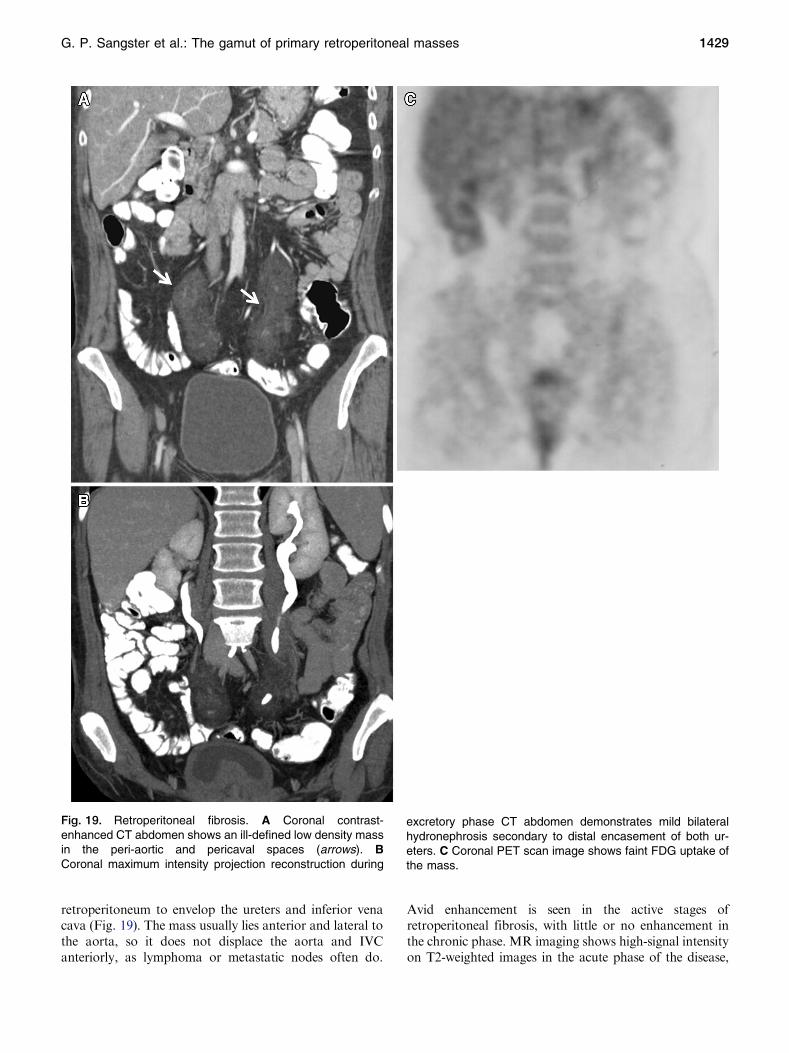
Fig. 19. Retroperitoneal fibrosis. A Coronal contrast-enhanced CT abdomen shows an ill-defined low density massin the peri-aortic and pericaval spaces (arrows). BCoronal maximum intensity projection reconstruction during
excretory phase CT abdomen demonstrates mild bilateralhydronephrosis secondary to distal encasement of both ur-eters. C Coronal PET scan image shows faint FDG uptake ofthe mass.
G. P. Sangster et al.: The gamut of primary retroperitoneal masses 1429

and low-signal intensity in the chronic fibrosing phase. TheFDG-PET scan shows increased uptake of FDG in theretroperitoneal space and is useful for detecting metabolicactivity and distant disease. A lack of radiotracer uptake inthe mass on FDG-PET imaging indicates a metabolicallyinactive disease. Due to its low specificity, 18F-FDG-PETis not useful for diagnosis of idiopathic and other benignforms of RF. Furthermore, FDG uptake in the aortic wallis nonspecific for RF and can also occur in elderly patients,especially those with hyperlipidemia and a history of car-diovascular disease [2, 22].
Conclusions
Primary retroperitoneal neoplasms are notable for theirwidely disparate histologies and presentations. While aspecific diagnosis of a retroperitoneal malignancy can bechallenging due to an overlap of imaging findings, CTand MR imaging can demonstrate important character-istics of these tumors. Combined with clinical anddemographic information, CT and MR imaging mayhelp in narrowing the differential diagnosis.
Compliance with ethical standards
Conflict of Interest Guillermo P. Sangster, Matias Migliaro, MaureenG. Heldmann, Peeyush Bhargava, Alireza Hamidian, and JaiyeolaThomas-Ogunniyi declare that they have no conflict of interest.
Ethical approval All procedures performed in studies involving humanparticipants were in accordance with the ethical standards of theInstitutional and/or National Research Committee and with the 1964Helsinki declaration and its later amendments or comparable ethicalstandards.
References
1. Neville A, Herts BR (2004) CT characteristics of primaryretroperitoneal neoplasms. Crit Rev Comput Tomogr 45(4):247–270
2. Rajiah P, Sinha R, Cuevas C, et al. (2011) Imaging of uncommonretroperitoneal masses. RadioGraphics 31:949–976
3. Francis IR, Cohan RH, Varma DGK, Sondak VV (2005)Retroperitoneal sarcomas. Cancer Imaging 5:89–94
4. Osman S, Lehnert BE, Elojeimy S, et al. (2013) A comprehensivereview of the retroperitoneal anatomy, neoplasms, and pattern ofdisease spread. Curr Probl Diagn Radiol 42(5):191–208
5. Craig WD, Fanburg-Smith JC, Henry LR, Guerrero R, Barton JH(2009) From the archives of the AFIP. Fat-containing lesions of theretroperitoneum: radiologic–pathologic correlation. RadioGraph-ics 29:261–290
6. Nishino M, Hayakawa K, Minami M, et al. (2003) Primaryretroperitoneal neoplasms: CT and mr imaging findings withanatomic and pathologic diagnostic clues. RadioGraphics 23:45–57
7. Scali E, Chandler T, Heffernan E, et al. (2015) Primary retroperi-toneal masses: what is the differential diagnosis? Am J Roentgenol40(6):1887–1903
8. Kim EY, Kim SJ, Choi D, et al. (2008) Recurrence of retroperi-toneal liposarcoma: imaging findings and growth rates at follow-upCT. Am J Roentgenol 191:1841–1846
9. Kitajima K, Kono A, Konishi J, et al. (2013) 18F-FDG-PET/CTfindings of retroperitoneal tumors: a pictorial essay. Jpn J Radiol31:301–309
10. Casella C, Villanacci V, D’Adda F, Codazzi M, Salerni B (2012)Primary extra-gastrointestinal stromal tumor of retroperitoneum.Clin Med Insights 6:189–197
11. Takao H, Yamahira K, Doi I, Watanabe T (2004) Gastrointestinalstromal tumor of the retroperitoneum: CT and MR findings. EurRadiol 14:1926–1929
12. Ozkavukcu E, Aygun S, Erden A, Savas B (2009) Pelvicretroperitoneal angioleiomyoma mimicking a uterine mass. DiagnInterv Radiol 15:262–265
13. Rha SE, Byun JY, Jung SE, et al. (2003) Neurogenic tumors in theabdomen: tumor types and imaging characteristics. RadioGraphics23(1):29–43
14. Lonergan GJ, Schwab CM, Suarez ES, Carlson CL (2002) Neu-roblastoma, ganglioneuroblastoma, and ganglioneuroma: radio-logic–pathologic correlation. RadioGraphics 22:911–934
15. Brennan C, Kajal D, Khalili K, Ghai S (2014) Solid malignantretroperitoneal masses—a pictorial review. Insights Imaging 5:53–65
16. Goh BK, Tan YM, Chung YF, et al. (2006) Retroperitonealschwannoma. Am J Surg 192:14–18
17. Kalra N, Vijayanadh O, Lal A, et al. (2005) Retroperitonealplexiform neurofibroma mimicking psoas abscesses. AustralasRadiol 49:330–332
18. Bass JC, Korobkin M, Francis IR, Ellis JH, Cohan RH (1994)Retroperitoneal plexiform neurofibromas: CT findings. Am JRoentgenol 163:617–620
19. Wasa J, Nishida Y, Tsukushi S, et al. (2010) MRI features in thedifferentiation of malignant peripheral nerve sheath tumors andneurofibromas. Am J Roentgenol 194:1568–1574
20. Ueno T, Tanaka YO, Nagata M, et al. (2004) Spectrum of germ celltumors: from head to toe. RadioGraphics 24:387–404
21. Soydinc HE, Sak ME, Evsen MS, Bozkurt Y, Keles A (2012)Unusual case of extraovarian granulosa cell tumor. Eur Rev MedPharmacol Sci 16(4):30–31
22. Caiafa RO, Vinuesa AS, Izquierdo RS, et al. (2013) Retroperi-toneal fibrosis: role of imaging in diagnosis and follow-up.Radiographics 33:535–552
1430 G. P. Sangster et al.: The gamut of primary retroperitoneal masses