Correlation Between Retinopathy of Prematurity and Levels Of
Upload
mithila-das-mazumderCategory
view
313download
1

Retinopathy of prematurity is a multifactorial
vasoproliferative retinal disorder that
Increases in incidences with decreasing gestational age .
Its a developmental vascular proliferative disorder
that occurs in the incompletely vascularized retina of
primarily premature infants.

• First described by TERRY in 1941
• Originally known as Retrolental fibroplasia
• Term RETINOPATY OF PREMATURITY was
coined by Health in 1951.
• Campbell suggested the relationship of
intensive oxygen therapy & subsequent
development of ROP.
• Kinsey: ROP was inversely proportional to
birth weight.

• Retina is a thin, semitransparent, multilayered sheet of neural tissue that lines the inner aspect of the posterior two-thirds of the wall of the globe.
• It is a thin delicate layer of nervous tissue of surface area 266mm2.
• It extends from optic disc to ora serrata

Retinal Vascularization begins at optic nerve head by 16 weeks of gestation, which migrate from the optic disc
at posterior pole and move towards periphery by 21-22 weeks of gestation.
Before the retinal vessels develops the avascular retina receives it oxygen supply by diffusion across the retina from choroidal vessels.
Choroidal Vascularization begins at 6th weeks of gestation and it is completed by 21 weeks

Retinal vessels grow out of the optic disc as a
wave of mesenchymal spindle cells.
Mesenchymal spindle cells lead the shunt,
endothelial proliferation and capillary
formation follow.
These new capillaries will form the mature
retinal vessels.
6 wks- The choroidal vessels supply the rest of
the avascularized retina.
32 Wks- The nasal portion of the retina is
completely vascularized to the ora serrata .
40-42 weeks - The larger temporal area usually
is completed


The onset of ROP progresses in two phases:
PHASE 1 : It involves an initial insult such as hyperoxia , hypoxia
or hypotension at a critical point in retinal vascularization. Which
causes vasoconstriction and decreased blood supply to the
developing retina with subsequent arrest in vascular
development.
The relative hyperoxia after birth is hypothesized to downregulate
the production of growth factor such as vascular endothelial
growth factor (VEGF)essential for normal development of Retinal
vessels.
PHASE 2: during the second phase Neovascularization
occurs. This abberant retinal vessel growth is thought to
be driven by excess angiogenic factors (VGEF) upregulated
by hypoxic avascular retina.
New vessels grow through the retina in to the vitreous .
These vessels are permeable , thus hemorrhage and edema
occurs.
Extensive and severe extraretinal fibrovascular proliferation
can lead to retinal detachment .


ROP pathogenesis and suggested treatments. (a) Retina vessels in the process of their
formation and progressive covering of the retina surface. (b) Hyperoxia at this formative
stage suppresses VEGF and, consequently, results in regression of newly formed
vessels. (c) Upon return to normal air, the ischemic retina upregulates VEGF to high
levels, causing excessive formation of leaky vessels. To antagonize VEGF at this stage
has been suggested as a strategy to reduce adverse vessel formation. (d) An alternative
strategy proposed by Shih et al. (2) is to protect retina vessels from oxygen-induced
obliteration through administration of PlGF-1.

• prematurity / low gestational age < 30 weeks
• Low birth weight
• Hyperoxia / Hypoxia
• High Light exposure
• Vitamin E deficiency
• Postnatal sepsis
• Mechanical ventilation
• Intraventricular haemorrhage
• Apneic attacks
• PDA

• Breast feeding is proctective against development of
ROP.
• Infant born to mother with pregnancy induced
hypertension have a reduced risk of developing ROP
due to accelerated fetal maturity of retinal vessels.

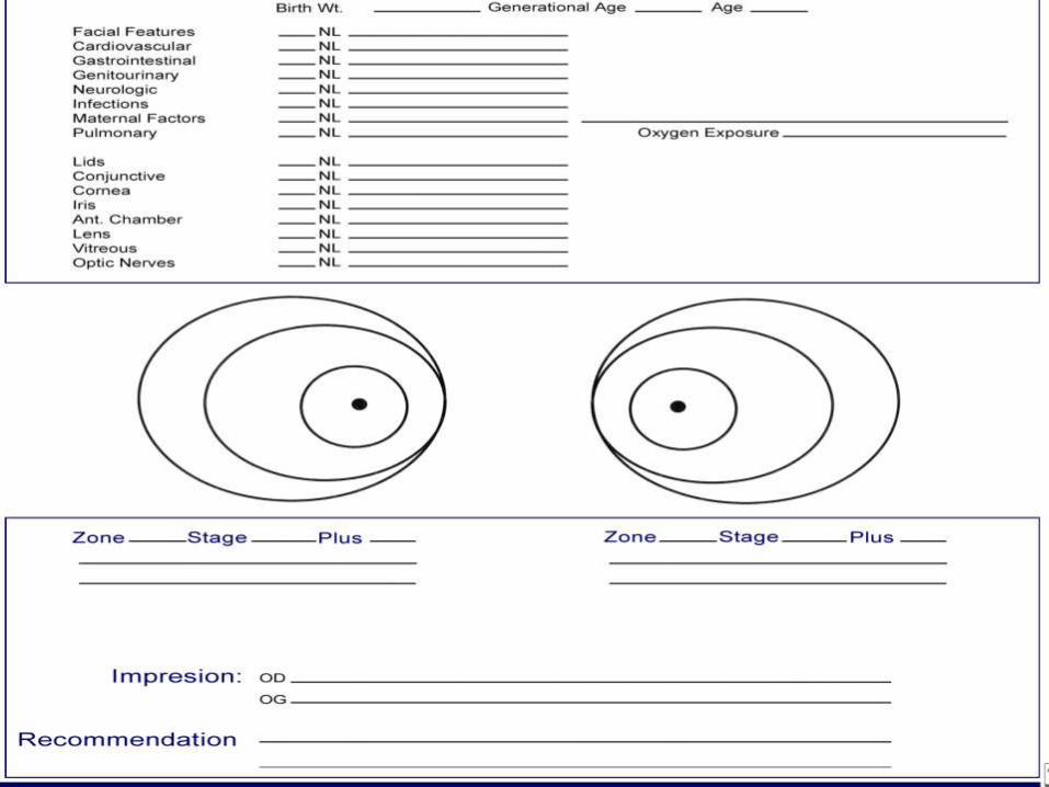
The International Classification of retinopathy of prematurity
(ICROP).
The classification consists of four components : Location
Severity
Extent
Plus Dieasese

Refers to how far the developing retinal vessels have
progressed . The retina is divided into three concentric circles or
zones :

Consist of an imaginary circle with optic
nerve at the centre and a radius is twice
the distance from optic nerve to the macula
.
Extents from the edge of zone 1 to the ora
serrata on the nasal side of the eye and
approximately half the distance to
the ora serrata on the temporal side.
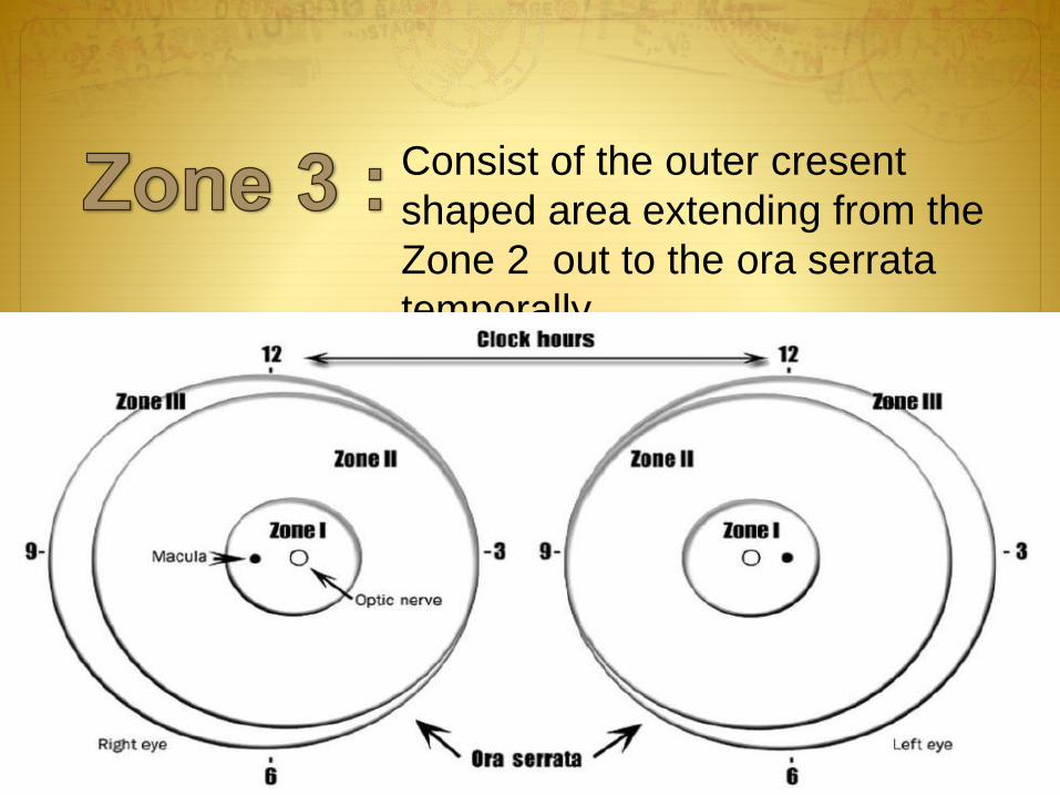
Consist of the outer cresent
shaped area extending from the
Zone 2 out to the ora serrata
temporally.
It refers to the stage of the diesease :
A demarcation line appears as a thin white line that
separates the normal retina from underdeveloped
avascular retina


A ridge of Fibrovascular tissue wight height and
width replaces the demarcation line . It extends
inwards from the plane of retina .
Ridge of fibrovascular tissue

Popcorn Isolated tufts of neovascular tissue
posterior to ridge at the level of retina

The ridge has extraretinal fibrovascular proliferation .
Abnormal blood vessels nd fibrous tissue develop on the
edge of the ridge and extend in to the vitreous.


Partial retinal detachment may result when the
scar tissue pulls on the retina.
-Tractional, as part of the change over from acute to cicatricial disease.
-Rhegmatogenous detachments, years later

STAGE 4 A
Macula Spared
STAGE 4 B
Macula involved

Complete retinal detachment occurs . The retina assmes a
funnel shaped appearance and is described as open or
narrow in the anterior and posterior zone .

REPORTED AS CLOCK HOURS IN THE APPROPRIATE ZONES

• Additional designation that refers to the presence of vascular dilation and arteriolar tortuosity of posterior retinal vessels at least 2 quadrants
• This indicates the severe form of the disease
• It may be associated with Iris vascular engorgement, pupillary rigidity, and vitreous haze.

It’s a vascular abnormalities of the posterior pole more than normal, less than PLUS
The newly accepted preplus serves as a warning

THRESHOLD ROP: • CRYO ROP study
• Zone I stage III with Plus
• Zone II Stage III with Plus
• ( 5 contigous or total 8 clock hours
• Risk of blindness is 50%

PRETHRESHOLD ROP :
• High risk Prethreshold
• Zone I Stage I, II, III with plus
• Stage III without plus
• Zone II Stage II and III with plus

WHOM TO SCREEN ?
• The recommendation is to screen all babies with
a Birth Weight < 1500gm
• Gestational age < 30 weeks
• Babies born after 30 weeks of gestation may be
considered for screening if they have been ill (
RDS, hypotension, surgery ) .

When to screen ?
• Babies born at < 26 of gestation area examined at
the at the postnatal age of 6 weeks .
• Babies born between 27 – 28 weeks of gestation are
examined at the postnatal age of 5 weeks .
• Babies born between 29 – 30 weeks of gestation are
examined at the postnatal age of 4 weeks.
• Babies of gestation >30 weeks are examined at the
postnatal age of 3 weeks.

• Patients are screened every 2 weeks until their
vessels have grown out to the ora Serrata and the
retina is considered mature.
• If ROP is diagnosed the frequency of the
examination depends on the severity and
rapidity of the progression of the disease.

How to screen ?
• The procedure is performed at in NICU by pediatric
opthalmologist , under thesupervision of neonatologist so that
complication can be handled.
• The pupil are dilated with a mixture of phenyl phrine 2.5% and
tropicamide or cyclopentolate 0.5% instilled 3times at 10min
intervals before the scheduled examination.
• Indirect opthalmoscopy is performed with 20D / 30D lens using
fresh sterile instruments.
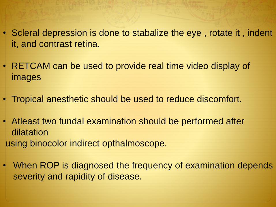
• Scleral depression is done to stabalize the eye , rotate it , indent
it, and contrast retina.
• RETCAM can be used to provide real time video display of
images
• Tropical anesthetic should be used to reduce discomfort.
• Atleast two fundal examination should be performed after
dilatation
using binocolor indirect opthalmoscope.
• When ROP is diagnosed the frequency of examination depends
severity and rapidity of disease.

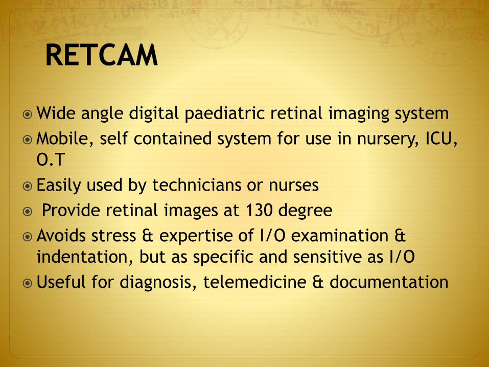
Wide angle digital paediatric retinal imaging system
Mobile, self contained system for use in nursery, ICU,
O.T
Easily used by technicians or nurses
Provide retinal images at 130 degree
Avoids stress & expertise of I/O examination &
indentation, but as specific and sensitive as I/O
Useful for diagnosis, telemedicine & documentation


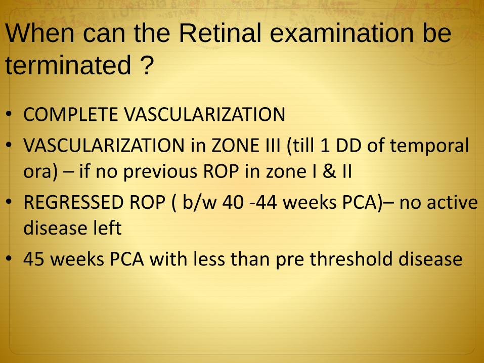
When can the Retinal examination be
terminated ?
• COMPLETE VASCULARIZATION
• VASCULARIZATION in ZONE III (till 1 DD of temporal ora) – if no previous ROP in zone I & II
• REGRESSED ROP ( b/w 40 -44 weeks PCA)– no active disease left
• 45 weeks PCA with less than pre threshold disease

Cryotherapy ( mostly
outdated)
Laser treatment (gold
standard)
Anti-VEGF (adjuvant)
before laser and surgery
Surgery

Cryotherapy significantly improves the outcome of
severe ROP
Superceded by laser photocoagulation.
Cyroprobe is applied to the external surface of the
sclera and areas peripheral to the ridge are frozen
until in avascular retina has been treated.
The procedure is done general anesthesia.
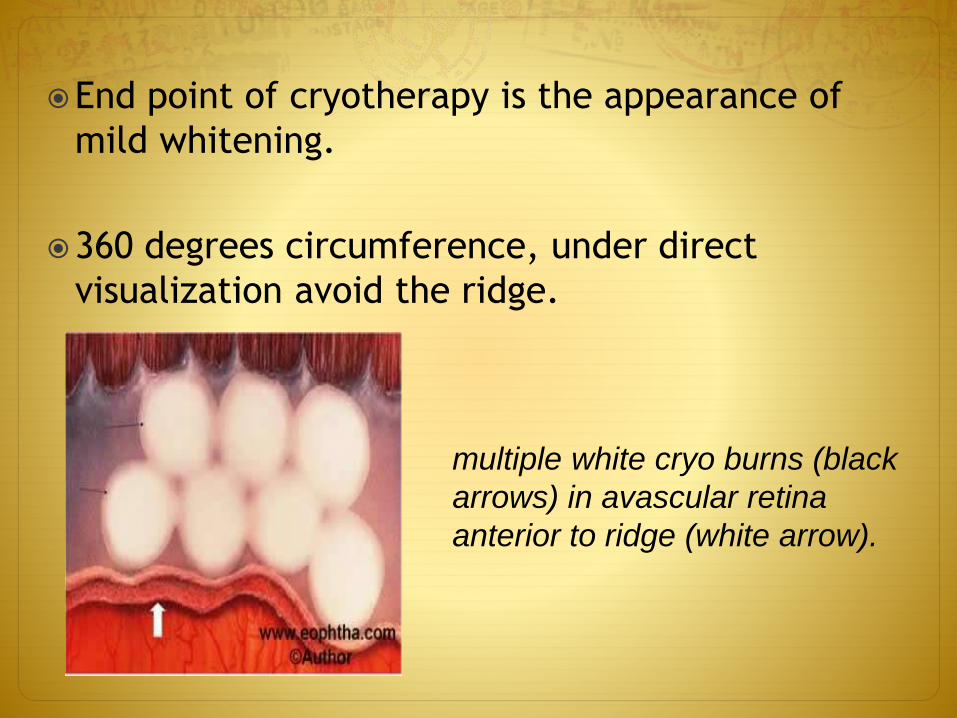
End point of cryotherapy is the appearance of
mild whitening.
360 degrees circumference, under direct
visualization avoid the ridge.
multiple white cryo burns (black
arrows) in avascular retina
anterior to ridge (white arrow).

Complications of cryotherapy

Procedure of choice, being less invasive, less
traumatic and causes less discomfort to the
infant.
Laser treatment is delivered through an
indirect opthalmoscope .
It can be performed in NICU and under local
anesthesia
Easy to treat posterior located lesion.
Argon green and Diode red LASER has been
used

An average of 1000 to 2000 spots of 100 mm
size 1½ burn width apart can be placed in
each eye.
Entire avascular retina till ora, avoid the
ridge.
Fundus picture of RE showing
laser scars (black arrows)

Complications of laser therapy
Burns in cornea and iris. Other
complications include cataract, and
retinal and vitreous haemorrhage.
Development of cataracts and glaucoma
or anterior segment ischemia following
laser has been reported.


Monotherapy
Single injections
Multiple injections for recurrence
Less desirable if periphery not perfused
Adjunctive therapy
Injections to allow regression beyond Zone 1
Laser for recurrent ROP
Anti-VEGF as a Bridge to laser peripherally
Treatment after laser / cryotherapy failure
Perioperative therapy before surgery
Reduce bleeding
Promote regression of neovascularization
Vitrectomy and scleral buckles

• Once the macula detaches in stage 4b or 5 ROP retinal
reattachment is done.
• It may include vitrectomy with or without lensectomy.
• Membrane peeling is done to remove the tractional force
causing the retinal detachment.
• A Scleral buckling procedure is useful for perpheral
detachments with drainageof subretinal fluid for effusional
detachment.
• Sucessful retinal reattachment outcome of the visual acuity
is in the range of the Legal blindness.
• Despite low vision outcome it is considered superior to
untreated stage 5 .

SCLERAL BUCKLE
It is done under GA
Peritomy
2.5 mm encircling band passed beneath 4 Recti
One anchoring mattress suture applied in all quadrants
Removal after 3-6 months

VITRECTOMY
Necessary in advanced cases
Lensectomy avoided
Peeling of membranes
Relieve of traction
No attempt to drain Sub Retinal Fluid
AIM : Ambulatory vision ie being able to see objects and move around a room without stumbling or bumping into obstacles.


Randomn Control Trial study , conducted on 172 preterm
infants
Peripheral Cryotherapy vs. Observation
“Threshold Disease”
Stage 3 (neovascularization)
5 contiguous, 8 noncontiguous clock hours
Zone I or II
Plus Disease
Cryotherapy superior to Observation:
Reduced unfavorable outcomes
Related improved visual acuity results

Randomn control trial study was conducted on
(n=317 bilateral; n=84 asymmetric unilateral infants)
Early peripheral laser vs conventional treatment
“High Risk Prethreshold” ROP disease – Type 1 or Type
2
Type 1 ROP
Zone I: Any Stage, plus / Stage 3, no plus
Zone II: Stage 2 or 3, plus
Finding: Early Peripheral laser superior to
conventional treatment .

• Type 2 ROP
Zone I: Stage 1 or 2, no plus
Zone II: Stage 3, no plus
Finding: Observation advised until Type 1 or
Regression
• Peripheral laser better than conventional
treatment for Type 1:
It concludes reduced unfavorable anatomic
outcome from 15.6% to 9.1% .
Reduced unfavorable visual acuity grating from
19.5% to 14.5%

Bevacizumab Eliminates the Angiogenic Threat in ROP”
Randomn Control Trial was conducted on 150 infants, 300
eyes
Stage 3, plus
Zone 1 and posterior Zone 2
Comparison : Intravitreal Bevacizumab v/s Peripheral
Laser (ETROP)
Summary
Bevacizumab reduced recurrence of ROP
Bevacizumab benefit over laser in Zone 1
Bevacizumab allowed continued peripheral vascularization
into avascular retina


• AVRY BOOK OF NEONATOLOGY
• PERSONS BOOK OPTHALMOLOGY
• MANUAL OF NEWBORN , CLOHERTY
• CARE OF NEWBORN BY MEHRBAN SINGH
• INTERNET