Retinoids in the treatment of skin aging: an overview of ...
22
Clinical Interventions in Aging 2006:1(4) 327–348 © 2006 Dove Medical Press Limited. All rights reserved 327 REVIEW Retinoids in the treatment of skin aging: an overview of clinical efficacy and safety Siddharth Mukherjee 1 Abhijit Date 2 Vandana Patravale 3 Hans Christian Korting 4 Alexander Roeder 4 Günther Weindl 5 1 Department of Pharmacology, Bombay College of Pharmacy, Kalina, Santacruz (E.), Mumbai, India; 2 Pharmaceutical R & D, Nicholas Piramal Research Center, Goregaon, Mumbai, India; 3 Department of Pharmaceutical Sciences and Technology, University Institute of Chemical Technology, Matunga, Mumbai, India; 4 Department of Dermatology and Allergology, Ludwig-Maximilians University, Munich, Germany; 5 Department of Dermatology, Eberhard Karls University Tübingen, Tübingen, Germany Correspondence: Alexander Roeder Department of Dermatology and Al- lergology, Ludwig-Maximilians University, Frauenlobstrasse 9-11, D-80337 Munich, Germany Tel + 49 89 5160 6151 Fax + 49 89 5160 6007 Email alexander.roeder@lrz. uni-muenchen.de Abstract: Aging of skin is an intricate biological process consisting of two types. While intrinsic or chronological aging is an inevitable process, photoaging involves the premature aging of skin occurring due to cumulative exposure to ultraviolet radiation. Chronological and photoaging both have clinically differentiable manifestations. Various natural and synthetic retinoids have been explored for the treatment of aging and many of them have shown histological and clinical improvement, but most of the studies have been carried out in patients presenting with photoaged skin. Amongst the retinoids, tretinoin possibly is the most potent and certainly the most widely investigated retinoid for photoaging therapy. Although retinoids show promise in the treatment of skin aging, irritant reactions such as burning, scaling or dermatitis associated with retinoid therapy limit their acceptance by patients. This problem is more prominent with tretinoin and tazarotene whereas other retinoids mainly represented by retinaldehyde and retinol are considerably less irritating. In order to minimize these side effects, various novel drug delivery systems have been developed. In particular, nanoparticles have shown a good potential in improving the stability, tolerability and efficacy of retinoids like tretinoin and retinol. However, more elaborate clinical studies are required to confirm their advantage in the delivery of topical retinoids. Keywords: photoaging, chronological aging, tretinoin, retinaldehyde, tazarotene, nanoparticles Introduction Skin – the largest organ of the body – protects all the other organs from the external environment. The skin is a complex organ with multiple structures and cell types and divided into three layers: epidermis, dermis, and the subcutaneous tissue. The epidermis is mainly composed of keratinocytes, pigment-producing melanocytes, and antigen-presenting Langerhans cells. A basement membrane separates the epidermis from the dermis, which primarily contains extracellular proteins produced by the fibroblasts below. The vascular supply to the skin resides in the dermis. The subcutaneous tissue consists of fat cells that underline the connective tissue network. Type I collagen is the most abundant protein in the skin connective tissue. The other extracellular matrix proteins, which are a part of the skin connective tissue, are collagens (III, V, and VII), elastin, proteoglycans, fibronectin, etc. The newly synthesized type I procollagen is secreted into the dermal extracellular space where it undergoes enzymatic processing to arrange itself into a triple helix configuration (Rittié and Fisher 2002). Apart from environmental protection against radiation, functions of the skin include heat regulation, immune response, biochemical synthesis, sensory detection, regulation of absorption/loss of water and electrolytes. The stratum corneum formed from nonviable corneocytes plays the major role. Keratin is aligned in the intercrossed disulfidic macrofibres along with filaggrin, the main protein component of the keratolytic granule. The cells develop a cornified involucre resulting from the intercrossing of involucrin and keratohyalin. Lamellar lipids accumulate in
Transcript of Retinoids in the treatment of skin aging: an overview of ...

Clinical Interventions in Aging 2006:1(4) 327–348© 2006 Dove Medical Press Limited. All rights reserved
327
R E V I E W
Retinoids in the treatment of skin aging: an overview of clinical effi cacy and safety
Siddharth Mukherjee1
Abhijit Date2
Vandana Patravale3
Hans Christian Korting4 Alexander Roeder4
Günther Weindl5
1Department of Pharmacology, Bombay College of Pharmacy, Kalina, Santacruz (E.), Mumbai, India; 2Pharmaceutical R & D, Nicholas Piramal Research Center, Goregaon, Mumbai, India; 3Department of Pharmaceutical Sciences and Technology, University Institute of Chemical Technology, Matunga, Mumbai, India; 4Department of Dermatology and Allergology, Ludwig-Maximilians University, Munich, Germany; 5Department of Dermatology, Eberhard Karls University Tübingen, Tübingen, Germany
Correspondence: Alexander RoederDepartment of Dermatology and Al-lergology, Ludwig-Maximilians University, Frauenlobstrasse 9-11, D-80337 Munich, GermanyTel + 49 89 5160 6151Fax + 49 89 5160 6007Email [email protected]
Abstract: Aging of skin is an intricate biological process consisting of two types. While intrinsic
or chronological aging is an inevitable process, photoaging involves the premature aging of skin
occurring due to cumulative exposure to ultraviolet radiation. Chronological and photoaging
both have clinically differentiable manifestations. Various natural and synthetic retinoids
have been explored for the treatment of aging and many of them have shown histological and
clinical improvement, but most of the studies have been carried out in patients presenting with
photoaged skin. Amongst the retinoids, tretinoin possibly is the most potent and certainly the
most widely investigated retinoid for photoaging therapy. Although retinoids show promise in
the treatment of skin aging, irritant reactions such as burning, scaling or dermatitis associated
with retinoid therapy limit their acceptance by patients. This problem is more prominent with
tretinoin and tazarotene whereas other retinoids mainly represented by retinaldehyde and
retinol are considerably less irritating. In order to minimize these side effects, various novel
drug delivery systems have been developed. In particular, nanoparticles have shown a good
potential in improving the stability, tolerability and effi cacy of retinoids like tretinoin and
retinol. However, more elaborate clinical studies are required to confi rm their advantage in the
delivery of topical retinoids.
Keywords: photoaging, chronological aging, tretinoin, retinaldehyde, tazarotene, nanoparticles
IntroductionSkin – the largest organ of the body – protects all the other organs from the external
environment. The skin is a complex organ with multiple structures and cell types
and divided into three layers: epidermis, dermis, and the subcutaneous tissue. The
epidermis is mainly composed of keratinocytes, pigment-producing melanocytes,
and antigen-presenting Langerhans cells. A basement membrane separates the
epidermis from the dermis, which primarily contains extracellular proteins produced
by the fi broblasts below. The vascular supply to the skin resides in the dermis. The
subcutaneous tissue consists of fat cells that underline the connective tissue network.
Type I collagen is the most abundant protein in the skin connective tissue. The
other extracellular matrix proteins, which are a part of the skin connective tissue,
are collagens (III, V, and VII), elastin, proteoglycans, fi bronectin, etc. The newly
synthesized type I procollagen is secreted into the dermal extracellular space where
it undergoes enzymatic processing to arrange itself into a triple helix confi guration
(Rittié and Fisher 2002).
Apart from environmental protection against radiation, functions of the skin
include heat regulation, immune response, biochemical synthesis, sensory detection,
regulation of absorption/loss of water and electrolytes. The stratum corneum
formed from nonviable corneocytes plays the major role. Keratin is aligned in the
intercrossed disulfi dic macrofi bres along with fi laggrin, the main protein component
of the keratolytic granule. The cells develop a cornifi ed involucre resulting from
the intercrossing of involucrin and keratohyalin. Lamellar lipids accumulate in
Clinical Interventions in Aging 2006:1(4)328
Mukherjee et al
the intercellular spaces, which are strongly hydrophobic.
The combination of the cornifi ed hydrophilic cells with
the hydrophobic intercellular material forms a barrier for
the external hydrophilic and hydrophobic substances.
With age the skin’s natural rejuvenation process slows
drastically and the skin becomes thinner, drier, and less
elastic (Ramos-E-Silva et al 2001).
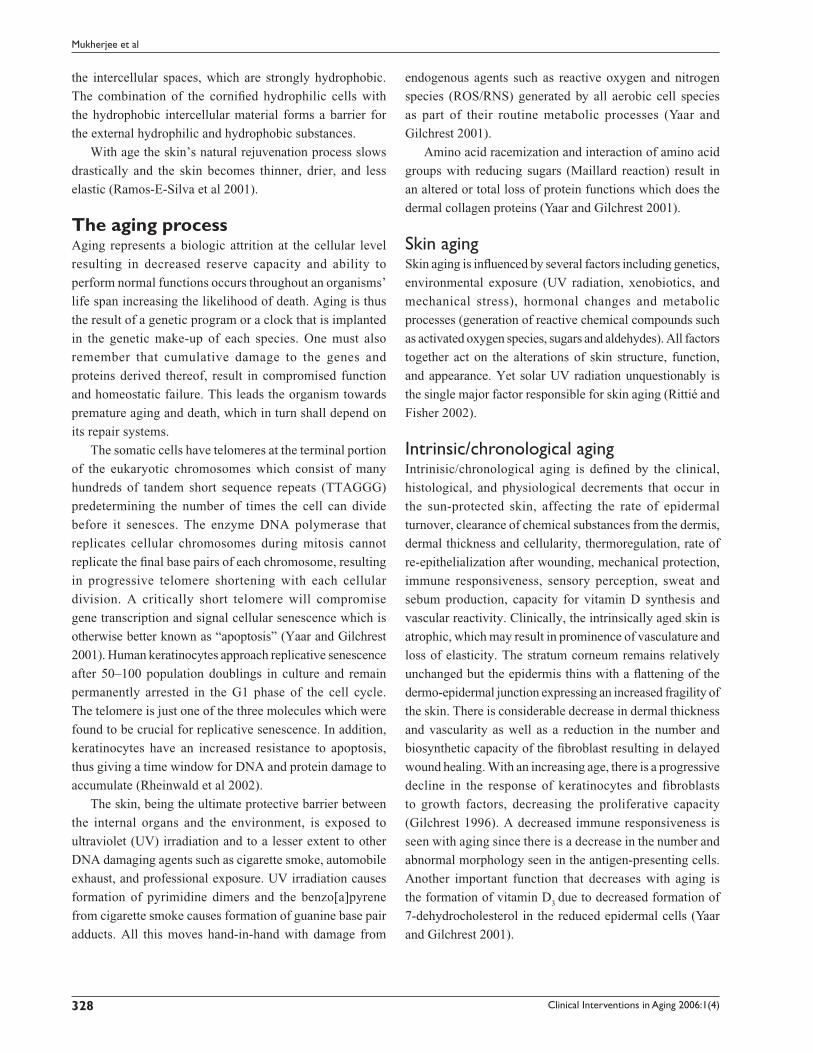
The aging processAging represents a biologic attrition at the cellular level
resulting in decreased reserve capacity and ability to
perform normal functions occurs throughout an organisms’
life span increasing the likelihood of death. Aging is thus
the result of a genetic program or a clock that is implanted
in the genetic make-up of each species. One must also
remember that cumulative damage to the genes and
proteins derived thereof, result in compromised function
and homeostatic failure. This leads the organism towards
premature aging and death, which in turn shall depend on
its repair systems.
The somatic cells have telomeres at the terminal portion
of the eukaryotic chromosomes which consist of many
hundreds of tandem short sequence repeats (TTAGGG)
predetermining the number of times the cell can divide
before it senesces. The enzyme DNA polymerase that
replicates cellular chromosomes during mitosis cannot
replicate the fi nal base pairs of each chromosome, resulting
in progressive telomere shortening with each cellular
division. A critically short telomere will compromise
gene transcription and signal cellular senescence which is
otherwise better known as “apoptosis” (Yaar and Gilchrest
2001). Human keratinocytes approach replicative senescence
after 50–100 population doublings in culture and remain
permanently arrested in the G1 phase of the cell cycle.
The telomere is just one of the three molecules which were
found to be crucial for replicative senescence. In addition,
keratinocytes have an increased resistance to apoptosis,
thus giving a time window for DNA and protein damage to
accumulate (Rheinwald et al 2002).
The skin, being the ultimate protective barrier between
the internal organs and the environment, is exposed to
ultraviolet (UV) irradiation and to a lesser extent to other
DNA damaging agents such as cigarette smoke, automobile
exhaust, and professional exposure. UV irradiation causes
formation of pyrimidine dimers and the benzo[a]pyrene
from cigarette smoke causes formation of guanine base pair
adducts. All this moves hand-in-hand with damage from
endogenous agents such as reactive oxygen and nitrogen
species (ROS/RNS) generated by all aerobic cell species
as part of their routine metabolic processes (Yaar and
Gilchrest 2001).
Amino acid racemization and interaction of amino acid
groups with reducing sugars (Maillard reaction) result in
an altered or total loss of protein functions which does the
dermal collagen proteins (Yaar and Gilchrest 2001).
Skin agingSkin aging is infl uenced by several factors including genetics,
environmental exposure (UV radiation, xenobiotics, and
mechanical stress), hormonal changes and metabolic
processes (generation of reactive chemical compounds such
as activated oxygen species, sugars and aldehydes). All factors
together act on the alterations of skin structure, function,
and appearance. Yet solar UV radiation unquestionably is
the single major factor responsible for skin aging (Rittié and
Fisher 2002).
Intrinsic/chronological agingIntrinisic/chronological aging is defi ned by the clinical,
histological, and physiological decrements that occur in
the sun-protected skin, affecting the rate of epidermal
turnover, clearance of chemical substances from the dermis,
dermal thickness and cellularity, thermoregulation, rate of
re-epithelialization after wounding, mechanical protection,
immune responsiveness, sensory perception, sweat and
sebum production, capacity for vitamin D synthesis and
vascular reactivity. Clinically, the intrinsically aged skin is
atrophic, which may result in prominence of vasculature and
loss of elasticity. The stratum corneum remains relatively
unchanged but the epidermis thins with a fl attening of the
dermo-epidermal junction expressing an increased fragility of
the skin. There is considerable decrease in dermal thickness
and vascularity as well as a reduction in the number and
biosynthetic capacity of the fi broblast resulting in delayed
wound healing. With an increasing age, there is a progressive
decline in the response of keratinocytes and fi broblasts
to growth factors, decreasing the proliferative capacity
(Gilchrest 1996). A decreased immune responsiveness is
seen with aging since there is a decrease in the number and
abnormal morphology seen in the antigen-presenting cells.
Another important function that decreases with aging is
the formation of vitamin D3 due to decreased formation of
7-dehydrocholesterol in the reduced epidermal cells (Yaar
and Gilchrest 2001).

Clinical Interventions in Aging 2006:1(4) 329
Retinoids in the treatment of skin aging
PhotoagingPhotoaging is the superimposition of photodamage on
intrinsically aged skin generally bringing about premature
aging. This specifi c damage occurs by chronic (multiple)
exposure of the skin to UV light. Clinically, the skin
becomes coarse; epidermis thickens (hyperplasia) initially
and then thins (atrophy), there is laxity, sallowness with
wrinkles, irregular hyperpigmentation, lentigines, and
telangiectasias (Gilchrest 1996). The pores of the skin are
larger, fi lled with horny material and have a tendency to
develop Favre-Racouchot’s syndrome (nodular elastoidosis
with cysts and comedones). There is also an increase in
development of benign neoplasms (seborrheic keratosis,
fi broma, acrochordon, and ruby spots), “premalignant”
lesions (actinic keratosis, lentigo maligna), and malignant
lesions (basal and squamous cell carcinomas and malignant
melanomas) on chronically exposed skin found in the
face, hands and neck regions (Torras 1996, Oppel and
Korting 2004). In severely damaged skin, there is loss of
epidermal polarity (orderly maturation) and individual
keratinocytes may show atypia, especially the lower
epidermal layers. More profound changes occur in the
dermis, where photodamage is characterized by degeneration
of collagen and deposition of abnormal elastotic material,
refl ected by wrinkles, furrows, and yellow discoloration
of the skin. The greater the photodamage, the more the
accumulation of thickened, tangled and degraded elastic
fi bers (Gilchrest 1996). The surface roughness is not only
attributed to the changes in the stratum corneum but also
to the changes in the glycosoaminoglycan (GAG) content
of the skin. With increase in age, there is a decrease in
the GAG content. Contradictorily, Bernstein and Uitto
(1995) found that there is an increase in the GAG content
in the photoaged skin. Yet GAG does not deposit in the
papillary dermis, instead it accumulates on the abnormal
elastotic material, which makes it unavailable as a source
of hydration resulting in a dull, leathery appearance of
the skin (Kang, Fisher, et al 2001). The microcirculation
is also affected by sun exposure. Blood vessels become
dilated and twisted (telangiectasia) and fi nally very sparse,
while their walls are initially thickened and later thinned
(Gilchrest 1996). UV irradiation of the skin increases the
reactive oxygen species and decreases the endogenous
antioxidant enzymes. The superoxide anion is produced
by energy transfer from several endogenous UV-absorbing
chromophores including NADH-/NADPH, tryptophan,
ribofl avin, or trans-urocanic acid (Rittié and Fisher 2002) in
the presence of molecular water present within the cell. The
superoxide anion is then converted to hydrogen peroxide,
which in the presence of transition metal ions such as iron and
copper undergoes conversion to a highly reactive hydroxyl
radical. This increased production of ROS alters gene and
protein structure and function leading to skin damage.
Table 1 gives an overview of the various epidermal,
dermal, and clinical signs with which one can differentiate
between chronological aging and photoaging.
Mechanism of collagen degradationMature collagen in skin undergoes continuous turnover, which
is required for optimal connective tissue function. The unique
molecular structure of collagen renders it largely resistant to
nonspecifi c proteolytic attack. The matrix metalloproteinases
(MMPs) are a group of enzymes responsible for degradation
of collagen. The MMPs are members of a large subfamily of
proteinases with certain common structural features. The human
Table 1 Comparison of chronological aging and photoaging
Identifi cation characteristics
Ageing types Epidermis Dermis Clinical
Chronological Thinner than normal with lower cell growth, Elastin fi bers appear irregular in their Skin is smooth, aging minor abnormalities in keratinocyte regularity arrangement, whereas collagen fi bers begin unblemished, but Normal stratum corneum to lower in number and thickness shows saggy appearance There is loss of rete pegs here as well
Photoaging Thick skin, with acanthosis followed by Excessive production of elastin fi bers in an Smooth, leathery, atrophy of the cells improper orientation, collagen fi bres reddened appearance High basal keratinocyte irregularity Stratum appear to thicken and then wear out soon with initially light wrinkles, corneum appears compact Appearance of grenzzone which later deepen, There is loss of rete pegs here as well thus showing loss of collagen fi bers
Clinical Interventions in Aging 2006:1(4)330
Mukherjee et al
family of MMPs is composed of at least 16 members who can
be classifi ed into 4 different subfamilies: 1) collagenases,
2) gelatinases, 3) stromelysins, and 4) membrane MMPs. The
fi rst three can cleave native, undenatured interstitial helical
collagens found in the skin within the triple-helical domain.
The cleavage site is specifi c in type I collagen generating
three-quarter and one-quarter length fragments. Following
this initial unequal split by collagenase, the resultant denatured
collagen called gelatin is further degraded by gelatinases and
stromelysins (Kang, Fisher, et al 2001).
Biochemical pathways that are triggered after UV irradiation activating cell surface cytokine and growth factor receptorsHuman skin cells respond to UV radiation by activation of
multiple cytokine and growth factor receptors. These include
epidermal growth factors receptors (EGF-R), tumor necrosis
factor (TNF)-α receptors, platelet activating factor (PAF)
receptor, interleukin (IL)-1 receptor, insulin receptor and
platelet derived growth factor. Amongst these, the EGF-R
activation has been the most studied. It is a single chain
180 kDa transmembrane protein. The extracellular domain
possesses high affi nity binding for EGF and EGF-like
ligands (transforming growth factor [TGF]-α, amphiregulin
and heparin binding-EGF) (Rittié and Fisher 2002). The
intracellular domain possesses intrinsic tyrosine kinase
activity. EGF-R also known as ErbB1 undergoes homo- or
heterodimerization with either ErbB2 or ErbB3 resulting
in the transphosphorylation of specifi c tyrosine residues.
EGF-R tyrosine phosphorylation is a well-characterized
marker for receptor activation and occurs within 10 minutes
of UV irradiation. Notably, UV fails to induce EGF-R
tyrosinase phosphorylation in cells expressing mutant
EGF-R lacking tyrosine kinase activity. UV irradiation
of EGF-R, like ligand activation, is dependent on
EGF-R tyrosine kinase-catalysed trans-phosphorylation.
Alternatively, it has been proposed that UV-induced
EGF-R tyrosine phosphorylation results from inactivation
of protein tyrosine phosphatases (PTPs) that function
to maintain EGF-R in a dephosphorylated basal state.
Inhibition by specific tyrosine kinase inhibitors results
in a very rapid dephosphorylation of EGF-R. Treatment
of the cells with UV irradiation substantially prolonged
the life of the EGF-R phosphorylated tyrosinases, thus
suggesting an inhibitory effect of UV on PTPs. This
inhibitory activity by UV was sensitive to N-acetyl cysteine,
a scavenger of reactive oxygen intermediates and could
be mimicked by treating cells with H2O
2. UV-induced
inactivation of PTP activity is postulated to result from
oxidation of a critical cysteine residue that is present in
the catalytic active site of all PTP’s to sulfenic acid. This
oxidation occurs by the exposure of the cysteine residue
on the PTP to reactive oxygen species which are generated
within the cells by UV irradiation (Rittié and Fisher 2002).
This inactivation of PTPs may result in the activation of
other cell surface receptors and cytokine receptors which
in turn leads to activation of small GTP-binding protein
families such as the Rac, Ras, and Cdc42. These are either
direct or indirect (via other GTP-binding proteins or ROS)
upstream regulators of mitogen-activated protein kinases
(MAPKs). The UV irradiation causes increased ROS
production and simultaneous increase in ceramide levels
which may also contribute to the activation of MAPK
pathways. A major effector of the MAPK pathways is
the transcription factor activator protein-1 (AP-1). AP-1
is constitutively composed of c-Fos and JunD proteins or
the other Jun and Fos family proteins (c-Jun, Junb, FosB,
Fra1, and Fra2) in the nonirradiated skin. The activation of
MAPKs indirectly activates the transcription factors for AP-1
formation ie, transcription of the c-Fos and c-Jun genes.
UV irradiation induces c-Jun mRNA and protein in human
skin in vivo within 30 min and 1 hour, respectively, and
protein levels remain elevated for at least 24 hours post UV
irradiation. Increased levels of c-Jun compete with JunD
for forming complexes with c-Fos resulting in c-Jun: c-Fos
AP-1 complexes (Rittié and Fisher 2002). Transcription of
several MMP family members is regulated by this AP-1
complex formed throughout the epidermal and dermal cells.
MMPs are a large family of zinc-requiring endoprotreases
with a broad range of specifi cities that together have the
capacity to degrade all the extracellular matrix proteins.
Initially, MMPs are synthesized as zymogens (proenzymes)
which undergo proteolytic degradation to be active. These
are inhibited by tissue inhibitors of metalloproteinases
(TIMPs). Several MMPs are upregulated by AP-1 including
MMP-1 (interstitial collagenase or collagenase1), which
intitiates the degradation of types I & III fi brillar collagens,
MMP-9 (92 kDa gelatinase or gelatinase B) degrades the
collagen fragments (gelatin) generated by collagenases and
MMP-3 (stromelysin 1) further degrades collagen type IV of
the basement membrane and activates proMMP-1. MMP-1,
MMP-3, and MMP-9 transcripts are induced within 8 hours

Clinical Interventions in Aging 2006:1(4) 331
Retinoids in the treatment of skin aging
following UV irradiation. Thus, together MMPs have the
capacity to completely degrade mature fi brillar collagen in
the skin within 24 hours of UV exposure via the induction
of transcription factor AP-1. In addition to causing collagen
breakdown, UV radiation impairs new type I collagen
synthesis and organization of collagen fi brils in skin in
vivo. Down-regulation of type I collagen is mediated by
down-regulation of the transcription of genes that encode for
type I procollagen. Type I procollagen mRNA and protein
expression levels are decreased within 8 hours following
UV irradiation of the human skin in vivo and become
essentially absent in the upper dermis within 24 hours after
UV irradiation, consistent with the sustained induction of
c-Jun and thus AP-1 activation.
The TGF-β is a major profibrotic cytokine, which
regulates multiple cellular functions including differentiation,
proliferation and induction of synthesis of major extracellular
matrix (ECM) proteins – collagen and elastin (Massague
1998). In human skin, TGF-β inhibits growth of epidermal
keratinocytes and stimulates growth of dermal fi broblasts
(Massague 2000). TGF-β inhibits the expression of MMP-1
and MMP-3 by binding to a certain cell surface receptor
complex (TGF-β receptor proteins: TβR I/II/III), thus
preventing the breakdown of collagens. UV irradiation
has been shown to impair the TGF-β signaling pathway
by reducing TβRII expression and to a lesser extent the
inhibitory Smad 7. Moreover, the connective tissue growth
factor is down-regulated after UV irradiation (Rittié and
Fisher 2002).
In this article, we critically compare the clinical effi cacy
and safety of various retinoids that have been in the treatment
and protection of skin aging.
RetinoidsThe importance of retinol (vitamin A) was discovered
during World War I and subsequent research showed
that its deficiency gives rise to xerosis and follicular
hyperkeratosis. The retinoid drug project was launched
in 1968 to synthesize compounds similar to vitamin A by
chemical manipulation of its molecule to improve clinical
effi cacy and safety. The use of these substances in therapy
dates back some 3000 years to ancient Egypt, where liver
was used to treat endemic night blindness. The modern
history of retinoids, however, began in 1909 when an
essential factor in the viability of an embryo in the fatty
extract of the egg yolk, called vitamin A, was discovered.
Retinoids fi nally were introduced into the treatment of
dermatoses including photoaging more than two decades
ago (Ramos-E-Silva et al 2001).
The retinoid family comprises vitamin A (retinol) and
its natural derivatives such as retinaldehyde, retinoic acid,
and retinyl esters, as well as a large number of synthetic
derivatives (Antille et al 2004). Retinol is a 20-carbon
molecule that consists of a cyclohexenyl ring, a side chain with
four double bonds (all in trans confi guration), and an alcohol
end group, hence the name all-trans-retinol. The oxidation
of the alcohol end group in retinol results in the formation
of an aldehyde (all-trans retinaldehyde or retinal), which can
be further oxidized to a carboxylic acid (all-trans retinoic
acid or tretinoin). Vitamin A cannot be synthesized by the
body; hence it needs to be supplied to the body. Naturally,
it is present as retinyl esters and beta-carotene. The retinyl
esters are converted to retinol before absorption from the
intestine and back to retinyl esters for storage in the liver. In
the plasma, retinol is bound to plasma-retinol binding proteins.
Retinol is be metabolized to four important products: retinyl
esters, all-trans retinoic acid, 14-hydroxy-4, 14-retro retinol,
and all-trans 3, 4-didehydroretinol, and its esters. Retinoids
are required for a vast number of biological processes. In
particular, they are involved in embryogenesis, reproduction,
vision, growth, infl ammation, differentiation, proliferation,
and apoptosis. Retinal is an essential part of the rhodopsin
pigment, necessary for vision (Roos et al 1998). Retinoids
are found in the keratinocytes in two forms: retinol and
retinyl esters – probably the storage form. This esterifi cation is
catalysed by two enzymes, acyl CoA: retinol acyltransferase and
lecithin: retinol acyltransferase (Törmä and Vahlquist 1990).
The metabolism of retinyl esters to retinol is catalysed by
retinyl ester hydrolase (Törmä and Vahlquist 1990).
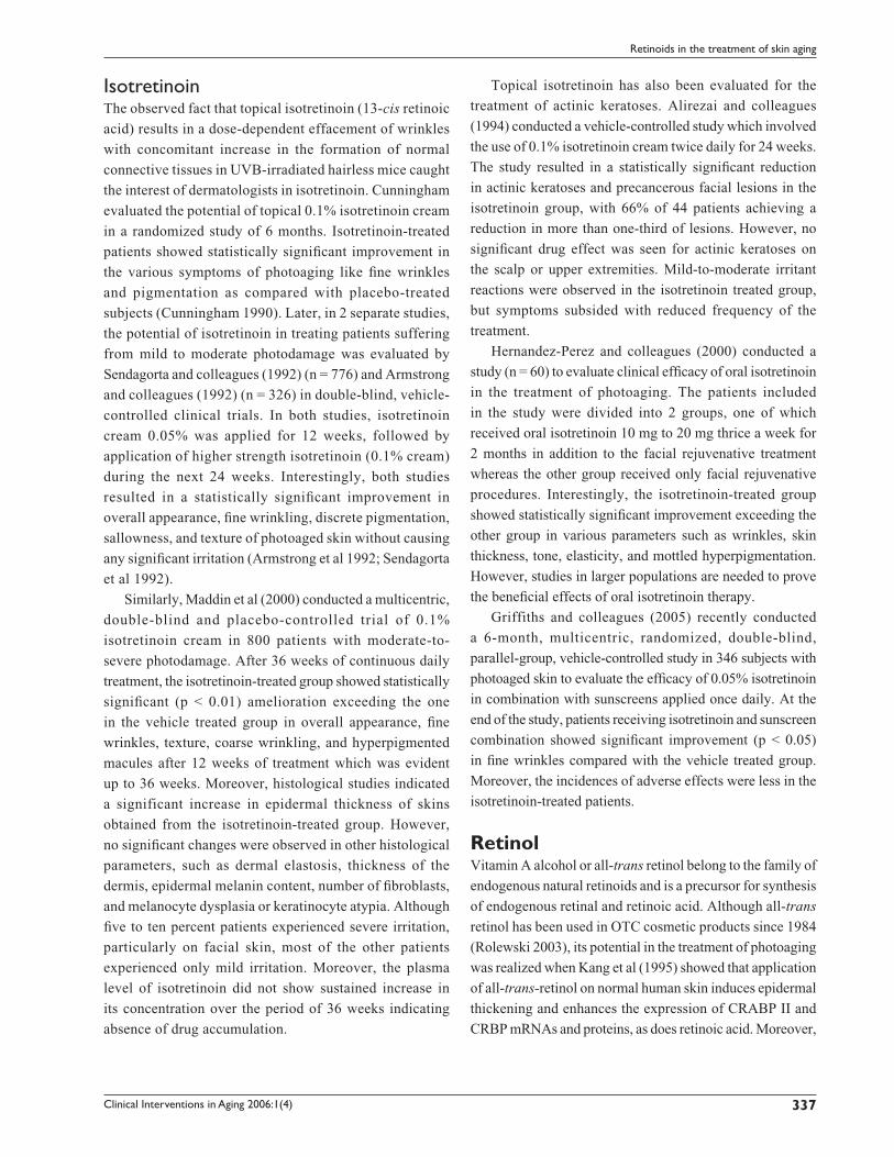
Retinoid classifi cation Based on the structural features and refl ecting the time
of introduction, retinoids can be classifi ed into various
generations. The chemical structures of various retinoids
are shown in Figure 1.
Mechanism of action of topical retinoidsRetinoids are very well known to infl uence a variety of
cellular processes, such as cellular growth and differentiation,
cell surface alterations, and immune modulation. Many of
their tissue effects are mediated by their interaction with
specifi c cellular and nucleic acid receptors. The cellular
or cytoplasmic receptors include the Cellular Retinoic

Clinical Interventions in Aging 2006:1(4)332
Mukherjee et al
Acid Binding Protein (CRABP) types I and II and the
cellular retinol binding protein (Astrom et al 1991). The
nucleic acid receptors were discovered in 1987 to reveal
the mechanism of action by which tretinoin and several of
its analogues would bring about their biological effects.
This discovery of the existence of a tretinoin specifi c gene
transcription factor lead to the realization that tretinoin is a
hormone. These nuclear receptors are related to a super
family of nuclear DNA transcription factors, which include
steroid, thyroid hormone, and vitamin D receptors. They
comprise two families, each of which are encoded by three
genes. The nuclear retinoic acid receptor family called RARs
was the fi rst to be described and consists of three forms
(RAR-α, RAR-β, RAR-γ) that are activated by RAR specifi c
SELETINOID G
OHO
OOO
O
O
Fourth Generation (pyranones)
Third Generation (poly-aromatics)
Second generation (mono-aromatics)
First Generation (non-aromatics)
ADAPALENE TAZAROTENE
COOH
COOH
COOC2H5
CH2OH
COOH
COOH
COOH
CHO
S
N
O
OE1
CH3O
H3COH3CO
ETRETINATE
ISOTRETINOIN ALITRETINOIN
TRETINOIN
ACITRETIN
RETINALDEHYDERETINOL
Figure 1 Chemical structures of retinoids.
Clinical Interventions in Aging 2006:1(4) 333
Retinoids in the treatment of skin aging
all-trans-retinoic acid (tretinoin). As such the RARs
have distinct DNA and retinoid-binding domains and
they function in pairs, either pairs of identical receptors
called homodimers or pairs of different receptors called
heterodimers. In the human skin, RARs partner with retinoid
X receptors (RXRs) to form heterodimers (Giguére et al
1987; Petkovich et al 1987; Brand et al 1988; Fisher et al
1994; Xiao et al 1995). The retinoid X receptors or RXRs
are the second family of nuclear receptors which interact
with 9-cis retinoic acid. Both RARs and RXRs are present
in the normal skin providing the necessary machinery for
the retinoid repair process of the photodamaged skin. The
RAR-γ subtype accounts for nearly 90% of RARs in the
human epidermis, whereas the RXR-α subtype accounts
for nearly 90% of the RXRs. Therefore, for the most part,
the normal human skin is regulated by paired heterodimers
composed of RAR-γ and RXR-α. The heterodimer complex
binds to specifi c elements in the DNA known as retinoic
acid response elements (RARE) in the promoter region
of the genes that are regulated by that specifi c retinoid
thus regulating the transcriptional activity of that retinoid-
responsive gene. The heterodimer requires only RAR
specifi c retinoid (tretinoin) to bind to RARE and initiate
transcriptional activity; the presence of a RXR binding
retinoid (9-cis retinoic acid) does not confer additional
trans-activation induced by the RAR retinoid. However,
for the heterodimer to function, the RXR protein must be
physically present to associate with the RAR protein. This
is probably the way topical retinoids improve photoaging
by modifying cellular differentiation programs: 1) initiating
the increase of epidermal proliferation leading to epidermal
thickening; 2) compaction of the stratum corneum; and
3) biosynthesis and deposition of the glycosoaminoglycans
(Griffi ths et al 1993).
New retinoids are selective for different RAR’s such
as the third generation retinoid Adapalene for RAR-β. The
newest retinoids are antagonists, which have potent anti-
infl ammatory activity and look promising as topical treatment
for psoriasis (Griffi ths et al 1998).
TretinoinTretinoin happens to be the retinoid that is investigated
more than any other retinoid implicated in the treatment of
intrinsic or photoaging. Although tretinoin has been used in
dermatology since the 1960s, its potential in the treatment
of aging was realized no earlier than in the 1980s. The
effi cacy of tretinoin in the treatment of photoaging was
fi rst demonstrated by Kligman and colleagues (1984) using
an animal model of photoaging. The authors observed
that treatment of photoaged mouse skin with tretinoin
for 10 weeks resulted in a signifi cant repair zone of new
collagen in the papillary dermis, which also correlated with
wrinkle effacement. This interesting observation prompted
researchers to investigate the potential of tretinoin in the
treatment of photoaging. Much later ex-vivo investigations
carried out by Fisher and colleagues (1996) helped in
understanding the molecular basis of this observation.
Fisher and colleagues (1996) found that pretreatment of
UV irradiated excised (photoaged) skin with 0.1% tretinoin
cream results in complete blockade of interstitial collagenase
and gelatinases synthesis thus preventing collagen
degradation. Moreover, application of 0.1% tretinoin also
blocked UV-induced activation of the nuclear transcription
factors AP-1 and NF-κB.
Following the ex-vivo observations, Kligman and
colleagues (1986) conducted a vehicle-controlled open
study to evaluate the clinical effi cacy of 0.05% tretinoin.
The study involved application of 0.05% tretinoin on the
photoaged facial and forearm skin for the duration of
3–12 months. Interestingly, tretinoin resulted in clinical
improvement of the photoaged skin. Moreover, histological
examination showed deposition of reticulin fi bers and
new dermal collagen formation (type I and III) accompanied
by angiogenesis in the papillary dermis. Encouraging
results obtained from this study stimulated researchers
to conduct a vast number of clinical trials to confirm
the clinical efficacy of tretinoin in the treatment of
photoaging.
Considering the exorbitant number of the reports
available in the literature, we have divided this part in several
subsections.
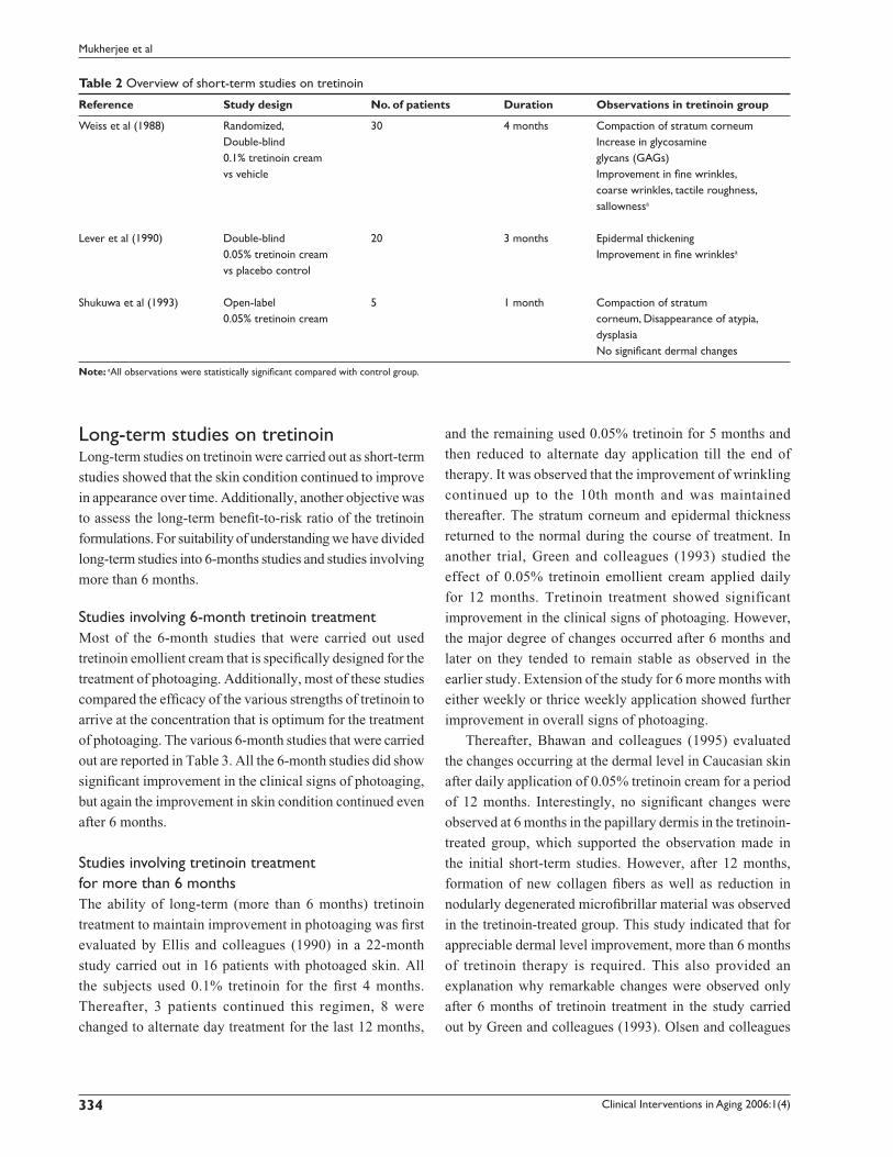
Short-term studies on tretinoinThis section should deal with the short-term studies that
were carried out immediately after the reports by Kligman
and colleagues (1986). Table 2 provides an overview of
those studies. In the two double-blind studies a statistically
signifi cant clinical improvement of various parameters was
observed. Furthermore, the tretinoin treated group had a
“rosy glow” not seen in the control group. Moreover, it was
observed that the skin condition continued to improve when
the follow-up assessment was performed after cessation
of treatment. Hence, studies involving longer duration of
tretinoin treatment were designed.
Clinical Interventions in Aging 2006:1(4)334
Mukherjee et al
Long-term studies on tretinoinLong-term studies on tretinoin were carried out as short-term
studies showed that the skin condition continued to improve
in appearance over time. Additionally, another objective was
to assess the long-term benefi t-to-risk ratio of the tretinoin
formulations. For suitability of understanding we have divided
long-term studies into 6-months studies and studies involving
more than 6 months.
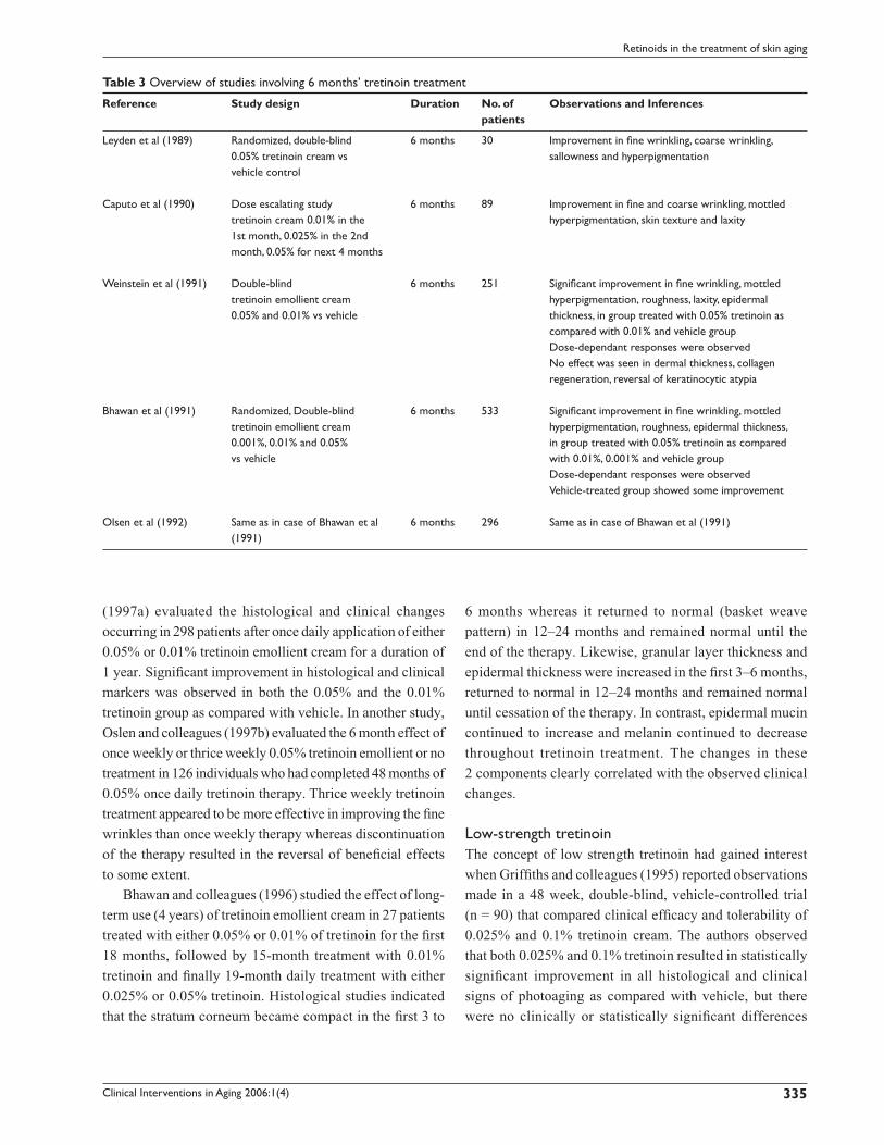
Studies involving 6-month tretinoin treatmentMost of the 6-month studies that were carried out used
tretinoin emollient cream that is specifi cally designed for the
treatment of photoaging. Additionally, most of these studies
compared the effi cacy of the various strengths of tretinoin to
arrive at the concentration that is optimum for the treatment
of photoaging. The various 6-month studies that were carried
out are reported in Table 3. All the 6-month studies did show
signifi cant improvement in the clinical signs of photoaging,
but again the improvement in skin condition continued even
after 6 months.
Studies involving tretinoin treatment for more than 6 monthsThe ability of long-term (more than 6 months) tretinoin
treatment to maintain improvement in photoaging was fi rst
evaluated by Ellis and colleagues (1990) in a 22-month
study carried out in 16 patients with photoaged skin. All
the subjects used 0.1% tretinoin for the fi rst 4 months.
Thereafter, 3 patients continued this regimen, 8 were
changed to alternate day treatment for the last 12 months,
and the remaining used 0.05% tretinoin for 5 months and
then reduced to alternate day application till the end of
therapy. It was observed that the improvement of wrinkling
continued up to the 10th month and was maintained
thereafter. The stratum corneum and epidermal thickness
returned to the normal during the course of treatment. In
another trial, Green and colleagues (1993) studied the
effect of 0.05% tretinoin emollient cream applied daily
for 12 months. Tretinoin treatment showed significant
improvement in the clinical signs of photoaging. However,
the major degree of changes occurred after 6 months and
later on they tended to remain stable as observed in the
earlier study. Extension of the study for 6 more months with
either weekly or thrice weekly application showed further
improvement in overall signs of photoaging.
Thereafter, Bhawan and colleagues (1995) evaluated
the changes occurring at the dermal level in Caucasian skin
after daily application of 0.05% tretinoin cream for a period
of 12 months. Interestingly, no signifi cant changes were
observed at 6 months in the papillary dermis in the tretinoin-
treated group, which supported the observation made in
the initial short-term studies. However, after 12 months,
formation of new collagen fi bers as well as reduction in
nodularly degenerated microfi brillar material was observed
in the tretinoin-treated group. This study indicated that for
appreciable dermal level improvement, more than 6 months
of tretinoin therapy is required. This also provided an
explanation why remarkable changes were observed only
after 6 months of tretinoin treatment in the study carried
out by Green and colleagues (1993). Olsen and colleagues
Table 2 Overview of short-term studies on tretinoin
Reference Study design No. of patients Duration Observations in tretinoin group
Weiss et al (1988) Randomized, 30 4 months Compaction of stratum corneum Double-blind Increase in glycosamine 0.1% tretinoin cream glycans (GAGs) vs vehicle Improvement in fi ne wrinkles, coarse wrinkles, tactile roughness, sallownessa
Lever et al (1990) Double-blind 20 3 months Epidermal thickening 0.05% tretinoin cream Improvement in fi ne wrinklesa
vs placebo control Shukuwa et al (1993) Open-label 5 1 month Compaction of stratum 0.05% tretinoin cream corneum, Disappearance of atypia, dysplasia No signifi cant dermal changes
Note: aAll observations were statistically signifi cant compared with control group.
Clinical Interventions in Aging 2006:1(4) 335
Retinoids in the treatment of skin aging
(1997a) evaluated the histological and clinical changes
occurring in 298 patients after once daily application of either
0.05% or 0.01% tretinoin emollient cream for a duration of
1 year. Signifi cant improvement in histological and clinical
markers was observed in both the 0.05% and the 0.01%
tretinoin group as compared with vehicle. In another study,
Oslen and colleagues (1997b) evaluated the 6 month effect of
once weekly or thrice weekly 0.05% tretinoin emollient or no
treatment in 126 individuals who had completed 48 months of
0.05% once daily tretinoin therapy. Thrice weekly tretinoin
treatment appeared to be more effective in improving the fi ne
wrinkles than once weekly therapy whereas discontinuation
of the therapy resulted in the reversal of benefi cial effects
to some extent.
Bhawan and colleagues (1996) studied the effect of long-
term use (4 years) of tretinoin emollient cream in 27 patients
treated with either 0.05% or 0.01% of tretinoin for the fi rst
18 months, followed by 15-month treatment with 0.01%
tretinoin and fi nally 19-month daily treatment with either
0.025% or 0.05% tretinoin. Histological studies indicated
that the stratum corneum became compact in the fi rst 3 to
6 months whereas it returned to normal (basket weave
pattern) in 12–24 months and remained normal until the
end of the therapy. Likewise, granular layer thickness and
epidermal thickness were increased in the fi rst 3–6 months,
returned to normal in 12–24 months and remained normal
until cessation of the therapy. In contrast, epidermal mucin
continued to increase and melanin continued to decrease
throughout tretinoin treatment. The changes in these
2 components clearly correlated with the observed clinical
changes.
Low-strength tretinoinThe concept of low strength tretinoin had gained interest
when Griffi ths and colleagues (1995) reported observations
made in a 48 week, double-blind, vehicle-controlled trial
(n = 90) that compared clinical effi cacy and tolerability of
0.025% and 0.1% tretinoin cream. The authors observed
that both 0.025% and 0.1% tretinoin resulted in statistically
signifi cant improvement in all histological and clinical
signs of photoaging as compared with vehicle, but there
were no clinically or statistically signifi cant differences
Table 3 Overview of studies involving 6 months’ tretinoin treatment
Reference Study design Duration No. of Observations and Inferences patients
Leyden et al (1989) Randomized, double-blind 6 months 30 Improvement in fi ne wrinkling, coarse wrinkling, 0.05% tretinoin cream vs sallowness and hyperpigmentation vehicle control
Caputo et al (1990) Dose escalating study 6 months 89 Improvement in fi ne and coarse wrinkling, mottled tretinoin cream 0.01% in the hyperpigmentation, skin texture and laxity 1st month, 0.025% in the 2nd month, 0.05% for next 4 months
Weinstein et al (1991) Double-blind 6 months 251 Signifi cant improvement in fi ne wrinkling, mottled tretinoin emollient cream hyperpigmentation, roughness, laxity, epidermal 0.05% and 0.01% vs vehicle thickness, in group treated with 0.05% tretinoin as compared with 0.01% and vehicle group Dose-dependant responses were observed No effect was seen in dermal thickness, collagen regeneration, reversal of keratinocytic atypia
Bhawan et al (1991) Randomized, Double-blind 6 months 533 Signifi cant improvement in fi ne wrinkling, mottled tretinoin emollient cream hyperpigmentation, roughness, epidermal thickness, 0.001%, 0.01% and 0.05% in group treated with 0.05% tretinoin as compared vs vehicle with 0.01%, 0.001% and vehicle group Dose-dependant responses were observed Vehicle-treated group showed some improvement
Olsen et al (1992) Same as in case of Bhawan et al 6 months 296 Same as in case of Bhawan et al (1991) (1991)

Clinical Interventions in Aging 2006:1(4)336
Mukherjee et al
between the two concentrations of tretinoin. However, the
incidences of adverse effects were signifi cantly greater
in the 0.1% tretinoin group as compared with the 0.025%
tretinoin group. Thus, it was speculated that low strength
tretinoin might be a good option for those patients who
can not tolerate standard therapy (0.05%). Thereafter,
Nykady and colleagues (2001) conducted two 24 weeks,
double-blind and vehicle-controlled trials to evaluate the
effi cacy and tolerability of 0.02% tretinoin cream applied
once daily in 328 patients with moderate to severely
photodamaged skin. Interestingly, both studies showed
that there is signifi cantly greater improvement in clinical
signs of photoaging like fi ne wrinkling, coarse wrinkling,
sallowness, and mottled hyperpigmentation (only in one
study) as compared with vehicle. Moreover, the treatment
was safe and well tolerated in most of the patients.
Tretinoin cream 0.02% is now recognized by the FDA for
the treatment of photoaging.
High strength tretinoinHigh strength tretinoin treatment has been evaluated in the
treatment of photoaging as the conventional tretinoin therapy
has following disadvantages:
1. Benefi cial effects of tretinoin are seen slowly and over a
long period of time, which often leads to discontinuation
of therapy.
2. Retinoid related adverse effects like irritation, erythema
and dermatitis.
Hence, in order to minimize or avoid these disadvantages,
Kligman and colleagues (1998) evaluated the potential of
high strength tretinoin (0.25% solution in a fast penetrating
vehicle) for the treatment of photoaging in 50 females.
The treatment regimen consisted of application of highly
concentrated tretinoin solution on alternate nights for
2 weeks and then every night thereafter until the end of
the treatment. Interestingly, just 4 to 6 week treatment
with high strength tretinoin resulted in improvement in
fine wrinkling, mottled hyperpigmentation, elasticity,
hydration, angiogenesis, and new collagen deposition above
the zone of solar elastosis and the extent was similar to the
results observed after 6 to 12 months of standard tretinoin
therapy (0.05%). Moreover, the high strength tretinoin
treatment was well tolerated in all patients. Subsequently,
Cuce and colleagues (2001) evaluated effi cacy of the
1% tretinoin solution applied twice a week in 15 women
with photodamaged skin. Histological studies carried out
after 15 days showed compaction of stratum corneum and
increased epidermal thickness. Additionally, surface
imaging studies showed improvement in skin texture and
appearance.
In another study, Kligman and colleagues (2004)
investigated the effect of high strength solution applied every
night in 32 women with photodamaged skin. Treatment for
4 weeks resulted in signifi cant improvement in fi ne wrinkles,
mottled hyperpigmentation, and roughness as observed in
their earlier study. The most noteworthy, but unexpected
observation in this study was the rapid accommodation of
the skin to retinoid side effects occurring within just 2 weeks.
Hence, although typical retinoid associated side-effects were
observed, they diminished very soon resulting in tolerance
and better acceptance of the therapy in the patients. Yet,
although high strength tretinoin has shown a good potential
in photoaging, the reported studies have been carried out in
a smaller population. Hence, large scale, multicentric and
standard tretinoin therapy (0.05%) controlled studies are
required to confi rm the effi cacy.
Tretinoin in intrinsic agingTo date only one vehicle-controlled clinical study has been
undertaken to evaluate the use of topical tretinoin for the
treatment of chronologically aged skin. In this study, 0.025%
tretinoin cream was applied once daily on chronologically
aged inner thigh skin of six women (mean age, 74 years) for
a period of 9 months (Kligman et al 1993). The cream was
applied to one inner thigh and vehicle to the other. Clinically,
the improvement with thigh skin was modest; showing a less
scaly, a less wrinkled, and a little fi rmer skin with a pink hue.
In contrast, histological changes associated with tretinoin
treatment were much more marked, when compared with the
vehicle. Tretinoin resulted in marked increase in epidermal
and granular cell layer thickness and a highly undulating
dermo-epidermal junction through the development of rete
pegs and produced uniformity in keratinocyte density while
it decreased melanocyte vacuolization. Ultrastructurally,
an increase in anchoring fi brils was noted at the level of the
dermo-epidermal junction. In the dermis, development of
several new micro vasculatures (angiogenesis) and production
of new elastic material and GAGs was observed. These
morphological changes suggested that the magnitude of
effect of tretinoin might be greater for chronologically aged
than for photoaged skin. However, large scale, multicentric
clinical trials need to be conducted for confirming the
utility of tretinoin for chronological aging.
Clinical Interventions in Aging 2006:1(4) 337
Retinoids in the treatment of skin aging
IsotretinoinThe observed fact that topical isotretinoin (13-cis retinoic
acid) results in a dose-dependent effacement of wrinkles
with concomitant increase in the formation of normal
connective tissues in UVB-irradiated hairless mice caught
the interest of dermatologists in isotretinoin. Cunningham
evaluated the potential of topical 0.1% isotretinoin cream
in a randomized study of 6 months. Isotretinoin-treated
patients showed statistically signifi cant improvement in
the various symptoms of photoaging like fi ne wrinkles
and pigmentation as compared with placebo-treated
subjects (Cunningham 1990). Later, in 2 separate studies,
the potential of isotretinoin in treating patients suffering
from mild to moderate photodamage was evaluated by
Sendagorta and colleagues (1992) (n = 776) and Armstrong
and colleagues (1992) (n = 326) in double-blind, vehicle-
controlled clinical trials. In both studies, isotretinoin
cream 0.05% was applied for 12 weeks, followed by
application of higher strength isotretinoin (0.1% cream)
during the next 24 weeks. Interestingly, both studies
resulted in a statistically signifi cant improvement in
overall appearance, fi ne wrinkling, discrete pigmentation,
sallowness, and texture of photoaged skin without causing
any signifi cant irritation (Armstrong et al 1992; Sendagorta
et al 1992).
Similarly, Maddin et al (2000) conducted a multicentric,
double-blind and placebo-controlled trial of 0.1%
isotretinoin cream in 800 patients with moderate-to-
severe photodamage. After 36 weeks of continuous daily
treatment, the isotretinoin-treated group showed statistically
signifi cant (p < 0.01) amelioration exceeding the one
in the vehicle treated group in overall appearance, fi ne
wrinkles, texture, coarse wrinkling, and hyperpigmented
macules after 12 weeks of treatment which was evident
up to 36 weeks. Moreover, histological studies indicated
a significant increase in epidermal thickness of skins
obtained from the isotretinoin-treated group. However,
no signifi cant changes were observed in other histological
parameters, such as dermal elastosis, thickness of the
dermis, epidermal melanin content, number of fi broblasts,
and melanocyte dysplasia or keratinocyte atypia. Although
fi ve to ten percent patients experienced severe irritation,
particularly on facial skin, most of the other patients
experienced only mild irritation. Moreover, the plasma
level of isotretinoin did not show sustained increase in
its concentration over the period of 36 weeks indicating
absence of drug accumulation.
Topical isotretinoin has also been evaluated for the
treatment of actinic keratoses. Alirezai and colleagues
(1994) conducted a vehicle-controlled study which involved
the use of 0.1% isotretinoin cream twice daily for 24 weeks.
The study resulted in a statistically signifi cant reduction
in actinic keratoses and precancerous facial lesions in the
isotretinoin group, with 66% of 44 patients achieving a
reduction in more than one-third of lesions. However, no
signifi cant drug effect was seen for actinic keratoses on
the scalp or upper extremities. Mild-to-moderate irritant
reactions were observed in the isotretinoin treated group,
but symptoms subsided with reduced frequency of the
treatment.
Hernandez-Perez and colleagues (2000) conducted a
study (n = 60) to evaluate clinical effi cacy of oral isotretinoin
in the treatment of photoaging. The patients included
in the study were divided into 2 groups, one of which
received oral isotretinoin 10 mg to 20 mg thrice a week for
2 months in addition to the facial rejuvenative treatment
whereas the other group received only facial rejuvenative
procedures. Interestingly, the isotretinoin-treated group
showed statistically signifi cant improvement exceeding the
other group in various parameters such as wrinkles, skin
thickness, tone, elasticity, and mottled hyperpigmentation.
However, studies in larger populations are needed to prove
the benefi cial effects of oral isotretinoin therapy.
Griffiths and colleagues (2005) recently conducted
a 6-month, multicentric, randomized, double-blind,
parallel-group, vehicle-controlled study in 346 subjects with
photoaged skin to evaluate the effi cacy of 0.05% isotretinoin
in combination with sunscreens applied once daily. At the
end of the study, patients receiving isotretinoin and sunscreen
combination showed signifi cant improvement (p < 0.05)
in fi ne wrinkles compared with the vehicle treated group.
Moreover, the incidences of adverse effects were less in the
isotretinoin-treated patients.
RetinolVitamin A alcohol or all-trans retinol belong to the family of
endogenous natural retinoids and is a precursor for synthesis
of endogenous retinal and retinoic acid. Although all-trans
retinol has been used in OTC cosmetic products since 1984
(Rolewski 2003), its potential in the treatment of photoaging
was realized when Kang et al (1995) showed that application
of all-trans-retinol on normal human skin induces epidermal
thickening and enhances the expression of CRABP II and
CRBP mRNAs and proteins, as does retinoic acid. Moreover,
Clinical Interventions in Aging 2006:1(4)338
Mukherjee et al
the authors also observed that retinol showed only minimal
signs of erythema and irritation unlike tretinoin. In another
study (n = 6; duration = 14 days), Fluhr and colleagues
(1999) confi rmed that retinol produces considerably less
transepidermal water loss, erythema and scaling than
retinoic acid. Interestingly, Fisher and colleagues (1996,
1997) further demonstrated that retinol inhibits UV
induction of MMP and stimulates collagen synthesis in
photoaged skin. However, it was observed that retinol is
20 times less potent than tretinoin and it requires further
conversion to retinoic acid (in vivo) to demonstrate its
action (Kurlandsky et al 1994; Kang et al 1995). Duell and
colleagues (1996) demonstrated that retinol could be as
effective as retinoic acid in producing ‘retinoid mediated
histological changes’ (like epidermal thickening and
keratinocyte proliferation), but with much less irritancy.
Pierard-Franchimont and colleagues (1998) fi rst conducted
a controlled clinical trial with retinol formulation. They
observed that retinol formulation resulted in signifi cant
improvement in fi ne wrinkles after 12 weeks of treatment.
Subsequently, Varani and colleagues (2000) studied the
effect of topical application of 1% retinol in 53 individuals
(80 years or above) with aged skin. The authors observed
that retinol application for 7 days reduced MMP (matrix
metalloproteinase), collagenase, and gelatinase expression
with concomitant increase in fi broblast growth and collagen
synthesis in the studied tissue specimens. Thus, it can be
concluded that retinol should be effective in the treatment of
aging and photoaging. However, the vehicle used for retinol
delivery would play a crucial role in eliciting its effi cacy,
as retinol is extremely unstable and easily gets degraded to
biologically inactive forms on exposure to light and air.
Retinol derivativesRetinol derivatives have been developed in order to improve
the chemical stability of retinol. Retinol derivatives like
retinyl acetate, retinyl propionate, and retinyl palmitate have
been widely used in cosmetic products instead of retinol.
In fact, retinol derivatives were thought to be useful for the
treatment of photoaging after the observation that retinyl
propionate induces epidermal thickening in mouse tail and
promoted collagen formation in UV-irradiated mice (Green
et al 1998). Based on these encouraging results, Green and
colleagues (1998) conducted a double-blind, randomized
and placebo-controlled trial for 48 weeks (n = 60).
Unfortunately, topical retinyl propionate cream (0.15%) did
not demonstrate any statistically signifi cant improvement
over placebo in any of the evaluated histopathological or
clinical symptoms of photoaging. However, in very few
subjects, actinic keratoses were reduced virtually to zero
by the end of 48 weeks, but this effect was not statistically
signifi cant.
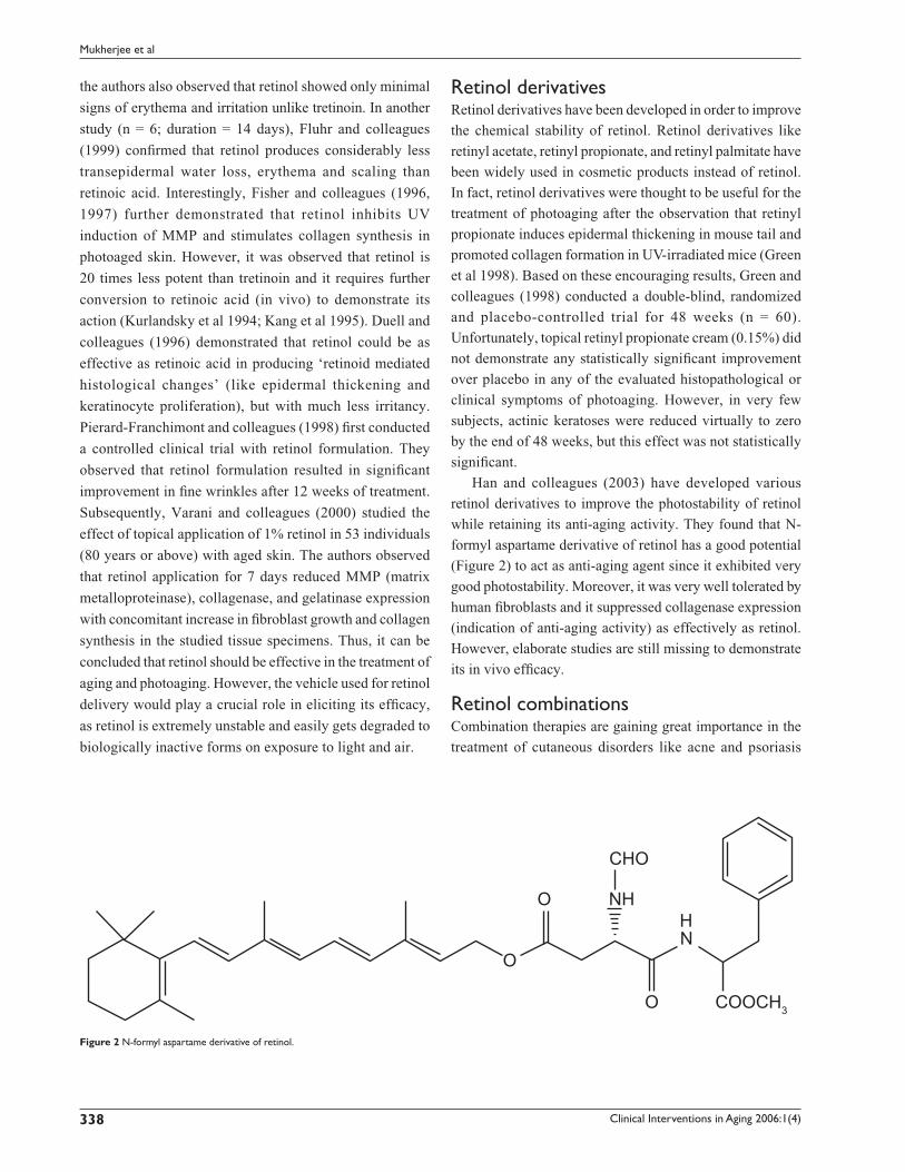
Han and colleagues (2003) have developed various
retinol derivatives to improve the photostability of retinol
while retaining its anti-aging activity. They found that N-
formyl aspartame derivative of retinol has a good potential
(Figure 2) to act as anti-aging agent since it exhibited very
good photostability. Moreover, it was very well tolerated by
human fi broblasts and it suppressed collagenase expression
(indication of anti-aging activity) as effectively as retinol.
However, elaborate studies are still missing to demonstrate
its in vivo effi cacy.
Retinol combinationsCombination therapies are gaining great importance in the
treatment of cutaneous disorders like acne and psoriasis
O
O
O COOCH3
CHO
NHHN
Figure 2 N-formyl aspartame derivative of retinol.

Clinical Interventions in Aging 2006:1(4) 339
Retinoids in the treatment of skin aging
as improved therapeutic effects have been observed
with combination product compared with the respective
monotherapy. Accordingly, researchers have recently
attempted the use of retinol in combination with other anti-
aging agents.
Seité and colleagues (2005) conducted two double-
blind vehicle-controlled clinical studies in postmenopausal
women to investigate the effects of a topically applied
retinol plus vitamin C combination on epidermal and dermal
compartments of aged or photoaged skin.
Study 1 involved a 3-month treatment with a combination
of retinol (0.07%) and vitamin C (3.5%) applied twice daily
in 8 volunteers. After the 3-month treatment, histological
changes such as thinning of stratum corneum, thickening
of the viable epidermis, and increase in interdigitation
index were observed. In the second study, volunteers with
photoaged skin were treated for 6-months with retinol
(0.04%) and vitamin C (3%) combination twice daily. After
the 6-month topical treatment, the observed histological
changes were mainly concentrated at the dermal level. Both
treated and control groups showed the same distribution
pattern of type I procollagen, however, the high level of
type III procollagen originally observed in photoaged
skin was reduced in the retinol- and vitamin C-treated
group, resulting in a lower type III-to-type I procollagen
ratio. Furthermore, a wide band of eosinophilic material
just beneath the epidermis, devoid of oxytalan fi bers and
forming the ‘grenz zone’, appeared more frequently and
was larger in the retinol- and vitamin C treated group.
Finally, the authors concluded that repeated topical
application of a preparation containing both retinol and
vitamin C could reverse, at least in part, skin changes induced
by both chronological and photoaging.
Draelos (2005) recently conducted a controlled
clinical trial to evaluate the efficacy of retinol (0.3%)
and hydroquinone (4%) in the treatment of photoaging
in comparison with 0.05% tretinoin emollient cream.
Interestingly, the retinol – hydroquinone combination
diminished the collective signs of photodamage more
effectively than 0.05% tretinoin emollient cream in terms
of dyspigmentation, fi ne wrinkles, and tactile roughness
within 16 weeks of treatment. Feinberg and colleagues
(2004) evaluated the efficacy of retinol (0.1%) and
glycolic acid (8%) combination in comparison with the
individual agents in the treatment of photoaging using the
photodamaged arm methodology. They observed that retinol-
glycolic acid combination offered signifi cant improvement
in the appearance of photoaged skin compared with glycolic
acid or retinol alone.
Barkovic and colleagues (2005) conducted a randomized,
placebo-controlled, double-blind clinical trial in
postmenopausal women to evaluate the effi cacy of retinol
– dimethylenolamine combination in the treatment of
chronological aging. The study demonstrated rapid and
signifi cant improvements in the appearance of aging skin
with daily application of the retinol – dimethylenolamine
combination.
RetinaldehydeRetinaldehyde is a retinoic acid precursor, which is formed
as an intermediate metabolite in the transformation of
retinol to retinoic acid in human keratinocytes. In the skin
retinaldehyde is metabolized to retinoic acid (which is a
well known anti-aging agent) as well as to retinol and retinyl
esters (which generally get depleted during photoaging),
indicating its use in the treatment of photoaging (Sass et al
1996). Moreover, metabolism of retinaldehyde to retinoic
acid occurs only by keratinocytes at a pertinent stage of
differentiation, leading to a more controlled delivery of
retinoic acid and weaker retinoid associated adverse effects
as compared to tretinoin and other synthetic retinoids
(Saurat et al 1994; Didierjean et al 1999; Sorg et al 1999).
Saurat and colleagues (1994) fi rst evaluated the biological
activity and tolerability profi le of retinaldehyde on human
skin. Noteworthy, topical retinaldehyde was well tolerated
on human skin and it also resulted in induction of CRABP
type II mRNA and protein, increased epidermal thickness,
increased keratin-14 expression, and enhanced keratinocyte
proliferation. However, later it was shown that retinaldehyde
exerts these biological activities only on transformation to
retinoic acid (Didierjean et al 1999).
In an open clinical trial, Ochando and colleagues
(1994) studied the effect of 0.05% retinaldehyde in
32 female volunteers showing symptoms of mild to
moderate photoaging. At the end of 4 months, considerable
reduction in the surface roughness and coarse wrinkling
was observed. Moreover, retinaldehyde treatment was
associated with very few adverse effects. Subsequently,
Creidi and colleagues (1998, 1999) conducted a randomized
and vehicle-controlled clinical trial (n = 125) to compare
the effi cacy of 0.05% retinaldehyde with 0.05% retinoic
acid for the treatment of photoaging. Optical profi lometry
studies on the volunteers indicated that both retinaldehyde
and retinoic acid were equally effective in reducing wrinkles

Clinical Interventions in Aging 2006:1(4)340
Mukherjee et al
and skin roughness. However, retinoic acid resulted in
higher incidences of local irritation than retinaldehyde
thus negatively affecting patient compliance. In another
study, Diridollou and colleagues (1999) evaluated the
effect of 0.05% retinaldehyde versus vehicle in 40 patients
showing symptoms of aging by ultrasound and rheological
techniques. Compared with the control group, the
retinaldehyde treated group showed a signifi cant increase
in epidermal thickness, as well as in cutaneous elasticity
(p < 0.01). Similarly, retinaldehyde treatment tended to
increase dermal thickness and reduce cutaneous stiffness
and was very well tolerated by the patients.
Recently, Mordon and colleagues (2004) conducted
a monocentric, comparative, randomized, double-blind
clinical trial (n = 16) to evaluate the effi cacy of retinaldehyde
versus excipient both in combination with nonablative laser
remodeling treatment. The study involved daily topical
application of 0.05% retinaldehyde (8 patients) immediately
after the fi rst laser treatment and up to 3 months after the
fi fth treatment. The control group (8 patients) was treated
under similar conditions, except with a daily application
of vehicle instead of retinaldehyde.
At the end of the study, an increase in dermal thickness
was observed for all patients treated by laser (both
retinaldehyde and control groups) on the forehead and neck.
However, the increase was greater for the retinaldehyde
group (p < 0.05) when compared with the control group
(vehicle). The increase in dermal thickness was 5.27% versus
1.13% for the forehead and 10.54% versus 3.57% for the
neck, respectively.
Thus, it can be concluded that retinaldehyde is a useful
topical agent for the treatment of aged and photoaged skin,
with a lower frequency of irritation.
TazaroteneTazarotene is a novel acetylenic retinoid known to be
effective in the topical treatment of psoriasis and acne.
Tazarotene is a prodrug, rapidly metabolized to its active
metabolite tazarotenic acid. Due to its rigid polyaromatic
structure, it does not undergo any isomerization or
conformational change in the skin. Although tazarotene
belongs to the retinoid family, it displays a receptor
selectivity pattern different from the one found with
tretinoin. Tretinoin directly activates all RAR subtypes
and indirectly RXRs whereas tazarotenic acid selectively
binds to RAR-β and RAR-γ but not to RXRs. Tazarotenic
acid modulates the expression of retinoid-responsive genes,
including those that regulate cell proliferation, cell
differentiation, and inflammation, corresponding to its
binding capacities to various RAR receptors. Tazarotene also
down-regulates the abnormal expression of keratinocytes,
epidermal growth factor receptor, and hyperproliferative
keratins (Nagpal et al 1995; Chandraratna 1996; DiSepio et al
1998; Roeder et al 2004).
Sefton and colleagues (2000) fi rst conducted a pilot,
double-blind, randomized trial to evaluate the effi cacy of
0.1% tazarotene gel in 10 healthy women with moderate
photodamage of the forearm skin. At the end of 12 weeks,
signifi cant reduction in pigmentary mottling, fi ne wrinkling,
and skin roughness were observed in the tazarotene
treated group as evidenced in silicon skin surface replicas.
Moreover, histopathological investigations indicated
a reduction of keratinocytic atypia and a restoration of
keratinocyte polarity.
Tazarotene was further evaluated by Kang, Leyden, and
colleagues (2001) who performed a multicentre, prospective,
randomized study in 349 facially photodamaged subjects,
to compare the effi cacy of four different concentrations
of tazarotene (0.01%, 0.025%, 0.05%, and 0.1%) with its
vehicle and with tretinoin 0.05% emollient cream. At the
end of 24 weeks, treatment success rates based on global
responses (>50% improvement compared with baseline)
were signifi cantly higher in the tazarotene- and tretinoin-
treated groups than in the vehicle-treated group (67%
with tazarotene 0.1%, 52% with tazarotene 0.05%, 36%
with tazarotene 0.025%, 41% with tazarotene 0.01%, 55%
with tretinoin 0.05% and 22% with vehicle). Interestingly,
a significantly greater proportion of tazarotene-treated
patients showed at least 50% improvement in various clinical
parameters than tretinoin-treated patients at weeks 12 and
20 thus indicating a trend towards a quicker response in
tazarotene-treated patients. However, at the end of 24 weeks,
there was no difference in overall improvement in the
photodamage in tazarotene- or tretinoin-treated groups. In
this study, both tazarotene 0.1% and tretinoin 0.05% cream
showed a similar degree of improvement in epidermal
thickness, fi ne wrinkling, lentigines, elastosis, and mottled
hyperpigmentation. The local adverse events observed were
generally of mild or moderate severity, and were greater
(mainly burning) with higher tazarotene concentrations.
Recently, Lowe and colleagues (2004) again compared
the efficacy of tazarotene 0.1% cream and tretinoin
0.05% emollient cream in the treatment of photoaging in
a double-blind, randomized, multicentre, 24 week study

Clinical Interventions in Aging 2006:1(4) 341
Retinoids in the treatment of skin aging
(n = 173). At week 16, the incidence of treatment success
(>50% global improvement) at the study endpoint was 78%
in the tazarotene group and 67% in the tretinoin emollient
group, with statistical signifi cance in favor of tazarotene. All
other signifi cant differences in effi cacy measures were also
in favor of tazarotene – for the overall integrated assessment
of photodamage at week 16, fi ne wrinkling at week 24,
mottled hyperpigmentation at weeks 12 and 16, and coarse
wrinkling at week 4. There were no signifi cant between-
group differences in the incidence of subjects achieving at
least a 1-grade improvement in irregular depigmentation,
lentigines, appearance of pore size, elastosis, tactile
roughness, telangiectasia, and actinic keratoses. The local
adverse events observed were generally of mild or moderate
severity and were greater (mainly burning) with higher
tazarotene concentrations. Thus, the effi cacy and tolerability
results obtained from this study are in broad agreement with
those reported by Kang, Leyden, and colleagues (2001).
Phillips and colleagues (2002) conducted a multicentre,
double-blind, randomized, vehicle-controlled clinical
trial that involved treatment of 563 subjects with
facial photodamage with tazarotene 0.1% cream for
24-weeks followed by a 28-week open-label extension. At
week 24, when compared with vehicle, tazarotene resulted
in a signifi cantly greater proportion of patients achieving
treatment success (>50% greater improvement) and at
least a 1 grade improvement in fi ne wrinkling, mottled
pigmentation, pore size, lentigines, elastosis, irregular
depigmentation, tactile roughness, coarse wrinkling, and
overall integrated assessment of photodamage. Pigmentary
changes were the fi rst to respond to treatment, showing
statistically signifi cant improvement over vehicle after
2 weeks of treatment. Fine wrinkling improved in the
tazarotene group after 4 weeks of treatment, and coarse
wrinkles, elastosis, and pore size were significantly
improved over vehicle by week 12. Telangiectasia and
actinic keratoses were not signifi cantly improved after
24 weeks of treatment, these two variables not being the
primary outcome measures of the study. After the fi rst
24 weeks, an open label extension of tazarotene 0.1%
cream was followed for another 28 weeks. Additional
clinical improvement was noted, which did not plateau
after 52 weeks of treatment, suggesting the value of long-
term treatment. Irritation was generally mild or moderate
and declined with ongoing treatment, whereas plasma
tazarotenic acid concentration did not exceed the plasma
levels of endogenous retinoids.
Recently, tazarotene has been evaluated once again
against photodamage in two subsequent studies. Machtinger
and colleagues (2004) evaluated the histological effects of
tazarotene cream in 50 patients with photodamaged facial
skin by conducting a multicentre, double-blind, randomized,
vehicle-controlled study. Blinded assessments showed
that the tazarotene-treated group showed amelioration
of keratinocytic and melanocytic atypia compared to the
vehicle-treated group. Between-group comparisons in
distribution of change from baseline categories of severity
were in favour of tazarotene (p = 0.055 for keratinocytic
atypia, p = 0.034 for melanocytic atypia, and p < 0.001 for
the number of granular cell layers). Compared with vehicle,
tazarotene was associated with an increase in epidermal
polarity (p = 0.008) and epidermal thickness (p = 0.012), and
a tendency for stratum corneum compaction. Tazarotene was
also associated with widened intercellular spaces relative to
vehicle (p < 0.001).
In another study, Kang and colleagues (2005) evaluated
the effi cacy and tolerability of tazarotene 0.1% cream in the
treatment of 568 patients with facial photodamage. Topical
tazarotene offered effi cacy in ameliorating multiple effects
of photodamage. Tazarotene cream was signifi cantly more
effective than vehicle in reducing fi ne wrinkles, mottled
hyperpigmentation, lentigines, irregular depigmentation,
apparent pore size, elastosis, tactile roughness, and an overall
integrated assessment of photodamage. Signifi cance was
achieved as early as at week 2 with mottled hyperpigmentation,
lentigines, irregular depigmentation and elastosis and had
not plateaued by week 24. The majority of patients reported
improvements in their photodamage as early as at week 4.
Adverse events were predominantly mild or moderate signs
or symptoms of skin irritation. Thus, topical tazarotene has
been shown to offer effi cacy in ameliorating multiple effects
of photodamage.
AdapaleneAdapalene is considered to be a third-generation synthetic
retinoid which contains a napthoic acid backbone. Unlike
retinoic acid, adapalene shows selectivity for the nuclear
retinoic acid receptor (RAR β/γ). It targets abnormal
desquamation of the skin, modulates cellular differentiation,
and possesses anti-infl ammatory properties (Leyden 2001).
Moreover, due to its receptor selectivity, it causes less skin
irritation. Adapalene is successfully being used for the
treatment of acne. However, not much has been done to
investigate its potential in aging/photoaging. So far only

Clinical Interventions in Aging 2006:1(4)342
Mukherjee et al
one study has been carried out to determine the potential of
adapalene in photoaging.
Kang and colleagues (2003) conducted a 2-center,
randomized, vehicle-controlled, investigator-masked,
parallel-group study in 83 patients suffering from actinic
keratoses and solar lentigines and other symptoms of
photoaging to evaluate the therapeutic potential of
adapalene gel (0.1% or 0.3%) or vehicle for 4 weeks,
followed by twice-daily applications, if tolerated, for up
to 9 months. After 9 months, 0.5 + 0.9% and 2.5 + 0.9%,
reduction in actinic keratoses was observed with adapalene
gel 0.1% and 0.3%, respectively. Whereas, with the vehicle
gel actinic keratoses increased by 1.5 + 1.3% (p < 0.005).
Signifi cant lightening of solar lentigines was observed
in patients treated with adapalene gel as compared with the
patients treated with vehicle gel (p < 0.05) within 1 month
of treatment. At the end of 9 months, 57% and 59% of the
patients had brighter lesions in the adapalene 0.1% and
0.3% groups, respectively, in comparison with only 36%
in the vehicle group (p < 0.05). Moreover, histological
evaluations revealed improved cellular atypia and reduced
epidermal melanin content in the adapalene-treated group
compared with the vehicle-treated group. A retrospective
evaluation of paired clinical photographs (before and after
9-month treatment) revealed signifi cant improvement in
wrinkles and other clinical features of photoaged skin
with adapalene as compared with its vehicle. As expected,
adapalene was very well tolerated by the patients involved
in the study. Thus, adapalene can be employed as a second
line treatment of photoaging mainly in patients demonstrating
extreme intolerance to conventional retinoids. However,
large scale clinical trials should be carried out to validate
the effi cacy of adapalene.
AlitretinoinAlitretinoin or 9-cis-retinoic acid is a naturally occurring
endogenous retinoid that binds to and activates all known
intracellular RAR and RXR subtypes (Cheer and Foster
2000). Its use in the topical treatment of AIDS-associated
Kaposi’s sarcoma is well documented in the literature
(Bodsworth et al 2001). Additionally, its safety and effi cacy
in the normal treatment of chronic hand dermatitis has also
been proved (Ruzicka et al 2004). Considering the RAR
binding activity of alitretinoin, Baumann and colleagues
(2005) recently conducted an open-label pilot study in 20
patients with photodamaged skin to evaluate the effi cacy
of 0.1% topical alitretinoin. The topical alitretinoin
treatment showed improvement in seborrheic keratoses,
actinic keratoses, and other symptoms of photoaging.
Moreover, the treatment was well tolerated by the patients.
However, larger, blinded, controlled trials are needed to
know the role and effi cacy of alitretinoin in the treatment
of photoaging.
Seletinoid GSeletinoid G represents a fourth generation of retinoids that
might fi nd an important place in the treatment of intrinsic/
photo aging. Like other synthetic retinoids, seletinoid G shows
receptor selectivity for RAR-γ, which is predominantly
expressed in the epidermis as compared with other RARs.
Recently, Kim and colleagues (2005) evaluated the safety
and effi cacy of seletinoid G in comparison with tretinoin
for the treatment of intrinsic/photo-aging after topical
application in 23 patients belonging to differing age groups.
Notably, intrinsically aged skin after topical treatment
with seletinoid G showed increase in the expressions
of type I procollagen, tropoelastin, and fi brillin-1, and
reduced MMP-1 similar to that of tretinoin demonstrating
its potential in the treatment of intrinsic aging. Moreover, in
the UV-irradiated young skin, topical seletinoid G treatment
inhibited UV-induced decrease of type I procollagen and
UV-induced increase of MMP-1 and c-Jun protein similar
to that seen with tretinoin. Interestingly, topical application
of seletinoid G under occlusion induced no skin irritation
in contrast to tretinoin, which caused severe erythema.
Thus, seletinoid G appears to be as effective as tretinoin
in the treatment of intrinsic/photo aging with the added
advantage of absence of skin irritation. However, larger,
blinded, controlled trials are needed to validate the role
and benefi t-to-risk ration of seletinoid G in the treatment
of intrinsic/photo aging.
Adverse effects of topical retinoidsThe most common and frequent adverse effect of topical
retinoids are known as ‘retinoid reaction’, characterized
by pruritus, burning sensation at the sites of application,
erythema, peeling. It is more common with tretinoin and
tazarotene than with isotretinoin, adapalene, retinol, and
retinaldehyde. The ‘retinoid reaction’ has been found to
be due to the free carboxylic acid in the polar end of the
retinoid, which is evident from the activity and toxicity
experiments done on CHO cells (Oda et al 1996). ‘Retinoid
reaction’ manifests itself generally within the fi rst few weeks
of treatment and is thought to get initiated by release of

Clinical Interventions in Aging 2006:1(4) 343
Retinoids in the treatment of skin aging
proinfl ammatory cytokines such as IL-1, TNF-α, IL-6, and
IL-8 (Torras 1996; Orfanos et al 1997). In order to validate
this hypothesis, Kim and colleagues (2003) evaluated the
changes in mRNA expression of inflammation-related
cytokines such as human monocyte chemoattractant
protein-1 (MCP-1), IL-8, TNF-α, interferon-γ (IFN-γ), IL-6,
IL-10, after treatment of epidermal cells with retinoic acid
and retinol. A minimum 3-fold increase in the mRNA and
protein levels of mainly MCP-1 and IL-8 were found, thus
validating the hypothesis to some extent.
The other side effect associated with retinoid therapy is
photosensitization, which normally occurs at the beginning of
the therapy. Patients on retinoid therapy are advised to avoid
excessive sun exposure and take precautionary measures
(like use of sunscreens) for sun protection. However, after
few months of therapy, the skin’s response to UV radiation
returns to normal. In certain cases, irritant conjunctivitis has
also been reported when the retinoid is applied close to the
eye (Torras 1996).
Over the past 30 years, no systemic side-effects on long-
term treatment with the topical retinoid have been observed
in young adults. Moreover, Lattarino and colleagues (1997)
found that topical tretinoin had no detectable effect on
endogenous plasma levels of tretinoin or its metabolites.
This is mainly because of the limited transdermal uptake
of these agents (Krautheim and Gollnick 2003). Pregnant
women or women of child-bearing age should use topical
retinoids with caution as retinoids on systemic exposure
are known to cause teratogenicity/embryotoxicity.
However, in over 25 years of topical tretinoin use for acne
therapy there have been no cases of related teratogenicity
(Kligman 1988). Additionally, in one case-control study of
215 mothers who were exposed to topical tretinoin in the
fi rst trimester of pregnancy, the prevalence of major fetal
malformations in the tretinoin-exposed group was 1.9%
versus 2.6% in the control group, indicating the safety of
topical tretinoin (Jick et al 1993). Even then, it is prudent
to advise women of childbearing age to avoid pregnancy
during treatment or, if pregnant, to discontinue the use of
topical retinoids.
Counteracting the adverse effectsTo counteract the symptoms of retinoid reaction, reduction
in the frequency of application or switching to a less
irritating retinoid is normally advised. Moreover, addition
of 3% indomethacin or 1% hydrocortisone to the retinoid
formulation, or concomitant treatment with a low to medium
potency topical corticosteroid has also been suggested to
relieve the symptoms (Torras 1996). However, if patients
cannot tolerate even the lowest concentration of retinoids,
then the treatment should be discontinued.
Kim and colleagues (2003) have demonstrated that
concomitant application of natural agents or extracts
like β-sitosterol, Magnoliae fl os, β-glycyrrhetinic acid,
Scleroglucan, Gingko extract, Raspberry extract, Schisandra
extract, Cola extract, Enna complex, or Vegetol red
grapevine extract could also be useful in counteracting
the irritant effects produced by topical retinoids. The
abovementioned natural agents or extracts reduced the
secretion of MCP-1 and IL-8 from human fi broblasts and
also showed a good protection against the retinoid-induced
irritation in the rabbit and human patch test. A different
approach to reduce adverse events of retinoids was applied
by Kambayashi et al (2005). Topical application of a new
synthetic retinoid (N-retinoyl-D-glucosamine) to hairless
mice showed good effi cacy in repair of photoaged skin
but did not induce skin irritations compared with retinoic
acid.
Although the aforementioned approaches have shown
potential in minimizing the side effects, they involve co-
administration of other therapeutic agents, which may
have its own effects on prolonged duration. The need of
hour is to have an approach that can effi ciently counteract
the adverse effects associated with retinoid therapy and
would not require co-administration of any therapeutic
agent. In fact, we believe that the same could be achieved
by modulating the delivery system/s employed for topical
retinoids. The further part of the review describes the
potential of delivery systems in optimizing topical retinoids
therapy with concomitant minimization in the adverse effects
associated with it.
Potential of delivery systems in topical retinoids therapyDrug delivery strategies are well known in pharmaceutical
research for their potential in optimizing efficacy
of therapeutic agents by either modulating their
physicochemical and biopharmaceutical properties or
minimizing/eliminating the side effects associated with
them, thus offering better patient compliance. The arrival
of controlled release systems, transdermal delivery systems,
implants, submicronic emulsions, and vesicular carriers
is suffi cient to substantiate the aforementioned advantage
of delivery systems. Since the last decade, there has been

Clinical Interventions in Aging 2006:1(4)344
Mukherjee et al
considerable interest in investigating the approaches for
improved delivery of retinoids.
The delivery system can be considered to be effi cient for
topical retinoids if it can:
1. Minimize/abolish the adverse effects of topical
retinoids.
2. Improve the stability (mainly photochemical) of retinoids
like retinoic acid and retinol.
3. Enhance the anti-aging effect of retinoids by modulating
their dermal transport or distribution.
Considering the above points, we believe that nanoparticles
can be utilized as an effi cient delivery system to optimize
the topical retinoid therapy.
Nanoparticles are solid colloidal particles, ranging in size
from 1 nm to 1000 nm, consisting of various biocompatible
matrices in which a therapeutic moiety can be adsorbed,
entrapped, or covalently attached (Lockman et al 2002).
Based on the nanoparticles matrix (or shell), they are be
classifi ed as:
1. Polymeric nanoparticles,
2. Solid lipid nanoparticles,
3. Inorganic nanoparticles.
Nanoparticles are extensively being investigated for drug
delivery in the pharmaceutical research from the last 3
decades on. Nanoparticles offer immense benefi ts such
as solubilization of hydrophobic actives, improvement in
bioavailability, improved (or altered) pharmacokinetics of
active pharmaceutical ingredient (API), protection of API
from physical, chemical or biological degradation, improved
cellular uptake, tissue targeting and controlled release of API.
However, most of the initial investigations on nanoparticles
dealt with the parenteral or oral delivery of APIs whereas
efforts to explore the potential of nanoparticles in topical
delivery were initiated in the last decade. Interestingly,
nanoparticles were found to improve the distribution
characteristics and stability of topically applied APIs such
as sunscreens (Wissing and Muller 2002).
In addition, they have been shown to offer localized and
targeted delivery of APIs which can be helpful in improving
their effi cacy with concomitant reduction in systemic side
effects associated.
Jenning and colleagues (Jenning, Gylser, et al 2000;
Jenning, Schäfer-Korting, et al 2000; Jenning and Gohla
2001) fi rst evaluated the potential of solid lipid nanoparticles
(SLNs) for the delivery of retinol. Interestingly, SLNs
offered a signifi cant improvement in the stability of retinol as
compared with the conventional emulsion. Moreover, highly
localized and controlled release of retinol could be observed
with the SLNs. Patravale and colleagues (2004) investigated
the potential of SLNs in counteracting the irritant effects and
in improving the physical stability of the most commonly
employed retinoid, ie, tretinoin. In fact, SLN-based tretinoin
gels showed drastic improvement in the tolerability of
tretinoin as compared with marketed products when
evaluated by the Draize patch test in rabbits. Furthermore,
photostability of tretinoin was markedly improved in SLNs
as compared with methanolic solution. Again as expected,
the tretinoin SLNs showed a high degree of localization in
skin when evaluated by in vitro skin permeation studies. All
these effects were attributed to the encapsulation of tretinoin
in nanoparticulate structures.
Recently, Yamaguchi and colleagues (2005) have
investigated the potential of inorganic nanoparticles of
tretinoin produced by boundary-organized reaction. As
observed earlier, tretinoin encapsulation in nanoparticles
resulted in significantly less irritation and inflammation
as compared with conventional formulation. In addition,
nanoparticulate tretinoin showed drastic improvement in
photostability as compared with conventional formulation,
which was retained even after 46 days of storage. However, the
most striking results were observed in the ex-vivo studies.
Histological evaluation in mouse epidermis after daily
application of nanoparticulate tretinoin cream for 4 days
revealed more than double increase in epidermal thickness
than conventional tretinoin vaseline preparation. Additionally,
nanoparticulate tretinoin treatment resulted in a signifi cant
increase in the mRNA levels of heparin-binding epidermal
growth factor. This observation was in good correlation
with the increased epidermal thickening observed with
nanoparticulate tretinoin. Interestingly, 4 days’ treatment
of aged skin of hairless mice with nanoparticulate tretinoin
showed signifi cant improvement in fi ne and coarse wrinkling
and texture in the neck area. To our knowledge, this is the fi rst
investigation, which clearly demonstrated that nanoparticles
could be an ideal approach in optimizing the topical retinoid
therapy with concomitant reduction in the side effects
associated with this therapy.
Other delivery strategies for retinoidsLiterature indicates that apart from nanoparticles, various
other formulation approaches like liposomes, microsponges,
microemulsions, and inclusion complexes with cyclodextrins
could also be employed for improving the topical delivery

Clinical Interventions in Aging 2006:1(4) 345
Retinoids in the treatment of skin aging
of retinoids. Their success in improving the stability,
tolerability, and effi cacy (in acne treatment) of retinoids is
well established. However, none of them have been evaluated
for improving the effi cacy of retinoids in the treatment of
aging. The detailed description of their potential in improving
the effi cacy and tolerability in the treatment of acne has been
described by Date and colleagues (2006).
Conclusion and outlookAging research is divided into 2 main streams the one being
the exploration of various pathophysiological and molecular
events responsible for aging and the other being investigation
on various anti-aging agents. Although much elaborate
mechanistic studies have been carried out for understanding
the pathophysiology of aging, they will still continue until
the complete cascade of molecular events responsible for
intrinsic/photoaging is elucidated. Amongst various anti-
aging agents, retinoids are the most promising agents that
are available for the treatment of aging. Amongst retinoids,
tretinoin is the most potent and best-studied retinoid.
However, its irritation potential has prompted dermatologists
to switch over to less irritating but comparably effective
retinoids like adapalene and to some extent retinol and
retinaldehyde. Receptor specifi c retinoids like seletinoid G
have been developed with the same vision and have been
found to be successful in small-scale studies.
We believe that future efforts in retinoid research will be
directed in following ways
1. Development of receptor selective synthetic retinoids
(like seletinoid G) or novel retinoid derivatives (like
N-formyl aspartamate derivative of retinol) or retinoid
co-drugs like retinyl ascorbate (Abdulmajed and Heard
2004) which may be superior in terms of tolerability,
stability and effi cacy.
2. Exploration of natural sources to identify agents or
extracts that may have retinoid like activity as in case of
PADMA 28 (Aslam et al 2005).
3. Evaluation of various combinations of anti-aging agents
having synergistic effects (analogous to combination
therapy in acne and psoriasis).
4. Development and clinical evaluation of nanoparticulate
carriers for retinoids.
Considering the developmental cost and the success rates
associated with the new chemical entities, there is limited
potential for developing novel synthetic retinoids. At the
same time, retinoid-like activity as shown by PADMA 28
opens a new era in the identification of natural products for
anti-aging treatment. As natural products have the well
known benefit of good acceptability we expect stimulation
in this area of research. Combination therapy has been
well established for cutaneous disorders like acne and
psoriasis. As relatively less developmental efforts are
required for commercializing new combinations, there is
scope for developing retinoid based combination therapies
for improved treatment of aging. Finally, in our opinion,
there is great scope for development of various drug
delivery systems (especially nanoparticulate systems)
to optimize the aging treatment with topical retinoids.
We believe that among various nanoparticulate carriers,
SLNs would have the greatest potential in optimizing
the retinoid therapy as apart from their advantage as a
carrier they are also known to have a UV-blocking effect,
which may help in reducing photosensitization induced
by retinoids. Interestingly, in one study, a SLN-based
anti-aging product was more effective in reducing the
depth of wrinkles (10.3%) as compared with the same
product based on conventional vehicle (4.1%) indicating
that SLNs themselves may have some effect on improving
wrinkling (Muller et al 2002). We believe that future
efforts in SLNs should be focused on proving its potential
to counteract photosensitivity and to identify the potential
of SLNs (blank or in combination with retinoids) in
improving the elasticity and wrinkling of intrinsically/
photo aged skin. Finally, complementary efforts from
clinicians are required to validate the potential of drug
delivery strategies in optimizing treatment of aging with
topical retinoids.
ReferencesAbdulmajed K, Heard C. 2004. Topical delivery of retinyl-ascorbate co-drug
1. Synthesis, penetration, into and permeation across human skin. Int J Pharm, 280:113–24.
Alirezai M, Dupuy R, Amblard P, et al. 1994. Clinical evaluation of topical isotretinoin in the treatment of actinic keratoses. J Am Acad Dermatol, 30:447–51.
Antille C, Tran C, Sorg O, et al. 2004. Penetration and metabolism of topical retinoids in ex-vivo organ-cultured full-thickness human skin explants. Skin Pharmacol Physiol, 17:124–8.
Armstrong RB, Lesiewicz J, Harvey G, et al. 1992. Clinical panel assessment of photodamaged skin treated with isotretinoin using photographs. Arch Dermatol, 128:352–6.
Aslam MN, Fligiel H, Lateef H, et al. 2005. PADMA 28: A multi-component herbal preparation with retinoid like dermal activity but without epidermal effects. J Invest Dermatol, 124:524–9.
Astrom A, Tavakkol A, Pettersson U, et al. 1991. Molecular cloning of two human cellular retinoic acid-binding proteins (CRABP). J Biol Chem, 266:17662–6.
Barkovic S, Leyden JJ, Wu IT, et al. 2005. Clinical evaluation of topical retinol-dimethylenolamine (DEMA) treatments for aging skin. J Am Acad Dermatol, 52(3:S1):P27.
Clinical Interventions in Aging 2006:1(4)346
Mukherjee et al
Baumann L, Vujevich J, Halem M, et al. 2005. Open-label pilot study of 0.1% alitretinoin gel in the treatment of photoaging. Cutis, 76:69–73.
Bernstein EF, Uitto J. 1995. Connective tissue alterations in photoaged skin and the effects of alphahydroxy acids. J Geriatr Dermatol, 3(Suppl. A):7–18A.
Bhawan J, Gonzalez-Serva A, Nehal K, et al. 1991. Effects of tretinoin on photodamaged skin. A histologic study. Arch Dermatol, 127:666-72.
Bhawan J, Palco MJ, Lee J, et al. 1995. Reversible histologic effects of tretinoin on photodamaged skin. J Geriatr Dermatol, 3:62-7.
Bhawan J, Olsen E, Lufrano L, et al. 1996. Histologic evaluation of the long-term effects of tretinoin on photodamaged skin. J Dermatol Sci, 11:177–82.
Bodsworth NJ, Bloch M, Bower M, et al. 2001. Phase III vehicle-controlled, multi-centered study of topical alitretinoin gel 0.1% in cutaneous AIDS-related Kaposi’s sarcoma. Am J Clin Dermatol, 2:77–87.
Brand MJ, Petkovich M, Krust A, et al. 1988. Identifi cation of a second human retinoic acid receptor. Nature, 332:850–3.
Caputo R, Monti M, Motta S, et al. 1990. The treatment of visible signs of senescence: the Italian experience. Br J Dermatol, 122 (Suppl 35):97–103.
Chandraratna RA. 1996. Tazarotene-first of a new generation of receptor-selective retinoids. Br J Dermatol, 135(Suppl. 49):18–25.
Cheer SM, Foster RH. 2000. Alitretinoin. Am J Clin Dermatol, 1:307–16.
Creidi P, Vienne MP, Ochonisky S, et al. 1998. Profi lometric evaluation of photodamage after topical retinaldehyde and retinoic acid treatment. J Am Acad Dermatol, 39:960–5.
Creidi P, Humbert P. 1999. Clinical use of topical retinaldehyde on photoaged skin. Dermatology, 199(Suppl 1):49–52.
Cuce LC, Bertino MC, Scattone L, et al. 2001. Tretinoin peeling. Dermatol Surg, 27:12–4.
Cunningham WJ. 1990. Topical isotretinoin and photodamage. Dermatologica, 181:350–1.
Date AA, Naik B, Nagarsenker MS. 2006. Novel drug delivery systems: potential in improving topical delivery of antiacne agents. Skin Pharmacol Physiol, 19:2–16.
Didierjean I, Tran C, Sorg O, et al. 1999. Biological activities of topical retinaldehyde. Dermatology, 1(Suppl 1):19–24.
Diridollou S, Vienne MP, Alibert M, et al. 1999. Effi cacy of topical 0.05% retinaldehyde in skin aging by ultrasound and rheological techniques. Dermatology, 199(Suppl 1):37–41.
DiSepio D, Ghosn C, Eckert RL, et al. 1998. Identification and characterization of a retinoid-induced class II tumor suppressor/growth regulatory gene. Proc Natl Acad Sci U S A, 95:14811–15.
Draelos S. 2005. Novel approach to the treatment of hyperpigmented photodamaged skin: 4% hydroquinone/0.3% retinol versus tretinoin 0.05% emollient cream. Dermatol Surg, 31:799–804.
Duell EA, Derguini F, Kang S, et al. 1996. Extraction of human epidermis treated with ret inol yields retro-ret inoids in addition to free retinol and retinyl esters. J Invest Dermatol, 107:178–82.
Ellis CN, Weiss JJ, Hamilton TA, et al. 1990. Sustained improvement with prolonged topical tretinoin (retinoic acid) for photoaged skin. J Am Acad Dermatol, 23:629–37.
Feinberg C, Hawkins S, Battaglia A, et al. 2004. Comparison of anti-aging effi cacy from cosmetic ingredients on photoaged skin. J Am Acad Dermatol, 50(3:S1):P27.
Fisher GJ, Talwar HS, Xiao JH, et al. 1994. Immunological identification and functional quantification of retinoic acid and retinoic X receptor proteins in the human skin. J Biol Chem, 269:20629–35.
Fisher GJ, Datta SC, Talwar HS, et al. 1996. The molecular basis of sun induced premature ageing and retinoid antagonism. Nature, 379:335–8.
Fisher GJ, Wang ZQ, Datta SC, et al. 1997. Pathophysiology of premature skin aging induced by ultraviolet light. N Engl J Med, 337:1419–28.
Fluhr JW, Vienne MP, Lauze C, et al. 1999. Tolerance profi le of retinol, retinaldehyde and retinoic acid under maximized and long-term clinical conditions. Dermatology, 199(Suppl 1):57–60.
Giguère V, Ong ES, Segui P, et al. 1987. Identifi cation of a receptor for the morphogen retinoic acid. Nature, 330:624–9.
Gilchrest BA. 1996. A review of skin aging and its medical therapy. Br J Dermatol, 135:867–75.
Green LJ, McCormick A, Weinstein GD. 1993. Photoaging and the skin: the effects of tretinoin. Dermatol Clin, 11:97–105.
Green C, Orchard G, Cerio R, et al. 1998. A clinicopathological study of the effects of topical retinyl propionate cream in skin photoaging. Clin Exp Dermatol, 23:162-7.
Griffiths CEM, Finkel IJ, Tranfaglia MG, et al. 1993. An in-vivo experimental model for topical retinoid effects on human skin. Br J Dermatol, 29:389–99.
Griffiths CEM, Kang S, Ellis CN, et al. 1995. Two concentrations of topical tretinoin (retinoic acid) cause similar improvement of photoaging but different degrees of irritation. Arch Dermatol, 131:1037–44.
Griffi ths CEM. 1998. Dowling Oration delivered at the Royal College of Physicians, London, Retinoids: renaissance and reformation. Clin Exp Dermatol, 24:329–35.
Griffi ths CE, Maddin S, Wiedow O, et al. 2005. Treatment of photoaged skin with a cream containing 0.05% isotretinoin and sunscreens. J Dermatol Treat, 16:79–86.
Han HS, Kwon YJ, Park MS, et al. 2003. Effi cacy validation of synthesized retinol derivatives in vitro: stability, toxicity, and activity. Bioorg Med Chem, 11:3839–45.
Hernandez-Perez E, Khawaja H, Alvarez T. 2000. Oral isotretinoin as part of the treatment of cutaneous aging. Dermatol Surg, 26:649–52.
Jenning V, Gysler A, Schäfer-Korting M, et al. 2000a. Vitamin A loaded solid lipid nanoparticles (SLN) for topical use: occlusive properties and drug targeting to the upper skin. Eur J Pharm Biopharm, 49:211–18.
Jenning V, Schäfer-Korting M, Gohla S. 2000b. Vitamin A loaded solid lipid nanoparticles (SLN) for topical use: drug release properties. J Control Rel, 66:115–26.
Jenning V, Gohla S. 2001. Encapsulation of retinoids in solid lipid nanoparticles (SLN). J Microencapsul, 18:149–58.
Jick SJ, Terris BZ, Jick H. 1993. First trimester topical tretinoin and congenital disorders. Lancet, 341:1181–2.
Kambayashi H, Odake Y, Takada K, et al. 2005. N-retinoyl-D-glucosamine, a new retinoic acid agonist, mediates topical retinoid effi cacy with no irritation on photoaged skin. Br J Dermatol, 153:30–36.
Kang S, Duell EA, Fisher GJ, et al. 1995. Application of retinol to human skin in vivo induces epidermal hyperplasia and cellular retinoid binding proteins characteristic of retinoic acid but without measurable retinoic acid levels or irritation. J Invest Dermatol, 105:549–56.
Kang S, Fisher G, Voorhees JJ. 2001. Photoaging: Pathogenesis, prevention and treatment. Clin Ger Med, 17:643–59.
Kang S, Leyden JJ, Lowe NJ, et al. 2001. Tazarotene cream for the treatment of facial photodamage. Arch Dermatol, 137:1597–604.
Kang S, Goldfarb M, Weiss J, et al. 2003. Assessment of adapalene gel for the treatment of actinic keratoses and lentigines: A randomized trial. J Am Acad Dermatol, 49:83–90.
Clinical Interventions in Aging 2006:1(4) 347
Retinoids in the treatment of skin aging
Kang S, Krueger GG, Tanghetti EA, et al. 2005. A multicenter, randomized, double-blind trial of tazarotene 0.1% cream in the treatment of photodamage. J Am Acad Dermatol, 52:268–74.
Kim BH, Lee YS, Kang KS. 2003. The mechanism of retinol-induced irritation and its application to anti-irritant development. Toxicol Lett, 146:65–73.
Kim MS, Lee SR, Rho HS, et al. 2005. The effects of a novel synthetic retinoid, seletinoid G, on the expression of extracellular matrix proteins in aged human skin in vivo. Clinica Chimica Acta, 362:161–9.
Kligman LH, Chen HD, Kligman AM. 1984. Topical retinoic acid enhances the repair of ultraviolet damaged dermal connective tissue. Connect Tissue Res, 12:139–50.
Kligman AM, Grove GL, Hirose R, et al. 1986. Topical tretinoin for photoaged skin. J Am Acad Dermatol, 15:836–59.
Kligman AM. 1988. Is topical tretinoin teratogenic? [letter]. JAMA, 259:2918.
Kligman AM, Dogadkina D, Lavker RM. 1993. Effects of topical tretinoin on the non-sun exposed protected skin of the elderly. J Am Acad Dermatol, 29:25–33.
Kligman DE, Sadiq I, Pagnoni A et al. 1998. High-strength tretinoin: a method for rapid retinization of facial skin. J Am Acad Dermatol, 39:S93–7.
Kligman DE, Draelos ZD. 2004. High-strength tretinoin for rapid retinization of photoaged skin. Dermatol Surg, 30:864–6.
Krautheim A, Gollnick H. 2003. Transdermal penetration of topical drugs used in the treatment of acne. Clin Pharmacokinet, 42:1287–304.
Kurlandsky SB, Xiao JH, Duell EA, et al. 1994. Biological activity of all-trans retinol requires metabolic conversion to all-trans-retinoic acid and is mediated through activation of nuclear retinoid receptors in human kertinocytes. J Biol Chem, 269:32821–7.
Latriano L, Tzimas G, Wong F et al. 1997. The percutaneous absorption of topically applied tretinoin and its effect on endogenous plasma tretinoin concentrations following single doses or long-term use. J Am Acad Dermatol, 36:S37–46.
Lever I, Kumar P, Marks R. 1990. Topical retinoic acid for treatment of solar damage. Br J Dermatol, 122:91–8.
Leyden JJ, Grove GL, Grove MJ, et al. 1989. Treatment of photodamaged facial skin with topical tretinoin. J Am Acad Dermatol, 21:638–44.
Leyden J. 2001. Adapalene in clincal practice. Cutis, 68(4S):7–9.Lockman PR, Mumper RJ., Khan MA, et al. 2002. Nanoparticle technology
for drug delivery across blood brain barrier. Drug Dev Ind Pharm, 28:1–13.
Lowe N, Gifford M, Tanghetti E, et al. 2004. Tarazotene 0.1% cream versus tretinoin 0.05% emollient cream in the treatment of photodamaged facial skin: a multicenter, double-blind, randomized, parallel-group study. J Cosmet Laser Ther, 6:79–85.
Machtinger LA, Kaidbey K, Lim J, et al. 2004. Histological effects of tazarotene 0.1% cream vs. vehicle on photodamaged skin: a 6-month, multicentre, double-blind, randomized, vehicle-controlled study in patients with photodamaged facial skin, Br J Dermatol, 151:1245–52.
Maddin S, Lauharanta J, Agache P, et al. 2000. Isotretinoin improves the appearance of photodamaged skin: results of a 36-week, multicenter, double-blind, placebo-controlled trial. J Am Acad Dermatol, 42:56–63.
Massague J. 1998. TGF-β signal transduction. Ann Rev Biochem, 67:753–91.
Massague J. 2000. How cells read TGF-β signals. Nat Rev, 1:169–78.Mordon S, Lagarde JM, Vienne MP, et al. 2004. Ultrasound imaging
demonstration of the improvement of non-ablative laser remodeling by concomitant daily topical application of 0.05% retinaldehyde. J Cosmet Laser Ther, 6:5–9.
Müller RH, Radtke S, Wissing SA. 2002. Solid lipid nanoparticles (SLN) and nanostructured lipid carriers (NLC) in cosmetic and dermatological preparations. Adv Drug Deliv Rev, 54(S1):S131–55.
Nagpal S, Athanikar J, Chandraratna RA. 1995. Separation of transactivation and AP1 antagonism functions of retinoic acid receptor alpha. J Biol Chem, 270:923–7.
Nyirady J, Bergfeld W, Ellis C, et al. 2001. Tretinoin cream 0.02% for the treatment of photodamaged facial skin: A review of 2 double-blind clinical trials. Cutis, 68:135–42.
Ochando N, LaGarde JM, Couval E, et al. 1994. Evaluation clinique et paraclinique des effets du rétinaldéhyde topique dans le photovieillissement cutané. Nouv Dermatol, 13:525–35.
Oda RM, Shimizu RW, Sabatine SC et al. 1996. Effects of structural changes on retinoid cytotoxity in the CHO clonal assay. In vitro Toxicol, 9:173–81.
Olsen EA, Katz I, Levine N, et al. 1992. Tretinoin emollient cream: a new therapy for photodamaged skin. J Am Acad Dermatol, 26:215–24.
Olsen EA, Katz HI, Levine N, et al. 1997a. Tretinoin emollient cream for photodamaged skin: Results of 48-week, multicenter, double-blind studies. J Am Acad Dermatol, 37:217–6.
Olsen EA, Katz HI, Levine N, et al. 1997b. Sustained improvement in photodamaged skin with reduced tretinoin emollient cream treatment regimen: Effect of once-weekly and three times-weekly applications. J Am Acad Dermatol, 37:227–30.
Oppel T, Korting HC. 2004. Actinic keratosis: the key event in the evolution from photoaged skin to squamous cell carcinoma. Therapy based on pathogenetic and clinical aspects. Skin Pharmacol Physiol, 17:67–76.
Orfanos CE, Zouboulis CC, Almond-Roesler B, et al. 1997. Current use and future potential role of retinoids in dermatology. Drugs, 53:358–88.
Patravale VB, Shah K. 2005. Solid lipid nanoparticles based tretinoin gels using novel solvent emulsifi cation-diffusion approach (abstract P-39). Proceedings of the International Symposium on Advances in Technology and Business Potential of Novel Drug Delivery Systems, 6:55.
Petkovich M, Brand MJ, Krust A, et al. 1987. A human retinoic acid receptor which belongs to the family of nuclear receptors. Nature, 330:444–50.
Phill ips TJ, Gottl ieb AB, Leyden JJ, et al . 2002. Efficacy of 0.1% tazarotene cream for the treatment of photodamage. A 12-month multicenter, randomized trial. Arch Dermatol, 138:1486–93.
Piérard-Franchimont C, Castelli D, Cromphaut IV. 1998. Tensile properties and contours of aging facial skin, A controlled double-blind comparative study of the effects of retinol, melibose-lactose and their association. Skin Res Technol, 4:237–43.
Ramos-E-Silva M, Hexsel DM, Rutowitsch MS, et al. 2001. Hydroxy acids and retinoids in cosmetics. Clin Dermatol, 19:460–6.
Rheinwald JG, Hahn WC, Ramsey MR, et al. 2002. A two-stage p16 (INK4A) and p53–dependent keratinocyte senescence mechanism that limits replicative potential independent telomere status. Mol Cell Bio, 22:5157–72.
Rittié L, Fisher G. 2002. UV-light-induced signal cascades and skin aging. Aging Res Rev, 1:705–20.
Roeder A, Schaller M, Schäfer-Korting M, et al. 2004. Tazarotene: Therapeutic strategies in the treatment of psoriasis, acne and photoaging. Skin Pharmacol Physiol, 17:111–18.
Rolewski SL. 2003. Clinical review: Topical retinoids. Dermatol Nurs, 15:447–65.
Roos TC, Jugert FK, Merk HF, et al. 1998. Retinoid metabolism in the skin. Pharmacol Rev, 50:315–33.

Clinical Interventions in Aging 2006:1(4)348
Mukherjee et al
Ruzicka T, Larsen FG, Galewicz D, et al. 2004. Oral alitretinoin (9-cis-retinoic acid) therapy for chronic hand dermatitis in patients refractory to standard therapy: results of a randomized, double-blind, placebo-controlled, multicenter trial. Arch Dermatol, 140:1453–9.
Sass Jo, Didierjean I, Carraux P, et al. 1996. Metabolism of topical retinaldehyde and retinol by mouse skin in vivo: predominant formation of retinyl esters and identifi cation of 14-hydroxy-4, 14-retro-retinol. Exp Dermatol, 5:267–71.
Saurat JH, Didierjean L, Masgrau E, et al. 1994. Topical retinaldehyde on human skin: biologic effects and tolerance. J Invest Dermatol, 103:770–4.
Sefton J, Kligman AM, Kopper SC, et al. 2000. Photodamage pilot study: A double-blind, vehicle-controlled study to assess the effi cacy and safety of tazarotene 0.1% gel. J Am Acad Dermatol, 43:656–63.
Seité S, Bredoux C, Compan D, et al. 2005. Histological evaluation of a topically applied Retinol-Vitamin C combination, Skin Pharmacol Physiol, 18:81–7.
Sendagorta E, Lesiewicz J, Armstrong RB. 1992. Topical isotretinoin for photodamaged skin. J Am Acad Dermatol, 27(Suppl):S15–8.
Shukuwa T, Kligman AM, Stoudemayer T, et al. 1993. The effect of Short-term (1month) topical tretinoin on photodamaged forearm skin. J Dermatol Treat, 4:139–43.
Sorg O, Didierjean L, Saurat J. 1999. Metabolism of topical retinaldehyde. Dermatology, 199(S 1):13–7.
Törmä H, Vahlquist A. 1990. Vitamin A esterifi cation in human epidermis: A relation to keratinocytes differentiation. J Invest Dermatol, 94:132–8.
Torras H. 1996. Retinoids in aging. Clin Dermatol, 74:207–15.Varani J, Warner RL, Gharaee-Kermani M, et al. 2000. Vitamin A
antagonizes decreased cell growth and elevated collagen-degrading matrix metalloproteinases and stimulates collagen accumulation in naturally aged human skin. J Invest Dermatol, 114:480–6.
Weinstein GD, Nigra TP, Pochi PE, et al. 1991. Topical tretinoin for treatment of photodamaged skin. Arch Dermatol, 127:659–65.
Weiss JS, Ellis CN, Headington JT, et al. 1988. Topical tretinoin improves photoaged skin: a double-blind vehicle-controlled study. JAMA, 259:527–32.
Wissing SA, Muller RH. 2002. Solid lipid nanoparticles as carrier for sunscreens: in vitro release and in vivo skin penetration. J Control Rel, 81:225–33.
Xiao JH, Durand B, Chambon P, et al. 1995. Endogenous retinoic acid receptor-retinoid X receptor heterodimers are the major functional forms regulating retinoid-responsive elements in adult human keratinocytes. J Biol Chem, 270:3001–11.
Yaar M, Gilchrest BA. 2001. Skin Aging: Postulated mechanisms and consequent changes in structure and function. Clin Ger Med, 17:617–30.
Yamaguchi Y, Nagasawa T, Nakamura N, et al. 2005. Successful treatment of photo-damaged skin of nano-scale atRA particles using a novel transdermal delivery. J Control Rel, 104:29–40.