Result - parliament.qld.gov.au · for the joints of the cervical, thoracic and !umbo-pelvic spine...
22
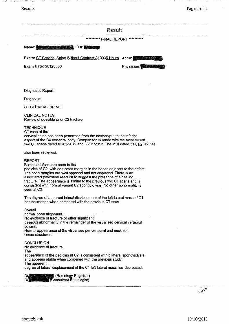
Results Result ********** FINAL REPORT ********** Name:······· ID#:--IJ Exam: CT Cervical Spine Without Contrast At 0936 Hours Ace#: Exam Date: 20120330 Diagnostic Report: Diagnosis: CT CERVICAL SPINE CLINICAL NOTES Review of possible prior C2 fracture. TECHNIQUE CT scan of the cervical spine has been performed from the basiocciput to the inferior aspect of the C4 vertebral body. Comparison is made with the most recent two CT scans dated 02/03/2012 and 30/01/2012. The MRI dated 31/01/2012 has also been reviewed. REPORT Bilateral defects are seen in the pedicles of C2, with corticated margins in the bones adjacent to the defect. The bone margins are well opposed and not displaced. There is no associated periosteal reaction to suggest the presence of a healing fracture. The appearance is similar to the previous two CT scans and is consistent with normal variant C2 spondylolysis. No other abnormality is seen at C2. The degree of apparent lateral displacement of the left lateral mass of C1 has decreased when compared with the previous CT scan. Overall normal bone alignment. No evidence of fracture or other significant osseous abnormality in the remainder of the visualised cervical vertebral column. Normal appearance of the visualised perivertebral and neck soft tissue structures. CONCLUSION No evidence of fracture. The appearance of the pedicles at C2 is consistent with bilateral spondylolysis and appears stable when compared with the previous study. The apparent degree of lateral displacement of the C1 left lateral mass has decreased. about:blank Page 1 of 1 --,---- 10/10/2013
Transcript of Result - parliament.qld.gov.au · for the joints of the cervical, thoracic and !umbo-pelvic spine...
Results
Result
********** FINAL REPORT **********
Name:······· ID#:--IJ
Exam: CT Cervical Spine Without Contrast At 0936 Hours Ace#:
Exam Date: 20120330
Diagnostic Report:
Diagnosis:
CT CERVICAL SPINE
CLINICAL NOTES Review of possible prior C2 fracture.
TECHNIQUE CT scan of the cervical spine has been performed from the basiocciput to the inferior aspect of the C4 vertebral body. Comparison is made with the most recent two CT scans dated 02/03/2012 and 30/01/2012. The MRI dated 31/01/2012 has
also been reviewed.
REPORT Bilateral defects are seen in the pedicles of C2, with corticated margins in the bones adjacent to the defect. The bone margins are well opposed and not displaced. There is no associated periosteal reaction to suggest the presence of a healing fracture. The appearance is similar to the previous two CT scans and is consistent with normal variant C2 spondylolysis. No other abnormality is seen at C2.
The degree of apparent lateral displacement of the left lateral mass of C1 has decreased when compared with the previous CT scan.
Overall normal bone alignment. No evidence of fracture or other significant osseous abnormality in the remainder of the visualised cervical vertebral column. Normal appearance of the visualised perivertebral and neck soft tissue structures.
CONCLUSION No evidence of fracture. The appearance of the pedicles at C2 is consistent with bilateral spondylolysis and appears stable when compared with the previous study. The apparent degree of lateral displacement of the C1 left lateral mass has decreased.
about:blank
Page 1 of 1
--,----
~
10/10/2013
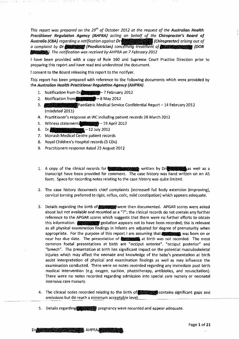
This report wos prepared on the 29'" of October 2012 at the request of the Australian Health Practitioner Regulation Agency (AHPRA) acting the Chiropractor's Boord of Australia {CBA) regarding a notification against arising aut of a complaint by Dr {Paediatrician) of (DOB lllllillllllllllllll- The notification was received by AHPRA an 7 February 2012
I have been provided with a copy of Rule 160 and Supreme Court Practice Direction prior to preparing this report and have read and understood the document.
I consent to the Board releasing this report to the notifyer.
This report has been prepared with reference to the following documents which were provided by the Australian Health Practitioner Regulation Agency (AHPRA).
D
1. Notification from [D~r::::'!- 7 February 2012 2. 1 - 8 May 2012
3. 1 Medical Service Confidential Report -14 February 2012
(misdated 2011)
4. Practitioner's response at lAC including patient records 28 March 2012
5. Witness statement{ £ -19 April 2012 6. Dr - 12 July 2012
7. Monash Medical patient records
8. Royal Children's Hospital records (3 COs)
9. Practitioners response dated 23 August 2012
1. A copy of the clinical records for written by Dr as well as a transcript have been provided for comment. The case history was hand written on an AS form. Space for recording notes relating to the case history was quite limited.
2. The case history documents chief complaints (increased full body extension (improving), cervical turning preferred to right, reflux, colic, mild constipation) which appears adequate.
3. Details regarding the birth of were then documented. APGAR scores were asked about but not available and recorded as a"?"; the clinical records do not contain any further reference to the APGAR scores which suggests that there were no further efforts to obtain this information. gestation appears not to have been recorded; this is relevant as all physical examination findings in infants are adjusted for degree of prematurity when appropriate. For the purpose of this report I am assuming that was born on or near her due date. The presentation of at birth was not recorded. The most common foetal presentations at birth are "occiput anterior''. "occiput posterior'' and "breech". The presentation at birth has significant impact on the potential musculoskeletal injuries which may affect the neonate and knowledge of the baby's presentation at birth assist interpretation of physical and examination findings as well as may influence the examination conducted. There were no notes recorded regarding any immediate post birth medical intervention (e.g. oxygen, suction, phototherapy, antibiotics, and resuscitation). There were no notes recorded regarding admission into special care nursery or neonatal intensive care nursery.
4. The clinical notes recorded relating to the birth oft•lll•ll contains significant gaps and omissions b ut_d_orE!i!C_h_arrli_nilllUillaccept~~lelt;>v_~L-
5. Details regardingl!ll!llllll pregnancy were recorded and appear adequate.
Page 1 of 21 AHPRA,.._

6. Details regarding previous chiropractic care at 4 weeks of age were recorded and appear adequate.
7. Details regarding the time period from leaving hospital to presentation at Dr 's clinic at-weeks and. days of age (date of birth and initial consultation 21/12/2011) appear to be included under the section titled "Neonate". The clinical records record ate of birth as -which appears to be an error.
8. Details regarding the sleep patterns of do not appear to have been obtained. Issues with sleep patterns in infancy may be important indicators of possible underlying conditions and provide a useful measure of response to interventions and treatment.
9. Details recorded regarding feeding are limited to "exclusively breast fed" and "vomiting after every feed". Further information regarding the infant's ability to breast feed and any issues relating to breast feeding are an important part of the case history. Information relating to the ability of the infant to suck, any issues with swallowing (e.g. gagging, choking, dribbling, prolonged feeds), any side preference , any fussiness or attachment issues whilst breastfeeding, length and frequency of breastfeeding should be obtained. Issues with breast feeding may be associated with issues with cranial nerve function and or issues affecting brainstem function which may need to be addressed during the examination and managment.
10. Details regarding systems review were not recorded. The systems review involves questions relating to major body systems and is an important component of the paediatric case history. In particular the system review acts as a screen for other issues which may be affecting the infant and which may indicate a need for further examination, assessment or referral to other health care practitioners. In particular further information relating to bowel function was not recorded. Concerns regarding constipation were noted in the records as one of the chief complaints.
11. Current and past medication uses were appropriately noted.
12. I would recommend that D review -case history form and provide increased space for note taking as well as develop an infant appropriate structure. I recommend that Dr pay more attention to infant behaviour in relationship to feeding, sleeping and bowel function when obtaining an infant case history. Deviation from expected behaviours with regards to sleeping, feeding and bowel function are the earliest, most sensitive and most common indicators clinicians use to identify potential issues with an infant and as a result clinicians need to pay particular attention to any changes with these behaviours. I recommend that Dr include a review of systems component to. case history taking. The review of systems is a crucial component of clinical information gathering which allows identification of other issues which may be relevant to the patient's examination and management.
13. The case history taken regarding an infant presenting for chiropractic care is a crucial and vital component of the clinical encounter. A well performed case history of an infant will need to consider the following items:
Case history with an infant typical structure • Jlr~s_en_ti11g COillQI~in1(~L--• prenatal history • perinatal history
Page 2 of21
AHPRA/Dr···
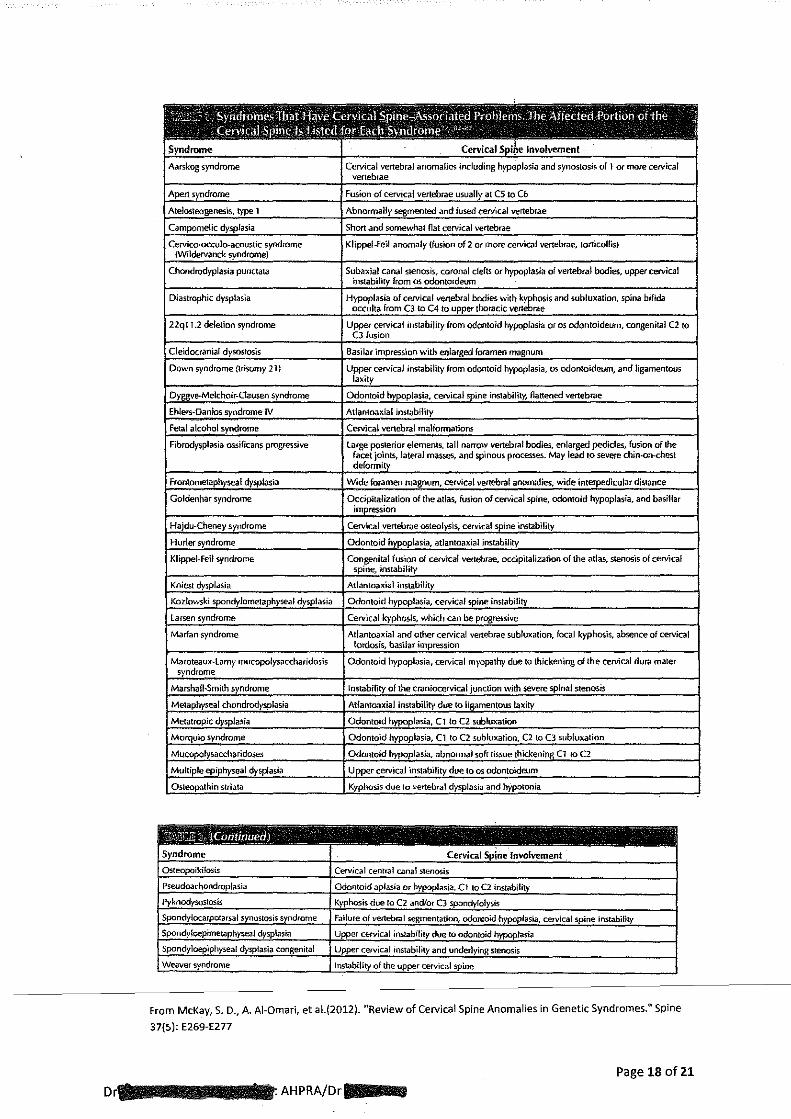
• neonatal history • infancy history • systems review • family history • assessment of development
When asking the parent about the prenatal history the following issues are usually addressed: • age of mother • previous pregnancies • number sibling • maternal health eg. hypertension, proteinuria, infection, stress etc. • maternal medication/ drugs use • X-ray or US exposure • foetal health, growth and positional issues
When asking about perinatal history the following issues are usually addressed: • length of gestation • intervention during labour e.g .. Forceps, ventouse suction • medication during labour eg. pethidine, epidural • duration of labour stages • details of membrane rupture • type of delivery caesarian or vaginal • presentation of neonate ie. occiput, brow, face, breech
When asking about neonatal history the following issues are usually addressed: • need for respirator, resuscitation, suction • APGAR score • weight, length, head circumference • Length of hospital stay, admission into special care nursery or intensive care. • Medication used • Post natal interventions such as need for oxygen, use of antibiotics, need for
phototherapy.
When asking about the infancy history the following issues are usually addressed: Sleeping Night
• Length of sleep
• How often wakes
• How well feeds
• How well settles Day
0 Length of day sleep
• Easily disturbed
• Restless
Feeding • Breast or Bottle • Attachment- difficulty attaching, preference for football hold • Fussiness
-------------·-s~cking-abHitv-
• _ Swallow I.e. gagging, coughing, choking, dribbling
D~AHPRA/Dr·-~~ Page 3 of 21
• Time taken- fast due to pain, slow due to poor suck or pain, normall0-20 mins • How often- pain relief effect offeed lasts approx 2 hrs. • Reflux and or projectile vomiting- how often, during or after or between feeds
• Name of formula
System review is crucial to avoid missing other conditions or diagnoses. When performing a review of systems the following issues are usually addressed:
Eyes, ears nose and throat Eyes: sticky eyes, conjunctivitis, tracks face, responds to smile Nose: snuffly breathing, mucous Ears: infections, responds to voice and noise Throat: infections
Respiratory system • Rattles, wheezes, vibrations • Chest infections e.g. bronchiolitis, croup, pneumonia • Apnoea • Difficulty breathing • Respiration rate I.e. increased or decreased
Cardiovascular system • Blue tongue or lips • Change in colour of arms or legs
Gastrointestinal system • How often has bowel motion • Is there straining if so for how long • Diarrhoea, hard stools • Bloating of tummy • Lot of bowel gas • Blood or mucous in stools
Genitourinary system • Smelly urine • How often wet nappy
Skin
• Rashes
• Spots
• Marks
• Bruises
When asking about the family history the following issue are usually addressed: • Allergies • Genetic disorders • Any significant health issue
When assessing_d.eJLelopment.tbe.foJio.wi.ng-acEUJsually.aaeFessecl::--------------
• Ask about use and symmetry movement of arms, hands, legs.
Page4 of21
D~AHPRMD·--
• Hand preference should not be evident prior to 12 months of age
• Head control
14. The standard examination of an infant would include noting or measuring birth weight, head
circumference and length. The anterior fontanel should be measured and plotted against
normal data. The posterior fontanel should be assessed and a note of it being either open or
closed made in the patient's record. Cranial nerve assessment should be conducted. Upper
and lower extremity muscle stretch reflexes should be assessed and graded. The
scapulohumeral reflex should be assessed. Respiratory and cardio-vascular function should
be assessed. Primitive reflexes including, but not limited to, Mora reflex, plantar grasp
reflex, palmar grasp reflex, rooting and sucking reflex, asymmetric tonic neck reflex, vertical
suspension and ventral suspension reflexes, Perez reflex and Galant's reflex should be
assessed. Direct, consensual as well as red pupillary reflex need to be assessed. An
assessment of muscle tone should be performed. Active and passive range of joint motion
for the joints of the cervical, thoracic and !umbo-pelvic spine including the sacrum and
coccyx should be assessed. The hip joints should be assessed for dysplasia and associated
changes with hip joint flexion and abduction. The skin needs to be assessed for rashes, birth
marks, hairy patches or any other blemish. The shape of the head and state of the cranial
sutures needs to be assessed.
15. Dr-s examination noted the resting posture ol 'lias right head tilt and
head rotation to the right, the anterior fontanel as open and medium, moderate right
plagiocephaly, right frontal bossing, right eye superior and mild anterior, tension over
superior ascending colon, blink with More reflex, Palmar and Plantar grasp reflex present,
Rooting reflex as absent, Sucking reflex was reported, Placing and Walking reflex as absent
just arched backwards, Babinski as present with one tick for either left or right side
(uncertain), Cranial nerves 2,3,4,6,7,8,9,10 were assessed as normal. The following
developmental milestones were recorded as having being attained: able to hold head up
from table momentarily, head and shoulder can be supported by the forearms, smiles,
doesn't reach for familiar objects, primitive grasp reflex present was reported, makes cooing
sounds and laughs. Persistent fisting, scissoring of legs, frog leg position, rapid tremors
sustained opisthotonus, facial ptosis, asymmetrical movement and ankle clonus were all
recorded as being absent.
16. The examination recorded by Dr4lilll •• lllldoes not include testing of upper or lower
extremity muscle stretch reflexes (also known as deep tendon reflexes); it is normal to test
Achilles reflex, quadriceps reflex, biceps reflex, brachioradialis reflex and triceps reflex
bilaterally. The muscle stretch reflexes provide important information regarding function of
some ofthe cervical and lumbar spinal nerves. Abnormal muscle stretch reflexes may be
associated with upper motor neuron issues. Testing muscle stretch reflexes can assist with
detecting clonus which may also be associated with upper motor neuron issues.
17. Pull to sitting test was not recorded. Pull to sitting test involves the supine infant being
---------lgeAHy-i'Jel<l-by-tlle-hands-and-puHerl-to-a-si tti ng positilm-:-The mfa nt s al51hty t"'o~s"'u"'p"'p"'o~rt"t"'h"'e"irc------
head, the degree of head extension, any head tilt or rotation and degree of arm use are
noted. This test provides important information concerning neck flexor function as well as
Page 5 of 21
AHPRAA····
upper extremity muscle tone. Given the presenting concerns of head asymmetry and head
posturing assessing the infant's neck and upper extremity function during the Pull to sitting
test would be expected as part of the examination o·ftti!IIIIIIIIIIIIIIIIIP
18. Upper extremity muscle stretch reflexes assist assessment of the lower cervical spine nerves.
Upper cervical nerve roots can be assessed by testing for the presence oft he
scapulohumeral reflex. The scapulohumeral reflex is not normally present and if there is a
positive response indicates involvement of one or more of the four upper cervical nerve
roots on the side of the positive test. The scapulohumeral reflex may assist in detecting
pathology above the C3 disc. (1, Appendix A. 2, Appendix B.)
19. Direct and consensual pupillary reflexes should be tested and recorded. Pupillary reflexes
may be altered with brainstem dysfunction and are important in assessing the infant's
neurological status.
20. DrJIIIIIIII!IIIIIIIrecorded dysfunction affecting the Cl vertebra, 51 vertebra, T4/51evel of the
spine, right suboccipital, left temporal, right sphenoid and right occiput. No supporting
physical examination findings were recorded. Cervical spine active or passive range of
motion testing was not recorded in the examination notes. Normally the dysfunction
identified at Cl by Dr (ClRP) can be expected to be associated with decreased
head rotation to the left as well as decreased upper cervical spine right lateral flexion with
these findings most apparent during active range of motion assessment.
21. Dr~~::;ha;s conducted an examination which has included and noted a number of tests o status. There are significant gaps in the examination, in particular
assessment of the Pull to sitting test, pupillary reflexes, scapulohumeral reflex as well as
upper and lower extremity muscle stretch reflexes. Performance of these tests would have
allowed a much better understanding ofl\l!lllllllla''s neurological function and status at the
start of treatment as well as possibly assisting in early identification of any issues such as
contra indications to adjusting or treatment. Upper cervical assessment findings supporting
the diagnosis of a ClRP were not recorded.
22. Weinstein states that the most common deficit with abnormalities of the craniovertebral
junction in children is myelopathy. The most common symptom is neck pain, present in
85%. The child may present with hemiparesis, monoparesis, paraparesis and quadriplegia.
Brain stem and cranial nerve deficits were exhibited by abnormalities such as sleep apnea,
dysphagia and aspiration pneumonia. Internuclear ophthalmoplegia (impaired external eye
muscle function) may be present as downbeat nystagmus. The most common cranial nerve
dysfunction was hearing loss which occurred in 25%. There may be bilateral or unilateral
paralysis of the soft palate and pharynx which is associated with aspiration pneumonia, poor
feeding and poor weight gain. (3) Appendix C
Page 6 of21
AHPRAA·--·
23. The initial treatment notes recorded for the 21 December 2011 document adjustment per
examination by hold, CST (craniosacral technique), D/R (dural release technique). These
notes indicate low force techniques involving finger pressure were used to correct the
identified spinal and cranial dysfunctions.
24. The second treatment occurred on the 23 December 2011 with the clinical notes recording
great sleep day after the adjustment, bit of tummy pain today as well as using bowels now
after almost every feed. The spinal and cranial areas treated at the initial visit were again
treated except for the right suboccipital(? This is referring to muscles). The type of
correction or technique used is not noted. The written statement provided by
infers but does not clearly state that the same techniques were used. The statement
provided by Mrs 'ndicates that similar treatment was provided. Abdominal
assessment was noted and recorded as no abnormality detected.
25. The next consultation was recorded as occurring on the 16 January 2012 at which time
···~·as approximately 3.5 months of age. The clinical notes report thathas been much improved post adjustment but had gone backwards over three weeks.
Treatment of C1RP, left temporal, right sphenoid, right occiput, T5 and lumbosacral
mobilisation was noted. The type of correction or technique used is not noted. The written
statement provided by Dr states that the same techniques used at the initial visit
were used. The statement provided indicates that similar treatment was
provided. Decreased left rotation and right lateral flexion are noted in the clinical notes. It
is not clear if this is referring to the cervical spine or elsewhere such as the lumbar spine, it is
also not clear as to whether the restricted motion was assessed before or after the
treatment provided on the 16 January. There is also a note that there was 7 days between
poos (bowel motions) and that was exclusively breastfed still.
26. I note that at the 16 January consultation, after not seeing-for a period of three
weeks, no further examination or assessment was noted other than confirming the spinal
and cranial dysfunction. ••••1s resting posture, degree of head control, presence or
absence of the Mora reflex, hip joint function, upper and lower extremity reflexes, presence
or absence of clonus were not noted. It is important when involved in the care of infants that regular assessment of neurological status and neuromuscular development occurs. In
addition regular assessment of hip joint function with reference to detection of dysplasia or
dislocation is performed. Physical growth parameters such a weight, length and head
circumference should be regularly assessed. It would be expected that not having seen an • infant under six months of age for a period of three weeks that a basic examination as noted
in this paragraph would be performed and recorded.
27. was next seen by Dr~ on the 18 January 2012. The clinical notes record
now nearly 4 months of age and that she had been 'pretty terrible' post
Left rotation is noted as having improved but still restricted. ClRP, right
---------,onc:cciriprrurtt,:-rr ighrsphenord, lumbosacral mobilrsation, ileocaecal valve and bowel massage were
reported as being treated/performed. Thoracic spine and hips were reported as having no
Page 7 of21 AHPRA/D·--
abnormality detected. Decreased movement of the transverse colon leading to(?) bowel
massage was noted. Big shift in facial asymmetry was noted; it is not clear if this is an
improvement or deterioration.
28. I note that at the 18 January consultation in spite of there being a report of worse behaviour
after the previous treatment that no attempt to identify a possible cause for the poor
response was recorded. Whenever infants respond poorly or are worse after a consultation
it is important for the chiropractor to try to ascertain the reason for the response as this
could be early signs of an underlying serious issue. There may be a simple explanation such
as failure to have a bowel motion, teething, upper respiratory infection, maternal diet
related upset or there may be a more serious issue such a urinary tract infection or
aggravation due to treatment. When there is poor response or an infant is worse after a
treatment typically some further physical examination is conducted to try to eliminate
possible common causes of irritability; neurological re-examination is conducted to identify
and possible neurological deterioration and further questions are asked of the mother to try
to ascertain any possible cause for the poor response. It would be prudent to check the
infant's temperature as a possible sign of infection. If the aggravation was possibly due to
the treatment provided at the previous consultation then future treatment should be
reassessed and the need for further investigation addressed.
29. On the 23 January 2012 Dr ecorded tha~showed definite
improvement with tummy time. Treatment was recorded as involving ClRP, right occiput
right coronal suture separation, T4, lumbosacral mobilisation and right sphenoid.
Improvement with facial balance was noted. The type of correction or technique used is not
noted. The written statement provided by Dr 3 IIIJoes not state that the same
techniques used at the initial visit were used but I do not see any reason to suspect that
different treatment was provided. The statement provided by~ indicates that
treatment similar to previous treatments was provided.
30. · was seen again by D~on the 25 January with-linical records
noting "cranky today- teething". Almost 4 months of age. Treatment of ClRP, right occiput,
right sphenoid, suture separation, Thoracic no abnormality detected, lumbosacral no abnormality detected, frontal lift. The written statement provided by D does not
state that the same techniques used at the initial visit were used but I do not see any reason
to suspect that different treatment was provided. The statement provided byiJIIIIIIIIIIIIIIIIII indicates that treatment similar to previous treatments was provided.
31. Clinical records note mother calling the clinic concerned regarding an issue with
poor head control on the 30 January 2012. The poor head control was noted in Dr
lli!llllllllllllllllllllt clinical records as being reported as becoming an issue from 27 /281h January
some two days after the last treatmentwith D Advice was provided by Dr
dlllilllllllllllllllttA.at should be taken to hospital emergency department. There is no
reference to history of recent accidents or trauma. statement reported
issues with s head control being noticed on Friday 27 January 2012 and that movement appeared to upsetlllllllllllllll!lllllaon Saturday 28 January 2012. also
Page 8 of21 Dr ______ IIJAHPRA/illil--11
Dr
states that on Thursday 26 Januarylllllllll!lllllllland her parents attended a friend's
barbeque. My personal experience of infants receiving care for musculoskeletal issues is
that there may be aggravation of their underlying condition as a result of increased handling
as well as handling by inexperienced people which may occur at parties and gatherings. It is
very difficult to exclude an incident which may have not been reported to the parent's
unless the parents are able to confirm that they were holding the entire time
they were at the function.
32. I note that Dr
January by
responded promptly to the contact received by the clinic on the 30
mother. I also note that the advice recorded as being provided by
Dr was appropriate, timely and adequate given the concerns expressed by
-smother. D clinical notes record a good level of contact being
maintained during that day.
33. The initial radiology opinion was that g11111111111thad suffered a bilateral fracture ofthe posterior arch of axis vertebra (C2). CT cervical spine without contrast performed on the 30
March 2012 was reported as follows": "Biloteral defects are seen in the pedicles of C2, with carticated margins in the bones adjacent to the defect. The bone margins are well opposed and not displaced. There is no associated periosteal reaction to suggest the presence of a healing fracture. The appearance is similar to the previous two CT scans and is consistent with normal variant C2 spondylolysis. No other abnormality is seen at C2. The degree of apparent lateral displacement of the left lateral mass of Cl has decreased when compared with the previous CT scan. Overall normal bone alignment. No evidence of fracture or other significant osseous abnormality in the remainder of the visualised cervical vertebral column. Normal appearance of the visualised perivertebral and neck soft tissue structures. CONCLUSION No evidence of fracture. The appearance of the pedic/es at C2 is consistent with bilateral spondylolysis and appears stable when compared with the previous study. The apparent degree of lateral displacement of the C1/eft lateral mass has decreased".
34. The final CT scan performed on the 6 July 2012 report recorded: "There are bilateral defects seen within the pedicles of C2 in keeping with developmental C2 spondy/olisis. However, the morphology of the /eft-sided defect has changed slightly since the previous study with overall narrowing of the defect and minor new bone formation which may indicate healing of o concurrent fracture. Unchanged appearances of the right sided defect. Unchanged appearances of the Cllateral masses with persistent but mild left sided asymmetry. Bony alignment is maintained. No other significant osseous abnormality demonstrated within the remainder of the visualised cervical column. Normal appearances of the adjacent soft tissues. Conclusion: Evidence of some healing involving the left C2 pedicle defect suggesting o likely previous underlying trauma/ fracture superimposed on a developmental spondylolisis."
35. I note that the response of the pars defect noted on the final CT scan on the 6 July 2012 as
minor new bone formation over four months after( was treated by
does not match well with the healing response of a fracture of the posterior arch of axis
reported by Parisi et at (8) after two months in a three month old infant and referred to in
paragraph 43 of this report.
AHPRA/Dr--Page 9 of 21
36. Development of C2 or axis vertebra. The axis develops from five primary centers of
ossification, which are usually present at birth. There is one center for the body oft he axis,
two for the odontoid process (which represents the body of the atlas), and two for the
vertebral arch. The neurocentra I synchondrosis, the cartilaginous structure that joins the
body to the two posterior centers of ossification, ossifies between the ages of three and six
years. Spondylolysis of the axis has been reported in adults and may represent a persistent
neurocentra I synchondrosis. With increasing ossification, the radiolucent image of the
synchondrosis becomes narrow. (4) Appendix D
37. Congenital defects, such as the absence of the pedicles, are rare but may be associated with
clinical symptoms or signs of instability. The synchondrosis between the body and the
posterior arch of the second cervical vertebra is a normal stage in the ossification of the
bone. The defect may be seen on a lateral roentgenogram, and there may be considerable
variation in its appearance. (4)
38. The C2 posterior arches fuse in the midline by 2 to 3 years and fuse with the body by 3 to 6
years. (S) Appendix E. Incomplete ossification and physiologic hypermobility of the pediatric
cervical spine contribute to imaging findings that can be confused with pathological
conditions. (5) On both CT and plain X-rays synchondroses can be mistaken for fracture
lines. Conversely, fractures through synchondroses can be misinterpreted as within the
realm of normal. (5)
39. Hangman's fractures and spondylolysis in infants. Hangman's fracture comprises an avulsion
fracture of the posterior elements of C2 in combination with an anterolisthesis of C2 on C3.
Congenital C2 spondylolysis consists of a division of the portio interacticularis with an
unclear etiology, as the site ofthe defect does not correspond to a primary ossification
center. (6) Appendix F
40. It has been suggested by Currarino that the underlying cause is a primitive defect of
chondrification of mesenchymal precursors of the vertebrae (7) Appendix G. Currarino eta!.
described three entities: first, a congenital defect, spondylolysis, with no abnormal motion;
secondly, an acquired traumatic spondylolysis with abnormal motion; and thirdly, a clear
hangman's fracture (7). This differential diagnosis may have important therapeutic
consequences and a large impact on medico-legal proceedings. (6)
Page 10 of21
AHPRA/D·--
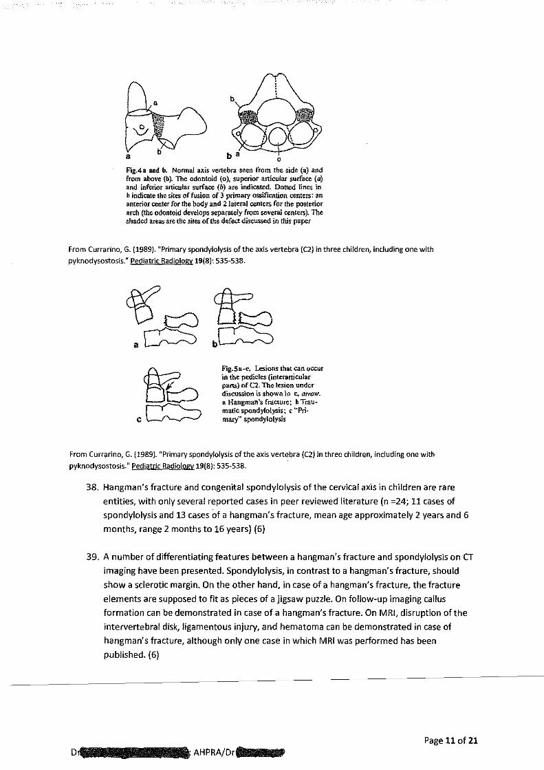
F'.g.4a and b. Normal axis vertebra seen from the side (a) and from above (b). The odontoid (o), superior articular surface (a} and inferior articular surface (b) are indicated. Dotted lines in b indicate the sires of fusion of 3 primary ossification centers: an anterior center for the body and 2lateral centers for the posterior arch (the odontoid develops separately from several <:enters). The shaded areas are the sites of the defec.:t discussed in this paper
From Currarino, G. (1989). "Primary spondylolysis of the axis vertebra {C2) in three children, including one with
pyknodysostosis." Pediatric Radiology 19(8): 535-538.
Fig.Sa~. Lesions that can occur in the pedicles (interarticular parts) of (.."2. The lesion under discussion is shown in c, arrow. a Hangman's fracture; b Traumatic spondylolysis; c ··Pri· mary'' spondylolysis
From Currarino, G. (1989). "Primary spondylolysis of the axis vertebra {C2) in three children, including one with
pyknodysostosis." Pediatric Radiology 19{8): 535-538.
38. Hangman's fracture and congenital spondylolysis of the cervical axis in children are rare
entities, with only several reported cases in peer reviewed literature (n =24; 11 cases of
spondylolysis and 13 cases of a hangman's fracture, mean age approximately 2 years and 6
months, range 2 months to 16 years) (6)
39. A number of differentiating features between a hangman's fracture and spondylolysis on CT
imaging have been presented. Spondylolysis, in contrast to a hangman's fracture, should
show a sclerotic margin. On the other hand, in case of a hangman's fracture, the fracture
elements are supposed to fit as pieces of a jigsaw puzzle. On follow-up imaging callus
formation can be demonstrated in case of a hangman's fracture. On MRI, disruption of the
intervertebral disk, ligamentous injury, and hematoma can be demonstrated in case of
hangman's fracture, although only one case in which MRI was performed has been
published. {6)
Page 11 of21
AHPRA/Drllllllilllll\lllll'
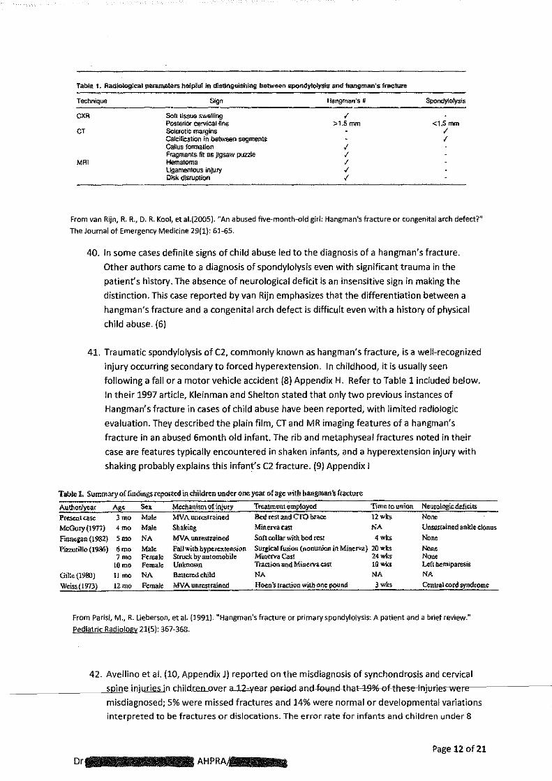
Table 1. Racliorogical ~rs helpful in d'IStinguiSiling_ between SJ>Qndy1Qly$is and hangman's fi1Jdure
Technique Sign Hangm;~;~n·s li SpondYlolysis
CXR Soft tissue sw&lling < Post(lrtor cervical tine >1.5mm <1.5 mm
CT Scte:rotiC margins I cak:iftcation in between segments I Gallus formation I Fragments frt as jigsaw puzzle I
MAl Hematoma I Ligamentous injury I Olsk disruption I
From van Rijn, R. R., D. R. Kool, et al.(2005). "An abused five-month-old girl: Hangman's fracture or congenital arch defect?11
The Journal of Emergency Medicine 29(1}: 61-65.
40. In some cases definite signs of child abuse led to the diagnosis of a hangman's fracture.
Other authors came to a diagnosis of spondylolysis even with significant trauma in the
patient's history. The absence of neurological deficit is an insensitive sign in making the
distinction. This case reported by van Rijn emphasizes that the differentiation between a
hangman's fracture and a congenital arch defect is difficult even with a history of physical
child abuse. (6)
41. Traumatic spondylolysis of C2, commonly known as hangman's fracture, is a well-recognized
injury occurring secondary to forced hyperextension. In childhood, it is usually seen
following a fall or a motor vehicle accident (8) Appendix H. Refer to Table 1 included below.
In their 1997 article, Kleinman and Shelton stated that only two previous instances of
Hangman's fracture in cases of child abuse have been reported, with limited radiologic
evaluation. They described the plain film, CT and MR imaging features of a hangman's
fracture in an abused 6month old infant. The rib and metaphyseal fractures noted in their
case are features typically encountered in shaken infants, and a hyperextension injury with
shaking probably explains this infant's C2 fracture. (9) Appendix I
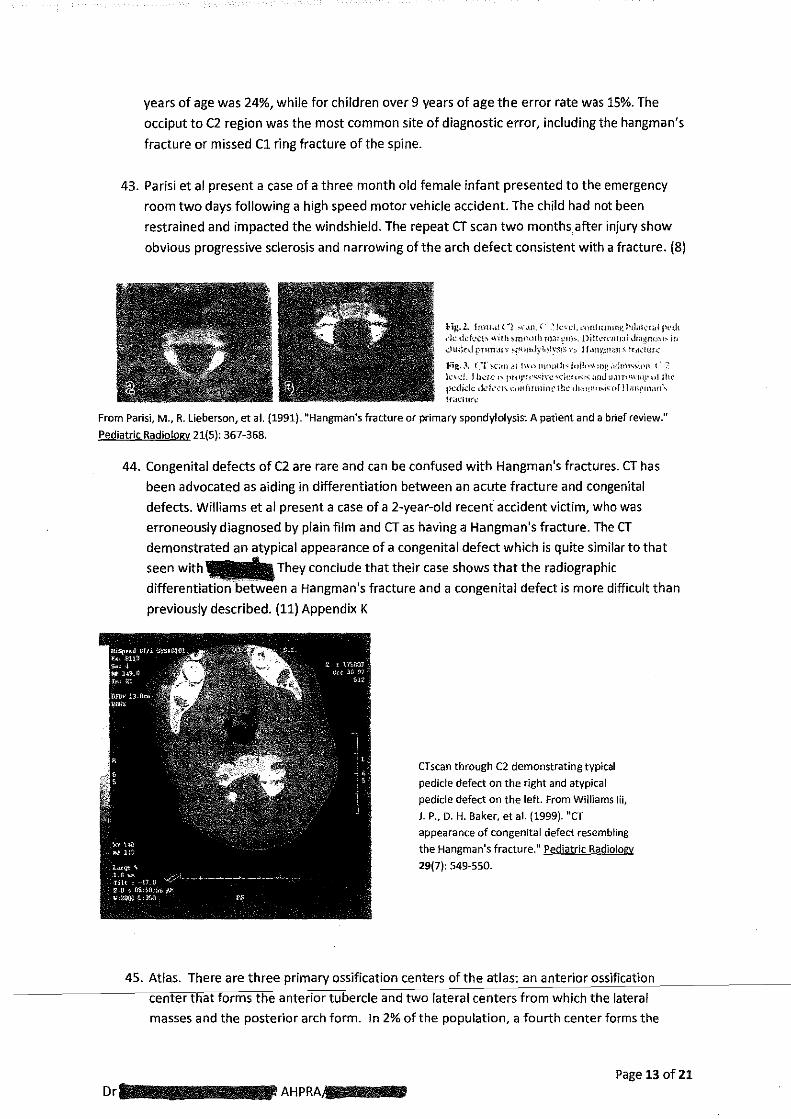
Tobie I. Summary of findings repot!<d in <:hil.dren under on< yeor Mage with ~angmon's frO<wre
Author/year Ag< s.. Mechanism of inji.Jr}' Treatment f::mptoyed Time to union Neurolcg,icdeficiu Present case 3mo Male MVAunrcstrained B<d=t and CTO brace 12wk$ None McGory (1977) 4mo Male Shaking Minerva east NA UnstlSt~ined ankle clonus
Finnegan (1982) 5mo NA MVA unfe.'\lrained So!\ collar with bed rest 4Wts None
P.inutillo (1986) 6mo Male FalJ Wilb. bypercxrensian Surgical fusion (noouffion in Miflerv,a} 20 wks NOne 7mo Female Struck by automobile Min~ cast 24wks None
tOmo Female Unknown Tta.:ti-on and Minef'V.a east 1G wks Left bem.iparests
Oille(J9&l) 11 rno NA Battered chlld NA NA NA Weiss{l973) 12mo- FcmaJt: MVA unr-c.stfained Hoen 's traction wiib one pound 3wks Ceru~l oord syndrome
From Parisi, M., R. Ueberson, et al. (1991). "Hangman's fracture or primary spondylolysis: A patient and a brief review."
Pediatric Radiology 21(5): 367-368.
42. Avellino et al. (10, Appendix J) reported on the misdiagnosis of synchondrosis and cervical
---------"s,p·"m"e'-'i'!Jn'llju"'r-"ie;;;sui"'n""cbiidren_mter a 12-year~feuflfl-#laHJ3-%-eHhes-e-injttries-we=-------
misdiagnosed; S% were missed fractures and 14% were normal or developmental variations
interpreted to be fractures or dislocations. The error rate for infants and children under 8
Page 12 of21 Dr -----AHPRA--1111111111
years of age was 24%, while for children over 9 years of age the error rate was 15%. The
occiput to C2 region was the most common site of diagnostic error, including the hangman's
fracture or missed C1 ring fracture of the spine.
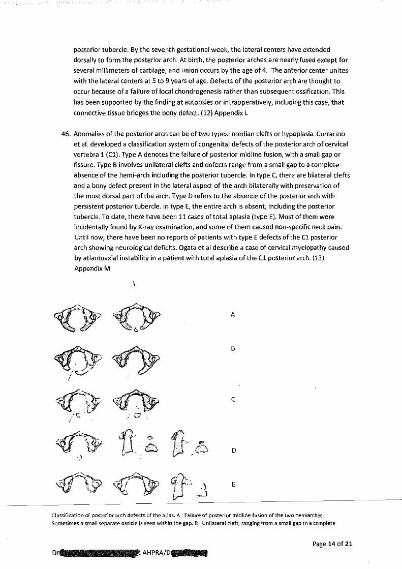
43. Parisi et al present a case of a three month old female infant presented to the emergency
room two days following a high speed motor vehicle accident. The child had not been
restrained and impacted the windshield. The repeat CT scan two months after injury show
obvious progressive sclerosis and narrowing of the arch defect consistent with a fracture. (8)
Hg.L lrnU.ll l ~1 ~,-,\n. ( · : k·.-d. <-'on!nm!lll: hlilkr;,l pi';,.h <k dck<:l'> v.ith ~mt~<lth m:n~ll),;., Dilt\'rt·rHhl Jr;t~n·-'~~~ ir• dtHkd prHn~H y ~~~·m.Jy1oJiy:w; r:,. j l.lfll'.H'an·~ !r.t>.:tur.::
1<1£, • .'. ( ·r· -..cul .~1 l~\(l !1\Jl!llh~ J<lil<•>':n!: ol<b)l'-,\\!)11, l. :'
ln..:!. I h.:;.;,-, !'"'!''1-'.'>~in: ~·.:ki<"'J" ;1nd n.lJT•'WIII)~''Ilh· p-cdid~.: tl'k·:h n•ni-irmint' ih1-' ,]r;,:,ll<l'-i~ of]l.itll'l!l<•tl\ lf;H.:ilHt_'
From Parisi, M., R.lieberson, et al. (1991}. "Hangman's fracture or primary spondylolysis: A patient and a brief review."
Pediatric Radiology 21(5): 367-368.
44. Congenital defects of C2 are rare and can be confused with Hangman's fractures. CT has
been advocated as aiding in differentiation between an acute fracture and congenital
defects. Williams et al present a case of a 2-year-old recent accident victim, who was
erroneously diagnosed by plain film and CT as having a Hangman's fracture. The CT
demonstrated an atypical appearance of a congenital defect which is quite similar to that
seen with ... They conclude that their case shows that the radiographic
differentiation between a Hangman's fracture and a congenital defect is more difficult than
previously described. (11) Appendix K
CTscan through C2 demonstrating typical
pedicle defect on the right and atypical
pedicle defect on the left. From Williams Iii,
J.P., D. H. Baker, et al. (1999). "CT
appearance of congenital defect resembling
the Hangman's fracture." Pediatric Radiology
29(7): 549·550.
45. Atlas. There are three primary ossification centers of the atlas: an anterior ossification
center that forms the anterior tubercle and two lateral centers from which the lateral
masses and the posterior arch form. In 2% of the population, a fourth center forms the
Page 13 of21
Dr·-----·AHPRA·--·
posterior tubercle. By the seventh gestational week, the lateral centers have extended
dorsally to form the posterior arch. At birth, the posterior arches are nearly fused except for
several millimeters of cartilage, and union occurs by the age of 4. The anterior center unites
with the lateral centers at 5 to 9 years of age. Defects of the posterior arch are thought to
occur because of a failure of local chondrogenesis rather than subsequent ossification. This
has been supported by the finding at autopsies or intraoperatively, including this case, that
connective tissue bridges the bony defect. (12) Appendix L
46. Anomalies of the posterior arch can be of two types: median clefts or hypoplasia. Currarino
et al. developed a classification system of congenital defects of the posterior arch of cervical
vertebra 1 (Cl). Type A denotes the failure of posterior midline fusion, with a small gap or
fissure. Type B involves unilateral clefts and defects range from a small gap to a complete
absence of the hemi-arch including the posterior tubercle. In type C, there are bilateral clefts
and a bony defect present in the lateral aspect of the arch bilaterally with preservation of
the most dorsal part of the arch. Type D refers to the absence of the posterior arch with
persistent posterior tubercle. In type E, the entire arch is absent, including the posterior
tubercle. To date, there have been 11 cases of total aplasia (type E). Most of them were
incidentally found by X-ray examination, and some of them caused non-specific neck pain.
Until now, there have been no reports of patients with type E defects of the Cl posterior
arch showing neurological deficits. Ogata et al describe a case of cervical myelopathy caused
by atlantoaxial instability in a patient with total aplasia of the Cl posterior arch. {13)
Appendix M
I '
A
B
c
1]·····.•. ,. 1 ..
'" ,• D
E
Classification of posterior arch defects of the atlas. A : Failure of posterior midline fusion of the two hemiarches. Sometimes a small separate ossicle is seen within the gap. B : Unilateral cleft, ranging from a small gap to a complete
Page 14 of21 AHPRA/D····
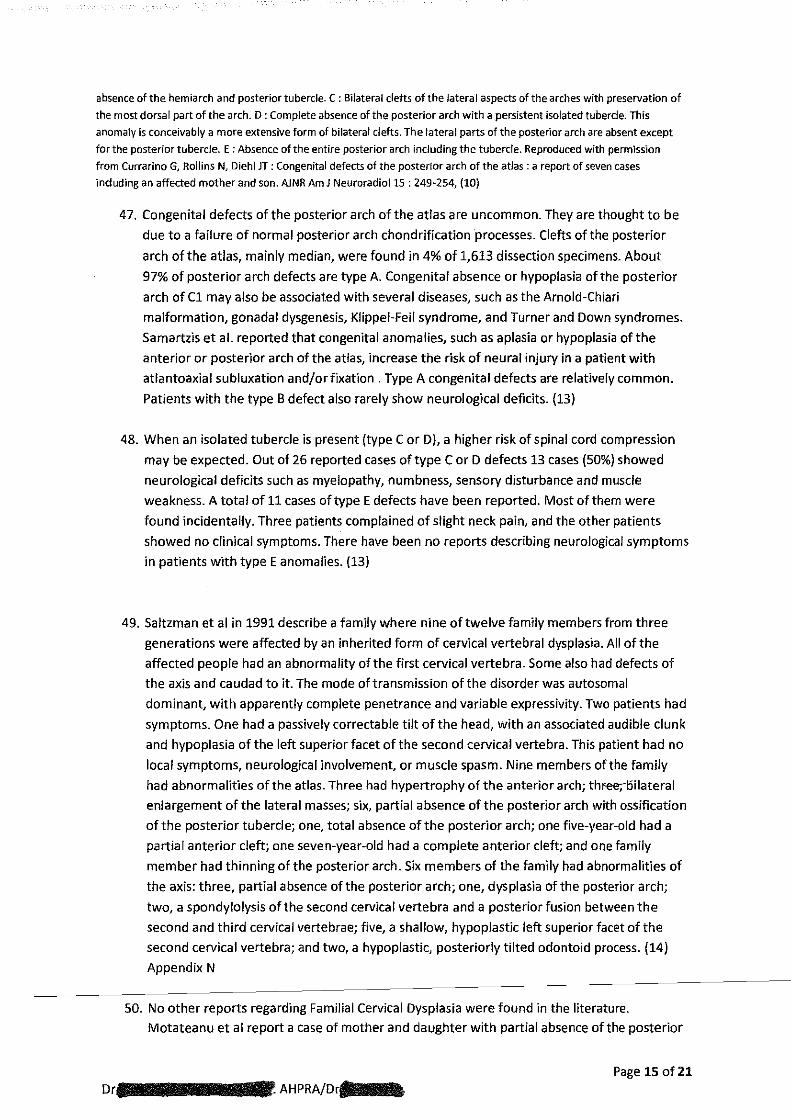
absence of the hemiarch and posterior tubercle. C : Bilateral clefts of the lateral aspects of the arches with preservation of
the most dorsal part of the arch. D: Complete absence of the posterior arch with a persistent isolated tubercle. This
anomaly is conceivably a more extensive form of bilateral clefts. The lateral parts of the posterior arch are absent except
for the posterior tubercle. E :Absence of the entire posterior arch including the tubercle. Reproduced with permlssion
from Currarino G, Rollins N, Diehl JT: Congenital defects of the posterior arch of the atlas : a report of seven cases
including an affected mother and son. AJNR Am J Neuroradio115 : 249-254, (10)
47. Congenital defects of the posterior arch of the atlas are uncommon. They are thought to be
due to a failure of normal posterior arch chondrification processes. Clefts of the posterior
arch of the atlas, mainly median, were found in 4% of 1,613 dissection specimens. About
97% of posterior arch defects are type A. Congenital absence or hypoplasia of the posterior
arch of C1 may also be associated with several diseases, such as the Arnold-Chiari
malformation, gonadal dysgenesis, Klippei-Feil syndrome, and Turner and Down syndromes.
Samartzis et al. reported that congenital anomalies, such as aplasia or hypoplasia of the
anterior or posterior arch of the atlas, increase the risk of neural injury in a patient with
atlantoaxial subluxation and/orfixation . Type A congenital defects are relatively common.
Patients with the type B defect also rarely show neurological deficits. (13)
48. When an isolated tubercle is present (type Cor D), a higher risk of spinal cord compression
may be expected. Out of 26 reported cases of type CorD defects 13 cases (50%) showed
neurological deficits such as myelopathy, numbness, sensory disturbance and muscle
weakness. A total of 11 cases of type E defects have been reported. Most of them were
found incidentally. Three patients complained of slight neck pain, and the other patients
showed no clinical symptoms. There have been no reports describing neurological symptoms
in patients with type E anomalies. (13)
49. Saltzman et al in 1991 describe a family where nine of twelve family members from three
generations were affected by an inherited form of cervical vertebral dysplasia. All of the
affected people had an abnormality of the first cervical vertebra. Some also had defects of
the axis and caudad to it. The mode of transmission of the disorder was autosomal
dominant, with apparently complete penetrance and variable expressivity. Two patients had
symptoms. One had a passively correctable tilt of the head, with an associated audible clunk
and hypoplasia of the left superior facet of the second cervical vertebra. This patient had no
local symptoms, neurological involvement, or muscle spasm. Nine members of the family
had abnormalities of the atlas. Three had hypertrophy of the anterior arch; thr-ee;i:lilateral
enlargement of the lateral masses; six, partial absence of the posterior arch with ossification
of the posterior tubercle; one, total absence of the posterior arch; one five-year-old had a
partial anterior cleft; one seven-year-old had a complete anterior cleft; and one family
member had thinning of the posterior arch. Six members of the family had abnormalities of
the axis: three, partial absence of the posterior arch; one, dysplasia of the posterior arch;
two, a spondylolysis of the second cervical vertebra and a posterior fusion between the
second and third cervical vertebrae; five, a shallow, hypoplastic left superior facet of the
second cervical vertebra; and two, a hypoplastic, posteriorly tilted odontoid process. (14)
Appendix N
50. No other reports regarding Familial Cervical Dysplasia were found in the literature. Motateanu et al report a case of mother and daughter with partial absence of the posterior
Page 15 of21
Dr·-----~~.AHPRA/D·-·

arch of the atlas. (15) Appendix 0. Currarino reported an affected mother and son,
suggesting an autosomal dominant inheritance. (Currarino G, Rollins N, Diehl JT. Congenital defects of
the posterior arch of the atlas :a report of seven cases including an affected mother and son. Am J Neuroradiol
1994 ; 15 : 249-254.]
51. Kaissi et al report two siblings and their mother, with congenital, persistent torticollis,
plagiocephaly, facial asymmetry, grooved tongues, and asymptomatic "dolicho-odontoid
process". All are of normal intelligence. No associated Neurological dysfunction, paresis,
apnoea, or failures to thrive were encountered. Radiographs of the cervical spine were non
contributory, but 3D CT scanning of this area allowed further visualisation of the cervico
cranial malformation complex in this family and might possibly explain the sudden early
juvenile mortality. Agenesis of the posterior arch of the atlas and bifidity/clefting of anterior
arch of the atlas associated with asymptomatic "dolicho-odontoid process" were the
hallmark in the proband and his female sibling. Some of the features were present in the
mother. All the family subjects were investigated. To the best of their knowledge the
constellation of malformation complex in this family has not been previously reported. (16)
Appendix P.
52. During the period of care under Dr~,-~ father, was x-rayed by Dr-( exact date not available to me) with the x-ray revealing absence of
the posterior arch of C1 with an ossified posterior tubercule. The radiological report for
--was not available to me whilst writing this report. This description appears to be
consistent with a type D congenital anomaly of Cl. This type of congenital anomaly affecting
Cl is not common and I was only able to find four published papers describing familial
inheritance of congenital posterior of Cl anomalies. It is recognised that a patient with one
congenital anomaly is more likely to have other congenital anomalies and that as a result it is
prudent to image other spinal areas prior to chiropractic care. In addition, as other body
systems may be involved a screening examination of these areas may be necessary.
53. The issue of x-raying all family members of people with posterior arch anomalies of Cl
without any other significant clinical indication prior to chiropractic care has not to my
knowledge been conclusively addressed in the literature. I am not aware of any guidelines
or recommendations suggesting that a child of a person with Type D orE absence of the
posterior arch of atlas should be x-rayed prior to chiropractic care. As a result of the
literature search conducted for this report articles by Saltzman(14), Motateanu (15),
Currarino (Paragraph 50) and Kaissi (16) were identified which report possible autosomal
dominant pattern of inheritance of some upper cervical spine anomalies. Given the rare
nature ofType E absence of the posterior arch of atlas and the possibility offamilial
inheritance occurring, I suggest that future guidelines and recommendations adopt the
position that when Type D orE atlas posterior arch anomalies are detected or known then
all close genetic relations of that individual undergo cervical spine x-ray examination prior to
receiving chiropractic care. This recommendation is balanced with the need to minimise
radiation exposure from x-rays in children which is well known and accepted within the
---------cllc•iil'• oop011r<arcctiticcllpmroo1ife-ss•on as well as all other health care professions.
Page 16 of 21
Drtlll88lilllllllilllllllillllllliiiiiiiSAHPRA/Dr-
54. When considering x-ray investigation of children the potential gain must be assessed as
being greater than the radiation exposure risk. It is my opinion that the presence of the
posterior arch defect inf r, father did not on its own provide enough clinical
justification for obtaining immediate x-rays of-in light of guidelines and
recommendations applying at that time. The clinical presentation and examination findings
reporte"d~i;n~D~r:~:~~ clinical notes do not provide sufficient clinical reason to obtain x-rays of I prior to 18 January 2012. It is possible that the poor response to
treatment provided on the 18 January may have been sufficient to warrant obtaining
cervical spine x-rays. This is difficult to determine as no physical examination notes were
made at the consultation on the 18 January 2012.
55. A family history of a genetic syndrome or physical examination findings suggestive of a
genetic syndrome indicate need for spinal x-rays in infants. See Table 1 (Page 17) below
from McKay et al (17) Appendix Q
56. Posterior arch fractures of C2 are generally felt to be the result of forced hyperextension of
the cervical spine with reported cases mostly occurring as a result of motor vehicle accidents
or child abuse. The treatment described by DrlliiBBBis conducted with the infant
supine; this position will result in the infant's cervical spine being placed in 10 to 15 degrees
of flexion due to the prominent occipital bone and as a result avoiding any extension stress
on upper cervical spine structures during treatment. (3. Weinstein SL. (2001) "The Pediatric
Spine". 2"d Edition. Lippincott Williams and Wilkins. 553-4.) Recommendations relating to
spine immobilisation in infants with suspected spinal injury include elevating the supine
infant's thorax 2-3 em above the head to straighten the cervical spine and remove any
cervical spine flexion. The treatment described as being used by Dr is regarded as
low force and does not involve cervical spine hyper-extension when correctly performed. It
is my opinion that the treatment described by Drl.lllllllllwould not create sufficient
force to cause a C2 fracture. The forces expected with the treatment described by Dr
811111111111111.would not create more force on the upper cervical spine than dressing and
undressing the infant. It is likely that trying_to force tight fitting clothing over the head of an
infant would create more stress on upper cervical spinal structures as well as possible
extension of the cervical spine when compared to the treatment described by DrAIIJIIIIIIIIIIIIIIII
57. Typically examination ofthe cervical spine in infants involves assessment of active as well as
passive cervical spine range of motion. Active range of motion is usually assessed by
attracting the infant's attention, typically by means of a light or high contrast object and
getting the infant to follow with the degree of head rotation to the left and right as well as
flexion and extension noted. Asymmetry of head rotation is clinically significant. Passive
cervical spine range of motion is usually assessed by having the infant supported by the
parent's hands and held in a sitting position whilst the chiropractor gently holds the infants
head and turns the infant's head to the left and the right noting the degree of rotation
possible before there is either tissue tension or the infant reacts. Cervical spine flexion,
extension as well as left and right lateral flexion range of motion are all typically assessed.
Page 17 of21
Dr~~~-----·AHPRA/Dr{--
Dr
---- -- ---- - -~-~------------~- --- ------~------------ ~- -- - -- -~~:.;_.~"'1 Syn(h omes That Have Cervical Spine-Associated Problems. The Affected Portion of the
-- --- Ce_rvical Spine Is tiskil f!J! Each_ ~ynt]c'e_llle"- <"-o----~--~- ---- - -- - -- '
Syndrome Cet\llCal Spt Involvement ., . ._ Aarskog syndrome Cervical vertebral anomalies including hyp~plasia and synostosis of 1 or more awical
vertebrae
Apert syndrome Fusion of cervical vertebrae usually at CS to C6
Atelosteogenesis, type 1 Abnormally segmented and fused cervic;;~l vertebrae
Campomelic dysplasia Short and somewhat flat cervical vertebrae
Cervico-occulo-acoustic syndrome Klippei-Feil anomaly (fusion of 2 or more cervical vertebrae, torticollis} {Wildervanck syndrome)
Chondrodyplasia punctata Subaxial canal stenosis, coronal defu or hypoplasia of vertebral bodies, uppet cervical instability fmm os odontoideum
Diastrophic dysplasia Hypoplasia of cervical vertebral bodies with kybhosis and subluxation, spina bifida occulta from C3 to C4 to upper thoracic verte rae
22q1 1.2 deletion syndrome Upper cervical instability (10m odontoid hypopfasia or os odontoideum, congenital C2 to C3 fusion
Cleidocranial dysostosis Basilar impression with enlarged foramen magnum
Down syndrome (trisomy 21) Upper cervical instability from odontoid hypoplasia, os odontoideum, and ligamentous laxity
Oyggve-Melchoir-C!al)sen syndrome Odontoid hypoplasia, cervical spine instability, flattened vertebrae
Ehlers~Danlos syndrome IV Atlcm!oaxial instability
Fetal alcohol syndrome Cervical vertebral malformations
Fibrodysplasia ossificans progressive Large posterior elements, tall narrow vertebr<~l bodies, enlarged pedides, fusion of the facet joints, lateral masses, and spinous processes. May lead to severe chin-on-chest deformity
Frontometaphyseal dysplasia Wide foramen magnum, cervical ~rt~ral anomalies, wide interpedicular dislance
Goldenhar syndrome Occipitalization of the atlas, fusion of cervical spine, odontoid hypoplasia, and basillar impression
Hajdu-Cheney syndrome Cervical vertebrae osteolysis, cervical spine instability
Hurler syndrome Odontoid hypoplasia, atlantoaxial instability
Klippel-Feil syndrome Congenital fusion of cervkal vertebrae, occipltalization of the atlas, stenosis of cervical spine, instability
Kniest dysplasia Atlantoaxial instability
Koz:lowski spondylometaphyseal dysplasia Odontoid hypoplasia, cervical spine instability
larsen syndrome Cervical kyphosis, which can be progressive
Marfan syndtome Atlantoaxial and other cervical vert.ebrae subluxation, focal kyphosis, absence of cen~ical lordosis, basilar impression
Maroteaux~lamy mucopolysaccharidosis syndrome
Odontoid hypoplasia, cervical myopathy due to thickening of the cervical dura mater
Marshall-Smith syndrome Instability of the craniocervical junction with severe spinal stenosis
Metaphyseal chondrodysplasia Atlantoaxial instability due to ligamentous laxity
Metatropic dysplasia Odontoid hypoplasia, Cl to C2 subluxation
Morquio syndrome Odontoid hypoplasia, Cl to C2 subluxation, 0 to C3 subluxation
Mucopolysaccharidoses Odontoid hypoplasia, abnormal soft tissue thickening C1 to C2
Multiple-epiphyseal dysplasia Upper cervical instability due to os odontoideum
Osleopathin striata Kyphosis due to vertebral dysplasia and hfpotonia
~----- ----- -- ----~~ ~ ---- M~-- --~-------~- ~-~-~~-'• -- • ---- -------- - - ---q L }i}~~~~PJ.e. (Cm~tJnl!e_d) __ ----- ----- -- -- -- -- - -- - - -- --Syndrome Cervical Spine fnYolvement
Osteopoikilosis Cervical central canal stenosis
Pseudoachondmplasia Odontoid aplasia or hypoplasia. C1 to (2 instability
Pyknodysostosis Kyphosis due to C2 and/or C3 spondylolysis
Spondylocarpotarsal synostosis syndrome Failure of vertcl11al segmentation, odomoid hypoplasia, cervical spine instdbllity
Spondy!oepimetaphyseal dysplasia Upper cervical instability due to odontoid hypoplasia
Spondyloeplphyseal dysplasia congenital Upper cervical instability and underlying stenosis
Weaver syndrome Instability of the upper cervical spine
From McKay, S.D., A. Al-Omari, et al.{2012). "Review of Cervical Spine Anomalies in Genetic Syndromes." Spine
37(5): E269-E277
Page 18 of21
----··· AHPRA/Dr·-·

58. Loss of range of motion in one or more direction is important clinical information for the
chiropractor and an important indicator for the need for treatment, with improved range of
motion an important clinical outcome measure. Restricted cervical spine movement in any
direction is typically associated with pain response. In the infant a pain response is typically
noted as tensing of muscles, stiffening, frowning and crying. Infants are not able to verbally
communicate pain other than by crying. Many physical examination procedures used within
both chiropractic and medicine are designed to detect abnormal findings with one of the
most common abnormal response being pain which generally is associated with crying in an
infant. All clinicians are and should be aware that any discomfort experienced by the patient
needs to be kept to the minimum required to obtain the necessary clinical information.
59. Passive cervical range of motion assessment of infants is part of a typical and standard
examination and is typically performed before and usually after any treatment. Dr
-appears to have followed standard examination procedure when examining
passive cervical spine range of motion in s case.
60. The reported development of decreased head control on the Friday, two days after the last
treatment provided by Dr •. l!lllllllll-n the preceding Wednesday, as well as the "very grizzly" behaviour reported on the Saturday suggest either aggravation of an underlying
issue or another separate issue affecting the infant. _..reported that 1 a appeared to react more strongly than previously to the examination and treatment provided
by Dr on the 25 January but appeared to settle over the next three hours.
61. Given the lowforce techniques used by D which may be exceeded by or matched
by forces produced by normal handling as well as inability to completely rule out any other
occurrence during the period 25 January to 27 January it is difficult to link without
reasonable doubt the examination and treatment provided by Dr and
•111111111s loss of head control. Temporal relationship does not automatically confirm
cause and effect.
62. The response of the pars defect noted on the final CT scan on the 6 July 2012 as minor new
bone formation over four months later does not match well with the healing response of a fracture of the posterior arch of axis reported by Parisi et al (8) after two months in a three
month old infant and referred to in paragraph 43 of this report.
63. The presenting complaint and presenting concerns which brough IIIIIIBto see Dr
diiiiiiii!IIIIIIII!Jare commonly Sf!en and,managed by chiropractors. The diagnosis and treatment
provided by Dr .. isconsistent with and appropriate given the limited examination
findings recorded in clinical records from the initial examination.
Page 19 of 21
AHPRA/D·--

Summary:
Dr
1. Dr did not allocate sufficient space to record paediatric case history details in
-linical form.
2. Dr failed to conduct and record a systems review as part of. paediatric
3.
case history.
did not record adequate progress treatment notes which clearly document
subjective reported information, objective findings, treatment provided, post treatment
examination findings and recommendations given to the patient.
4. Dr failed to include assessment of muscle stretch reflexes, scapulohumeral
reflex, pupillary reflexes and pull to sitting test as part of her standard infant
examination.
5. Or-did not record physical examination findings which support her diagnosis.
6. or( J failed to perform a re-examination and/or record re-examination findings
when her patient's presentation worsened.
7. responded rapidly and appropriately when notified of concerns regarding
.-.head control. 8. Dr 's decision not to x-ray was appropriate.
9. The treatment reported as provided by DC would not be expected to produce
sufficient force to cause a fracture to Cl or C2 vertebra in an infant.
10. The Joss of head control apparent with wo days after treatment by Dr
J & could have been the result of unrelated factors.
Yours sincerely,
Signed: Date:
Dr
Page 20 of21
•••••••. AHPRA/Dr·lflllll!illllllliiiii!IP

References:
1. Shimizu, T., H. Shimada, et al. (1993). "Scapulohumeral reflex (Shimizu) Its clinical
significance and testing maneuver." Spine 18(15): 2182-2190 Appendix A
2. Davies NJ. (2010). "Chiropractic Pediatrics: A clinical handbook". 2"' Edition Elselvier. 278-79
Appendix B
3. Weinstein SL. (2001) The Pediatric Spine. 2"' Edition Lippincott Williams and Wilkins. 220-
221 Appendix C
4. Smith JT, et a1.(1993) "Persistent Synchondrosis of the Second Cervical Vertebra Simulating a
Hangman's Fracture in a Child". JBJS 1228-30 Appendix D
5. Mortazavi, M., P. Gore, et al. (2011) "Pediatric cervical spine injuries: a comprehensive
review." Child's Nervous System 27(5): 705-717. Appendix E
6. van Rijn, R. R., D. R. Kool, et al.(2005). "An abused five-month-old girl: Hangman's fracture or
congenital arch defect?" The Journal of Emergency Medicine 29{1): 61-65. Appendix F
7. Currarino, G. (1989). "Primary spondylolysis of the axis vertebra (C2) in three children,
including one with pyknodysostosis." Pediatric Radiology 19(8): 535-538. Appendix G
8. Parisi, M., R.Lieberson, et al. (1991). "Hangman's fracture or primary spondylolysis: A
patient and a brief review." Pediatric Radiology 21(5): 367-368 Appendix H
9. Kleinman, P. K. andY. A. Shelton (1997). "Hangman's fracture in an abused infant: imaging
features." Pediatric Radiology 27(9): 776-777. Appendix I
10. Avellino, A., F. Mann, et al. (2005). "The misdiagnosis of acute cervical spine injuries and
fractures in infants and children: the 12-year experience of a Ievell pediatric and adult
trauma center." Child's Nervous System 21(2): 122-127 Appendix J
11. Williams, J.P., D. H. Baker, et al. (1999). "CT appearance of congenital defect resembling the
Hangman's fracture." Pediatric Radiology 29(7): 549-550. Appendix K
12. Klima, P., Jr., D. T. Blumenthal, et al. (2003). "Congenital Partial Aplasia of the Posterior Arch
of the Atlas Causing Myelopathy: Case Report and Review of the Literature." Spine 28(12):
E224-E228 Appendix l
13. Ogata, T., T. Morino, et al. (2012) "Cervical myelopathy caused by atlantoaxial instability in a
patient with an os odontoideum and total aplasia of the posterior arch of the atlas: a case
report." Journal of Medical Case Reports C7 -171 6(1): 1-6. Appendix M
14. Saltzman Cl, Hensinger RN, et al. (1991). "Familial cervical dysplasia." The Journal of Bone &
Joint Surgery 73(2): 163-171. Appendix N
15. Motateanu M, Gudinchet F, et al. (1991). "Case report 665: Familial partial absence of
posterior arch of C1". Skeletal Radiology 20:231-232 Appendix 0
16. Kaissi, A., F. Chehida, et al. (2007). "Persistent torticollis, facial asymmetry, grooved tongue,
and dolicho-odontoid process in connection with atlas malformation complex in three family
subjects." European Spine Journal16(3): 265-270. Appendix P
17. McKay, S.D., A. Al-Omari, et al.(2012). "Review of Cervical Spine Anomalies in Genetic
Syndromes." Spine 37(5): E269-E277 Appendix Q
Page 21 of21 Dr ______ IIIIJIIIAHPRA/I'J·-·