Resting Membrane Potential Review Recall there is an uneven distribution of charged substances...
58
Resting Membrane Potential Review • Recall there is an uneven distribution of charged substances (mainly ions) across the cell membrane of every cell in the body which creates an electrical potential between the ICF and ECF • The ICF is negatively charged compared to the ECF whereby a typical resting membrane potential (RMP) of –70 mV is established
-
Upload
geoffrey-howard -
Category
Documents
-
view
226 -
download
0
Transcript of Resting Membrane Potential Review Recall there is an uneven distribution of charged substances...

Resting Membrane Potential Review
• Recall there is an uneven distribution of charged substances (mainly ions) across the cell membrane of every cell in the body which creates an electrical potential between the ICF and ECF
• The ICF is negatively charged compared to the ECF whereby a typical resting membrane potential (RMP) of –70 mV is established

Basis of the Resting Membrane Potential
• In all cells, Na+ and K+ are constantly pumped across the cell membrane by the Na+,K+-ATPase maintaining: – a high Na+ concentration in the ECF and a low Na+
concentration in the ICF– a high K+ concentration in the ICF and a low K+
concentration in the ECF• There is a constant diffusion of Na+ into the cell by:
• Na+ channels that are always open (leaky)• There is a constant diffusion of K+ out of the cell by:
• open K+ channels that are always open (leaky)• When a cell is at rest, the pumping of the Na+,K+-ATPase,
exactly equals the diffusion of Na+ and K+
– results in a steady state condition
Changes in the Resting Membrane Potential• Many cells of the body use the electric potential across the
cell membrane to function– the membrane potential changes from its resting value
due to a change in the environment of the cell• the change in the membrane potential causes the cell
to “respond” to the change in its environment• Changes in the membrane potential from resting values are
due to the function of gated ion channels– these channels remain closed (while a cell is at rest) until
a change in the environment of the cell (STIMULUS) causes them to open

Types of Gated Ion Channels
• Gated ion channels only allow the diffusion of 1 (sometimes 2) type of ion across the cell membrane– Ligand-gated channels
• open when a specific chemical binds to the extracellular portion of the channel
– Stretch-gated channels • open when the plasma membrane is stretched
– Voltage-gated channels • open when the membrane potential deviates from
resting and reaches a specific voltage

Gated Channels
• Channel types include some of the following examples– Voltage-gated Ca2+ channels– Stretch-gated Cl- channels– Voltage-gated K+ channels– Ligand-gated Na+ channels
• The diffusion of any additional ions across the plasma membrane occurs at a much faster rate than the rate of pumping of the Na+,K+-ATPase – this causes the cell membrane potential to deviate from
the resting value

Example: ligand-gated Na+ channel • Closed when a chemical is NOT bound to the extracellular
portion of the channel– Na+ cannot enter the cell
• Opens when a specific chemical attaches to the extracellular portion of the channel– Na+ diffuses into the cell
Operation of a Ligand-Gated Channel
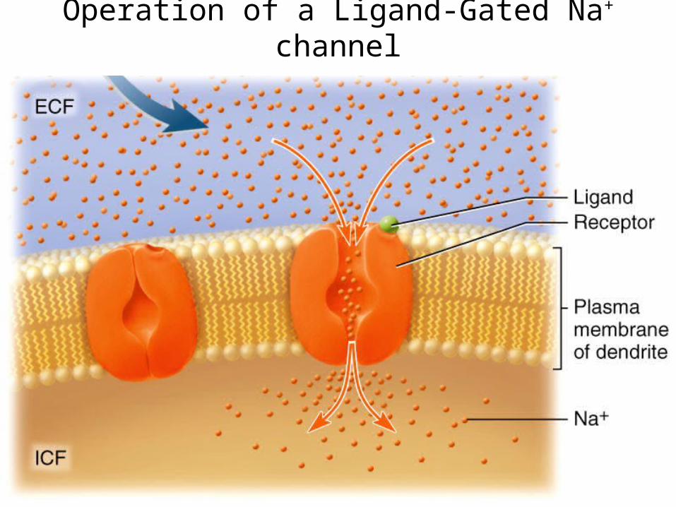
Operation of a Ligand-Gated Na+ channel

Deviations in the Resting Membrane Potential
• The opening of a gated ion channel will allow a specific ion to diffuse down its respective gradient across the cell membrane
• The membrane potential will deviate from the resting value (-70mV) based on 2 criteria:– the charge of the diffusing ion
• either positive (cation) or negative (anion)– the direction of the diffusion
• either into or out of the cell
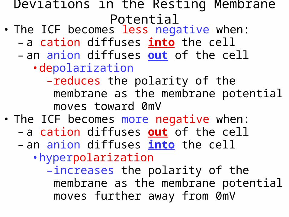
Deviations in the Resting Membrane Potential• The ICF becomes less negative when:
– a cation diffuses into the cell– an anion diffuses out of the cell
• depolarization– reduces the polarity of the membrane as the
membrane potential moves toward 0mV• The ICF becomes more negative when:
– a cation diffuses out of the cell– an anion diffuses into the cell
• hyperpolarization– increases the polarity of the membrane as the
membrane potential moves further away from 0mV

Deviations in the Resting Membrane Potential
• When the gated ion channels close, the cell membrane potential returns to its resting value
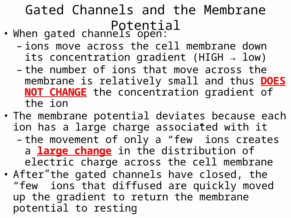
• When gated channels open: – ions move across the cell membrane down its
concentration gradient (HIGH → low)– the number of ions that move across the membrane is
relatively small and thus DOES NOT CHANGE the concentration gradient of the ion
• The membrane potential deviates because each ion has a large charge associated with it– the movement of only a “few” ions creates a large
change in the distribution of electric charge across the cell membrane
• After the gated channels have closed, the “few” ions that diffused are quickly moved up the gradient to return the membrane potential to resting
Gated Channels and the Membrane Potential
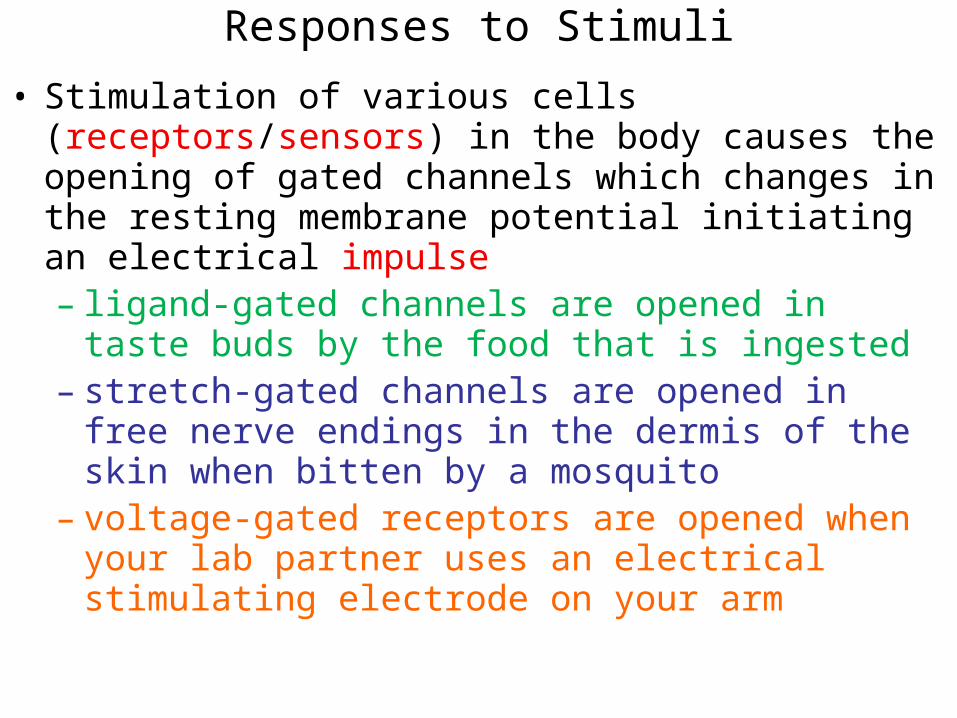
Responses to Stimuli
• Stimulation of various cells (receptors/sensors) in the body causes the opening of gated channels which changes in the resting membrane potential initiating an electrical impulse– ligand-gated channels are opened in taste buds by the
food that is ingested – stretch-gated channels are opened in free nerve endings
in the dermis of the skin when bitten by a mosquito– voltage-gated receptors are opened when your lab partner
uses an electrical stimulating electrode on your arm
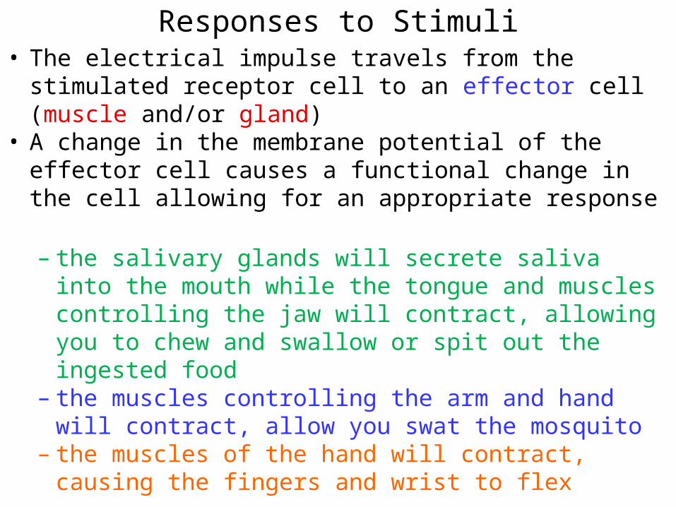
Responses to Stimuli• The electrical impulse travels from the stimulated receptor
cell to an effector cell (muscle and/or gland)• A change in the membrane potential of the effector cell
causes a functional change in the cell allowing for an appropriate response
– the salivary glands will secrete saliva into the mouth while the tongue and muscles controlling the jaw will contract, allowing you to chew and swallow or spit out the ingested food
– the muscles controlling the arm and hand will contract, allow you swat the mosquito
– the muscles of the hand will contract, causing the fingers and wrist to flex
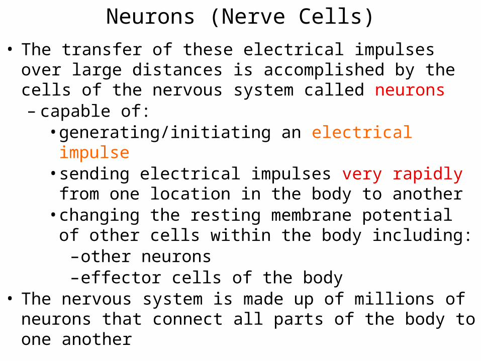
• The transfer of these electrical impulses over large distances is accomplished by the cells of the nervous system called neurons – capable of:
• generating/initiating an electrical impulse• sending electrical impulses very rapidly from one
location in the body to another• changing the resting membrane potential of other cells
within the body including:– other neurons – effector cells of the body
• The nervous system is made up of millions of neurons that connect all parts of the body to one another
Neurons (Nerve Cells)

The Nervous System
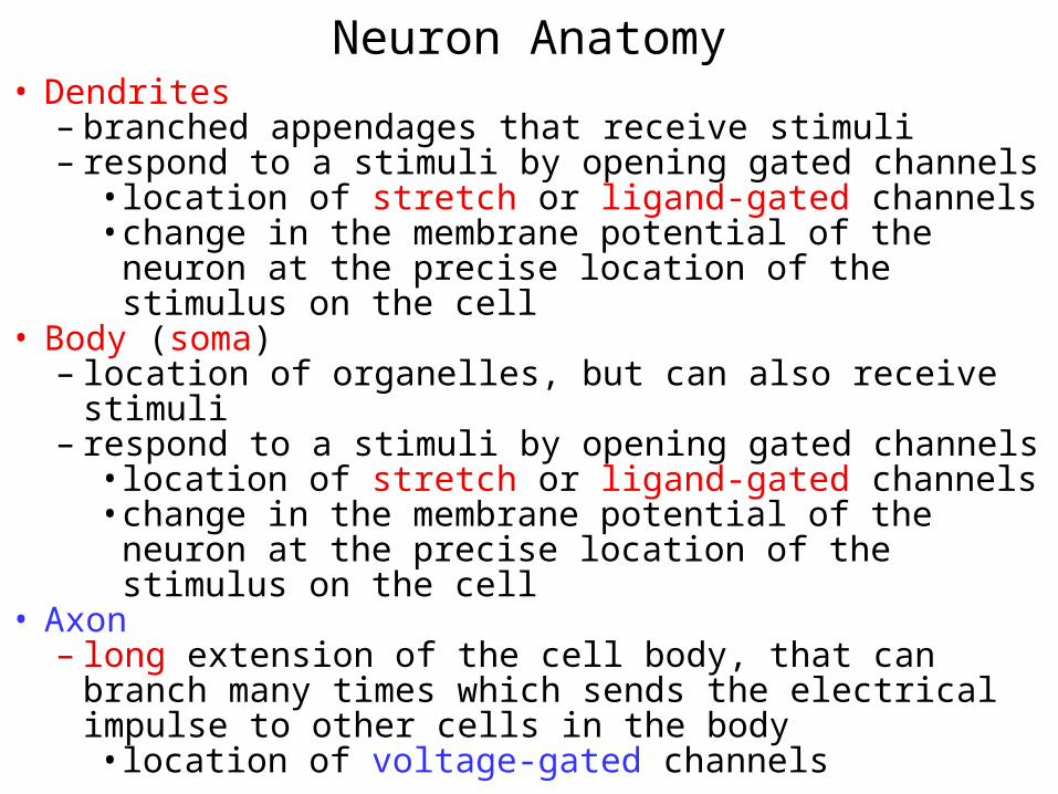
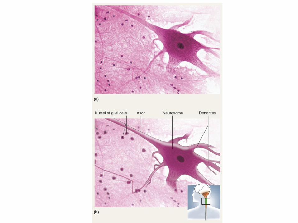
Neuron Anatomy• Dendrites
– branched appendages that receive stimuli– respond to a stimuli by opening gated channels
• location of stretch or ligand-gated channels• change in the membrane potential of the neuron at the
precise location of the stimulus on the cell• Body (soma)
– location of organelles, but can also receive stimuli– respond to a stimuli by opening gated channels
• location of stretch or ligand-gated channels• change in the membrane potential of the neuron at the
precise location of the stimulus on the cell • Axon
– long extension of the cell body, that can branch many times which sends the electrical impulse to other cells in the body• location of voltage-gated channels

Neuron

Initiation of an electrical impulse
• The initiation of an electrical impulse occurs at either the dendrites or the body of a neuron– the opening of stretch or ligand-gated channels causes
EITHER a depolarization or a hyperpolarization, depending on the charge and the direction of movement of the ion at the location of the opened gated channels • this type of membrane potential change is called a
graded (local) potential– a brief, localized change in the membrane potential

• The grade or magnitude of depolarization or hyperpolarization is directly related to the size of the stimulus – determines the number of gated channels that is opened
• determines the number of ions that cross the plasma membrane
Graded Potentials

Graded Potentials of Stretch-gated Channels
• A small pressure applied to the skin:– causes a small amount of stretch of the cell membrane of
the pressure sensing cells of the skin• causes few stretch-gated channels to open
– allows few ions to cross the cell membrane• causes a small change of the membrane
potential from the resting value• A large pressure applied to the skin:
– causes a large amount of stretch of the cell membrane of the pressure sensing cells of the skin
– causes more stretch-gated channels to open– allows more ions to cross the cell membrane
• causes a larger change of the membrane potential from the resting value

Graded Potentials
• Decrease in magnitude with distance from the site of stimulation– as ions move into/out of the cell through opened gated
channels, they diffuse away from the opened gated channel• as the ions diffuse away from the opened gated
channel the concentration of the ion decreases– as the ion concentration decreases, so does it’s
influence on the membrane potential • the further away from the stimulus, the closer
the membrane potential is to the resting value
Function of Graded Potentials
• The purpose of graded potentials in the dendrites or soma is to cause (or prevent) the opening of voltage-gated ion channels in the axon of the neuron– open when the membrane potential in the axon has been
depolarized to a minimum value– the opening of voltage-gated channels in the axon will
create a membrane potential change in the axon called an action potential• the action potential will “travel” down the length of
the axon and all of its branches to the axon terminus
• A very rapid sequence of membrane potential changes due to the opening and closing of voltage-gated Na+ and voltage-gated K+ channels
• There are 3 sequential phases to an AP in a neuron:– Depolarization
• a reduction in the polarity of the membrane potential– Repolarization
• a return of the membrane potential towards the resting value
– Hyperpolarization• the membrane potential reaches values more negative
than the resting value• All APs in a neuron have the same magnitude regardless of
the size of the stimulus (not graded)
Action Potentials (APs)

Action potential
• The initiation of an AP occurs at the beginning of the axon called the initial segment and requires that the membrane potential at the axon be depolarized to threshold– the minimum amount of depolarization required to
initiate an action potential • typically -55mV • causes the opening of voltage-gated Na+ channels
Threshold and Action Potentials

Threshold and Action Potentials
• Threshold can be reached by a depolarizing graded potential in the dendrites or soma of a neuron– small (weak) stimuli DO NOT initiate an AP because the
magnitude of the graded potential at the axon is TOO SMALL to depolarize the membrane at the axon to threshold • subthreshold stimuli
– large (strong) stimuli DO initiate an AP • threshold stimuli
• All-or-none phenomenon– action potentials either completely, or not at all

Ionic Basis of Action Potential (Resting State)
• Na+ and K+ channels are closed
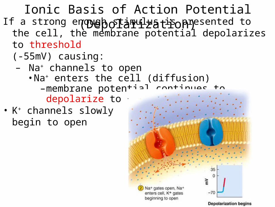
Ionic Basis of Action Potential (Depolarization)If a strong enough stimulus is presented to the cell, the
membrane potential depolarizes to threshold (-55mV) causing:– Na+ channels to open
• Na+ enters the cell (diffusion)– membrane potential continues to depolarize to
+30mV• K+ channels slowly
begin to open

Ionic Basis of Action Potential (Repolarization)
Membrane potential reaches peak depolarization of +30mV causing:
• Na+ channels to close• K+ channels to open
– K+ exits the cell (diffusion)• the membrane potential
returns toward resting values (repolarization)

Ionic Basis of Action Potential (Hyperpolarization and Return to Resting)
• K+ channels remain open • This causes more than enough K+ to leave the cell resulting
in hyperpolarization of the membrane potential• Eventually, the K+
channels close, allowing the membrane potential to return to resting
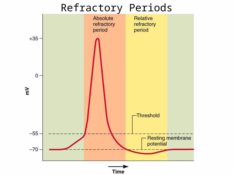
Refractory Periods
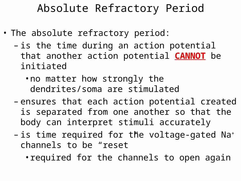
• The absolute refractory period:– is the time during an action potential that another action
potential CANNOT be initiated • no matter how strongly the dendrites/soma are
stimulated– ensures that each action potential created is separated
from one another so that the body can interpret stimuli accurately
– is time required for the voltage-gated Na+ channels to be “reset” • required for the channels to open again
Absolute Refractory Period

• The relative refractory period:– is the time after the absolute refractory period until the
membrane potential returns to the resting value
• During this time another action potential CAN be initiated – requires a stronger than normal stimulus at the dendrites
• during this time some of the voltage-gated Na+ channels have been “reset” while others have not
Relative Refractory Period

Propagation of an Action Potential
• Once an action potential has been initiated at the beginning of the axon, it must “travel” (propagate) along the length of the axon to the axon terminus
• The influx of Na+ into the cell during depolarization causes the membrane potential in “front” of the opened Na+ channels to depolarize to threshold
• Reaching threshold opens up the Na+ channels in “front” of the site of the action potential causing an action potential to be created in this new location
• As the next group of Na+ channels begins to open, the ones “behind” them are closing
• The impulse continues to propagate away from its point of origin to the axon terminus
• “the domino effect”

Propagation of an Action Potential

• The propagation velocity is the speed at which the action potential propagates along the length of the axon
• Conduction velocity depends on:– axon diameter (thickness)
• the larger the diameter, the greater the conduction velocity
– presence of a myelin sheath • dramatically increases impulse speed
– to speeds up to 300 mph• more effective than increasing axon diameter
• The human body uses both methods to maximize propagation velocity
Propagation Velocity of an Action Potential

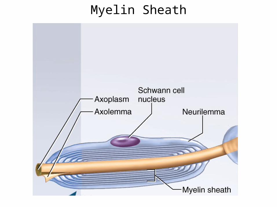
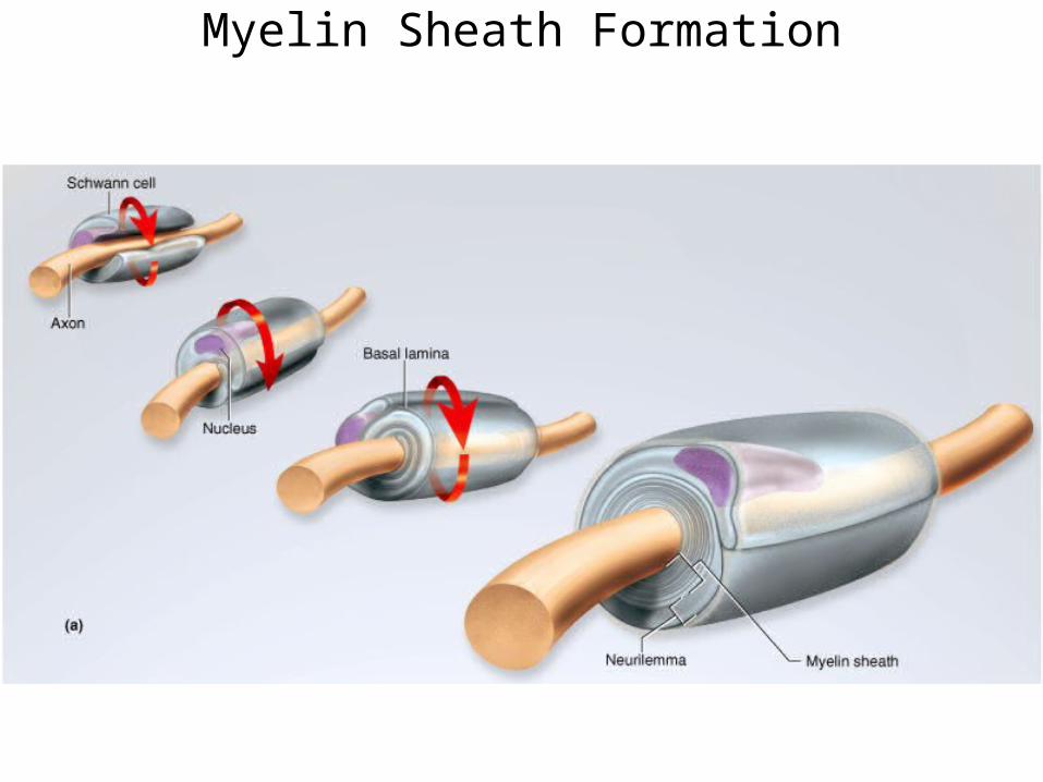
• White, fatty (lipid), segmented covering around most long axons
• Increases propagation velocity of APs by electrically insulating the axon
• Formed by Schwann cells– wraps around the axon many times with its plasma
membrane– encloses the axon with many concentric layers of lipid
bilayers
Myelin Sheath
Myelin Sheath
Myelin Sheath Formation
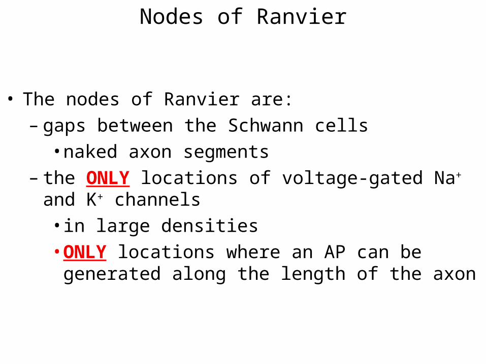
• The nodes of Ranvier are:– gaps between the Schwann cells
• naked axon segments– the ONLY locations of voltage-gated Na+ and K+
channels • in large densities• ONLY locations where an AP can be generated along
the length of the axon
Nodes of Ranvier
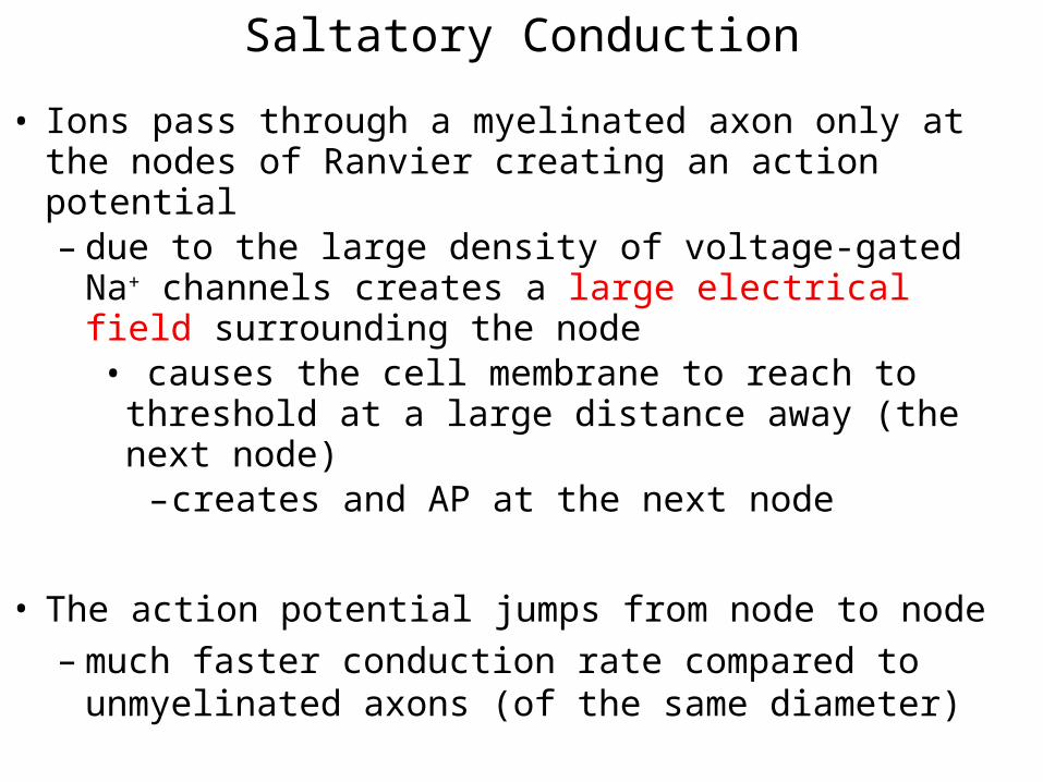
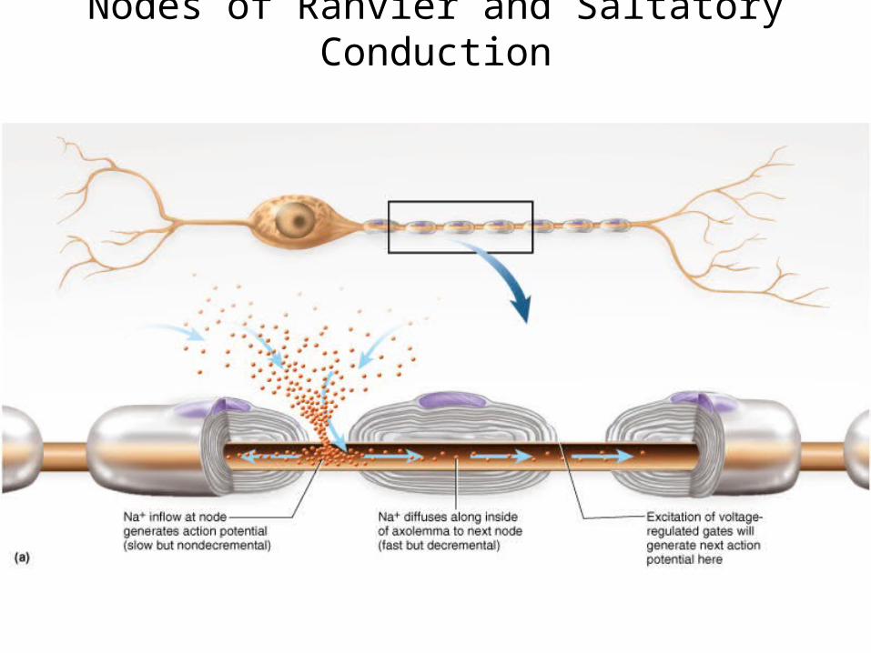
• Ions pass through a myelinated axon only at the nodes of Ranvier creating an action potential– due to the large density of voltage-gated Na+ channels
creates a large electrical field surrounding the node• causes the cell membrane to reach to threshold at a
large distance away (the next node)– creates and AP at the next node
• The action potential jumps from node to node– much faster conduction rate compared to unmyelinated
axons (of the same diameter)
Saltatory Conduction
Nodes of Ranvier and Saltatory Conduction

Saltatory Conduction

• When the AP reaches the axon termini the impulse must be transmitted to the next cell in the path to the effector
• A synapse is the junction between 2 cells where the impulse is transmitted from one cell to another :– Presynaptic cell (before synapse) – Postsynaptic cell (after synapse)
– found between:• 2 neurons• a neuron and an effector cell (muscle or gland)
– 2 general types include: – chemical – electrical
Axon Termini and Synapses
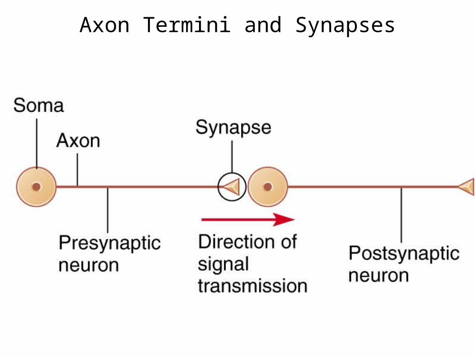
Axon Termini and Synapses

• Composed of 3 parts: – axonal terminal of the presynaptic neuron
• contains synaptic vesicles – filled with a neurotransmitter (chemical/ligand)
– receptor region on the postsynaptic cell which contains ligand-gated channels
– fluid-filled space between the cells (synaptic cleft) • separates the presynaptic and postsynaptic cells
Chemical Synapses

Chemical Synapse

• An action potential that arrives at the axon terminus of the presynaptic cell causes the opening of voltage-gated Ca2+ channels– causes Ca2+ to diffuse into the cytoplasm of the
presynaptic cell • triggers the exocytosis of neurotransmitters into the
synaptic cleft • The neurotransmitters diffuse across the cleft and open the
ligand-gated channels on the postsynaptic cell– causes ions to cross the cell membrane and result in a
graded potential • postsynaptic potential
– depolarization or hyperpolarization
Synaptic Cleft: Information Transfer

Synaptic Cleft: Information Transfer


The 2 types of postsynaptic potentials are: • EPSP (excitatory postsynaptic potentials)
– depolarizing graded potentials– causes the membrane potential move towards threshold
which increases the chances that an AP will be initiated in an axon
• IPSP (inhibitory postsynaptic potentials)– hyperpolarizing graded potentials
• causes the membrane potential move away from threshold which reduces the chances that an AP will be initiated in an axon
Postsynaptic Potentials
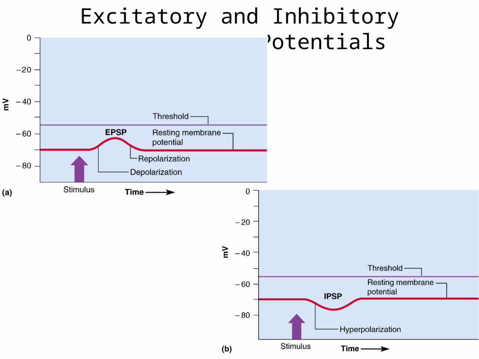
Excitatory and Inhibitory Postsynaptic Potentials


• A single EPSP CANNOT initiate an action potential– EPSPs must summate (add) to bring the membrane
potential to threshold at the axon• Temporal summation
– postsynaptic potentials are generated at a single location at a high frequency
• Spatial summation – postsynaptic potentials are generated at different
locations at the same time• IPSPs can also summate with EPSPs
– cancel each other out
EPSPs and IPSPs Summate
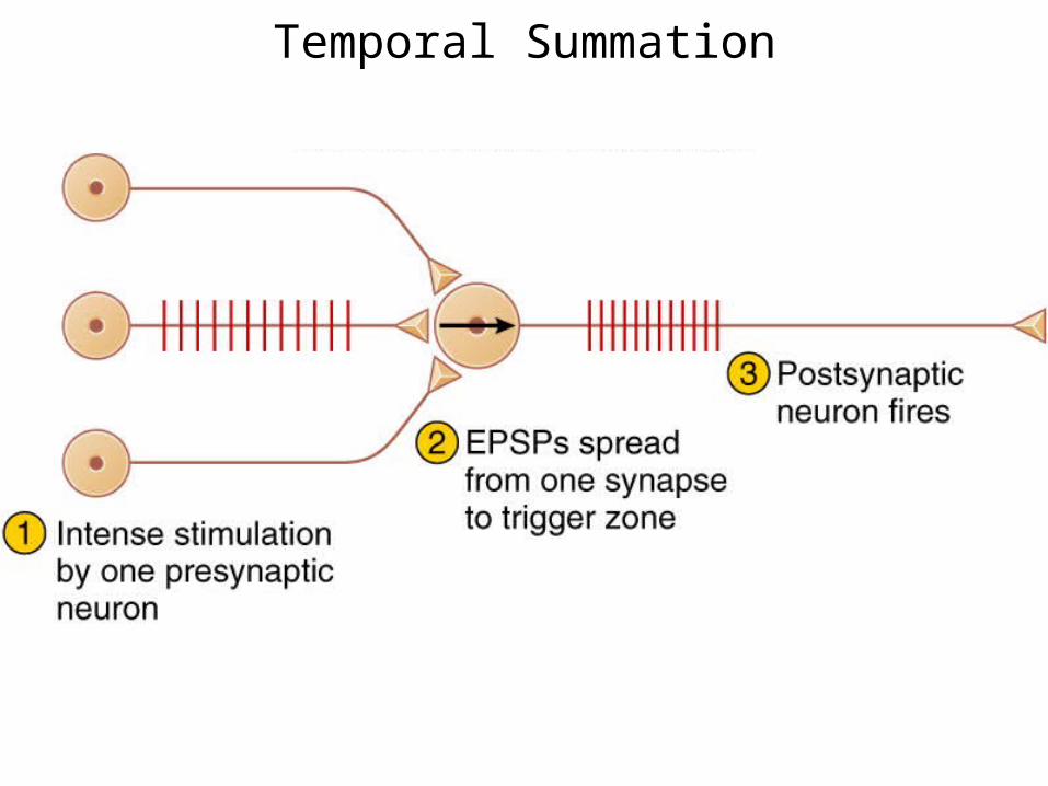
Temporal Summation
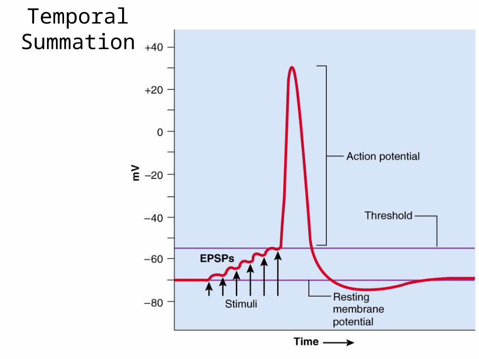
Temporal Summation
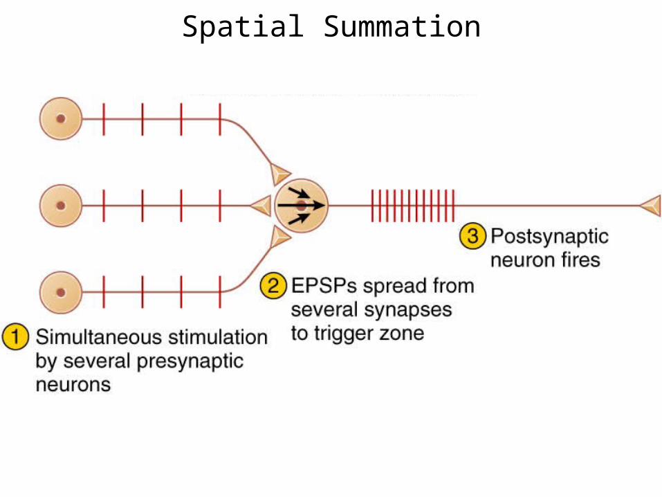
Spatial Summation