Response of swine spleen to Streptococcus suis infection revealed by transcription analysis
12
RESEARCH ARTICLE Open Access Response of swine spleen to Streptococcus suis infection revealed by transcription analysis Ran Li 1,2† , Anding Zhang 1,2† , Bo Chen 2† , Liu Teng 2 , Ya Wang 2 , Huanchun Chen 1,2 , Meilin Jin 1,2* Astract Background: Streptococcus suis serotype 2 (SS2), a major swine pathogen and an emerging zoonotic agent, has greatly challenged global public health. Systematical information about host immune response to the infection is important for understanding the molecular mechanism of diseases. Results: 104 and 129 unique genes were significantly up-regulated and down-regulated in the spleens of pigs infected with SS2 (WT). The up-regulated genes were principally related to immune response, such as genes involved in inflammatory response; acute-phase/immune response; cell adhesion and response to stress. The down-regulated genes were mainly involved in transcription, transport, material and energy metabolism which were representative of the reduced vital activity of SS2-influenced cells. Only a few genes showed significantly differential expression when comparing avirulent isogenic strain (ΔHP0197) with mock-infected samples. Conclusions: Our findings indicated that highly pathogenic SS2 could persistently induce cytokines mainly by Toll- like receptor 2 (TLR2) pathway, and the phagocytosis-resistant bacteria could induce high level of cytokines and secrete toxins to destroy deep tissues, and cause meningitis, septicaemia, pneumonia, endocarditis, and arthritis. Background Streptococcus suis ( S. suis ) is an important pathogen associated with many diseases in pigs, including menin- gitis, septicaemia, pneumonia, endocarditis, and arthritis. S. suis serotype 2 (SS2) is considered the most patho- genic as well as the most prevalent capsular type among thirty-three serotypes (types 1 to 31, 33, and 1/2) in dis- eased pigs, and it is also the causative agent of serious infections in humans, especially in people in close con- tact with pig or pork byproducts [1-3]. Two recent large-scale outbreaks of human SS2 epidemics in China (one had 25 cases with 14 deaths in Jiangsu in 1998, the second had 204 cases with 38 deaths in Sichuan in 2005), featured clinical streptococcal toxic shock syn- drome, have greatly challenged the global public health [4-7]. Recently, S. suis infection has also caused sporadic human illness in other countries, including Thailand [8,9], United Kingdom [10], Portugal [11], Australia [12], Netherlands [13] and United States [14,15], and been recognized as the third most common cause of community acquired bacterial meningitis in Hong Kong and as the leading cause of adult meningitis in Vietnam [5,16]. The past pathogenesis studies were performed mainly on the pathogenic bacteria and as a result, a few virulence- associated factors have been successfully identified. Poly- saccharide capsule has been considered essential for the virulence of the bacterium [17,18], and other factors, such as suilysin, the so-called extracellular protein factor and muramidase-released protein have been shown to be linked to, but not essential for the full virulence of S. suis [19]. GapdH [20], Enolase [21,22], FbpS [19], Adhesin [23-27] have been proved to be involved in the adherence and virulence of S. suis. Recently, serum opacity-like factor [28], IgA1 protease [29], D-Alanylation of Lipoteichoic Acid [30] and pgdA [31] were identified as important fac- tors in S. suis virulence. In addition, SalK/SalR [32] and CovR [33] were found to affect the virulence of S. suis Chi- nese isolates. These studies have contributed to the under- standing of S. suis pathogenesis and also suggested that host responses also play essential roles in the development of the diseases. * Correspondence: [email protected] † Contributed equally 1 Unit of Animal Infectious Diseases, National Key Laboratory of Agricultural Microbiology, Huazhong Agricultural University, Wuhan, Hubei, China Full list of author information is available at the end of the article Li et al. BMC Genomics 2010, 11:556 http://www.biomedcentral.com/1471-2164/11/556 © 2010 Li et al; licensee BioMed Central Ltd. This is an Open Access article distributed under the terms of the Creative Commons Attribution License (http://creativecommons.org/licenses/by/2.0), which permits unrestricted use, distribution, and reproduction in any medium, provided the original work is properly cited.
Transcript of Response of swine spleen to Streptococcus suis infection revealed by transcription analysis
RESEARCH ARTICLE Open Access
Response of swine spleen to Streptococcus suisinfection revealed by transcription analysisRan Li1,2†, Anding Zhang1,2†, Bo Chen2†, Liu Teng2, Ya Wang2, Huanchun Chen1,2, Meilin Jin1,2*
Astract
Background: Streptococcus suis serotype 2 (SS2), a major swine pathogen and an emerging zoonotic agent, hasgreatly challenged global public health. Systematical information about host immune response to the infection isimportant for understanding the molecular mechanism of diseases.
Results: 104 and 129 unique genes were significantly up-regulated and down-regulated in the spleens of pigsinfected with SS2 (WT). The up-regulated genes were principally related to immune response, such as genesinvolved in inflammatory response; acute-phase/immune response; cell adhesion and response to stress. Thedown-regulated genes were mainly involved in transcription, transport, material and energy metabolism whichwere representative of the reduced vital activity of SS2-influenced cells. Only a few genes showed significantlydifferential expression when comparing avirulent isogenic strain (ΔHP0197) with mock-infected samples.
Conclusions: Our findings indicated that highly pathogenic SS2 could persistently induce cytokines mainly by Toll-like receptor 2 (TLR2) pathway, and the phagocytosis-resistant bacteria could induce high level of cytokines andsecrete toxins to destroy deep tissues, and cause meningitis, septicaemia, pneumonia, endocarditis, and arthritis.
BackgroundStreptococcus suis (S. suis) is an important pathogenassociated with many diseases in pigs, including menin-gitis, septicaemia, pneumonia, endocarditis, and arthritis.S. suis serotype 2 (SS2) is considered the most patho-genic as well as the most prevalent capsular type amongthirty-three serotypes (types 1 to 31, 33, and 1/2) in dis-eased pigs, and it is also the causative agent of seriousinfections in humans, especially in people in close con-tact with pig or pork byproducts [1-3]. Two recentlarge-scale outbreaks of human SS2 epidemics in China(one had 25 cases with 14 deaths in Jiangsu in 1998, thesecond had 204 cases with 38 deaths in Sichuan in2005), featured clinical streptococcal toxic shock syn-drome, have greatly challenged the global public health[4-7]. Recently, S. suis infection has also caused sporadichuman illness in other countries, including Thailand[8,9], United Kingdom [10], Portugal [11], Australia[12], Netherlands [13] and United States [14,15], and
been recognized as the third most common causeof community acquired bacterial meningitis in HongKong and as the leading cause of adult meningitis inVietnam [5,16].The past pathogenesis studies were performed mainly
on the pathogenic bacteria and as a result, a few virulence-associated factors have been successfully identified. Poly-saccharide capsule has been considered essential for thevirulence of the bacterium [17,18], and other factors, suchas suilysin, the so-called extracellular protein factor andmuramidase-released protein have been shown to belinked to, but not essential for the full virulence of S. suis[19]. GapdH [20], Enolase [21,22], FbpS [19], Adhesin[23-27] have been proved to be involved in the adherenceand virulence of S. suis. Recently, serum opacity-like factor[28], IgA1 protease [29], D-Alanylation of LipoteichoicAcid [30] and pgdA [31] were identified as important fac-tors in S. suis virulence. In addition, SalK/SalR [32] andCovR [33] were found to affect the virulence of S. suis Chi-nese isolates. These studies have contributed to the under-standing of S. suis pathogenesis and also suggested thathost responses also play essential roles in the developmentof the diseases.
* Correspondence: [email protected]† Contributed equally1Unit of Animal Infectious Diseases, National Key Laboratory of AgriculturalMicrobiology, Huazhong Agricultural University, Wuhan, Hubei, ChinaFull list of author information is available at the end of the article
Li et al. BMC Genomics 2010, 11:556http://www.biomedcentral.com/1471-2164/11/556
© 2010 Li et al; licensee BioMed Central Ltd. This is an Open Access article distributed under the terms of the Creative CommonsAttribution License (http://creativecommons.org/licenses/by/2.0), which permits unrestricted use, distribution, and reproduction inany medium, provided the original work is properly cited.
Inducing excessive inflammation is recognized as oneof the reasons why highly invasive SS2 strain could causesevere diseases [31,34]. A few previous studies indicatedthat high level of cytokines and chemokines could bereleased by human brain microvascular endothelial cells[35], a whole-blood culture system [36], macrophages[37] and monocytes [38] stimulated by SS2, and haveimportant roles in the initiation and development ofinflammation and meningitis [39]. More direct proofswere the studies on mice with different genetic back-ground, which indicated that IL-10 was responsible, atleast in part, for the high survival, which suggested thataberrant innate immune response contributed to SS2diseases [40].To be aware of the information about host immune
response would enable people to better understand thedisease. Transcriptional response of alveolar macro-phages to SS2 has been performed and the results indi-cated that NF-kB and MAP-kinases signaling pathwayswere induced upon interaction with SS2 [41]. However,it is not easy to get more information since the primarymacrophages are so sensitive to the interference. Spleenplays an important role in immune response and couldbe an ideal target to study host immune responseagainst infection [42,43]. In the present study, the geneexpression profiles of swine spleens which suffered fromhighly pathogenic SS2, avirulent isogenic strain and PBSrespectively were investigated to reveal the host immuneresponse to SS2 and the contributions of host responseto SS2 diseases.
ResultsTranscriptome analysisThe transcriptome analysis indicated that 14,992, 15,487and 15,757 probe sets, corresponding to 62.1%, 64.2%and 65.3% of all probe sets, were detected in WT,ΔHP0197 and mock-infected pig spleens respectively(Additional file 1). The expression profiles of porcinespleens challenged with WT 3 days post inoculationwere compared with those of the mock-infected group.After quantile normalization and statistical analysis,1014 transcripts were identified at the global false dis-covery rate (FDR) of 10% (Additional file 2). Further-more, the criteria of a two-fold or greater change indifferential expression and a FDR of 10% were chosento determine up-regulated and down-regulated genesin the WT infected replicates. Using these criteria, 120and 132 transcripts, representing 104 and 129 uniquegenes, were significantly up-regulated and down-regulated respectively (Additional file 3). However, onlya few genes showed significantly differential expressionswhen comparing ΔHP0197 with mock-infected samples(Figure 1A).
Of the 233 unique DE transcripts, 158 transcripts couldbe determined based on BLASTX searches and anno-tated with DAVID or by searching against the GenBankdatabase (Figure 1B). Among these, 135 unique geneswere grouped into 39 categories based on biological pro-cess Gene Ontology (GO) terms or according to theirpotential Biology Process Classification by referring torecent publications (Figure 1C). Unsurprisingly, themajority of genes were related to the immune response,Transcription, Transport, material and energy metabo-lism, etc. (Table 1).
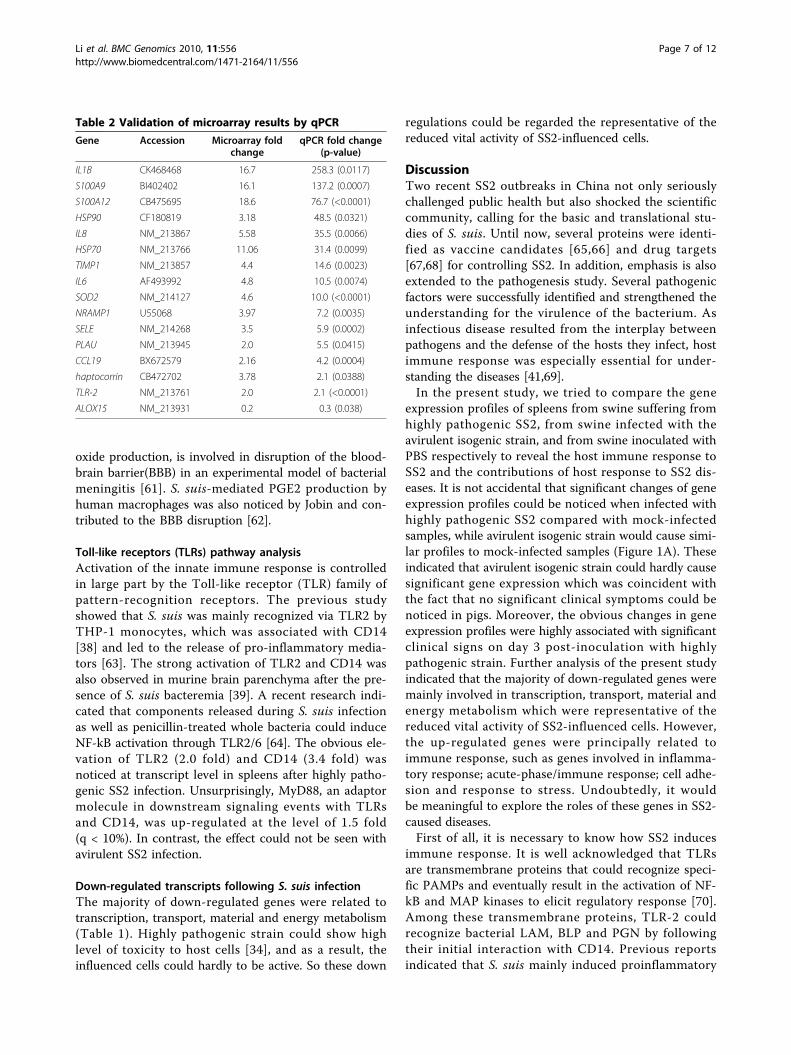
Validation of microarray data by quantitative real-timePCR (qPCR)The qPCR was performed to validate the expression pat-terns during infection for specific genes identified in themicroarray assay. In order to validate the differentialexpression of various identified genes, 16 up-regulatedgenes, with the increase ranging from 2.0-fold to 18.6-fold, and 3 down-regulated genes, with the decrease ran-ging from 2.5-fold to 5.9-fold, were selected for qPCRanalysis. All the selected down-regulated genes could beamplified from the control samples but failed to achievesignificant detectable signs from WT-infected spleens,except for ALOX15 which showed 3.2-fold down-regu-lated expression. All selected up-regulated genes showedhigher expression in WT-infected samples than in thecontrol samples (Table 2). Though variation in foldchanges could be observed between qPCR and microar-ray (Table 2), the differential expression patterns werecoincident between the results of the two techniques,which indicated the reliability of the microarray analysis.
Induction of inflammasomes and acute phase proteins bySS2 infectionHighly pathogenic SS2 infection could cause up-regu-lated expression of a large set of genes involved in theinflammatory response and acute phase proteins bymicroarray analysis. IL-1B, IL-6 and IL-8 could beinduced by foreign pathogens and play essential roles incontrolling infections [5,44]. However, they may alsocause pathology when these productions are excessiveor uncontrolled [45]. Ye et al. also found that signifi-cantly high level of cytokines could be induced by highlypathogenic SS2 strain and play important roles in sepsis[34], which is in coincidence with ours. In addition,quite a few genes related to inflammatory response werefound up-regulated, such as S100 family proteins(S100A8, S100A9 and S100A12) [46], Pentraxin 3 [47]and Resistin [48,49]. They play important roles in med-iating inflammatory responses, recruiting inflammatorycells to sites of tissue damage or contributing to resist-ing the invasion of various pathogens.
Li et al. BMC Genomics 2010, 11:556http://www.biomedcentral.com/1471-2164/11/556
Page 2 of 12

Acute phase proteins (APPs), such as Lactotransferrin[50], Haptoglobin [51], Serum amyloid A 2 [52] andcoagulation factor XIII, were involved in physiologicreactions initiated early in the inflammatory process[53], and could be a response to S. suis infection [54].CEBPD belonging to the CCAAT-enhancer binding pro-tein (CEBP) family which is crucial in the regulation ofgenes involved in immunity and inflammation. Theseup-regulated genes are the representative of host acuteresponse struggling to eliminate invading pathogens.
Induction of genes related in cell adhesion and stressresponseCell adhesion molecules (CAMs) have been implicatedin the regulation of a wide variety of fundamental cellu-lar processes, such as cell adhesion, cell polarization,survival, movement, and proliferation [55]. E-selectin isa cell adhesion molecule expressed on endothelial cellsactivated by cytokines, and plays an important role in
recruiting leukocytes to the site of injury [56]. Versicancan bind adhesion molecules on the surface of inflam-matory leukocytes [57] and act as a TLR2 agonist ininducing the release of proinflammatory cytokines [58].Thrombospondin 1 is an adhesive glycoprotein thatmediates cell-to-cell and cell-to-matrix interactions andit could interact with numerous proteases involved inangiogenesis [59]. Mucosal vascular addressin cell adhe-sion molecule 1 is predominantly expressed on highendothelial venules in inflamed tissues, and could assistthe extravasations of leucocyte [60]. The up-regulationof cell adhesion molecules after SS2 infection wouldcontribute to recruiting leukocytes to the site of infec-tion, which could control infection.Genes related to oxidative stress and homeostasis were
also identified to be up-regulated. SOD2 provides vitalprotection against reactive oxygen species (ROS), thusprotecting tissues from damage in a broad range of dis-ease states. The secretion of PGE2, together with nitric
Figure 1 Clustering and characterization of the differential expression of genes. (A) 233 genes were selected for cluster analysis which isdescribed in methods. Each row represents a separate transcript and each column represents a separate piglet. Color legend is on the left, thecolor scale ranges from saturated green for log ratios -3.0 and above to saturate red for log ratios 3.0 and above. Red indicates increasedtranscript expression levels, green indicates decreased levels compared with normal samples. (B) Percentage distribution of unique genes wasfrom 233 differentially regulated transcripts after BLASTX searches and annotation. 158 unique genes had significant similarities based on BLASTXsearches. 135(126+9) unique genes had been annotated by Biological Process (BP) Classification. (C) Categories of annotated genes genes basedon biological process GO term. Many categories shared the same transcripts.
Li et al. BMC Genomics 2010, 11:556http://www.biomedcentral.com/1471-2164/11/556
Page 3 of 12
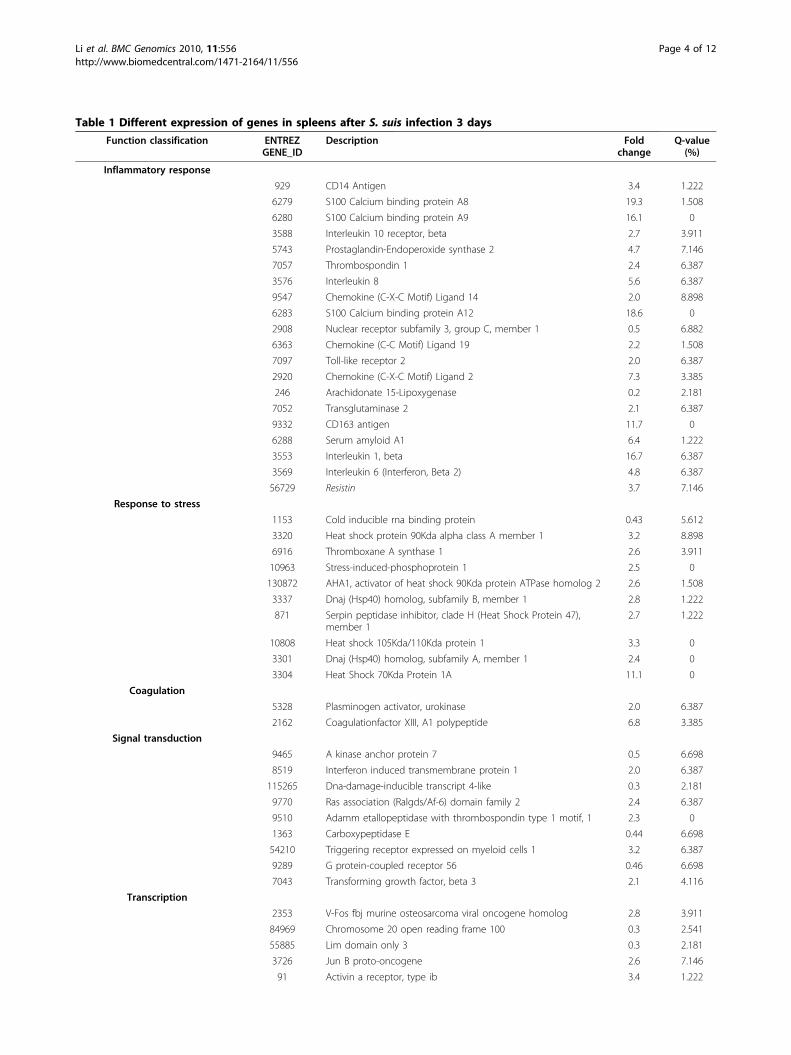
Table 1 Different expression of genes in spleens after S. suis infection 3 days
Function classification ENTREZGENE_ID
Description Foldchange
Q-value(%)
Inflammatory response
929 CD14 Antigen 3.4 1.222
6279 S100 Calcium binding protein A8 19.3 1.508
6280 S100 Calcium binding protein A9 16.1 0
3588 Interleukin 10 receptor, beta 2.7 3.911
5743 Prostaglandin-Endoperoxide synthase 2 4.7 7.146
7057 Thrombospondin 1 2.4 6.387
3576 Interleukin 8 5.6 6.387
9547 Chemokine (C-X-C Motif) Ligand 14 2.0 8.898
6283 S100 Calcium binding protein A12 18.6 0
2908 Nuclear receptor subfamily 3, group C, member 1 0.5 6.882
6363 Chemokine (C-C Motif) Ligand 19 2.2 1.508
7097 Toll-like receptor 2 2.0 6.387
2920 Chemokine (C-X-C Motif) Ligand 2 7.3 3.385
246 Arachidonate 15-Lipoxygenase 0.2 2.181
7052 Transglutaminase 2 2.1 6.387
9332 CD163 antigen 11.7 0
6288 Serum amyloid A1 6.4 1.222
3553 Interleukin 1, beta 16.7 6.387
3569 Interleukin 6 (Interferon, Beta 2) 4.8 6.387
56729 Resistin 3.7 7.146
Response to stress
1153 Cold inducible rna binding protein 0.43 5.612
3320 Heat shock protein 90Kda alpha class A member 1 3.2 8.898
6916 Thromboxane A synthase 1 2.6 3.911
10963 Stress-induced-phosphoprotein 1 2.5 0
130872 AHA1, activator of heat shock 90Kda protein ATPase homolog 2 2.6 1.508
3337 Dnaj (Hsp40) homolog, subfamily B, member 1 2.8 1.222
871 Serpin peptidase inhibitor, clade H (Heat Shock Protein 47),member 1
2.7 1.222
10808 Heat shock 105Kda/110Kda protein 1 3.3 0
3301 Dnaj (Hsp40) homolog, subfamily A, member 1 2.4 0
3304 Heat Shock 70Kda Protein 1A 11.1 0
Coagulation
5328 Plasminogen activator, urokinase 2.0 6.387
2162 Coagulationfactor XIII, A1 polypeptide 6.8 3.385
Signal transduction
9465 A kinase anchor protein 7 0.5 6.698
8519 Interferon induced transmembrane protein 1 2.0 6.387
115265 Dna-damage-inducible transcript 4-like 0.3 2.181
9770 Ras association (Ralgds/Af-6) domain family 2 2.4 6.387
9510 Adamm etallopeptidase with thrombospondin type 1 motif, 1 2.3 0
1363 Carboxypeptidase E 0.44 6.698
54210 Triggering receptor expressed on myeloid cells 1 3.2 6.387
9289 G protein-coupled receptor 56 0.46 6.698
7043 Transforming growth factor, beta 3 2.1 4.116
Transcription
2353 V-Fos fbj murine osteosarcoma viral oncogene homolog 2.8 3.911
84969 Chromosome 20 open reading frame 100 0.3 2.541
55885 Lim domain only 3 0.3 2.181
3726 Jun B proto-oncogene 2.6 7.146
91 Activin a receptor, type ib 3.4 1.222
Li et al. BMC Genomics 2010, 11:556http://www.biomedcentral.com/1471-2164/11/556
Page 4 of 12

Table 1 Different expression of genes in spleens after S. suis infection 3 days (Continued)
116448 Oligodendrocyte transcription factor 1 2.4 6.387
79365 Basic helix-loop-helix domain containing, class B, 3 0.4 2.181
64919 B-cell cll/lymphoma 11B 0.5 2.181
23635 Single-stranded dna binding protein 2 0.4 2.541
23414 Zinc finger protein, multitype 2 0.5 6.698
7552 Zinc finger protein 6 (Cmpx1) 0.5 3.385
6920 Transcription elongation factor A (SII), 3 2.2 6.387
4783 Nuclear factor, interleukin 3 regulated 2.4 3.911
1052 CCAAT/Enhancer binding protein (C/EBP), delta 3.1 0
Cell adhesion
6401 Selectin E 3.5 1.222
8174 Mucosal vascular addressin cell adhesion molecule 1 2.3 6.387
5067 Contactin 3 0.5 6.794
4867 Nephronophthisis 1 (Juvenile) 0.4 3.911
1462 Chondroitin sulfate proteoglycan 2 (Versican) 9.1 0
960 CD44 antigen 2.3 2.541
Ubiquitin cycle
115123 Membrane-associated ring finger (C3HC4) 3 4.4 1.222
7317 Ubiquitin-activating enzyme E1 0.4 2.181
11274 Ubiquitin specific peptidase 18 0.4 6.698
9666 Zinc finger daz interacting protein 3 0.5 6.882
Transport
6556 Solute carrier family 11, member 1 4.0 2.051
4057 Lactotransferrin 5.9 3.385
1356 Ceruloplasmin (Ferroxidase) 2.3 2.541
1410 Crystallin, alpha B 2.8 6.387
283652 Solute carrier family 24, member 5 0.4 2.181
54843 Synaptotagmin-like 2 0.4 6.698
6947 Haptocorrin 8.9 0
3949 Low density lipoprotein receptor 2.1 5.612
3043 Hemoglobin, beta 0.4 6.698
3042 Hemoglobin, alpha pseudogene 2 0.2 2.181
3040 Hemoglobin, alpha 1 0.2 2.181
2554 Gamma-aminobutyric acid (Gaba) a receptor, alpha 1 0.3 3.911
2288 Fk506 binding protein 4, 59Kda 2.2 1.222
6557 Solute carrier family 12, member 1 0.4 6.882
152789 Janus kinase and microtubule interacting protein 1 0.5 6.794
Nucleic acid metabolic process
401251 Muts homolog 5 0.5 6.698
56952 Phosphoribosyl transferase domain 0.4 2.181
51251 5’-Nucleotidase, cytosolic Iii 0.4 6.698
10492 Synaptotagmin binding, cytoplasmic rna interacting protein 0.4 4.116
8347 Histone 1,H2bd 2.6 6.387
8334 Histone 1, H2ac 5.6 1.222
6430 Splicing factor, arginine/serine-rich 5 0.5 0
4302 Myeloid/Lymphoid or mixed-lineage leukemia translocated to, 6 0.4 6.882
Response to stimulus
5806 Pentraxin-related gene, rapidlyinduced by il-1 beta
14.1 1.222
6372 Chemokine (C-X-C motif) ligand 6 5.5 1.222
64135 Interferon induced with helicase c domain 1 0.4 6.698
3240 Haptoglobin 4.6 0
6648 Superoxide dismutase 2, mitochondrial 4.6 1.508
Li et al. BMC Genomics 2010, 11:556http://www.biomedcentral.com/1471-2164/11/556
Page 5 of 12
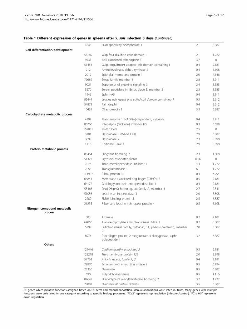
Table 1 Different expression of genes in spleens after S. suis infection 3 days (Continued)
1843 Dual specificity phosphatase 1 2.1 6.387
Cell differentiation/development
58189 Wap four-disulfide core domain 1 2.1 1.222
9531 Bcl2-associated athanogene 3 3.7 0
51454 Gulp, engulfment adaptor ptb domain containing1 0.4 2.181
212 Aminolevulinate, delta-, synthase 2 0.4 6.698
2012 Epithelial membrane protein 1 2.0 7.146
79689 Steap family member 4 2.8 3.911
9021 Suppressor of cytokine signaling 3 2.4 3.385
5270 Serpin peptidase inhibitor, clade E, member 2 2.3 3.385
1946 Ephrin-A5 0.4 3.911
85444 Leucine rich repeat and coiled-coil domain containing 1 0.5 5.612
54873 Palmdelphin 0.4 5.612
10439 Olfactomedin 1 3.3 6.387
Carbohydrate metabolic process
4199 Malic enzyme 1, NADP(+)-dependent, cytosolic 0.4 3.911
80760 Inter-alpha (Globulin) inhibitor H5 0.3 6.698
152831 Klotho beta 2.5 0
3101 Hexokinase 3 (White Cell) 2.9 6.387
3099 Hexokinase 2 2.3 8.898
1116 Chitinase 3-like 1 2.9 8.898
Protein metabolic process
85464 Slingshot homolog 2 2.3 1.508
51327 Erythroid associated factor 0.06 0
7076 Timp metallopeptidase inhibitor 1 4.4 1.222
7053 Transglutaminase 3 6.1 1.222
114907 F-box protein 32 0.4 6.794
64844 Membrane-associated ring finger (C3HC4) 7 0.5 2.181
64172 O-sialoglycoprotein endopeptidase-like 1 0.4 2.181
55466 Dnaj (Hsp40) homolog, subfamily A, member 4 2.7 2.541
51056 Leucine aminopeptidase 3 2.0 8.898
2289 Fk506 binding protein 5 2.5 6.387
26235 F-box and leucine-rich repeat protein 4 0.5 6.698
Nitrogen compound metabolicprocess
383 Arginase 0.2 2.181
64850 Alanine-glyoxylate aminotransferase 2-like 1 0.2 6.882
6799 Sulfotransferase family, cytosolic, 1A, phenol-preferring, member2
2.0 6.387
8974 Procollagen-proline, 2-oxoglutarate 4-dioxygenase, alphapolypeptide ii
3.2 6.387
Others
129446 Cardiomyopathy associated 3 0.3 2.181
128218 Transmembrane protein 125 2.0 8.898
57763 Ankyrin repeat, family A, 2 0.4 2.181
29970 Schwannomin interacting protein 1 0.5 6.794
23336 Desmuslin 0.5 6.882
590 Butyrylcholinesterase 0.5 4.116
84649 Diacylglycerol o-acyltransferase homolog 2 3.2 1.222
79887 Hypothetical protein Flj22662 3.5 6.387
DE genes which putative functions assigned based on GO term and manual annotation. Manual annotations were listed in italics. Many genes with multiplefunctions were only listed in one category according to specific biology processes. “FC≥2” represents up regulation (infection/control), “FC ≤ 0.5” representsdown regulation.
Li et al. BMC Genomics 2010, 11:556http://www.biomedcentral.com/1471-2164/11/556
Page 6 of 12
oxide production, is involved in disruption of the blood-brain barrier(BBB) in an experimental model of bacterialmeningitis [61]. S. suis-mediated PGE2 production byhuman macrophages was also noticed by Jobin and con-tributed to the BBB disruption [62].
Toll-like receptors (TLRs) pathway analysisActivation of the innate immune response is controlledin large part by the Toll-like receptor (TLR) family ofpattern-recognition receptors. The previous studyshowed that S. suis was mainly recognized via TLR2 byTHP-1 monocytes, which was associated with CD14[38] and led to the release of pro-inflammatory media-tors [63]. The strong activation of TLR2 and CD14 wasalso observed in murine brain parenchyma after the pre-sence of S. suis bacteremia [39]. A recent research indi-cated that components released during S. suis infectionas well as penicillin-treated whole bacteria could induceNF-kB activation through TLR2/6 [64]. The obvious ele-vation of TLR2 (2.0 fold) and CD14 (3.4 fold) wasnoticed at transcript level in spleens after highly patho-genic SS2 infection. Unsurprisingly, MyD88, an adaptormolecule in downstream signaling events with TLRsand CD14, was up-regulated at the level of 1.5 fold(q < 10%). In contrast, the effect could not be seen withavirulent SS2 infection.
Down-regulated transcripts following S. suis infectionThe majority of down-regulated genes were related totranscription, transport, material and energy metabolism(Table 1). Highly pathogenic strain could show highlevel of toxicity to host cells [34], and as a result, theinfluenced cells could hardly to be active. So these down
regulations could be regarded the representative of thereduced vital activity of SS2-influenced cells.
DiscussionTwo recent SS2 outbreaks in China not only seriouslychallenged public health but also shocked the scientificcommunity, calling for the basic and translational stu-dies of S. suis. Until now, several proteins were identi-fied as vaccine candidates [65,66] and drug targets[67,68] for controlling SS2. In addition, emphasis is alsoextended to the pathogenesis study. Several pathogenicfactors were successfully identified and strengthened theunderstanding for the virulence of the bacterium. Asinfectious disease resulted from the interplay betweenpathogens and the defense of the hosts they infect, hostimmune response was especially essential for under-standing the diseases [41,69].In the present study, we tried to compare the gene
expression profiles of spleens from swine suffering fromhighly pathogenic SS2, from swine infected with theavirulent isogenic strain, and from swine inoculated withPBS respectively to reveal the host immune response toSS2 and the contributions of host response to SS2 dis-eases. It is not accidental that significant changes of geneexpression profiles could be noticed when infected withhighly pathogenic SS2 compared with mock-infectedsamples, while avirulent isogenic strain would cause simi-lar profiles to mock-infected samples (Figure 1A). Theseindicated that avirulent isogenic strain could hardly causesignificant gene expression which was coincident withthe fact that no significant clinical symptoms could benoticed in pigs. Moreover, the obvious changes in geneexpression profiles were highly associated with significantclinical signs on day 3 post-inoculation with highlypathogenic strain. Further analysis of the present studyindicated that the majority of down-regulated genes weremainly involved in transcription, transport, material andenergy metabolism which were representative of thereduced vital activity of SS2-influenced cells. However,the up-regulated genes were principally related toimmune response, such as genes involved in inflamma-tory response; acute-phase/immune response; cell adhe-sion and response to stress. Undoubtedly, it wouldbe meaningful to explore the roles of these genes in SS2-caused diseases.First of all, it is necessary to know how SS2 induces
immune response. It is well acknowledged that TLRsare transmembrane proteins that could recognize speci-fic PAMPs and eventually result in the activation of NF-kB and MAP kinases to elicit regulatory response [70].Among these transmembrane proteins, TLR-2 couldrecognize bacterial LAM, BLP and PGN by followingtheir initial interaction with CD14. Previous reportsindicated that S. suis mainly induced proinflammatory
Table 2 Validation of microarray results by qPCR
Gene Accession Microarray foldchange
qPCR fold change(p-value)
IL1B CK468468 16.7 258.3 (0.0117)
S100A9 BI402402 16.1 137.2 (0.0007)
S100A12 CB475695 18.6 76.7 (<0.0001)
HSP90 CF180819 3.18 48.5 (0.0321)
IL8 NM_213867 5.58 35.5 (0.0066)
HSP70 NM_213766 11.06 31.4 (0.0099)
TIMP1 NM_213857 4.4 14.6 (0.0023)
IL6 AF493992 4.8 10.5 (0.0074)
SOD2 NM_214127 4.6 10.0 (<0.0001)
NRAMP1 U55068 3.97 7.2 (0.0035)
SELE NM_214268 3.5 5.9 (0.0002)
PLAU NM_213945 2.0 5.5 (0.0415)
CCL19 BX672579 2.16 4.2 (0.0004)
haptocorrin CB472702 3.78 2.1 (0.0388)
TLR-2 NM_213761 2.0 2.1 (<0.0001)
ALOX15 NM_213931 0.2 0.3 (0.038)
Li et al. BMC Genomics 2010, 11:556http://www.biomedcentral.com/1471-2164/11/556
Page 7 of 12
cytokines by TLR2 of human macrophages and murinebrain [39,63], and several proinflammatory cytokines,such as IL-1B, IL-6, IL-8, TNF-a and MCP-1 could betriggered [35,36,38,41]. In our study, large doses of bac-teria could be isolated from spleens of WT-infected pigswhile no bacterium could be found to exist in pigsinfected with ΔHP0197. In coincidence with these, TLR-2 pathway and several proinflammatory cytokines wereinduced only in WT-infected pigs. ΔHP0197 showedsimilar transcript profile as control pigs due to eitherfailing to invading or being easily eliminated by host. Incontrast, the large doses of bacteria effected maximalcytokines release in WT-infected pigs [37]. The exagger-ated high levels of cytokines perhaps exacerbate theinflammation and were considered to be responsible forS. suis caused diseases [39]. So the successful lethalpathogens could persistently induce cytokines secretedoriginally to clear the foreign invader, and as a result,the host’s defense was utilized by S. suis to cause dis-eases, and to some extent to death.As we all know that the secreted cytokine is an impor-
tant part of a host defense system, which could recruitinflammatory cells to sites of tissue damage and help toeliminate the pathogens. However, this innate defensesystem is a double edged sword. If the recruiting inflam-matory cells could kill the invader, the disease could becontrolled. On the opposite side, if the recruiting phago-cytes could not efficiently kill the bacteria, the tidewould be turned to pathogen’s favor, and the persis-tently induced cytokines would result in the exacerbatedinflammation and lead to the death during the septicphase of infection. These might be the reason why thesurvival rate could be elevated when inflammation wasinhibited by IL-10 [40], and why the level of cytokinewas correlated inversely with survival time in patientswith sepsis [45]. In coincidence with our analysis, patho-genic S. suis could effectively resist the uptake by phago-cytes and CPS could inhibit activation of signalingpathways involved in phagocytosis [17,71,72]. In addi-tion, several virulence-associated proteins such as FBPS[19], PDGA [31], LTA [30], HP0197 (unpublished data),serine protease [73] etc. were also contributed to thephagocytosis resistance, and the up-regulation of theseproteins in vivo may suggest the better phagocytosisresistance [31,74,75]. Due to failing phagocytosis, bac-teria could not only cause exacerbated inflammation butalso contribute to its survival in the bloodstream in“modified Trojan Horse” theory in which bacteria travelextracellularly while attached to, but not phagocytosed[17,72], and then cause bacteremia and even septemia.One of the key questions to be answered is how
S. suis crosses the blood-brain barrier to cause meningi-tis, which was observed in all WT-infected pigs. Thefindings of the reported study presented that suilysin-
positive strain could show toxin to produce functionalalteration and increase the permeability of BBB; and Sui-lysin-negative strain might stimulate the production ofproinflammatory cytokines resulting in alteration of BBBpermeability [76,77]. And they also indicated that thishighly pathogenic strain could produce high level of tox-ins in vivo-Suilysin, MRP, hyl [74], and undoubtedly itwould contribute to the penetration of deep tissue andBBB. In addition, the stimulated production of proin-flammatory cytokines would result in the alteration ofBBB permeability, and it would be more feasible forS. suis to break through BBB. From our understanding,WT strain could utilize the synergic effect of toxins andhigh level of cytokines to accelerate the penetration ofdeep tissue and BBB. These might be the reason whythe strain could cause severe human diseases in Sichuan,2005.
ConclusionsMicroarray technology has been used to analyse thegloble porcine transcriptional response to infection withvarious pathogenic microorganisms recently. Studyon the transcriptional response to the Gram-positivebacterium SS2 by using the Affymetrix GeneChip PorcineGenome Array has not been reported until now.Although great efforts have been made to understand themolecular basis of this infection, the response to SS2infection is still largely unknown. Transcriptome analysisbased on S. suis-infected spleens could improved theinterference received by the cells analysis, and also supplythe solid supplementary for analysis on alveolar macro-phages. Highly pathogenic S. suis could persistentlyinduce cytokines mainly by TLR2 pathway, and even-tually the high level of cytokines and toxins secreted byphagocytosis-resistant bacteria could destroy deep tis-sues, and cause meningitis, septicaemia, pneumonia,endocarditis, and arthritis.
MethodsBacterial strainsSS2 strain 05ZY (WT) which was isolated from thebrain of a diseased piglet collected in Sichuan outbreakin China 2005 showed high virulence to pigs [4,78], andwas applied to infect pigs. An isogenic HP0197 mutant(ΔHP0197) derived strain 05ZY showed no obviousvirulence to pigs (unpublished data) was applied as acontrol.
Animals infection and tissue collectionAll the experimental protocols were approved by theLaboratory Animal Monitoring Committee of HubeiProvince and performed accordingly. A total of 12 pigsof high-health status (ages 4-5 weeks) were assignedto three groups, within four in each. The pigs were
Li et al. BMC Genomics 2010, 11:556http://www.biomedcentral.com/1471-2164/11/556
Page 8 of 12
determined to be SS2-free by antibody-based ELISA andnasal swabs-based bacteriologic test. One hour beforeinoculation, all pigs were given 2 ml of 1% acetic acid(pH 2.9) intranasally to enhance the sensitivity of theS. suis challenge. Two groups were inoculated intrana-sally with 1 ml of 2×10⁶CFU of WT strain or ΔHP0197respectively, and the rest group inoculated with PBS wasserved as control. All pigs inoculated with WT showedtypical symptoms at day 3 while pigs inoculated withΔHP0197 or PBS showed no significant clinical signs.Blood samples from each group were detected for bac-terial burden. Bacteria could be found in the blood ofpigs in the WT group at day 3 post-inoculation whileno bacterium was found from the blood of pigs inocu-lated with isogenic mutant strain or PBS at the sametime point. All pigs were sacrificed at day 3, and theirtissue samples were cultured to prove in vivo bacterialburden. Bacteria were found in the spleens of the WT
group, and no bacterium was found in the other twogroups. Spleen samples were aseptically collected andimmediately frozen in liquid nitrogen for future RNAisolation. Total RNA was isolated from approximately200 mg of each sample by using the TRIzol (Invitrogen)and RNeasy Midi kit (QIAGEN) based on the manufac-turer’s protocols. The integrity, quality, and quantity ofRNA were assessed using the Agilent Bioanalyser 2100.
Microarray hybridizations and data analysisThe RNA labelling and hybridization were conducted bya commercial Affymetrix array service (CapitalBio Corp.Beijing, China). An aliquot of 2 μg of total RNA wasconverted to double-stranded cDNA with the one-cyclecDNA Synthesis Kit (Affymetrix), and then biotin-taggedcRNA was produced with MessageAmp™ II aRNAAmplification Kit. The resulting bio-tagged cRNA wasfragmented to strands of 35 to 200 bases in length
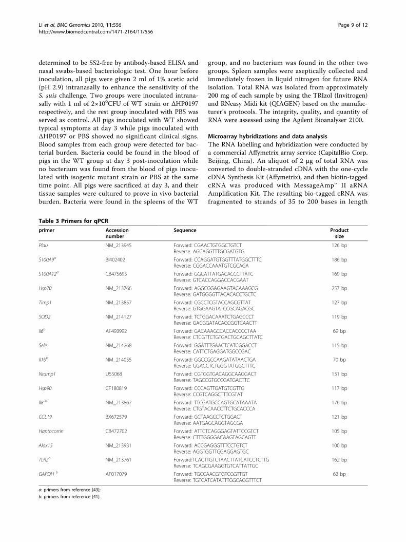
Table 3 Primers for qPCR
primer Accessionnumber
Sequence Productsize
Plau NM_213945 Forward: CGAACTGTGGCTGTCTReverse: AGCAGGTTTGCGATGTG
126 bp
S100A9a BI402402 Forward: CCAGGATGTGGTTTATGGCTTTCReverse: CGGACCAAATGTCGCAGA
186 bp
S100A12a CB475695 Forward: GGCATTATGACACCCTTATCReverse: GTCACCAGGACCACGAAT
169 bp
Hsp70 NM_213766 Forward: AGGCGGAGAAGTACAAAGCGReverse: GATGGGGTTACACACCTGCTC
257 bp
Timp1 NM_213857 Forward: CGCCTCGTACCAGCGTTATReverse: GTGGAAGTATCCGCAGACGC
127 bp
SOD2 NM_214127 Forward: TCTGGACAAATCTGAGCCCTReverse: GACGGATACAGCGGTCAACTT
119 bp
Il6b AF493992 Forward: GACAAAGCCACCACCCCTAAReverse: CTCGTTCTGTGACTGCAGCTTATC
69 bp
Sele NM_214268 Forward: GGATTTGAACTCATCGGACCTReverse: CATTCTGAGGATGGCCGAC
115 bp
Il1bb NM_214055 Forward: GGCCGCCAAGATATAACTGAReverse: GGACCTCTGGGTATGGCTTTC
70 bp
Nramp1 U55068 Forward: CGTGGTGACAGGCAAGGACTReverse: TAGCCGTGCCGATGACTTC
131 bp
Hsp90 CF180819 Forward: CCCAGTTGATGTCGTTGReverse: CCGTCAGGCTTTCGTAT
117 bp
Il8 b NM_213867 Forward: TTCGATGCCAGTGCATAAATAReverse: CTGTACAACCTTCTGCACCCA
176 bp
CCL19 BX672579 Forward: GCTAAGCCTCTGGACTReverse: AATGAGCAGGTAGCGA
121 bp
Haptocorrin CB472702 Forward: ATTCTCAGGGAGTATTCCGTCTReverse: CTTTGGGGACAAGTAGCAGTT
105 bp
Alox15 NM_213931 Forward: ACCGAGGGTTTCCTGTCTReverse: AGGTGGTTGGAGGAGTGC
100 bp
TLR2b NM_213761 Forward:TCACTTGTCTAACTTATCATCCTCTTGReverse: TCAGCGAAGGTGTCATTATTGC
162 bp
GAPDH b AF017079 Forward: TGCCAACGTGTCGGTTGTReverse: TGTCATCATATTTGGCAGGTTTCT
62 bp
a: primers from reference [43];
b: primers from reference [41].
Li et al. BMC Genomics 2010, 11:556http://www.biomedcentral.com/1471-2164/11/556
Page 9 of 12

according to Affymetrix’s protocols and then it washybridized to GeneChip Porcine Genome Array. Hybri-dization was performed at 45°C with rotation for 16 h(Affymetrix GeneChip Hybridization Oven 640). TheGeneChip arrays were washed and then stained (strepta-vidin-phycoerythrin) on an Affymetrix Fluidics Station450 followed by scanning on GeneChip Scanner 3000.The hybridization data were analyzed using GeneChip
Operating software (GCOS 1.4). A global scaling factor of500 was used to normalize the different arrays. We identi-fied the differentially expressed genes according to changep-value calculated by GCOS 1.4, and 2-fold change as anempirical criterion. Then all DE genes were performed forhierarchical cluster (Ver.3.0) and TreeView (Ver.1.1.1)analyses. Genes with significant similarities to transcriptsin nr database based on BLASTX searches were selectedfor GO analysis with DAVID http://david.abcc.ncifcrf.gov/home.jsp. Annotation results were obtained by inputtingthe gene list of ENTREZ_GENE_ID as identifier. Allmicroarray results from this study were deposited inNCBI’s Gene Expression Omnibus (GEO) database, acces-sion numbers are: Platform, GPL3533; Series, GSE23596;Samples, GSM578704, GSM578705, GSM578706,GSM578707, GSM578708, GSM578709, GSM578710,GSM578711, GSM578712.
qPCR analysisAll tested RNAs from swine spleens were reversely tran-scribed to cDNA with the M-MLV Reverse Transcrip-tase (Promega). Each cDNA sample was used as atemplate for qPCR and the amplification mixture con-tained SYBR Green (TOYOBO, Japan), forward andreverse primers. Some primers were designed by theprogram Primer 5.0, the primer names, accession num-ber, primer sequence and product size are shown inTable 3. The efficiency of the PCR reaction was 91-99%for all reactions (slope standard line between -3.3 and-3.6). The standard line consisted of five 10-fold dilu-tions of the samples. Analysis was performed using theABI7500 Software (Applied Biosystems). PCRs were per-formed in ABI PRISM 7500 sequence detection systemas follows: 1 cycle at 95°C for 10 min; 45 cycles at 95°Cfor 30 s, 60°C for 30 s and 72°C for 30 s. Melting curveswere performed at the end of amplification for validat-ing data quality by increasing the temperature from 65°C to 95°C, read every 0.2°C, hold 2 sec, then cooling at25°C for 30 s. The PCR products were confirmed usingagarose gel electrophoresis (1.5%). Amplification of thegapdh gene was used as internal control. All the testedgenes are shown in Table 3. All reactions were per-formed in triplicate. For each run, to normalize theamount of sample cDNA added to each reaction, the Ctvalue of each test gene was subtracted by the Ct value
of the endogenous control gapdh gene (delta Ct = Cttested gene - Ct gapdh), and then for a comparisonbetween the expression of the gene in treated samplesand in control samples. The delta Ct values of the genein treated samples were subtracted by the delta Ct valueof the gene in control samples (delta-delta Ct = delta Cttreatment - delta Ct control). The fold changes were cal-culated by the formula of 2-delta-delta Ct described byLivak & Schmittgen [79]. Data were means ± SD of tri-plicate reactions for each gene transcript.
Additional material
Additional file 1: Spleen transcriptome analysis following S. suisinfection using the Affymetrix Porcine Genechip. Data of each probeis from the three piglets of the control group (NC-1P, NC-2P, NC-3P), theWT group (WT-1P, WT-2P, WT-3P) and the △HP0197 group (M97-1P, M97-2P, M97-3P). “P”, present; “A”, absent; “M”, marginal; select while Count (P)≥ 2 in two groups. Totally, 15,757, 14,992 and 15,487 probesets weredetected expression in the control group, the WT group and △HP0197group respectively.
Additional file 2: transcripts expressed in porcine spleen followingS. suis (WT) infection. “FC”, Fold change, gene expression levelfollowing WT infection compared to the control. “≥2” represents upregulation, “<1” represents down regulation. “q-value”, significance levelof differential expression for a particular gene. “Gene description”, topinformative BLASTX hit.
Additional file 3: Differentially expressed transcripts in porcinespleen following S. suis (WT) infection (q < 10%, FC≥2). 120transcripts (row 5-124) were significantly up-regulated, and 132 (row 125-256) were significantly down-regulated, “FC”, Fold change, geneexpression level following WT infection compared to the control.
AbbreviationsSS2: Streptococcus suis serotype 2; DE: differentially expressed; FC: foldchange; GO: Gene Ontology; FDR: false discovery rate; qPCR: quantitativereal-time PCR; TLR: Toll-like receptors; PRRs: pattern-recognition receptors.
AcknowledgementsWe thank Professor Yanxiu Liu for her revision of the language of thismanuscript.This work was supported by National Basic Research Program of China(program 973, grant 2006CB504404), the National Transgenic Major Program(2009ZX08009-141B), the National Natural Science Foundation of China(30871870), and Program for Changjiang Scholars and Innovative ResearchTeam in University (IRT0726).
Author details1Unit of Animal Infectious Diseases, National Key Laboratory of AgriculturalMicrobiology, Huazhong Agricultural University, Wuhan, Hubei, China.2College of Veterinary Medicine, Huazhong Agricultural University, Wuhan,Hubei, China.
Authors’ contributionsRL and BC carried out all works and drafted the manuscript. AZ madesubstantial contributions to bioinformatics and statistical analysis. LT and YWparticipated in the animal challenge experiment. HC participated in theexperiment design and coordination. MJ helped to revise and finalize themanuscript. All authors read and approved the final manuscript.
Received: 4 May 2010 Accepted: 11 October 2010Published: 11 October 2010
Li et al. BMC Genomics 2010, 11:556http://www.biomedcentral.com/1471-2164/11/556
Page 10 of 12
References1. Staats JJ, Feder I, Okwumabua O, Chengappa MM: Streptococcus suis: past
and present. Vet Res Commun 1997, 21(6):381-407.2. Lun ZR, Wang QP, Chen XG, Li AX, Zhu XQ: Streptococcus suis: an
emerging zoonotic pathogen. Lancet Infect Dis 2007, 7(3):201-209.3. Hill JE, Gottschalk M, Brousseau R, Harel J, Hemmingsen SM, Goh SH:
Biochemical analysis, cpn60 and 16S rDNA sequence data indicate thatStreptococcus suis serotypes 32 and 34, isolated from pigs, areStreptococcus orisratti. Vet Microbiol 2005, 107(1-2):63-69.
4. Tang J, Wang C, Feng Y, Yang W, Song H, Chen Z, Yu H, Pan X, Zhou X,Wang H, et al: Streptococcal toxic shock syndrome caused byStreptococcus suis serotype 2. PLoS Med 2006, 3(5):e151.
5. Vanier G, Segura M, Lecours MP, Grenier D, Gottschalk M: Porcine brainmicrovascular endothelial cell-derived interleukin-8 is first induced andthen degraded by Streptococcus suis. Microb Pathog 2009, 46(3):135-143.
6. Haas G, Karaali G, Ebermayer K, Metzger WG, Lamer S, Zimny-Arndt U,Diescher S, Goebel UB, Vogt K, Roznowski AB, et al: Immunoproteomics ofHelicobacter pylori infection and relation to gastric disease. Proteomics2002, 2(3):313-324.
7. Holden MT, Hauser H, Sanders M, Ngo TH, Cherevach I, Cronin A,Goodhead I, Mungall K, Quail MA, Price C, et al: Rapid evolution ofvirulence and drug resistance in the emerging zoonotic pathogenStreptococcus suis. PLoS One 2009, 4(7):e6072.
8. Wangsomboonsiri W, Luksananun T, Saksornchai S, Ketwong K,Sungkanuparph S: Streptococcus suis infection and risk factors formortality. J Infect 2008, 57(5):392-396.
9. Rusmeechan S, Sribusara P: Streptococcus suis meningitis: the newestserious infectious disease. J Med Assoc Thai 2008, 91(5):654-658.
10. Watkins EJ, Brooksby P, Schweiger MS, Enright SM: Septicaemia in a pig-farm worker. Lancet 2001, 357(9249):38.
11. Taipa R, Lopes V, Magalhaes M: Streptococcus suis meningitis: first casereport from Portugal. J Infect 2008, 56(6):482-483.
12. Tramontana AR, Graham M, Sinickas V, Bak N: An Australian case ofStreptococcus suis toxic shock syndrome associated with occupationalexposure to animal carcasses. Med J Aust 2008, 188(9):538-539.
13. van de Beek D, Spanjaard L, de Gans J: Streptococcus suis meningitis inthe Netherlands. J Infect 2008, 57(2):158-161.
14. Smith TC, Capuano AW, Boese B, Myers KP, Gray GC: Exposure toStreptococcus suis among US swine workers. Emerg Infect Dis 2008,14(12):1925-1927.
15. Fittipaldi N, Collis T, Prothero B, Gottschalk M: Streptococcus suisMeningitis, Hawaii. Emerg Infect Dis 2009, 15(12):2067-2069.
16. Wertheim HF, Nguyen HN, Taylor W, Lien TT, Ngo HT, Nguyen TQ,Nguyen BN, Nguyen HH, Nguyen HM, Nguyen CT, et al: Streptococcus suis,an important cause of adult bacterial meningitis in northern Vietnam.PLoS One 2009, 4(6), e5973.
17. Chabot-Roy G, Willson P, Segura M, Lacouture S, Gottschalk M:Phagocytosis and killing of Streptococcus suis by porcine neutrophils.Microb Pathog 2006, 41(1):21-32.
18. Smith HE, Damman M, van der Velde J, Wagenaar F, Wisselink HJ,Stockhofe-Zurwieden N, Smits MA: Identification and characterization ofthe cps locus of Streptococcus suis serotype 2: the capsule protectsagainst phagocytosis and is an important virulence factor. Infect Immun1999, 67(4):1750-1756.
19. de Greeff A, Buys H, Verhaar R, Dijkstra J, van Alphen L, Smith HE:Contribution of fibronectin-binding protein to pathogenesis ofStreptococcus suis serotype 2. Infect Immun 2002, 70(3):1319-1325.
20. Brassard J, Gottschalk M, Quessy S: Cloning and purification of theStreptococcus suis serotype 2 glyceraldehyde-3-phosphatedehydrogenase and its involvement as an adhesin. Vet Microbiol 2004,102(1-2):87-94.
21. Esgleas M, Li Y, Hancock MA, Harel J, Dubreuil JD, Gottschalk M: Isolationand characterization of alpha-enolase, a novel fibronectin-bindingprotein from Streptococcus suis. Microbiology 2008, 154(Pt 9):2668-2679.
22. Zhang A, Chen B, Mu X, Li R, Zheng P, Zhao Y, Chen H, Jin M:Identification and characterization of a novel protective antigen, Enolaseof Streptococcus suis serotype 2. Vaccine 2009, 27(9):1348-1353.
23. Haataja S, Tikkanen K, Hytonen J, Finne J: The Gal alpha 1-4 Gal-bindingadhesin of Streptococcus suis, a gram-positive meningitis-associatedbacterium. Adv Exp Med Biol 1996, 408:25-34.
24. Haataja S, Tikkanen K, Liukkonen J, Francois-Gerard C, Finne J:Characterization of a novel bacterial adhesion specificity ofStreptococcus suis recognizing blood group P receptor oligosaccharides.J Biol Chem 1993, 268(6):4311-4317.
25. Haataja S, Tikkanen K, Nilsson U, Magnusson G, Karlsson KA, Finne J:Oligosaccharide-receptor interaction of the Gal alpha 1-4Gal bindingadhesin of Streptococcus suis. Combining site architecture andcharacterization of two variant adhesin specificities. J Biol Chem 1994,269(44):27466-27472.
26. Tikkanen K, Haataja S, Francois-Gerard C, Finne J: Purification of agalactosyl-alpha 1-4-galactose-binding adhesin from the gram-positivemeningitis-associated bacterium Streptococcus suis. J Biol Chem 1995,270(48):28874-28878.
27. Tikkanen K, Haataja S, Finne J: The galactosyl-(alpha 1-4)-galactose-binding adhesin of Streptococcus suis: occurrence in strains of differenthemagglutination activities and induction of opsonic antibodies. InfectImmun 1996, 64(9):3659-3665.
28. Baums CG, Kaim U, Fulde M, Ramachandran G, Goethe R, Valentin-Weigand P: Identification of a novel virulence determinant with serumopacification activity in Streptococcus suis. Infect Immun 2006,74(11):6154-6162.
29. Zhang A, Mu X, Chen B, Liu C, Han L, Chen H, Jin M: Identification andcharacterization of IgA1 protease from Streptococcus suis. Vet Microbiol2010, 140(1-2):171-175.
30. Fittipaldi N, Sekizaki T, Takamatsu D, Harel J, Dominguez-Punaro Mde L, VonAulock S, Draing C, Marois C, Kobisch M, Gottschalk M: D-alanylation oflipoteichoic acid contributes to the virulence of Streptococcus suis. InfectImmun 2008, 76(8):3587-3594.
31. Fittipaldi N, Sekizaki T, Takamatsu D, Dominguez-Punaro Mde L, Harel J,Bui NK, Vollmer W, Gottschalk M: Significant contribution of the pgdAgene to the virulence of Streptococcus suis. Mol Microbiol 2008,70(5):1120-1135.
32. Li M, Wang C, Feng Y, Pan X, Cheng G, Wang J, Ge J, Zheng F, Cao M,Dong Y, et al: SalK/SalR, a two-component signal transduction system, isessential for full virulence of highly invasive Streptococcus suis serotype2. PLoS ONE 2008, 3(5):e2080.
33. Pan X, Ge J, Li M, Wu B, Wang C, Wang J, Feng Y, Yin Z, Zheng F, Cheng G,et al: The orphan response regulator CovR: a globally negativemodulator of virulence in Streptococcus suis serotype 2. J Bacteriol 2009,191(8):2601-2612.
34. Ye C, Zheng H, Zhang J, Jing H, Wang L, Xiong Y, Wang W, Zhou Z, Sun Q,Luo X, et al: Clinical, Experimental, and Genomic Differences betweenIntermediately Pathogenic, Highly Pathogenic, and EpidemicStreptococcus suis. J Infect Dis 2009, 199(1):97-107.
35. Vadeboncoeur N, Segura M, Al-Numani D, Vanier G, Gottschalk M: Pro-inflammatory cytokine and chemokine release by human brainmicrovascular endothelial cells stimulated by Streptococcus suis serotype2. FEMS Immunol Med Microbiol 2003, 35(1):49-58.
36. Segura M, Vanier G, Al-Numani D, Lacouture S, Olivier M, Gottschalk M:Proinflammatory cytokine and chemokine modulation by Streptococcussuis in a whole-blood culture system. FEMS Immunol Med Microbiol 2006,47(1):92-106.
37. Segura M, Stankova J, Gottschalk M: Heat-killed Streptococcus suis capsulartype 2 strains stimulate tumor necrosis factor alpha and interleukin-6production by murine macrophages. Infect Immun 1999, 67(9):4646-4654.
38. Segura M, Vadeboncoeur N, Gottschalk M: CD14-dependent and-independent cytokine and chemokine production by human THP-1monocytes stimulated by Streptococcus suis capsular type 2. Clin ExpImmunol 2002, 127(2):243-254.
39. Dominguez-Punaro MC, Segura M, Plante MM, Lacouture S, Rivest S,Gottschalk M: Streptococcus suis serotype 2, an important swine andhuman pathogen, induces strong systemic and cerebral inflammatoryresponses in a mouse model of infection. J Immunol 2007,179(3):1842-1854.
40. Dominguez-Punaro Mde L, Segura M, Radzioch D, Rivest S, Gottschalk M:Comparison of the susceptibilities of C57BL/6 and A/J mouse strains toStreptococcus suis serotype 2 infection. Infect Immun 2008,76(9):3901-3910.
41. de Greeff A, Benga L, Wichgers Schreur PJ, Valentin-Weigand P, Rebel JM,Smith HE: Involvement of NF-kappaB and MAP-kinases in the
Li et al. BMC Genomics 2010, 11:556http://www.biomedcentral.com/1471-2164/11/556
Page 11 of 12

transcriptional response of alveolar macrophages to Streptococcus suis.Vet Microbiol 2009, 141(1-2):59-67.
42. Zhao SH, Kuhar D, Lunney JK, Dawson H, Guidry C, Uthe JJ, Bearson SM,Recknor J, Nettleton D, Tuggle CK: Gene expression profiling inSalmonella Choleraesuis-infected porcine lung using a longoligonucleotide microarray. Mamm Genome 2006, 17(7):777-789.
43. Chen H, Li C, Fang M, Zhu M, Li X, Zhou R, Li K, Zhao S: UnderstandingHaemophilus parasuis infection in porcine spleen through atranscriptomics approach. BMC Genomics 2009, 10:64.
44. van der Poll T, Keogh CV, Guirao X, Buurman WA, Kopf M, Lowry SF:Interleukin-6 gene-deficient mice show impaired defense againstpneumococcal pneumonia. J Infect Dis 1997, 176(2):439-444.
45. Norrby-Teglund A, Pauksens K, Norgren M, Holm SE: Correlation betweenserum TNF alpha and IL6 levels and severity of group A streptococcalinfections. Scand J Infect Dis 1995, 27(2):125-130.
46. Foell D, Wittkowski H, Vogl T, Roth J: S100 proteins expressed inphagocytes: a novel group of damage-associated molecular patternmolecules. J Leukoc Biol 2007, 81(1):28-37.
47. Hojo K, Tamura A, Mizoguchi C, Kato D, Ohshima T, Maeda N: Predominantbacteria recovered from a periodontitis site in a hamster model raisedby silk-ligature with Porphyromonas gingivalis infection. Biosci BiotechnolBiochem 2008, 72(5):1348-1351.
48. Patel L, Buckels AC, Kinghorn IJ, Murdock PR, Holbrook JD, Plumpton C,Macphee CH, Smith SA: Resistin is expressed in human macrophages anddirectly regulated by PPAR gamma activators. Biochem Biophys ResCommun 2003, 300(2):472-476.
49. Bokarewa M, Nagaev I, Dahlberg L, Smith U, Tarkowski A: Resistin, anadipokine with potent proinflammatory properties. J Immunol 2005,174(9):5789-5795.
50. Ward PP, Conneely OM: Lactoferrin: role in iron homeostasis and hostdefense against microbial infection. Biometals 2004, 17(3):203-208.
51. Knura-Deszczk S, Lipperheide C, Petersen B, Jobert JL, Berthelot-Herault F,Kobisch M, Madec F: Plasma haptoglobin concentration in swine afterchallenge with Streptococcus suis. J Vet Med B Infect Dis Vet Public Health2002, 49(5):240-244.
52. Thorn CF, Lu ZY, Whitehead AS: Tissue-specific regulation of the humanacute-phase serum amyloid A genes, SAA1 and SAA2, by glucocorticoidsin hepatic and epithelial cells. Eur J Immunol 2003, 33(9):2630-2639.
53. Baumann H, Gauldie J: The acute phase response. Immunol Today 1994,15(2):74-80.
54. Sorensen NS, Tegtmeier C, Andresen LO, Pineiro M, Toussaint MJ,Campbell FM, Lampreave F, Heegaard PM: The porcine acute phaseprotein response to acute clinical and subclinical experimental infectionwith Streptococcus suis. Vet Immunol Immunopathol 2006, 113(1-2):157-168.
55. Rikitake Y, Takai Y: Interactions of the cell adhesion molecule nectin withtransmembrane and peripheral membrane proteins for pleiotropicfunctions. Cell Mol Life Sci 2008, 65(2):253-263.
56. Al-Numani D, Segura M, Dore M, Gottschalk M: Up-regulation of ICAM-1,CD11a/CD18 and CD11c/CD18 on human THP-1 monocytes stimulatedby Streptococcus suis serotype 2. Clin Exp Immunol 2003, 133(1):67-77.
57. Wight TN: Versican: a versatile extracellular matrix proteoglycan in cellbiology. Curr Opin Cell Biol 2002, 14(5):617-623.
58. Wang W, Xu GL, Jia WD, Ma JL, Li JS, Ge YS, Ren WH, Yu JH, Liu WB:Ligation of TLR2 by versican: a link between inflammation andmetastasis. Arch Med Res 2009, 40(4):321-323.
59. Simantov R, Silverstein RL: CD36: a critical anti-angiogenic receptor. FrontBiosci 2003, 8:s874-882.
60. Nakache M, Berg EL, Streeter PR, Butcher EC: The mucosal vascularaddressin is a tissue-specific endothelial cell adhesion molecule forcirculating lymphocytes. Nature 1989, 337(6203):179-181.
61. Tilley SL, Coffman TM, Koller BH: Mixed messages: modulation ofinflammation and immune responses by prostaglandins andthromboxanes. J Clin Invest 2001, 108(1):15-23.
62. Jobin MC, Gottschalk M, Grenier D: Upregulation of prostaglandin E2 andmatrix metalloproteinase 9 production by human macrophage-like cells:synergistic effect of capsular material and cell wall from Streptococcussuis. Microb Pathog 2006, 40(1):29-34.
63. Graveline R, Segura M, Radzioch D, Gottschalk M: TLR2-dependentrecognition of Streptococcus suis is modulated by the presence ofcapsular polysaccharide which modifies macrophage responsiveness. IntImmunol 2007, 19(4):375-389.
64. Schreur PJ, Rebel JM, Smits MA, van Putten JP, Smith HE: Differentialactivation of the Toll-like receptor 2/6 complex by lipoproteins ofStreptococcus suis serotypes 2 and 9. Vet Microbiol 143(2-4):363-370.
65. Baums CG, Valentin-Weigand P: Surface-associated and secreted factors ofStreptococcus suis in epidemiology, pathogenesis and vaccinedevelopment. Anim Health Res Rev 2009, 10(1):65-83.
66. Feng Y, Zhang H, Ma Y, Gao GF: Uncovering newly emerging variants ofStreptococcus suis, an important zoonotic agent. Trends Microbiol 2010,18(3):124-31.
67. Zhang Q, Peng H, Gao F, Liu Y, Cheng H, Thompson J, Gao GF: Structuralinsight into the catalytic mechanism of gluconate 5-dehydrogenasefrom Streptococcus suis: Crystal structures of the substrate-free andquaternary complex enzymes. Protein Sci 2009, 18(2):294-303.
68. Zhang Q, Gao F, Peng H, Cheng H, Liu Y, Tang J, Thompson J, Wei G,Zhang J, Du Y, et al: Crystal structures of Streptococcus suis mannonatedehydratase (ManD) and its complex with substrate: genetic andbiochemical evidence for a catalytic mechanism. J Bacteriol 2009,191(18):5832-7.
69. Goldmann O, von Kockritz-Blickwede M, Holtje C, Chhatwal GS, Geffers R,Medina E: Transcriptome analysis of murine macrophages in response toinfection with Streptococcus pyogenes reveals an unusual activationprogram. Infect Immun 2007, 75(8):4148-4157.
70. Akira S, Uematsu S, Takeuchi O: Pathogen recognition and innateimmunity. Cell 2006, 124(4):783-801.
71. Segura M, Gottschalk M: Streptococcus suis interactions with the murinemacrophage cell line J774: adhesion and cytotoxicity. Infect Immun 2002,70(8):4312-4322.
72. Segura M, Gottschalk M, Olivier M: Encapsulated Streptococcus suis inhibitsactivation of signaling pathways involved in phagocytosis. Infect Immun2004, 72(9):5322-5330.
73. Hu Q, Liu P, Yu Z, Zhao G, Li J, Teng L, Zhou M, Bei W, Chen H, Jin M:Identification of a cell wall-associated subtilisin-like serine proteaseinvolved in the pathogenesis of Streptococcus suis serotype 2. MicrobPathog 2010, 48(3-4):103-9.
74. Tan C, Liu M, Jin M, Liu J, Chen Y, Wu T, Fu T, Bei W, Chen H: The keyvirulence-associated genes of Streptococcus suis type 2 are upregulatedand differentially expressed in vivo. FEMS Microbiol Lett 2008,278(1):108-114.
75. Zhang A, Chen B, Li R, Mu X, Han L, Zhou H, Chen H, Meilin J:Identification of a surface protective antigen, HP0197 of Streptococcussuis serotype 2. Vaccine 2009, 27(38):5209-5213.
76. Gottschalk M, Segura M: The pathogenesis of the meningitis caused byStreptococcus suis: the unresolved questions. Vet Microbiol 2000,76(3):259-272.
77. Vanier G, Segura M, Friedl P, Lacouture S, Gottschalk M: Invasion of porcinebrain microvascular endothelial cells by Streptococcus suis serotype 2.Infect Immun 2004, 72(3):1441-1449.
78. Zhang A, Xie C, Chen H, Jin M: Identification of immunogenic cell wall-associated proteins of Streptococcus suis serotype 2. Proteomics 2008,8(17):3506-3515.
79. Livak KJ, Schmittgen TD: Analysis of relative gene expression data usingreal-time quantitative PCR and the 2(-Delta Delta C(T)) Method. Methods2001, 25(4):402-408.
doi:10.1186/1471-2164-11-556Cite this article as: Li et al.: Response of swine spleen to Streptococcussuis infection revealed by transcription analysis. BMC Genomics 201011:556.
Li et al. BMC Genomics 2010, 11:556http://www.biomedcentral.com/1471-2164/11/556
Page 12 of 12