Respiratory System Module 2021-2022 Dr. Mohammad Odibate ...
20
Respiratory System Module 2021-2022 Dr. Mohammad Odibate Department of Microbiology and Pathology
Transcript of Respiratory System Module 2021-2022 Dr. Mohammad Odibate ...

Respiratory System Module
2021-2022
Dr. Mohammad Odibate
Department of Microbiology and Pathology

I
Laboratory Diagnosis of Group A Streptococcus
Steps of Laboratory Diagnosis of Group A
\: i,~~ Streptococcus ~ \~~ p
ro , li"J ---±;l> Specimen co 11 ecti on
J,«y,a >•• 2. Direct Antigen detection
3. Group A streptococci screening culture
4. Identification of GAS ,, & ro ~~ A s \-r~ p ~0 c 0 c, u.~ ''
5. Reporti ng results.

Laboratory Diagnosis of Group A Streptococcus
1- Specimen:
Throat swab of tonsillar area and/or posterior
pharynx (Avoid the tongue and uv~ la)
Swc1b
Throat is swabbed
in the area o f
the tonsi ls
Laboratory Diagnosis of Group A Streptococcus
~ . 2. Direct Antigen detection: 1. The pati ent's throat is first swabbed to collect a sampl e of mucu s. 2. The sample is applied to a strip of nitrocellulose film and, if GAS ant ige ns
are present, these will migrate along the film to form a vi sibl e line of antigen bound to labeled antibodies
3. Because a common probl em is the low se nsitivity. All negat ive res u lt s should be followed by culture.
(
i II ! I ~ I I
SA.MP\ f Wf:- il ·I f)11ll.\t1•v~ b1.1fk r · P. · ..
,. /; ~q r, p\e. .,.
1
1 () I 11< llft••,
A J J i u L'> I :., , ,,
-<)~"' 1
c} f .S,, L'- ~ ~ _y,:•
d ·· ~ -rr ~~ I ;~c .JJ
f 1
I
,.) ' u_.0 ~
S Cf h f' e f' ,_v-~ L
) I
P ,·I -. i,·v"
Laboratory Diagnosis of Group A Streptococcus L ~ - \il<: ¥V\ c) J ~ ,·c_
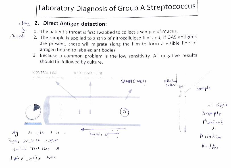
3. Group A streptococci screening culture Incubate cultures under atmospheric conditions (35°C for 18-24h) Examine the presence of hemolytic colonies on blood agar Re incubate negative cultures for an additional 18-24h ~ --==--------
~-hemolys is (Group A streptococci)
a -he mo lys is +-------iii~----.,._;.
cl e."r \ :: •
Z c' tt e q ro\.\" J ·. -hemolysis

Laboratory Diagnosis of Group A Streptococcus
4. 1ldentification of GAS:
Catalasetest - ve_
~acitracin susceptibilty (\(\ '--' b \·.)\-t'c_ - Pr inciple:
• Fo r identification of group A
• dist ingujsh between 5. pyogenes from other beta hemolytic streptococ€i
~ e,.,c; 1 h lJ e
• Strep. pyogenes is sensitive to Bacitracin giving zone of inhibition aro und disk
Group A streptococci is susceptible to
Ba cit ra c i n disk ( I eft); The right shows
resistance

Laboratory Diagnosis of Group A Streptococcus
5. Reporting results:
The results on the microbiology request form may include
- 5. pyogenes group A isolated
beta hemoltyic streptococci, not group A streptococci
isolated
No 5. pyogenes or beta hemolytic steptococci

DIAGNOSIS OF DIPHTHERIA
Diagnosis r')(/''7 br<H\~ J , u-tJ
1. The initial diagnosis of diphtheria is entirely cli nical ~ _)\ ~ ._3
(l qSo p1'tc. ~ I"\'/..._
2. Laboratory diagnosis C· c.1_,l;
A. Specimen: from the nose and throat and any other mucocuta neous
lesion. A portion of membrane should be removed and subm itted for
culture along with underlying exudate
B. Direct smear:
Gram stain: club shaped Gram positive bacilli with chinese lette r
arrangment
C. Culture media: cysteine-tellurite plate (Tisdale agar)
Results:
• C. diphtheriae : produce grayish -black coloni es, surround ed by a
brown/black halo.
D. Urease and oxidase negative, Catalase positive

UIAUNOSIS O F DIPHTHERIA
brown - bl«c.k 1,ti ~ lo 0
Palhade~
~ . ~i /j\ ' , I f'\. p1 ' :• , ~
f ·! ,:~ j ·. r.. <· ~11 • . . r'• _••. . , .. l 1f~ ~,
·, -,~ l ~I
'
- - -- --- • .-.r, ■ ft

DIAGNOSIS OF DIPHTHERIA
F. Toxin demonstration. As the pathogenesis is due to diphtheria toxin, isol ation of bacill i dose not complete the diagnosis. Toxin demonstration should be done following isolation, which can be of two types, in vivo and in vitro
□ In vitro test: Elek's test ~ od--c:...- , ..,, J , ~ ..> ~ ~
• __iJ __,. , )-- ,., ,; .. " ~
_j I lo ~
Js ' tc J..~,.. , c.,
. h-~t • .., ~
cYf ~l, ~ t) ,· f~~ \tt u,<-1
o-\J (\ v r-ri <-i \ f \ o r q
_) \ (JD }-o Xi r, ) I ......:::_.,.J.> I
~ r \.,J , Gt' ~
~ox,·~ ~-
Ab
DIAGNOSIS OF DIPHTHERIA L_ __ ~~:..:..:.~-=----=--=------------
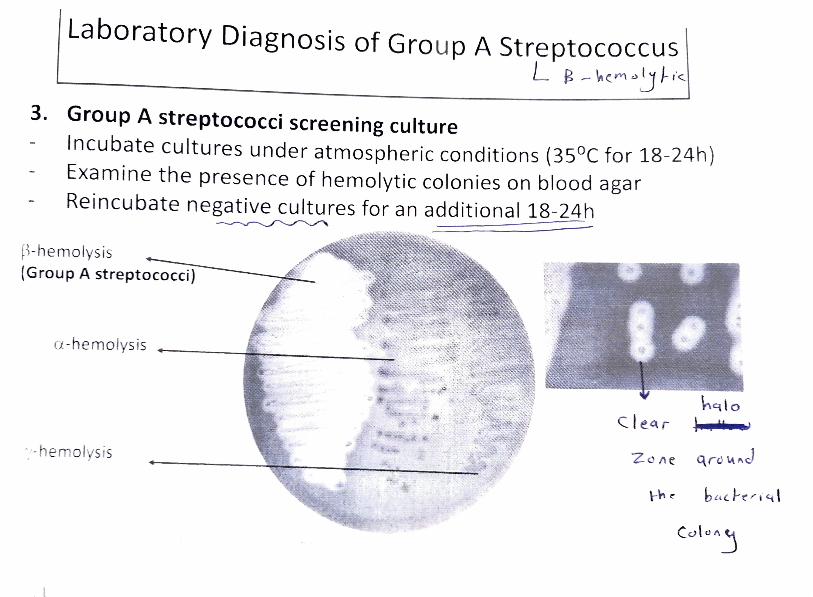
Elek 's test: rapid diagnosis (16-24 hrs) Sterile filter paper with C. diphtheriae antitoxin
~ ~
(_PJ(!__
d I H l.( S ,· 0 ,,
J •~
.. p) 4 ~ I Ab-Ag
_) I
A-b ...JI ~
-r.., y i "' ,,
I
\ \
, ,.,,_,,,,\
Known toxigenic C. diphtheriae ...---.;i
(positive contro l)
Unknown (patient's sample) I
) , Known nontoxigenic C. diphtheriae ~ ' (negative control)
lfc c~ I~~.~ 1"-- Result: positive toxigenic C. diphtheriae Nutrient agar with selective agent for Corynebacterium - v~ J , ~ + v,
C , , 1' ~ r-- v \
DIAGNOSIS OF DIPHTHERIA btl)JJ,::11 c? -
Elek 's test: rapid diagnosis (16-24 hrs)

DIAGNOSIS OF DIPHTHERIA t ~<~ ~ ), 1~~r
Elek 's t est: rapid d iagnosis (16-24 hrs)
Results:
Positive test: formation of four radiating lines resulting from the precipitation reaction between exotoxin and diphtheria antitoxin.
~-----·- -----""'-/'_ ,-- ~
/
/ ... • t ,1, .r1 ' 1t.';, ',.
r ~ ~ ,~.-vr r e'> ~t\-

Laboratory Diagn osis Lower Respiratory Infecti on
tt Pf ( r
Re» f'•,.,,r~putum culture 1' \ ~ C'c. ~.·v"' Th I · · . I e sputum cu ture 1s an important part of the diagnostic evalu at ion of potent1 a
lower respiratory tract infections . However, expectorated sputum specimens are
variably contaminated by colonizing oropha ryngeal flora , ma king resu Its hard to
interpret. Proper collection of the specimen is crucial to the re covery of the
etiological agent.
Specimen criteria: • If possible, specimen should be collected before antimicrobial treat m ent.
• First morning specimen is best.
• Spec imen must be collected in a sterile container.
• If multiple cultures are ordered they should be collected at least 24 hours
a pa rt.

Laboratory Diagnosis Lower Respiratory Infection
~ ~ xpectorated sputum ;.::,. l; • Specimen collection should be supervised by a trained
()e.,q> _h req H-, professional. 0 \.:_\ ~
~\ ~_J_) • Request the patient to remove any dentures and to
rinse the mouth or gargle with plain water before
specimen collection.
• Tell the patient to provide a specimen fro m a deep
cough, avoiding, as much as possible, mixing the
specimen with saliva or nasal secretions.
• Make sure the patient understands the difference
between saliva (from mouth) and sputum (from chest).

Laboratory Diagnosis Lower Respiratory Infection
Specimen Quality
Poor quality sputum Better quality
L w ;t-\.\ s~ Ii v et

_)\
ac..1·J
+~s ~ ~1-- ""'""
, · I'\
-r.B
r u u r LJUd ll LY spuLurn o e LL e r yud11 Ly
G w it-VI S '-\ l iv<,
... .. ~
Laboratory Diagnosis of H. lnfluenzae 1. Specimen collection and transport
D Depending on the site of infection, various specimn s m ay be collected such as CSF, blood, respiratory tract sputum, throat swabs, midd le ear, and sinuses
□ As H. influenze is highly sensitive to low tempertures, the specimen should neve r be refragutrated
□ Sample should be trasported and proscessed immdediatly w it hout any delay.
2. Direct detection: □ Gram staining : preparation from different sampls may show gram -negat ive
coccobacilli z;, \S \ )\ --0
c,,rs,, h ra--J 0 Capsule detection (Quellung reaction)
Antigen detection: The t yp e b ca psular anti gen can be detcted in CSF, urine , or other bdy fuids by
latex agg lutin ation using particl es co at ed with anti bodi es to type b antige n o r Direct im mun oflurcscnce tes t .

Laboratory Diagnosis of H. lnflu~r,_zpe . .. - - - ~- ,, .. . , .. . . ._.., - . . . .. "" .. ,.... ..,
Capsule detection (Quellung reaction}
)1
I C q f S4
1'A-~ <~~
Anti capsular )...,_ y "W Under
_j Ab microscope
i, ~~: '."·· \ ~• •. ! '~ (
~~-u~i i~o.<:tt® ,c"~~ t~Qtt~~
L
pctckJ r.;\I\.._J
<'
[
Ab I t'.'cc..J f.,
3.
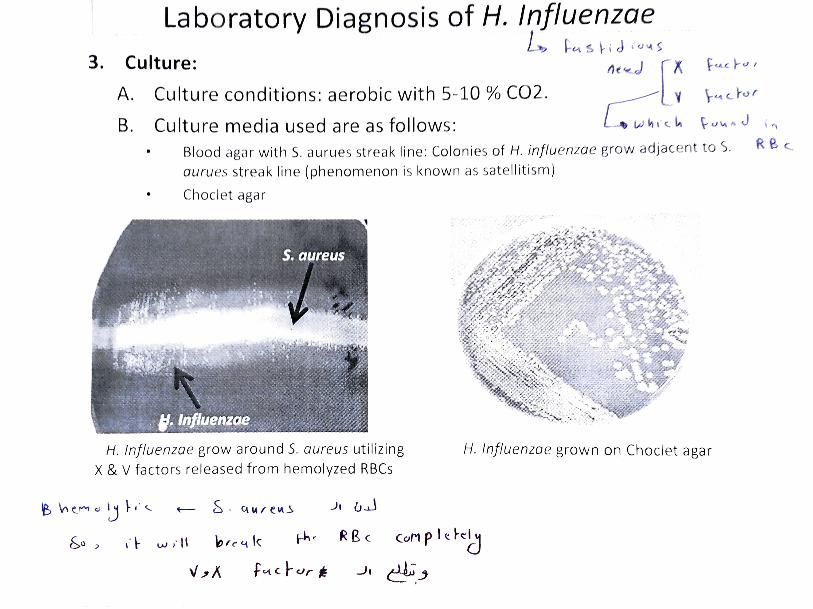
Laboratory Diagnosis of H. lnfluenzae
Culture: L~ ~ L'\ $ ~ i J I ,., \,\ )
A.
B.
Culture conditions: aerobic with 5-10 % CO2.
Culture media used are as follows:
\-<.-t c. h., r
\- U\.\ ~ J \ ,,
Blood aga r wi th 5. auru es strea k line : Co lo nies of H. influenzae grow adjacent to). RB <..
aurues strea k line (ph enomenon is known as sat elli ti srn)
Choclet agar
H. !nfluenzae grow around 5. aureus utilizing
X & V factors released from hemolyzed RBCs H. lnflu enzae grown on Choclet agar
~ "' c_r, d !J ~ 1· ' ~ S • c.uue" ~ J, u..J
&o > d· w i" l\ ~'e"' k ..-h ~ RBc Corl p I<.:. h::~
v,, J<. t CJt. C.~cJr* ..Jc ~--'

- ,.. • ••• • ""• • • • ,,..,-- • . .... .. -• • .. -• •u•, ••· ... •· • •• •• ,
Laboratory Diagnosis of H. lnfluenzae ~ ,
Growth requirements

Laboratory Diagnosis of H. I nfl uenzae
4. Biochemical tests:
• Reduces nitrate to nitrite.
• Catalase and Oxidase positive
• Fermentation of sugars: Glucose(+),
Sucrose(-), Lactose(-) Mannitol (-).