Respiratory -...
6
0 | Page slides sheet Respiratory handout PBL
Transcript of Respiratory -...

0 | P a g e
slides sheet
Respiratory
handout
PBL

1 | P a g e
The human respiratory system is a series of organs responsible for taking in oxygen and expelling carbon dioxide. The primary organs of the respiratory system are lungs, which carry out this exchange of gases as we breathe.
To perform this function, there should be harmonically
working components which include: 1. The lungs -> the main organ.
2. Conductive system -> airways including nasal openings, larynx, pharynx
and main bronchi + alveoli of the lungs.
3. Pump system -> rib cage + spine + respiratory ( inspiratory and expiratory) muscles ( diaphragm mainly and intercostal muscles) It is important for keeping a pressure gradient. *Bilateral diaphragmatic paralyses is incompatible with life.
4. Control system -> respiratory centers in the CNS and chemoreceptors. Chemoreceptors detect changes in pO2 , pCO2 and pH which is dependent on CO2. Respiratory centers get signals from the chemoreceptors and give orders to control the breathing rate to have normal breathing act, but if these centers fail to send orders hypoventilation will occur ( abnormal levels of O2 and CO2 )
*Hypoventilation may lead to respiratory failure
All these components work together by pumping gases from the atmospheric air to the lungs through the conductive system to reach the alveoli
All these components are needed for us to breathe, and any abnormality in one of them may lead to respiratory failure.

2 | P a g e
Respiratory Failure
A syndrome in which the respiratory system fails in one or both of the
gas exchange functions (O2 and CO2), it’s of two types:
Hypoxemic
Hypercapnic
o Type I respiratory failure. o Due to severe hypoxemia ( low pO2) with normal or low pCO2 o We consider the person
hypoxemic if pO2 is under 60mmHg based on the O2-Hb dissociation curve (when pO2 is 60mmHg, oxygen saturation is %90).
o Caused when the oxygen concentration in the air is low such as in high altitudes, and in lung diseases that affect alveolar parenchyma (pneumonia, pulmonary edema, lung fibrosis ) that prevents gas exchange through diffusion.
o Type II respiratory failure. o Due to hypercapnia (high pCO2). o pCO2 is above 50 mmHg. o Accompanies problems in
the control and pump systems (multifocal fibrosclerosis, rib fractures...)
Hypercapnia depends on the alveolar ventilation rate. Alveolar ventilation’s rate = tidal volume * respiratory
rate *Tidal volume-> the exhaled air volume in a respiration cycle. Tidal volume will be reduced if the patient doesn’t have normal functioning respiratory muscles.
Control system problems will cause hypoventilation due to apneas (inability to breathe).

3 | P a g e
Pump system problems will cause hypercapnia and decreased ventilation because of low tidal volume.
Low tidal volume-> Low alveolar ventilation rate -> Hypercapnia *abnormal breathing rate doesn’t necessarily mean respiratory failure.
Not every hypoxia is respiratory failure, it has to be severe ( pO2 < 60 mmHg )
Normal pO2 = (101-(1/3)*patient’s age) +/- 5
Example: if the patient is 21 years old then its 101-(1/3)*21= 94 So the normal range for this patient is 89-99
Oxygen saturation is measured by the pulsoximeter while pO2 is measured by arterial blood gas analysis
Respiratory Diseases
1) Control system problems Either in the chemoreceptors or respiratory centers. Congenital abnormalities in the chemoreceptors causes
primary hypoventilation syndrome.
Most common cause of suppression of the respiratory centers is drug overdose, mainly morphine, which is used as a post-operative pain killer and palliative treatment for cancer patients.
Other things that affect the respiratory centers include tumors, strokes, cerebral hemorrhages, trauma,… .
Remember: all of the above will lead to hypoventilation and hypercapnic respiratory failure.
2) Pump system problems For respiratory failure to occur, bilateral diaphragmatic paralysis
should be present such as in diaphragmatic fatigue which is caused by tetanus.

4 | P a g e
*Weakening of the respiratory muscles occurs when there is
muscular diseases ( myopathies ) such as neuromotor
diseases, myasthenia gravis, COPD secondary to muscle
wasting and cachexia, duchenne muscular dystrophy in
addition to problems in the ribs such as scoliosis.
Remember: all of the above will lead to hypercapnic respiratory failure.
3) Lung diseases Cause hypoxemic respiratory failure They include pulmonary edema, emphysema, fibrosis,
pneumonia, acute respiratory distress syndrome (ARDS) ARDS is a very common cause of severe hypoxemic respiratory
failure. It’s a secondary disease to sepsis and multiple trauma characterized by diffuse lung infiltrate. Patients are resistant to oxygen therapy meaning that they can never reach pO2 of 60 mmHg in spite of getting high concentrations of O2 (100% saturated)
Common Respiratory Symptoms
1) Cough (usually a sign of airways problems)
2) Dyspnea ( shortness of breath/SOB) 3) Cyanosis (due to lack of oxygen) 4) Chest pain asthma, COPD , bronchitis. 5) Sputum. 6) Can be associated with
fatigue.
5 | P a g e
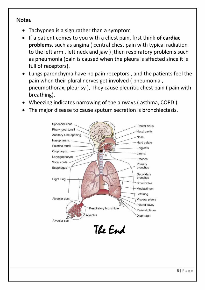
Notes:
Tachypnea is a sign rather than a symptom
If a patient comes to you with a chest pain, first think of cardiac problems, such as angina ( central chest pain with typical radiation to the left arm , left neck and jaw ) ,then respiratory problems such as pneumonia (pain is caused when the pleura is affected since it is full of receptors).
Lungs parenchyma have no pain receptors , and the patients feel the pain when their plural nerves get involved ( pneumonia , pneumothorax, pleurisy ), They cause pleuritic chest pain ( pain with breathing).
Wheezing indicates narrowing of the airways ( asthma, COPD ).
The major disease to cause sputum secretion is bronchiectasis.
The End