Respiratory Management in Pediatrics - Creighton University
68
Respiratory Management in Pediatrics Children’s Hospital Omaha Critical Care Transport Sue Holmer RN, C-NPT
Transcript of Respiratory Management in Pediatrics - Creighton University

Respiratory
Management in
Pediatrics
Children’s Hospital Omaha Critical Care Transport
Sue Holmer RN, C-NPT
Objectives
• Examine the differences between the
pediatric and adults airways.
• Recognize respiratory distress and
impending respiratory failure.
• Discuss management of respiratory
distress and respiratory failure.
• Case Scenarios.

Children are not small adults…
Children are still growing in every way.
Their bodies are different, they perceive
things and communicate differently, and
the long term implications of treatment
are not the same.

Respiratory Emergencies
Pediatric Respiratory
Emergencies • # 1 reason for
pediatric hospital
admissions.
• # 1 cause of death
during the first year of
life with the exception
of congenital
abnormalities.

Early Intervention is Critical
Respiratory Distress
Respiratory Failure/Shock
Cardiopulmonary Failure
Cardiopulmonary Arrest

Pediatric Cardiopulmonary
Arrests
Cardiac
10%
Shock
10%
Respiratory
80%
Most pediatric cardiopulmonary arrests begin as
respiratory failure or respiratory arrest.
Decrease respiratory reserve + Increased O2 demand =
Increased respiratory failure risk
Respiratory Emergencies in
Pediatrics
Airway Diseases
• Croup, epiglottitis, asthma, bronchiolitis,
foreign body aspiration, bronchopulmonary
dysplasia.
Lung Tissue Diseases
• pneumonia, ARDS, aspiration, pulmonary
contusion
Non-respiratory causes
• CNS depression, musculoskeletal disorders,
thoracic disorders or injuries, shock

Why are children more
vulnerable?
• Obligate nose breathers until 6 months
• Large tongue
• Lymphoid tissue achieves adult size at 2
• Large, anterior epiglottis
• Narrow subglottic region
• Fewer alveoli
• Smaller airways: Hagen-Pouiselle’s Law
• Decreased cartilage in airways
• Increased chest wall compliance
• Increased metabolic rate, increased O2 consumption
Typical oxygen consumption 6-8 ml/kg/min in a child vs. 3-4 ml/kg/min in adult

Pediatric Airway

Airway Resistance
Full Term Newborn Airway
1mm of edema, the diameter
will be 44% of normal.
Adult Airway
1mm of edema, the diameter
will be 81% of normal.
Poiseuille’s law
If radius is halved,
resistance increases 16fold
Resistance increases 3x in an adult
and 16x in an infant.
R = 8 n l
r4

Adult Airway VS. Pediatric
Airway
Adult Pediatric

The Licorice Airway…
• Please bite a small piece off to top and
bottom of your licorice. We will now
perform a test on your airway. – Breath in and out of the licorice for 30 seconds.

Airway Management
Position
Position
Position

Airway Positioning
“Sniffing Position”
In the child older than 2 years
Towel is placed under the head

Airway Position - Children
<2yrs

Airway Position - Children
<2yrs

Pediatric Respiratory
Management
Airway
Breathing
Circulation
“Without an “A” you will not get a “B”.

Airway Management
• “A” Open it correctly!
• Position the patient in the neutral, supine
position.
• Use the head tilt, chin lift to open the airway
and place the patient in a “sniffing” position.
Use a shoulder role.
• If you can not open the airway: Reposition!
• Clear the airway by suctioning any secretions
within the mouth or nose.

Airway Assessment
OPEN and CLEAR
Able to Maintain
Unable to Maintain

Maintaining the Pediatric
Airway
• Nasal Airway
• Oral Airway
• Bag Masking
• Intubation

Foreign Bodies
• ALWAYS consider a foreign body as a
cause of Respiratory Distress.
• Usually will have a SUDDEN onset.

Signs of Respiratory Distress
• Tachypnea
• Tachycardia
• Grunting
• Stridor
• Head bobbing
• Flaring
• Inability to lie down
• Agitation
• Retractions
• Accessory muscles
• Wheezing
• Sweating
• Prolonged
expiration
• Apnea
• Cyanosis
Signs of Impending Respiratory
Failure
• Reduced air entry
• Severe work
• Irregular breathing or apnea
• Cyanosis despite Oxygen delivery
• Altered Level of Consciousness
• Diaphoresis

Respiratory Failure
• Respiratory Failure is the inability of the airway and lungs to meet the metabolic demands of the body.
Hypoxic Respiratory Failure
Inadequate oxygenation
Can’t Get Oxygen in
Hypercarbic Respiratory Failure
Inadequate ventilation
Can’t Get CO2 out

Nasopharyngeal Airway
Contraindications:
Basilar skull
fracture
CSF leak
Coagulopathy
Length: Nostril to Tragus

Endotracheal tube as nasal airway
A regular ETT
can be cut and
used as a
nasal airway

Adjuncts: Oral Airway
Wrong size: Too Long

Adjuncts: Oral Airway
Wrong size: Too Short

Adjuncts: Oral Airway
Correct size

Bag Mask Ventilation

Intubation: Indications
• Failure to oxygenate
• Failure to remove CO2
• Increased WOB
• Cardiovascular failure
• Neuromuscular weakness
• CNS failure

Laryngoscope Blades
Macintosh
Miller

Airway
• Open?
• Able to maintain?
• Position and
Assess?
• Bag/Mask?
• Intubation?
Pediatric Airway
with inflammation

A Closer Look
Normal Pediatric Airway

A Closer Look
Abnormal Pediatric Airway


ET Tube Sizing
Age kg ETT Length (lip)
Newborn 3.5 3.5 9
3 mos 6.0 3.5 10
1 yr 10 4.0 11
2 yrs 12 4.5 12
Children > 2 years:
ETT size: (Age +16)/4
ETT depth (lip): Length of tube x 3 (Approximately)

Intubation Technique
Straight Laryngoscope Blade – used to
pick up the epiglottis
Better in
younger children
with a floppy
epiglottis

2/15/2012
Presentation Template 2
40
Rapid Sequence Intubation
• When: Intubation is emergent and there
is concern for aspiration
• Why: Obtain airway control rapidly and
minimize aspiration risk
• How:
– All necessary intubation equipment and
personnel
– Preoxygenate
– Rapidly acting sedative, analgesic and
neuromuscular blocking agent are
adiministered simultaneously

2/15/2012
Presentation Template 2
41
Rapid Sequence Intubation
• When: Intubation is emergent and there
is concern for aspiration
• Why: Obtain airway control rapidly and
minimize aspiration risk
• How:
– All necessary intubation equipment and
personnel
– Preoxygenate
– Rapidly acting sedative, analgesic and
neuromuscular blocking agent are
adiministered simultaneously

Deterioration after Intubation
• Displaced tube
• Obstructed tube
• Pneumothorax
• Equipment

Respiratory Case Scenarios
Let’s manage some
patients together…

Case scenario 1
• 3 month old is admitted to the hospital
with a runny nose, poor appetite, and
frequent coughing.
Classify patient

Scenario 1 Assessment

Scenario 1 Assessment
Vitals
H.R. = 136
R.R. = 60
WOB = Intercostal and subcostal retractions
B.S. = Noisy breathing (crackles and wheezing)
SpO2 on Room Air = 88%
Diagnosis

Respiratory Syncytial Virus
(RSV)
• RSV is a very common virus that infects half the children during their first year of life.
• Symptoms include wheezing, nasal congestion, rapid breathing, cough, irritability, retractions, poor feeding, sluggishness, and fever.
• Synagis is given as a prophalytic treatment to children with the highest risk for severe RSV.

RSV in the Airway
Premature lung with RSV

Scenario 1 Treatment
• “A” Airway Management • Secretion Management
* Suction before all feeds.
• “B” Breathing • Oxygen Therapy
• “C” Circulation • Hydration
• Treat symptoms
• Prophylaxis (Synagis)
SUCTION

The Nose
• Nose is responsible for 50% of total
airway resistance at all ages
Infant: blockage of nose = respiratory distress

Case Scenario 2
• A 2 year old patient is admitted to the
ED with lethargy, poor appetite for 3
days, fever, increasing respiratory
distress.
Classify Patient

Scenario 2 Assessment

Case Scenario 2
Vitals H.R. = 172
R.R. = 58
WOB = substernal retractions
B.S. = rales, diminished bases
SpO2 on Room Air = 80%
Diagnosis

Scenario 2 Treatment
• “A” Airway Management • Position child to Open Airway
• Clear Airway
• “B” Breathing • Oxygen Therapy
• “C” Circulation • Hydration
Case Scenario 2
• 30 minutes later:
Vitals H.R. = 186
R.R. = 66
WOB = substernal and intercostal retractions
B.S. = diminished
SpO2 on 10 liter O2 mask = 90%
X-ray = hyperinflation, right lower lobe atelectasis
Cap gas results:
pH = 7.26, CO2 = 75, O2 = 53

Case Scenario 2
• Chest X-ray

What is plan B?
L aryngeal
M ask
A irway
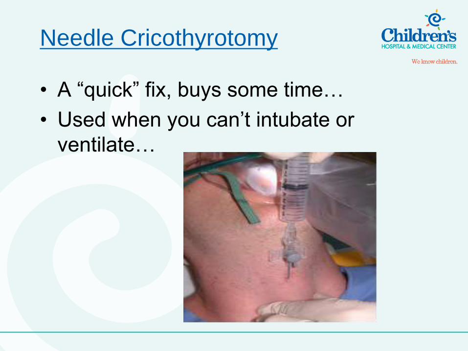
Needle Cricothyrotomy
• A “quick” fix, buys some time…
• Used when you can’t intubate or
ventilate…

King Airway
• Backup Airway…
• Inserted Blind….

Case Scenario 3
• 7 year old child is brought to the
Emergency Department with a chief
complaint of SOB.
Classify
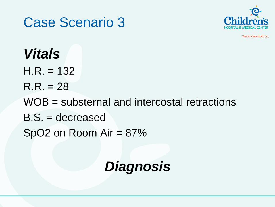
Case Scenario 3
Vitals H.R. = 132
R.R. = 28
WOB = substernal and intercostal retractions
B.S. = decreased
SpO2 on Room Air = 87%
Diagnosis
Asthma Statistics
• 23 million Americans currently have Asthma.
• Number of children who currently have Asthma: 7.0 million.
• Students with Asthma miss nearly 13 million school days every year due to illness.

Scenario 3 Treatment
• “A” Airway Management • Oxygen
• Sitting Position, Position of Comfort
• “B” Breathing • Albuterol 0.5 ml and more bronchodilators
• Steroids
• Encourage Coughing
• “C” Circulation • Hydration

Special Populations
• Tracheotomies
– Stay “CALM”!!

Special Populations
• Cystic Fibrosis
– SUCTION, Position of Comfort, Cough
– SUCTION
– SUCTION

It is all about the ABC’s…
• Airway
• Breathing
• Circulation
Recognize the signs of respiratory
distress and use your ABC’s.

Questions?

Thank You!!!