Respiratory infection following tracheostomy -...
8
Thorax (1964), 19, 89 Respiratory infection following tracheostomy M. S. GOTSMAN AND J. L. WHITBY From the Queen Elizabeth Hospital, Edgbaston, Birmineham. 15 Tracheostomy has been practised for centuries for the relief of respiratory tract obstruction. Its use has increased very markedly since the introduction of intermittent positive pressure respiration (I.P.P.R.) in the treatment of poliomyelitis (Galloway, 1943; Lassen, 1953). Its value has since been proved for other conditions associated with respiratory paralysis, such as tetanus (Cole, 1959; Smythe and Bull, 1961) and myasthenia gravis. It is also used in respiratory failure characterized by secretional ventilatory obstruc- tion (Nelson, 1958), as is found in severe head injuries (Bryce-Smith, 1950; Maciver, Frew, and Matheson, 1958; Cawthorne, Hewlett, and Ranger, 1959) or in chronic bronchitis (Hugh- Jones, 1958; Davidson, 1959) or in unconscious patients who are unable to cough up retained secretions (Nelson, 1958). These authors and numerous others (Atkins, 1960; Meade, 1961; Head, 1961) are enthusiastic advocates of tracheostomy, and they stress the urgency of performing the operation when indicated and the undoubted benefits obtained from its use. Little attention has been paid to the post-operative respiratory sequelae. We have been impressed by the frequency with which these patients have become infected with hospital cross- infecting organisms, particularly staphylococci. They are very susceptible to infection and, when infected, form a reservoir dangerous to other patients. We have traced all cases of tracheostomy performed in the Queen Elizabeth Hospital in 1961 in order to assess the incidence and significance of infection following tracheostomy. METHODS CLINICAL MATERIAL Patients on whom tracheostomy had been performed in 1961 were identified from the operating theatre records for that year. Tracheo- stomy is normally performed in an operating theatre by a surgical registrar, but any tracheostomy per- formed in the wards would not be recorded and could not be included in this survey. Patients whose clinical records were inadequate for assessment were also excluded. Four additional selected patients had a 89 tracheostomy performed in 1962 and have been used for illustrative but not statistical purposes. BACTERIOLOGICAL MATERIAL This was assessed from the patients' notes, laboratory records, and any epi- demiological data available. The phage types of all staphylococci isolated from patients were included and examined in relation to staphylococci isolated through- out the hospital. This was possible as all staphylococci are phage typed as a routine. NECROPSY MATERIAL Full reports and histological preparations were examined from all patients who had been submitted to necropsy. An attempt was made to correlate these findings with the incidence of post- operative infection. RESULTS OF THE STUDY INDICATIONS FOR TRACHEOSTOMY Forty patients underwent tracheostomy in the operating theatre but only 29 of these could be included in the survey. For the remaining 11 patients the data available were inadequate for an assessment to be made. The indications for which tracheostomy was performed are listed in Table I and illustrated in Figure 1. TABLE I INDICATION FOR TRACHEOSTOMY Respiratory failure ... 2 Mechanical obstruction 6 Respiratory muscle paralysis and bulbar palsy 2 Prophylactic I Unconscious with aspiration ... 7 Post-operative Neurosurgery...4 Thoracic surgery...3 Abdominal surgery...4 Six patients required tracheostomy for mechanical obstruction of the upper respiratory tract, and in two of these patients the obstruction was precipitated by direct endoscopy. Secretional ventilatory obstruction in one form or another was the indication for tracheostomy in all the remaining patients. These patients were either unwilling or unable to cough up retained secretions. Prolonged unconsciousness and an inability to cough up the retained secretions accounted for 11 on 3 June 2018 by guest. Protected by copyright. http://thorax.bmj.com/ Thorax: first published as 10.1136/thx.19.1.89 on 1 January 1964. Downloaded from
Transcript of Respiratory infection following tracheostomy -...

Thorax (1964), 19, 89
Respiratory infection following tracheostomyM. S. GOTSMAN AND J. L. WHITBY
From the Queen Elizabeth Hospital, Edgbaston, Birmineham. 15
Tracheostomy has been practised for centuries forthe relief of respiratory tract obstruction. Its usehas increased very markedly since the introductionof intermittent positive pressure respiration(I.P.P.R.) in the treatment of poliomyelitis(Galloway, 1943; Lassen, 1953). Its value hassince been proved for other conditions associatedwith respiratory paralysis, such as tetanus (Cole,1959; Smythe and Bull, 1961) and myastheniagravis. It is also used in respiratory failurecharacterized by secretional ventilatory obstruc-tion (Nelson, 1958), as is found in severe headinjuries (Bryce-Smith, 1950; Maciver, Frew, andMatheson, 1958; Cawthorne, Hewlett, andRanger, 1959) or in chronic bronchitis (Hugh-Jones, 1958; Davidson, 1959) or in unconsciouspatients who are unable to cough up retainedsecretions (Nelson, 1958).These authors and numerous others (Atkins,
1960; Meade, 1961; Head, 1961) are enthusiasticadvocates of tracheostomy, and they stress theurgency of performing the operation whenindicated and the undoubted benefits obtainedfrom its use. Little attention has been paid tothe post-operative respiratory sequelae. We havebeen impressed by the frequency with which thesepatients have become infected with hospital cross-infecting organisms, particularly staphylococci.They are very susceptible to infection and, wheninfected, form a reservoir dangerous to otherpatients.We have traced all cases of tracheostomy
performed in the Queen Elizabeth Hospital in1961 in order to assess the incidence andsignificance of infection following tracheostomy.
METHODS
CLINICAL MATERIAL Patients on whom tracheostomyhad been performed in 1961 were identified fromthe operating theatre records for that year. Tracheo-stomy is normally performed in an operating theatreby a surgical registrar, but any tracheostomy per-formed in the wards would not be recorded and couldnot be included in this survey. Patients whose clinicalrecords were inadequate for assessment were alsoexcluded. Four additional selected patients had a
89
tracheostomy performed in 1962 and have been usedfor illustrative but not statistical purposes.
BACTERIOLOGICAL MATERIAL This was assessed fromthe patients' notes, laboratory records, and any epi-demiological data available. The phage types of allstaphylococci isolated from patients were included andexamined in relation to staphylococci isolated through-out the hospital. This was possible as all staphylococciare phage typed as a routine.
NECROPSY MATERIAL Full reports and histologicalpreparations were examined from all patients who hadbeen submitted to necropsy. An attempt was madeto correlate these findings with the incidence of post-operative infection.
RESULTS OF THE STUDY
INDICATIONS FOR TRACHEOSTOMY Forty patientsunderwent tracheostomy in the operating theatrebut only 29 of these could be included in thesurvey. For the remaining 11 patients the dataavailable were inadequate for an assessment to bemade.The indications for which tracheostomy was
performed are listed in Table I and illustratedin Figure 1.
TABLE IINDICATION FOR TRACHEOSTOMY
Respiratory failure... 2Mechanical obstruction 6Respiratory muscle paralysis and bulbar palsy 2Prophylactic IUnconscious with aspiration... 7Post-operative
Neurosurgery...4Thoracic surgery...3Abdominal surgery...4
Six patients required tracheostomy formechanical obstruction of the upper respiratorytract, and in two of these patients the obstructionwas precipitated by direct endoscopy.
Secretional ventilatory obstruction in one formor another was the indication for tracheostomy inall the remaining patients. These patients wereeither unwilling or unable to cough up retainedsecretions.Prolonged unconsciousness and an inability to
cough up the retained secretions accounted for 11
on 3 June 2018 by guest. Protected by copyright.
http://thorax.bmj.com
/T
horax: first published as 10.1136/thx.19.1.89 on 1 January 1964. Dow
nloaded from
M. S. Gotsman and J. L. Whitby
patients. These have been listed under twoheadings. In one group unconsciousness persistedor occurred following a neurosurgical procedure.In the other group, neurological conditions wereoften present (head injuries and cerebrovascularaccidents) but neurosurgery was not undertaken.12I I
109.
8
7
65
4
3
2
.2_ o '-
Four~paiet onwo rchotm apefome afeupr aboiasugrhd
c~~~n.a
Q. D 2 0 ccLU.0
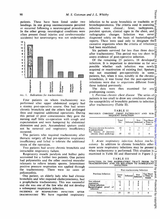
FIG. 1. Indications.for tracheostomy.
Four patients on whom tracheostomy was
performed after upper abdominal surgery had
a stormy post-operative course. One had severe
chronic bronchitis and the others had prolongedileus and required additional operations. Duringthis period of poor consciousness they gave thenursing staff little co-operation with cough andexpectoration and were hampered by abdominaldistension and pain. Accumulated sputum couldnot be removed and respiratory insufficiencyoccurred.The patients who required tracheostomy after
thoracic surgery all had pre-operative respiratoryinsufficiency and failed to tolerate the additionalstrain of the operation.Two patients had severe chronic bronchitis and
intractable respiratory failure.Respiratory muscle paralysis and bulbar palsy
accounted for a further two patients. One patienthad polyneuritis and the other received muscularrelaxants to relieve tetanic spasms. Intermittentpositive pressure respiration was applied throughthe tracheostomy. There were no cases ofpoliomyelitis.One patient, an elderly lady who had chronic
bronchitis and who required cholecystectomy, hada prophylactic tracheostomy. This was successfuland she was one of the few who did not developa subsequent respiratory infection.INCIDENCE OF RESPIRATORY INFECTION AFTER
TRACHEOSTOMY We have regarded respiratory
infection to be acute bronchitis or tracheitis orbronchopneumonia. The criteria used in assessinginfection were clinical; fever, tachypnoea,purulent sputum, clinical signs in the chest, andradiographic changes. Infection was neverdiagnosed solely on the basis of bacteriologicalreports. These were used only to designate thecausative organisms when the criteria of infectionhad been established.
Six patients survived for less than three daysafter tracheostomy. This period was too short toassess evidence of post-operative infection.Of the remaining 23 patients, 18 developed
infection. It is important to determine as far aspossible whether such infection was newlyacquired or reactivation of existing foci. Sputumwas not examined pre-operatively in somepatients, but, when it was, notably in the chronicbronchitics, it was found that the post-operativeinfections were due to organisms different fromthose isolated previously.The data were thert examined for any
predisposing causes.1. Previous chronic chest disease The series of
patients is too small to draw any conclusion aboutthe susceptibility of bronchitic patients to infectionafter tracheostomy (Table II).
TABLE IIPREVIOUS CHRONIC CHEST PATHOLOGY AND SUB-
SEQUENT INFECTION
Total Not Too ShortNo. Infected Infected a Time to
Assess
Previous chronic bronchitis 10 5 3 2Kyphoscoliosis .. I I _No history of previous
chronic chest pathology 18 12 2 4
2. Acute respiratory infectioni before trachle-ostomny In addition to chronic bronchitis othermore acute respiratory infections may be presentwhen tracheostomy is performed. This situation isexamined in Table III and illustrated in Figure 2.
TABLE IIIINFECTION IN THE RESPIRATORY TRACT PRIOR TOTRACHEOSTOMY AND ITS RELATION TO SUBSEQUENT
INFECTION
Post-tracheostomy Infection
Previous Infection Total Inetd Not Too ShortNo. | Infected a Time to
Not infected I.... 1 5 4 2Aspiration .. .. 7 5 0 2Clinical infection .. 6 5 0 IBacteriological proof of
infection .. 5 3 1 I
Total .. .. 29 18 5 6
90
on 3 June 2018 by guest. Protected by copyright.
http://thorax.bmj.com
/T
horax: first published as 10.1136/thx.19.1.89 on 1 January 1964. Dow
nloaded from

Respiratory infection following tracheostomy
Survival too short
Not I nfectedInfected
Infection of Infectioninfection in relation to post-
The patients were divided into four groups.Five had overt infection with positive bacterio-logical cultures from the sputa; those showinginfection after tracheostomy were infected withdifferent organisms. In six patients there wasclinical evidence of respiratory infection butbacteriological data were not obtained. Sevenpatients are considered to have aspirated vomitusor inhaled sputum during a pzriod ofunconsciousness. Aspiration in an unconsciouspatient must lead to a pneumonia. Eleven patientshad no evidence of any pre-existing infection.Where evidence of previous infection existed, it
was difficult to assess what role the tracheostomyhad played in any subsequent infection. Thenature of the infecting organisms in most casessuggests the occurrence of fresh infection, and thispoint is further considered later.
3. Intermittent positive pressure respirationThis was required in eight patients. Seven of themdeveloped respiratory tract infection.
4. Effect of previous antibiotic therapy Anti-biotic administration was examined in the 23patients who survived long enough for evidenceof post-operative infection to be assessed. Table IVshows the relation of therapy to subsequent infec-tion. Prophylactic administration of antibiotics inthe absence of infection, in the small group which
TABLE IVANTIBIOTIC ADMINISTRATION IN RELATION TO POST-
TRACHEOSTOMY INFECTION
Number Post- Noin operative Infection
Group Infection
Previous antibiotic administra-tion for any reason .. 9 8 1
No previous antibiotic admin-istration.9 8 1
Prophylactic administration ofantibiotic in the absence ofinfection.5 2 3
received them, appeared to confer some protection.The incidence of infection is so high that it is
difficult to identify causal factors. Previous infec-tion or aspiration of non-infected material appearsto predispose to further infection since the onlypatients who did not develop post-operative infec-tion were free from infection before thetracheostomy was undertaken.
BACTERIOLOGY OF THE RESPIRATORY INFECTION Infive patients no evidence of infection was obtained.
Six patients died before the question of post-tracheostomy infection could be assessed.
In three further patients with clinical infectionsevere enough to warrant antibiotic therapy nospecimens were submitted for bacteriologicalexamination.From the remaining 15 patients the following
organisms were found in association with clinicalevidence of infection: Staphylococcus aureus, 11;Proteus species, 4; Pseudomanas pyocyanea, 5;and other enterobacteriaceae, 9.
DISCUSSION OF SELECTED PATIENTS
The following four patients were admitted to theMedical Unit under the care of Professor W.Melville Arnott and have been subjected to hispersonal study. Since they were admitted during1962, they are not included in the statisticalanalysis but they illustrate important features ofthe epidemiology and the danger of secondaryinfection in the chest following tracheostomy.CASE 1 This Irish labourer, aged 49, was admittedwith acute bronchopneumonia superimposed onchronic bronchitis. He was semiconscious, unable togive a coherent history, and in severe cardiac failure.A sample of capillary blood analysed by the method
of Astrup showed a Pco2 of 65 mm. Hg, a pH of 7-22,and a base excess of - 1, indicating alveolar hypo-ventilation with respiratory acidosis. Haemophilusinfluenzae, sensitive to all antibiotics except penicillin.was grown from an initial sputum specimen. He hadalready received penicillin and streptomycin whichwas changed to chloramphenicol. He was uncoopera-tive and unable to cough up sputum. Oxygen wasadministered in slowly increasing concentrations buthis level of consciousness deteriorated and continuedto do so in spite of respiratory centre stimulants(nikethamide and diethylamine vanillic acid (Vandid)).
Tracheostomy was done and the patient was venti-lated on a Smith-Clarke respirator. The Pco2 fell to30 mm. Hg and his clinical condition improved sorapidly that the respirator was discontinued thefollowing day. The tracheostomy tube was removedafter 24 hours. A growth of Proteus sp. was obtainedfrom the sputum at the time of removal of thetracheostomy.
12
10 E98-i
6-
2
Not AspirationInfected
FIG. 2. Pre-existing chesttracheostomy infection.
91
on 3 June 2018 by guest. Protected by copyright.
http://thorax.bmj.com
/T
horax: first published as 10.1136/thx.19.1.89 on 1 January 1964. Dow
nloaded from

M. S. Gotsman and J. L. Whitby
The following day he started to produce a largevolume of foul-smelling purulent sputum and hismental state deteriorated. The Pco2 rose to 94 mm.Hg. The tracheostomy tube was re-inserted andI.P.P.R. was given -again. Two multiple resistantstaphylococci were then recovered from the sputum(phage types 77 and 29/80). These were present inother patients in the main ward. The patient had beennursed in isolation in a separate side ward. Theorganisms were sensitive to methicillin and erythro-mycin only. The chloramphenicol was stopped andmethicillin started.Two days later the sputum became green in colour
and Pseudomotnas pxyocyanea sensitive to colomycinwas isolated from several specimens. This wasassociated with clinical deterioration.The disease ran a stormy course but the patient
recovered and was discharged three weeks later.He illustrates several interesting features. The
initial infection was due to H. influenzae, anorganism commonly pathogenic in patients withchronic bronchitis. Chloramphenicol eliminatedH. influenzae which was not recovered from sub-sequent specimens of sputum. The staphylococciwere fresh invaders after the tracheostomy hadbeen performed. They were present in severalother patients in the main ward, and isolation andbarrier nursing in a side ward, but in the sameward area, were ineffective in preventing infectionof this patient. The staphylococcal infection wasas severe as the initial bronchopneumonia. As thestaphylococcal infection was eliminated it wasreplaced by Ps. pyocyanea. This was also asso-ciated with clinical manifestations of infection forwhich treatment was required.
CASE 2 A man, aged 46, had worked in an ironfoundry since the age of 14. He was in receipt of an80% disability pension for silicosis and massivepulmonary fibrosis.On a previous admission 10 specimens of sputum
had been examined on successive days. No tuberclebacilli were seen on direct examination of the sputa.Culture and guinea-pig inoculation of the sputa forMyco. tuberculosis were also negative. H. influenzaeand Neisseria sp. were cultured.He was readmitted to the unit on 13 February 1962.
Figure 3 illustrates relevant clinical and bacteriologicalfeatures of this illness. On admission he was short ofbreath and had developed atrial flutter; but he wasapyrexial. The blood pressure had fallen to 85/60mm. Hg. Acute cardiac failure was superimposed onhis previous respiratory insufficiency. The Pco2 was62 mm. Hg.On the day after admission his clinical condition
deteriorated and he became distressed and extremelydyspnoeic. In spite of intravenous aminophylline andVandid he did not improve. Tracheostomy was per-formed and intermittent positive pressure respiration
I.P.P.R.TRACHEOSTOMY
METHICILLINCHLORAMPHENICOL
Ui)
z
500
ProteusPs. pyocyoncaStaphylococcusCoilNormal CommensalsNil
Uatoa
m m
1001
Pc02 ( mm.Hg) 601
102 1
TEMP 0F 10019896 -~140l
PULSE 120 lN \ ARATE 100
80_12 14 16 18 202224 2628 2 4 6 8
FEBRUARY MARCH
FIG. 3. Data relaiting to Case 2 durinig his illness inFebruary and March, 1962.
was given with a Barnet respirator. A culture of hissputum taken shortly before the tracheostomy wasperformed yielded commensal organisms only.
After tracheostomy he regained consciousness, theatrial flutter reverted to sinus rhythm, and thecapillary blood Pco2 fell to 45 mm. Hg. A furtherspecimen of sputum aspirated directly from thetrachea produced no growth on culture. He was givenchloramphenicol, 500 mg. six-hourly. The nursingregime was strict and the patient was isolated.
Twelve hours after tracheostomy had been per-formed he had improved so much that it was possibleto wean him from the respirator.
Seventy-two hours later the patient suddenlydeteriorated and developed signs of carbon dioxidenarcosis. The tracheostomy tube was re-inserted andI.P.P.R. given again. He had developed acute broncho-pneumonia. This aggravated the existing alveolarhypoventilation and led to carbon dioxide retention.A swab of the tracheostomy wound yielded
Staphylococcus aureus (phage type 75/77) sensitiveonly to erythromycin and methicillin. The sameorganism was grown from the sputum, and he wasgiven methicillin, 500 mg. six-hourly. It may be signifi-cant that one of the medical attendants was a nasalcarrier of a staphylococcus of the same phage type.
After three days his temperature returned to normalalthough large quantities of sputum were still beingaspirated. The Pco2 fell to 46 mm. Hg. The sputumbecame foul-smelling, and Escherichia coli was cul-tured from it. The tracheostomy wound was nowseptic.
In the subsequent fortnight, the staphylococcidisappeared from the sputum. Citrobacter freundii was
92
n r--lM-
on 3 June 2018 by guest. Protected by copyright.
http://thorax.bmj.com
/T
horax: first published as 10.1136/thx.19.1.89 on 1 January 1964. Dow
nloaded from

Respiratory infection following tracheostomy
grown from the sputum and the tracheostomy wound,and later Proteus sp. was found in the sputum.There was a further clinical set-back associated
with infection when the Pco2 increased to 92 mm.Hg, but this improved after an hour on the respiratorand vigorous physiotherapy.
All antibiotics were discontinued after six weeks,and the patient was discharged to a convalescenthome.We were fortunate to have an opportunity to re-
assess the patient three months later. He wasreadmitted following an upper respiratory tractinfection. Prior to this he had been able to walkaround the house and had been free from purulentsputum.On this occasion he was apyrexial and the physical
signs in the chest had not altered. A culture ofsputum produced a heavy growth of pneumococcisensitive to penicillin and tetracycline. Pulmonaryfunction tests showed evidence of airways obstruction.
In the light of his previous history no antibioticswere given. It was felt that these should be reservedfor subsequent overt clinical infection of the chestassociated with constitutional signs.This man illustrates several problems. He was
a respiratory cripple. The cardiac failure asso-ciated with the atrial flutter (and the acute chestinfection) precipitated respiratory failure. Trache-ostomy saved his life. Subsequent to this heimproved, but the tracheostomy wound becameinfected, and a staphylococcal bronchopneumoniacaused dangerous deterioration. The staphylococ-cus was traced to a nasal carrier on the ward. Theinfection responded to methicillin. As the staphyl-ococci disappeared, they were replaced in turn byCit. freundii and Proteus sp. These disappearedwithout further antibiotic treatment.This man died six months later. At necropsy
the lungs were indurated and fibrotic with multipleapical cavities. The bronchi were fairly clear andthe tracheostomy opening had healed with minimalscarring.
CASE 3 A man, aged 27, injured his right thumb anddeveloped tetanus. It was necessary to paralyse himcompletely with curare, and institute I.P.P.R. to con-trol tetanic spasms. Tracheostomy was performed tofacilitate I.P.P.R. Artificial respiration was controlledwith frequent pH and Pco2 estimations. He receivedpenicillin, I M.U. b.d., for the control of his infection.Two days after the tracheostomy had been
performed he developed a chest infection.'Hospital' staphylococci (phage type 80) were cul-
tured from two specimens of sputa. The organismswere sensitive to methicillin and erythromycin. Hewas therefore treated with methicillin. The chest in-fection improved and two days later no organismscould be recovered from the sputum. Four days laterhis chest infection deteriorated and E. coli sensitive
to chloramphenicol was recovered from the sputum.Chloramphenicol was added to the antibiotic regime.
Irreversible brain damage occurred followingcardiac arrest, and he died a few days later as a resultof intractable hypotension.Necropsy showed marked cerebral softening with
bronchopneumonia in both lungs.Tracheostomy was obligatory since he required
prolonged muscular relaxation and I.P.P.R. Priorto the tracheostomy he had no pre-existing chestinfection. A high standard of barrier nursing carewas maintained. He received prophylactic peni-cillin therapy both for the local tetanus infectionand in order to prevent a respiratory infection. Yethe soon succumbed to a resistant staphylococcus.This was eliminated by methicillin, but once againsuperinfection with E. coli occurred. He died 14days after admission and, although the chestinfection was not the main cause of death, he hada severe bronchopneumonia.
CASE 4 A man, aged 67, with chronic bronchitishad an abdominal aneurysm resected. He had sufferedfrom severe obliterative arterial disease in his legs.The operation was followed by a prolonged periodof apnoea which required tracheostomy and I.P.P.R.A swab taken from the tracheostomy wound on the
following morning yielded Streptococcus viridans anda sensitive strain of Staph. aureus (phage type 55/71).In spite of prophylactic chloramphenicol he developeda profuse diarrhoea the following day, and Staph.aureus (phage type 53/77), sensitive only to methicillinand erythromycin, was cultured from the stool. Hewas therefore given methicillin. The sputum increasedin quantity and became purulent. A heavy growth ofthe same staphylococcus and E. coli was obtained.He was also pyrexial and developed clinical signs ofthe infection on the chest. He improved on methicillin,but Cit. freundii appeared in the sputum and this wassensitive only to neomycin and kanamycin. Neomycinand nystatin were added to the regime.
His clinical condition improved, and a smallervolume of sputum was produced, from which only ascanty growth of Staph. aureus and Cit. freundii wasisolated. Ps. pyocyanea was later cultured from thesputum but was not associated with fresh symptoms,and no further antibiotic therapy was given. He wasdischarged one month after operation.The patient had no chest infection when
tracheostomy was undertaken. He developed astaphylococcal enteritis with organisms present inother patients who were in the ward at the time.This occurred in spite of strict barrier nursing inan isolation cubicle, and chloramphenicol prophyl-axis. Staphylococcal pneumonia ensued; when thissubsided on methicillin, Cit. freundii, coliforms,and later Ps. pyocyanea colonized the sputum,illustrating how readily these organisms proliferate
93
on 3 June 2018 by guest. Protected by copyright.
http://thorax.bmj.com
/T
horax: first published as 10.1136/thx.19.1.89 on 1 January 1964. Dow
nloaded from

M. S. Gotsman and J. L. Whitby
when the normal bacterial flora is removed byantibiotics.
DISCUSSION
REVIEW OF THE LITERATURE There are manyproblems in the management of a tracheostomy;most of these have already been discussed veryadequately in the existing literature. They includemechanical factors, the problem of crusting oftracheal secretions, and the techniques to be usedfor aspiration (Negus, 1961; Crawford, 1961).Little attention has been paid to respiratoryinfection after tracheostomy. Nelson and Bowers(1957) reviewed 310 cases of tracheostomy butdid not mention pulmonary infection as a compli-cation. Other large series mention infection, butthe incidence and bacteriological characteristics ofthe infection received no attention. Atkins (1960)reviewed 526 cases, and a table in his paperrevealed that 129 of these cases came to necropsy.Pneumonia was found in 99 (76%) but there is nocomment upon this in the text. Head (1961), inanother exhaustive series of patients from theMassachussets General Hospital, described infec-tion as a late complication in 80 out of 462 cases;the nature of the infection was not classified andany manifestations from superficial sepsis topulmonary infection were included in these figures.The July number of Anesthesiology (1962) isdevoted to a symposium on inhalation therapy, butwhile tracheostomy and I.P.P.R. are discussed indetail, infectious complications are not considered.By contrast Lepper, Kofman, Blatt, Dowling, andJackson (1954) described the bacteriological find-ings in 72 poliomyelitis cases where respiratoryparalysis was treated with tracheostomy andI.P.P.R. Pulmonary infection occurred in everycase and the causative organisms were wide butincluded Staph. pyogenes, Pselidomonas sp., andProteus sp. It is not clear whether the infectedcases represented all the poliomyelitis cases withtracheostomy in one hospital, or only those whodeveloped respiratory infection, but the evidencefor repeated infection with change in the organismresponsible is formidable and illustrates the easewith which infection occurs in these patients.
In two other small but intensively studied series(Davidson, 1959; Nisbet, and Wilson, 1958) thebacteriology of infection is discussed since respira-tory infection was the indication for tracheostomy;the incidence of post-operative respiratory infec-tion is difficult to assess. Smythe and Bull (1961)and Cole (1959) found that some patients who hadtetanus and who were treated by tracheostomy and
I.P.P.R. developed a fatal pneumonia: but theygive no bacteriological data.
In an account of the management oftracheostomy, Frew (1961) makes the point thattracheostomy patients are readily cross-infectedwith organisms in the hospital environment andthat nursing of tracheostomy patients in individualcubicles should lead to a reduction in the incidenceof infection. He gives no illustrative cases and nospecific information on the causal organisms.
INCIDENCE OF INFECTION Our experience is thatcross infection with hospital pathogenic organismsis almost certain to occur within a week oftracheostomy being established. It is in accordwith the view that an abnormal respiratory tractis readily colonized by resistant staphylococci inhospitals as illustrated for bronchitics by Mitchell,Dunn, Lees, and Hedges (1961). Other organismscausing infection were also well recognized cross-infecting types such as Ps. pyocyanea and Proteulssp.
FACTORS IN CAUSATION OF INFECTION The problemis to decide whether the tracheostomy can beincriminated as the cause of the patient's infection.Many patients in our series had tracheostomy per-formed for conditions in which respiratory tractinfection was already established or its develop-ment inevitable. Aspiration of material and failureto clear secretions from the respiratory tractoccurred because these patients were unable tocough. Nelson has stressed the importance of thissyndrome of 'secretional ventilatory obstruction.
In two patients, who died shortly aftertracheostomy had been performed, and in whomthe period of survival was too short to assess therole of respiratory infection, the trachea andbronchi were congested and filled with muco-purulent secretion. There were numerous areas ofatelectasis and in some parts frank infection. Itcould be argued that infection is the inevitableresult of such aspiration and that this factor isthe indication for tracheostomy. The effectiveprotective mechanisms of the respiratory tract arelost and colonization may occur with organismsthat are not pathogenic as primary invaders.Under these conditions the tracheostomy cannotbe blamed for the infection but it appears probablethat the type of infection is influenced by thepresence of a tracheostomy. Bronchopneumoniaand aspiration pneumonia in hospitals are fre-quently complicated by staphylococcal infection,but other organisms such as H. influenzae andStrep. pneumoniae are also common causal agents.
94
on 3 June 2018 by guest. Protected by copyright.
http://thorax.bmj.com
/T
horax: first published as 10.1136/thx.19.1.89 on 1 January 1964. Dow
nloaded from

Respiratory infection following tracheostomy
In our series not a single case was infected withthese latter organisms while on tracheostomy. Inaddition in a number of cases bacteriological datashowed that one causal organism was present inthe infected sputum at the time tracheostomy wasperformed. This was subsequently replaced byStaph. aureus, Ps. pyocyanea, or other species in15 patients who lived for three days or more.Most patients with a tracheostomy received
routine antibiotic cover. The majority of staphy-lococcal infections were with organisms resistantto penicillin, streptomycin, and tetracycline, andin many units these are the most popular 'prophy-lactic' antibiotics. In this hospital the majority ofstaphylococci isolated were only sensitive toerythromycin, chloramphenicol, and methicillin.On the Professorial Medical Unit chlorampheni-col-resistant staphylococci were also common (Fig.4). Thus to protect in-patients from staphylococcal
1005
a,
ab
8o-
Medical unit mHospitale
60-
40
20-
Penicillin Tetracycline Chloramphenicol Erythromycin
FIG. 4. Percentage ofstrains ofStaph. aureus isolated inQueen Elizabeth Hospital at large and in the ProfessorialMedical Unit resistant to various antibiotics.
infection with a prophylactic antibiotic one ofthese latter antibiotics should be chosen in prefer-ence -to the former: the problem of antibioticprophylaxis however is not as simple as this. Inpatients who have an established staphylococcalinfection, treatment with an effective antibioticwas often followed by the establishment of afurther resistant infection usually caused by Ps.pyocyanea.
There is usually no question about the patho-genicity of Staph. aureus in pulmonary conditions,as measured by the volume of purulent sputum,the pulmonary changes, and the constitutionalsymptoms produced in the patient. In the caseof replacement infection with Gram-negativebacteria following successful anti-staphylococcaltherapy the association is not so clear. Often thereis much less constitutional disturbance, and someof our cases illustrate the difference.
In the normal person inspired air passes throughthe upper respiratory tract where it is warmed andfiltered so effectively that swabs from the mainbronchi taken through a bronchoscope are sterile(Brumfitt, Willoughby, and Bromley, 1957). Thetracheostomy bypasses the upper respiratory tract.Most respirators include a humidifier and to someit is possible to fit an air filter. Even when air isfiltered there are periods when the patient is dis-connected from the respirator and respires directlythrough his tracheostomy. Thus airborne infectionof the bronchial tree may be readily and directlyachieved. The catheter used for suction is anothermeans by which infection may be acquired. Thetracheostomy wound may also represent a portalof entry for bacteria.
Patients who have a tracheostomy cannot cougheffectively. An efficient cough requires a closedglottis, which allows the development ofmomentary, high intra-thoracic pressure. Theglottis is opened and an explosive current of air athigh velocity is directed up the trachea. Thevelocity of the blast is important since this deter-mines the effectiveness of the cough. This preventsthe tracheostomy patient from clearing his owntrachea and bronchi effectively since the cough isshallow. If the patient can cough, he does notproduce sputum in the pharynx but often expec-torates it through the tracheostomy opening. Thisis unhygienic and unpleasant; it soils the woundand the tracheostomy dressings, and is a way ofdisseminating and spraying the organisms in theward.
MANAGEMENT The epidemiology of the infectionprobably differs very little from that of woundinfection. In one respect it is similar to a burn.An exposed surface (the bronchial and trachealmucosa) is covered with dead material and thisprobably accounts for the patient's increasedsusceptibility to infection.
In the management of tracheostomy there aretwo main problems in relation to infection; thepatient must be protected from infection but ifinfection occurs other patients must be preventedfrom acquiring it. Both these objectives demandthe isolation of the patient.
In our series many of these patients wereisolated in single rooms within the ward area andbarrier nursing was attempted but could notalways be enforced. In spite of isolation patientsbecome infected with organisms present in theward at the time of their isolation. This impliesthat for isolation to be successful the patientshould be nursed in a true isolation unit uncon-nected with general hospital wards. Carriers of
95
on 3 June 2018 by guest. Protected by copyright.
http://thorax.bmj.com
/T
horax: first published as 10.1136/thx.19.1.89 on 1 January 1964. Dow
nloaded from

M. S. Gotsman and J. L. Whitby
dangerous organisms should be rigorously ex-cluded from the isolation room. The question ofvisitors is also difficult; they usually do not carrydangerous organisms but, if permitted to enter,they must adopt the same precautions as the staff.Attention must be paid to the hygiene of thetracheostome, and for this purpose disposablesterile catheters are the most suitable for trachealaspiration. It is important to be gentle with suctionwhich should never be applied while inserting thecatheter. The air supply for respiration in humidi-fiers should be bacteria-free, since any inspiredbacteria will be implanted directly into the lowerrespiratory tract. If antibiotics are to be admini-stered they should be appropriate to combat likelyinfecting organisms; other antibiotics may actuallyfavour such infection. Daily swabs of the trachealaspirate should be submitted for bacteriology. Inthis way any change in flora can be recognizedand combated at the earliest possible moment ifclinically indicated.Tracheostomy is such an effective procedure in
the management of many conditions that wouldotherwise prove fatal that it is essential for it tobe managed efficiently.Pulmonary infection is a dangerous hazard and
counteracts many of the benefits of tracheostomy.The main purpose of performing the tracheostomyis to reduce and remove the retained secretionsand reduce the amount of work performed by thelungs for a given amount of gas exchange.Pulmonary infection aggravates this problem andundoes the benefits which would accrue from thetracheostomy.
SUMMARY
Twenty-nine patients submitted to tracheostomyin the Queen Elizabeth Hospital in 1961 have beenexamined for evidence of pulmonary infection.
Twenty-three lived for more than three daysand, of these, 18 became infected following trache-ostomy. Eleven infections were due to antibiotic-resistant staphylococci and five to Ps. pyocyanea.
The nature of the infecting organisms clearlyindicated the hospital environment as the sourceof the infection.
Factors influencing -the establishment of infec-tion have been discussed and some recommenda-tions made about the management of tracheostomypatients.
We are indebted to Professor W. Melville Arnott forpermission to study the patients on his unit and forhis criticism and advice, and to Professor A. L.d'Abreu for encouragement and suggestions.
REFERENCES
Anesthesiology (1962). Symposium-Inhalational Therapy. 23, pp. 407575.
Atkins, J. P. (1960). Current utilization of tracheostcmy as a thera-peutic measure. Laryngoscope (St. Louis), 70, 1672.
Brumfitt, W., Willoughby, M. L. N., and Bromley, L. L. (1957). Anevaluation of sputum examination in chronic bronchitis. Lancet,ii, 1306.
Bryce-Smith, R. (1950). The management of head injuries from ananaesthetist's point of view. Brit. med. J., 2, 322.
Cawthorne, T., Hewlett, A. B., and Ranger, D. (1959). Tracheostomyin a respiratory unit in a neurological hospital. Proc. roy. Soc.Med., 52, 403.
Cole, L. B. (1959). Tracheostomy in tetanus. Ibid., 52, 41 1.Crawford, 0. B. (1961). The anesthesiologist's responsibilities in
tracheostomy. Anesthesiology, 22, 86.Davidson, L. A. G. (1959). Tracheotomy in acute respiratory disease.
Lancet, 1, 597.Frew, I. J. C. (1961). Tracheostomy and controlled respiration. J.
Laryng., 75, 136.Galloway, T. C. (1943). Tracheotomy in bulbar poliomyelitis. J.
Amer. med. Ass., 123, 1096.Head, J. M. (1961). Tracheostomy in the management of respiratory
problems. New Engl. J. Med., 264, 587.Hugh-Jones, P. (1958). Oligopnoea. Proc. roy. Soc. Med., 51, 104.Lassen, H. C. A. (1953). The 1952 epidemic of poliomyelitis in
Copenhagen, with special reference to the treatment of acuterespiratory insufficiency. Lancet, i, 37.
Lepper, M. H., Kofman, S., Blatt, N., Dowling, H. F., and Jackson,G. G. (1954). Effect of eight antibiotics used singly and in com-bination on the tracheal flora following tracheotomy in polio-myelitis. Antibiot. Chemother., 4, 829.
Maciver, I. N., Frew, I. J. C., and Matheson, J. G. (1958). The roleofrespiratory insufficiency in the mortality ofsevere head injuries.Lancet, i, 390.
Meade, J. W. (1961). Tracheotomy-its complications and theirmanagement. New Engl. J. Med., 265, 519.
Mitchell, A. A. B., Dunn, R. I. S., Lees, T. W., and Hedges, C. K.(1961). Staphylococcal pulmonary infection. Lancet, ii, 669.
Negus, V. (1961). Protection of the respiratory tract. Brit. med. J.,2, 723.
Nelson, T. G. (1958). Tracheotomv: A Clinical and ExperimentalStudv. Williams and Wilkins, Baltimore.
Nelson, T. G.,and Bowers, W. F. (1957). Tracheotomy-indications,advantages, techniques, complications, and results. J. Amer. med.Ass., 164, 1530.
Nisbet, H. I. Armstrong, and Wilson, F. (1958). The treatment ofacute respiratory infection in infants. Brit. J. Anaesth., 30, 419.
Smythe, P. M., and Bull, A. B. (1961). Treatment of tetanus withspecial reference to tracheotomy. Brit. med. J., 2, 732.
96
on 3 June 2018 by guest. Protected by copyright.
http://thorax.bmj.com
/T
horax: first published as 10.1136/thx.19.1.89 on 1 January 1964. Dow
nloaded from