Residual dipolar couplings and orientational effects · PDF fileResidual dipolar couplings and...
50
Residual dipolar couplings and orientational effects Markus Zweckstetter Max-Planck-Institute for Biophysical Chemistry, Göttingen [email protected]
Transcript of Residual dipolar couplings and orientational effects · PDF fileResidual dipolar couplings and...
Residual dipolar couplings and orientational effects
Markus ZweckstetterMax-Planck-Institute for Biophysical
Chemistry, Gö[email protected]
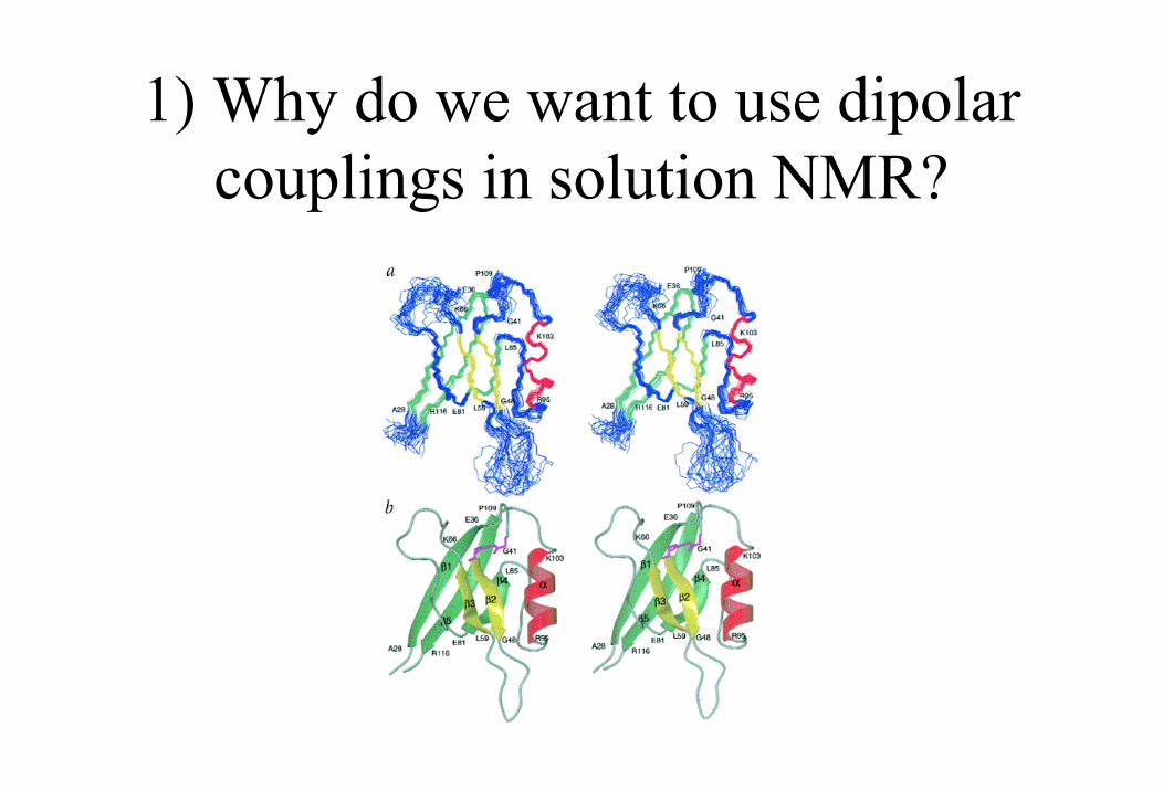
1) Why do we want to use dipolar couplings in solution NMR?
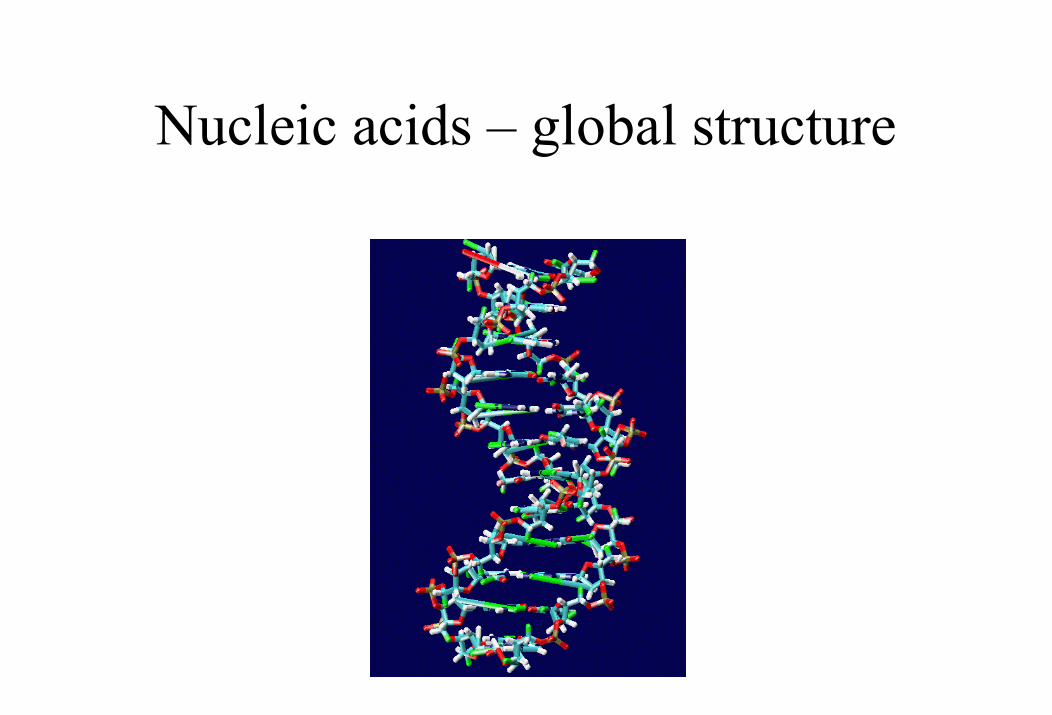
Nucleic acids – global structure
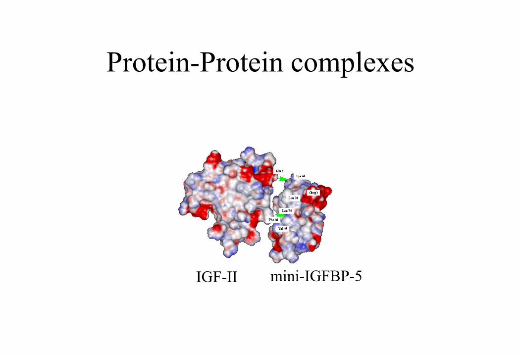
Protein-Protein complexes
IGF-II mini-IGFBP-5
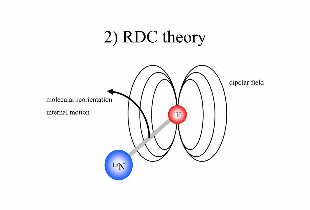
2) RDC theory
molecular reorientation
internal motion
15N
1H
dipolar field
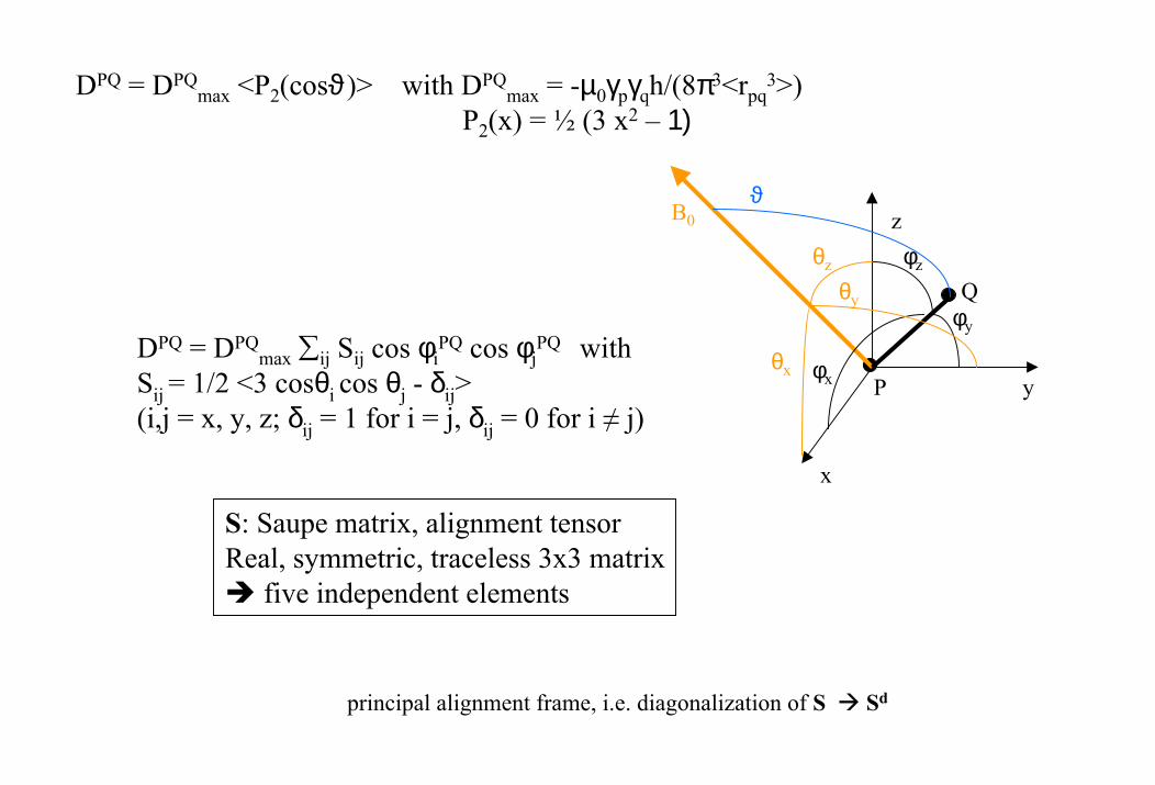
DPQ = DPQmax <P2(cosϑ)> with DPQ
max = -µ0γpγqh/(8π3<rpq3>)
P2(x) = ½ (3 x2 – 1)
x
z
P
Q
θx
θz
θy
φx
φy
φz
B0ϑ
DPQ = DPQmax ∑ij Sij cos φi
PQ cos φjPQ with
Sij = 1/2 <3 cosθi cos θj - δij> (i,j = x, y, z; δij = 1 for i = j, δij = 0 for i ≠ j)
y
S: Saupe matrix, alignment tensorReal, symmetric, traceless 3x3 matrix
five independent elements
principal alignment frame, i.e. diagonalization of S Sd
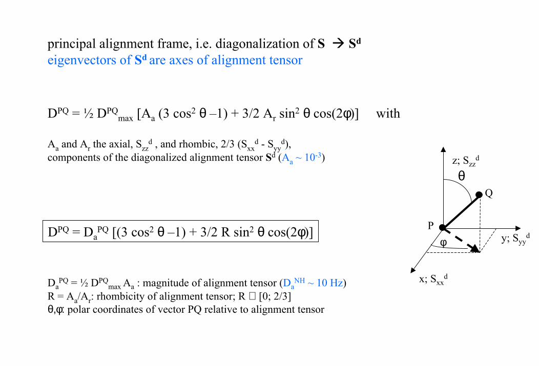
principal alignment frame, i.e. diagonalization of S Sd
eigenvectors of Sd are axes of alignment tensor
DPQ = ½ DPQmax [Aa (3 cos2 θ –1) + 3/2 Ar sin2 θ cos(2φ)] with
Aa and Ar the axial, Szzd , and rhombic, 2/3 (Sxx
d - Syyd),
components of the diagonalized alignment tensor Sd (Aa ~ 10-3)
x; Sxxd
P
θ
φ
z; Szzd
Q
DPQ = DaPQ [(3 cos2 θ –1) + 3/2 R sin2 θ cos(2φ)] y; Syy
d
DaPQ = ½ DPQ
max Aa : magnitude of alignment tensor (DaNH ~ 10 Hz)
R = Aa/Ar: rhombicity of alignment tensor; R ∈ [0; 2/3] θ,φ: polar coordinates of vector PQ relative to alignment tensor
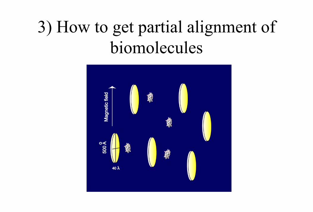
3) How to get partial alignment of biomolecules
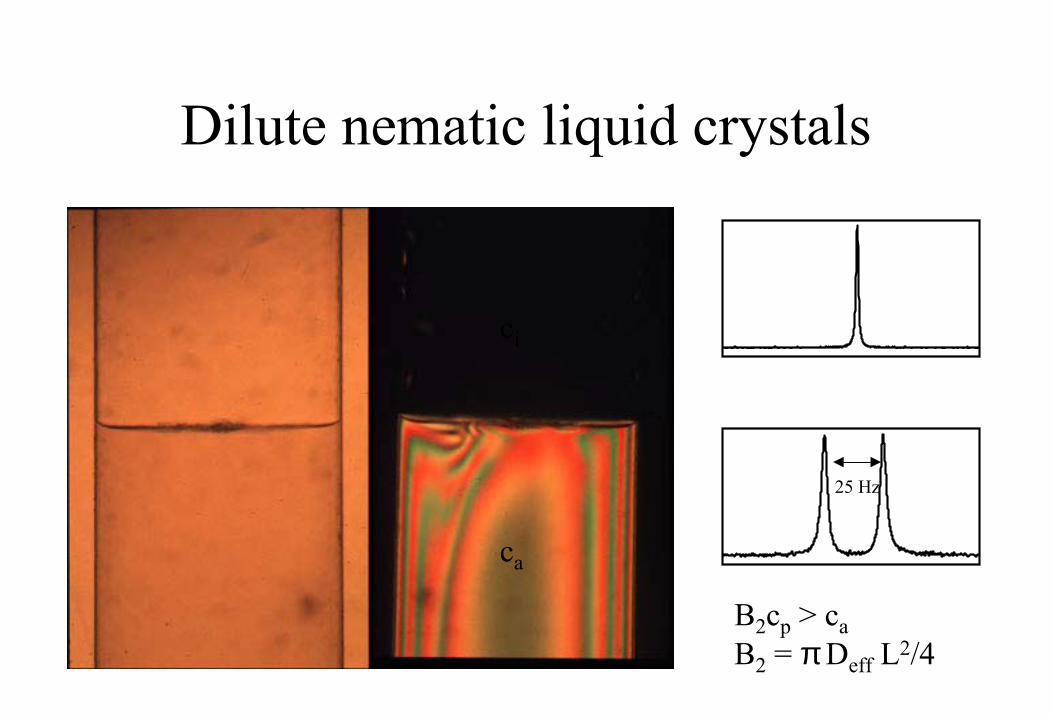
Dilute nematic liquid crystals
ca
ci
25 Hz
B2cp > caB2 = πDeff L2/4
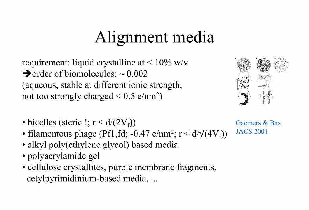
Alignment mediarequirement: liquid crystalline at < 10% w/v
order of biomolecules: ~ 0.002(aqueous, stable at different ionic strength,not too strongly charged < 0.5 e/nm2)
• bicelles (steric !; r < d/(2Vf))• filamentous phage (Pf1,fd; -0.47 e/nm2; r < d/√(4Vf))• alkyl poly(ethylene glycol) based media• polyacrylamide gel• cellulose crystallites, purple membrane fragments,
cetylpyrimidinium-based media, ...
Gaemers & Bax JACS 2001
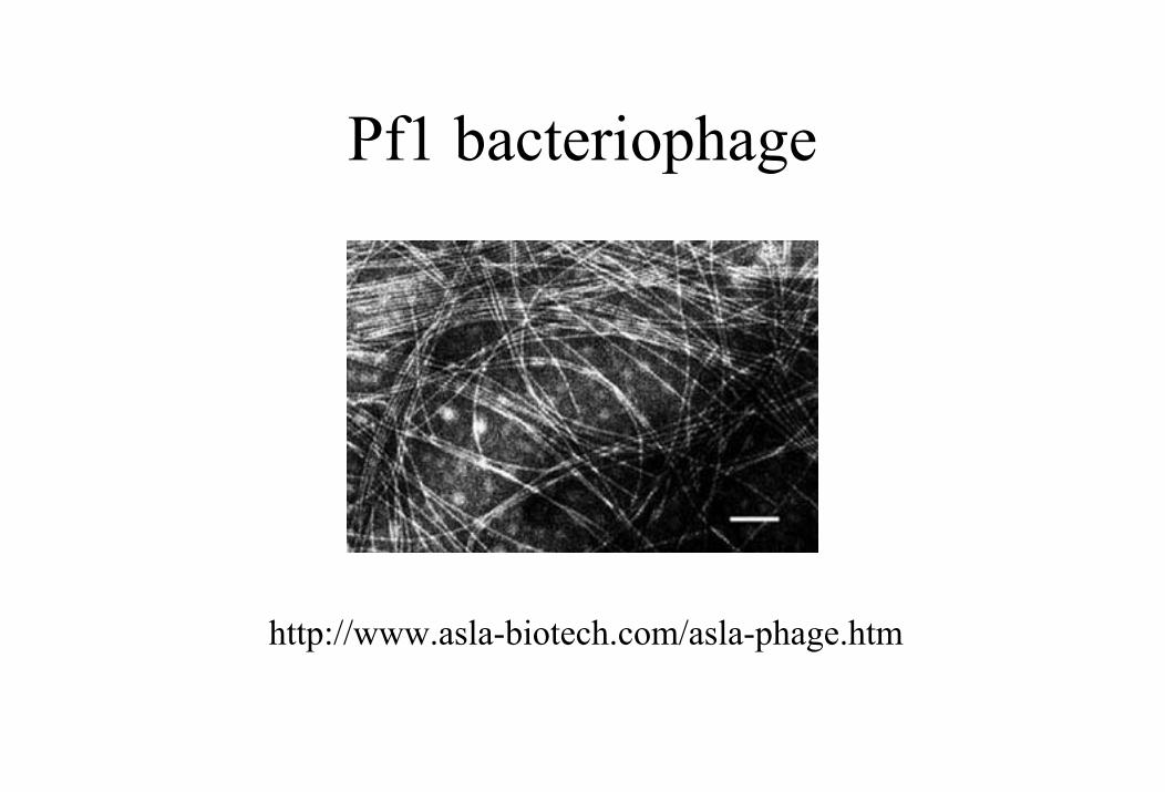
Pf1 bacteriophage
http://www.asla-biotech.com/asla-phage.htm
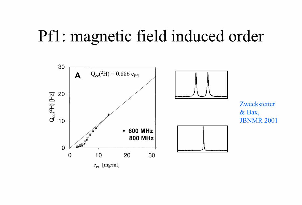
Pf1: magnetic field induced order
• 600 MHz� 800 MHz
Qcc(2H) = 0.886 cPf1
Zweckstetter& Bax, JBNMR 2001
cPf1 [mg/ml]
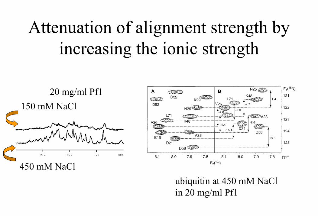
Attenuation of alignment strength by increasing the ionic strength
450 mM NaCl
150 mM NaCl20 mg/ml Pf1
7.08.09.0 ppm
ubiquitin at 450 mM NaCl in 20 mg/ml Pf1
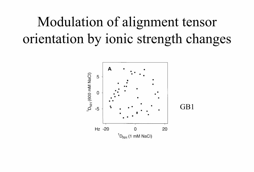
Modulation of alignment tensor orientation by ionic strength changes
GB1
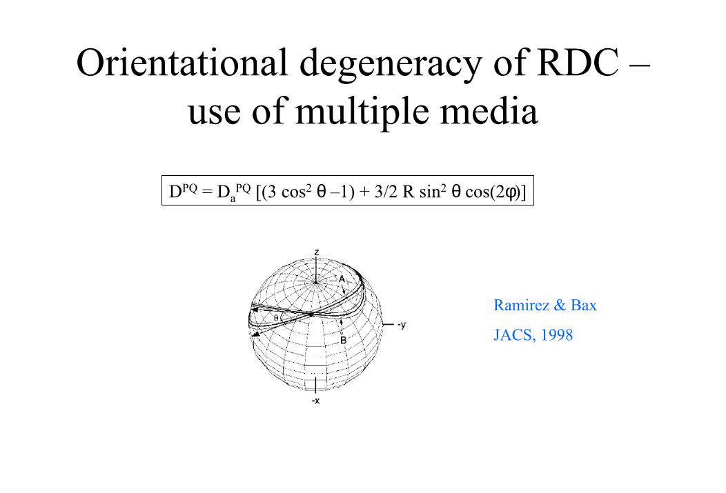
Orientational degeneracy of RDC –use of multiple media
DPQ = DaPQ [(3 cos2 θ –1) + 3/2 R sin2 θ cos(2φ)]
Ramirez & Bax
JACS, 1998

4) RDC measurement
NOESY HSQC
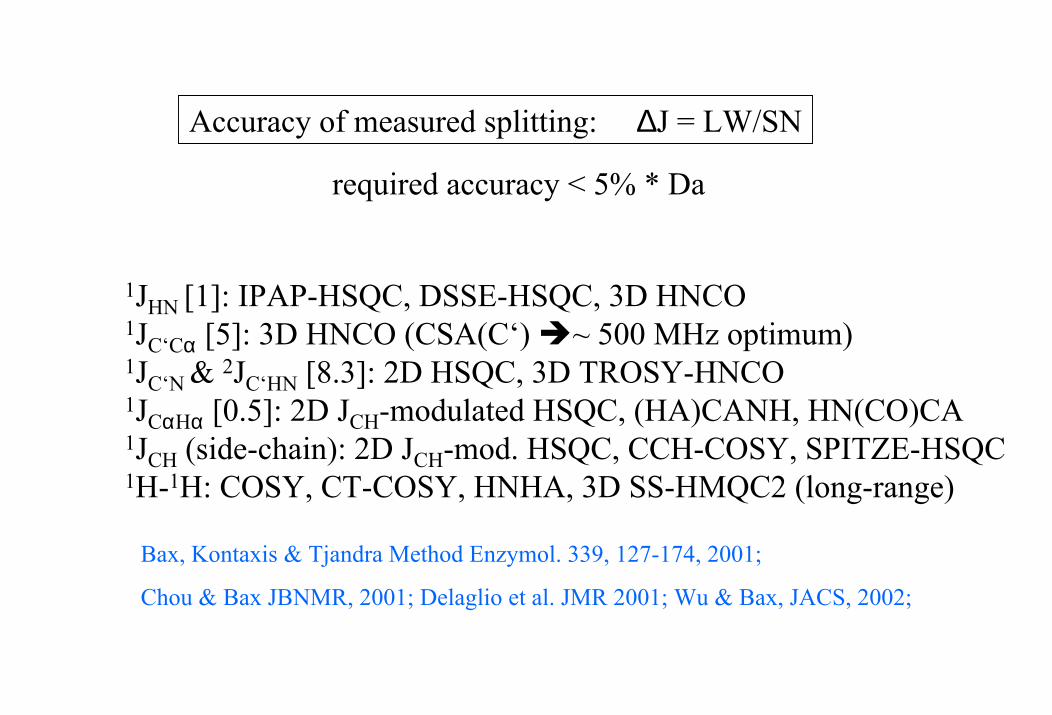
Accuracy of measured splitting: ∆J = LW/SN
required accuracy < 5% * Da
1JHN [1]: IPAP-HSQC, DSSE-HSQC, 3D HNCO1JC‘Cα [5]: 3D HNCO (CSA(C‘) ~ 500 MHz optimum)1JC‘N & 2JC‘HN [8.3]: 2D HSQC, 3D TROSY-HNCO1JCαHα [0.5]: 2D JCH-modulated HSQC, (HA)CANH, HN(CO)CA1JCH (side-chain): 2D JCH-mod. HSQC, CCH-COSY, SPITZE-HSQC1H-1H: COSY, CT-COSY, HNHA, 3D SS-HMQC2 (long-range)
Bax, Kontaxis & Tjandra Method Enzymol. 339, 127-174, 2001;
Chou & Bax JBNMR, 2001; Delaglio et al. JMR 2001; Wu & Bax, JACS, 2002;
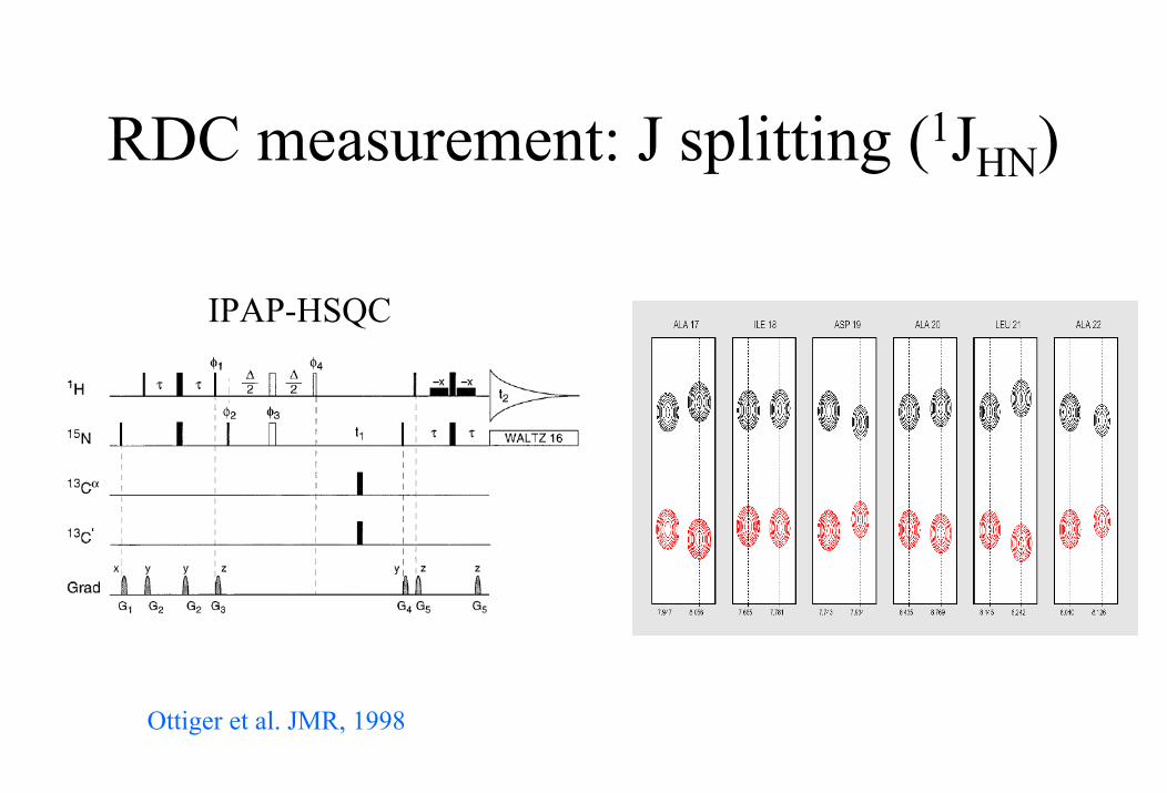
RDC measurement: J splitting (1JHN)
IPAP-HSQC
Ottiger et al. JMR, 1998
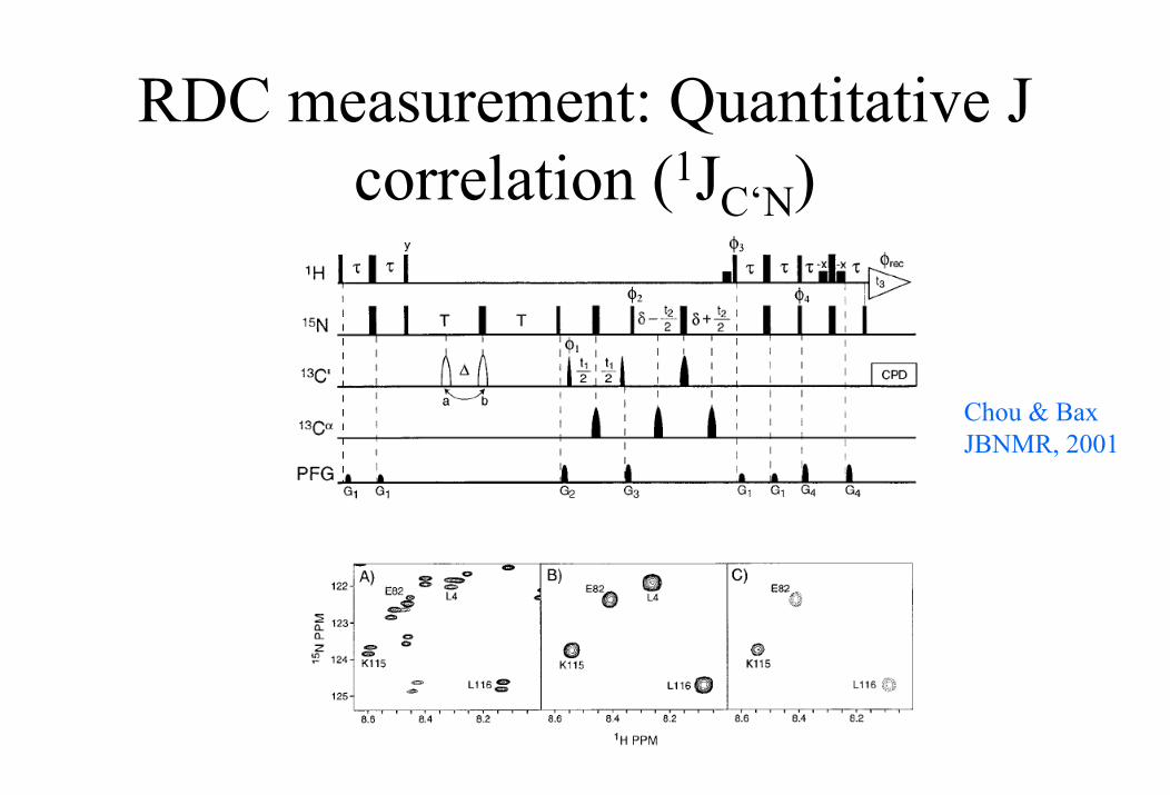
RDC measurement: Quantitative J correlation (1JC‘N)
Chou & BaxJBNMR, 2001
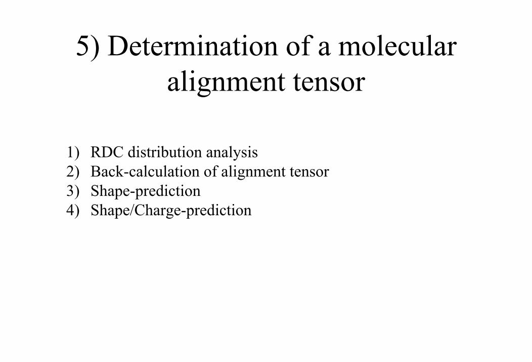
5) Determination of a molecular alignment tensor
1) RDC distribution analysis2) Back-calculation of alignment tensor3) Shape-prediction4) Shape/Charge-prediction
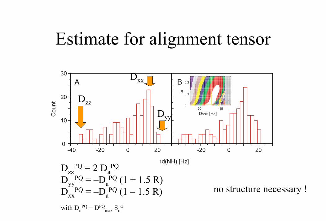
Estimate for alignment tensor
DzzPQ = 2 Da
PQ
DyyPQ = –Da
PQ (1 + 1.5 R)Dxx
PQ = –DaPQ (1 – 1.5 R) no structure necessary !
Cou
nt
-40 -20 0 200
10
20
30
1d(NH) [Hz]
-20 0 20
A B
D [Hz]aNH
-20 -150
0.1
0.2
R
Dzz
Dyy
Dxx
with DiiPQ = DPQ
max Siid
Back-calculation of alignment tensor
• singular value decomposition (SVD)
very stable & with a minimum of five RDCs possible
• iterative least squares procedure (Levenberg-Marquardt minimization) χ2 = ∑i=1,..,N [di
PQ(exp) – diPQ(calc)]2/(σi
PQ)2
fixing of alignment parameters (e.g. rhombic component zero due to three-fold or higher symmetry)
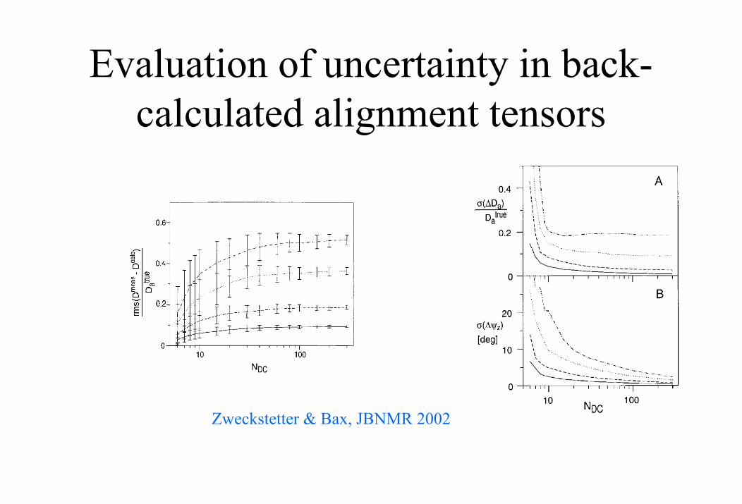
Evaluation of uncertainty in back-calculated alignment tensors
Zweckstetter & Bax, JBNMR 2002
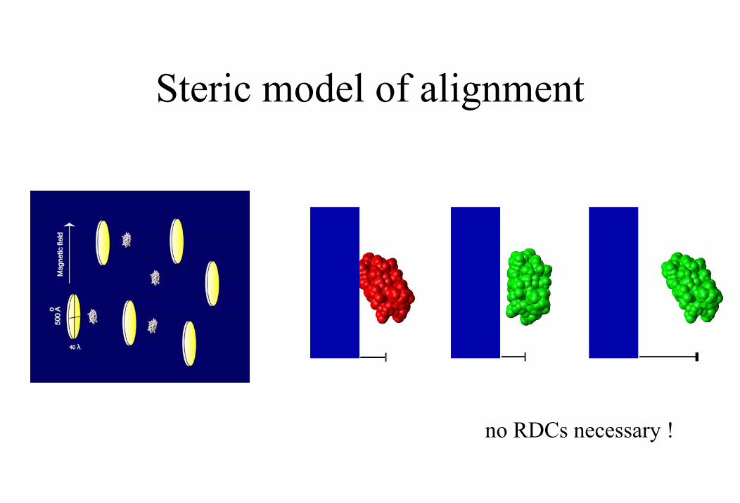
Steric model of alignment
no RDCs necessary !
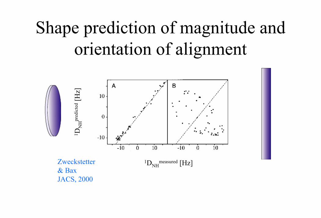
Shape prediction of magnitude and orientation of alignment
1 DN
Hpr
edic
ted
[Hz]
Zweckstetter& BaxJACS, 2000
1DNHmeasured [Hz]
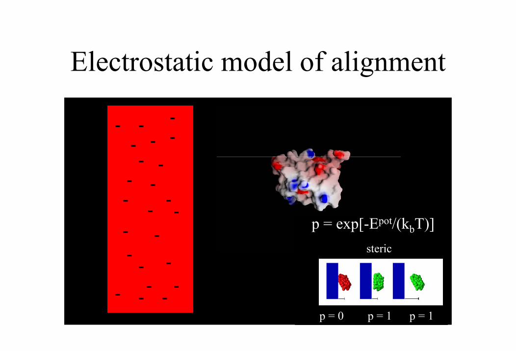
Electrostatic model of alignment
-
-
-
-
- -
-
-
- -
-
-
--
--
-
-
-
-
-
-
--
p = exp[-Epot/(kbT)]
p = 0 p = 1p = 1
steric
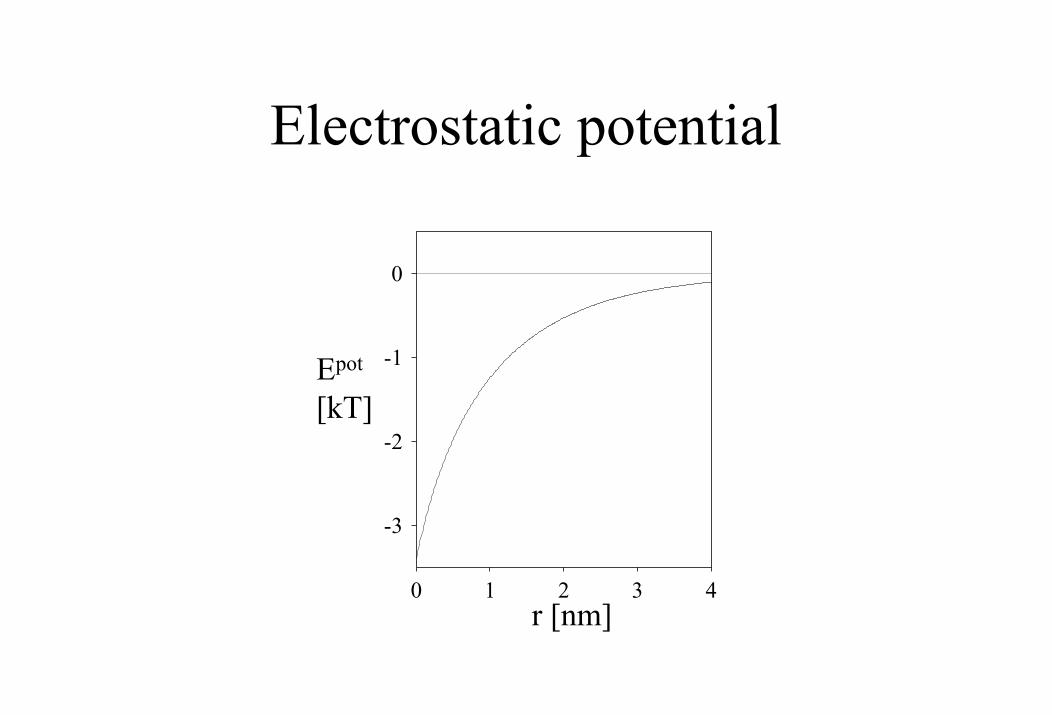
Electrostatic potential
-3
-2
-1
0
0 1 2 3 4r [nm]
Epot
[kT]
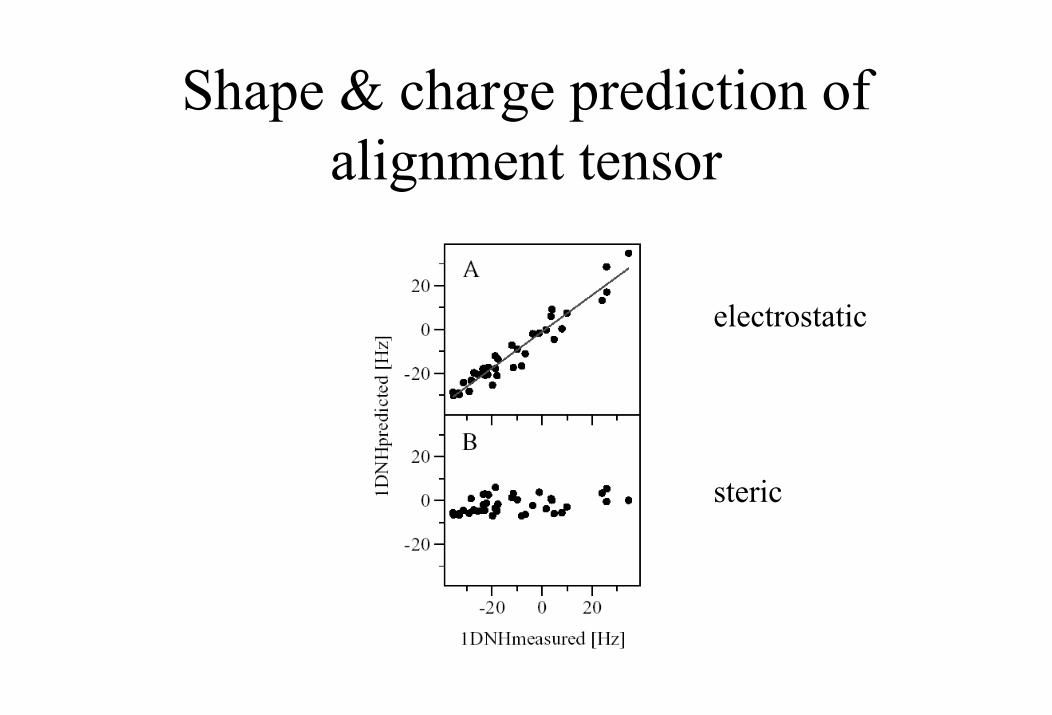
Shape & charge prediction of alignment tensor
electrostatic
steric
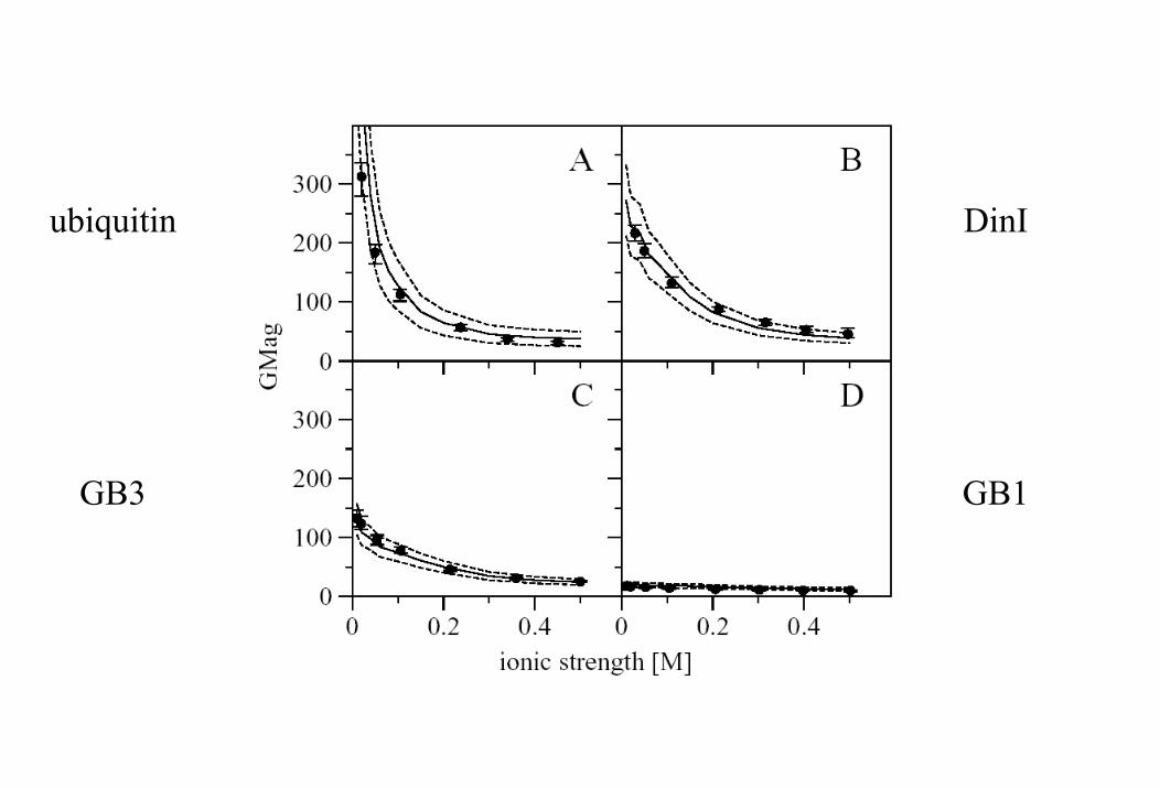
ubiquitin DinI
GB3 GB1
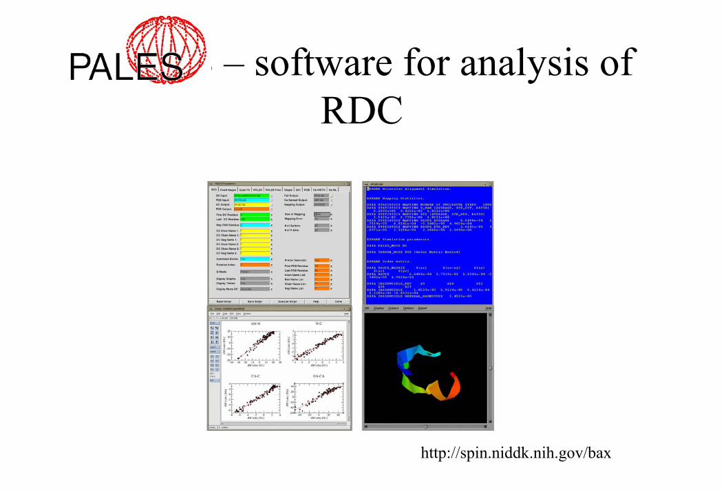
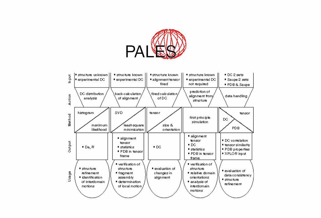
PALES – software for analysis of RDC
http://spin.niddk.nih.gov/bax
6) RDC applications
• validation of structures• analysis of inter-domain motion• structure refinement (proteins, nucleic acids, oligosaccharides)• identification of multimerization state• determination of relative domain orientations• structure determination of protein complexes• analysis of slow dynamics• improved assignment• rapid structure determination• ...
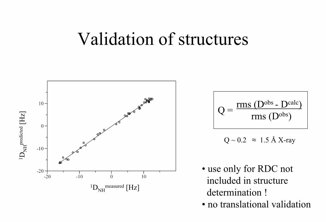
Validation of structures
rms (Dobs - Dcalc)rms (Dobs) Q =
• use only for RDC notincluded in structuredetermination !
• no translational validation
Q ~ 0.2 ≈ 1.5 Å X-ray
1DNHmeasured [Hz]
1 DN
Hpr
edic
ted
[Hz]
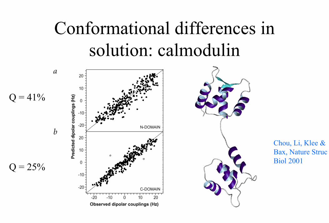
Conformational differences in solution: calmodulin
Q = 41%
Chou, Li, Klee & Bax, Nature Struc Biol 2001
Q = 25%

Flexibility of the inter-domain linker in solution
Baber et al. JACS, 2000
NH-dynamics from RDC: Peti et al. JACS 2002
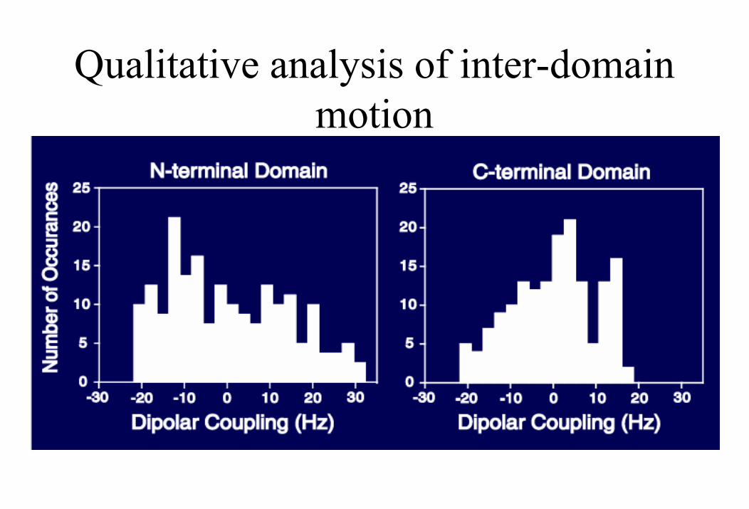
Qualitative analysis of inter-domain motion
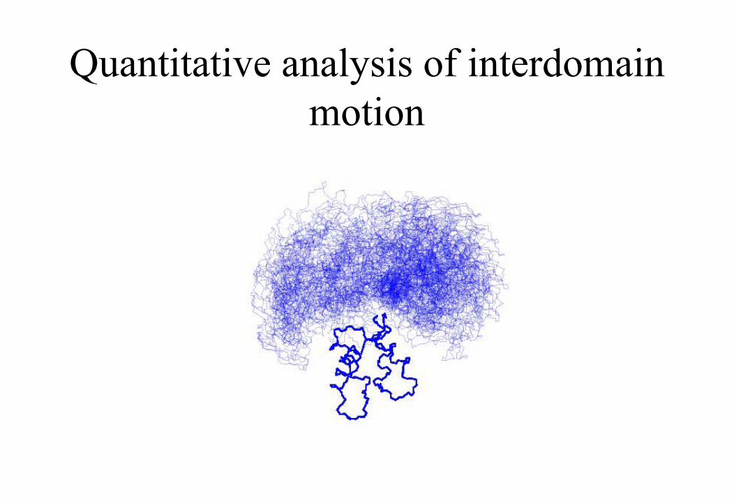
Quantitative analysis of interdomain motion
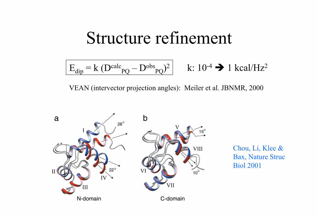
Structure refinementk: 10-4 1 kcal/Hz2Edip = k (Dcalc
PQ – DobsPQ)2
VEAN (intervector projection angles): Meiler et al. JBNMR, 2000
Chou, Li, Klee & Bax, Nature Struc Biol 2001
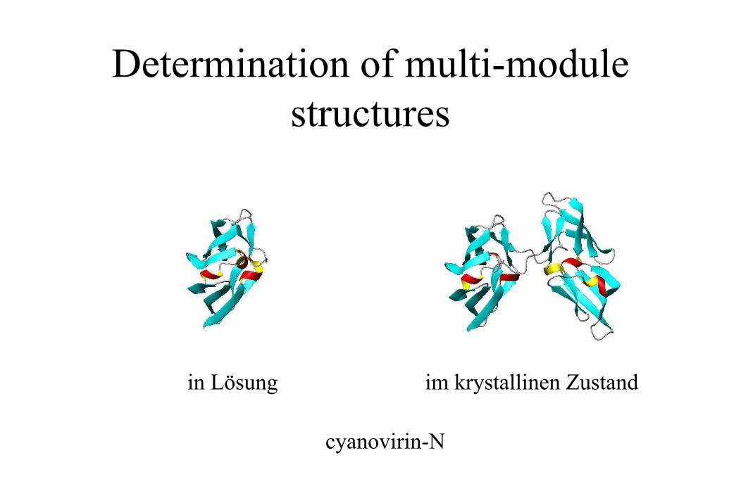
Determination of multi-module structures
in Lösung im krystallinen Zustand
cyanovirin-N
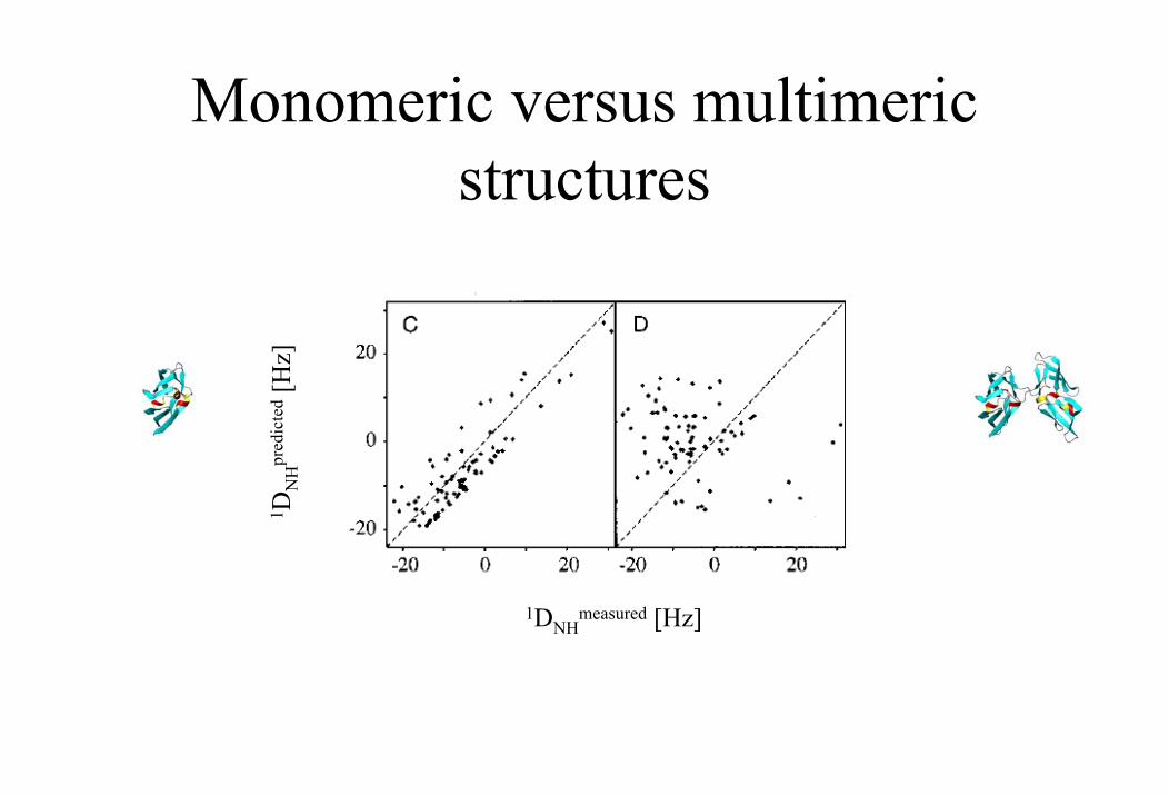
Monomeric versus multimeric structures
1 DN
Hpr
edic
ted
[Hz]
1DNHmeasured [Hz]
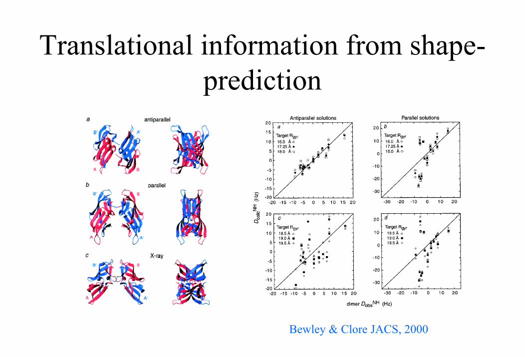
Translational information from shape-prediction
Bewley & Clore JACS, 2000
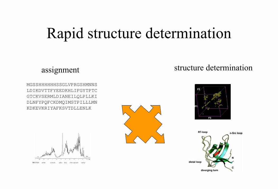
Rapid structure determination
structure determinationassignmentMGSSHHHHHHSSGLVPRGSHMNNSLDIKDVTTFYEEDKHLIFGYTPTCGTCKVSERMLDIANEILQLPLLKIDLNFYPQFCKDMQIMSTPILLLMNKDKEVKRIYAFKSVTDLLENLK
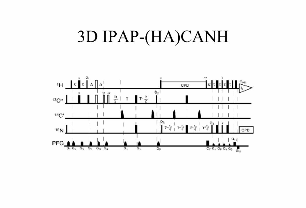
3D IPAP-(HA)CANH
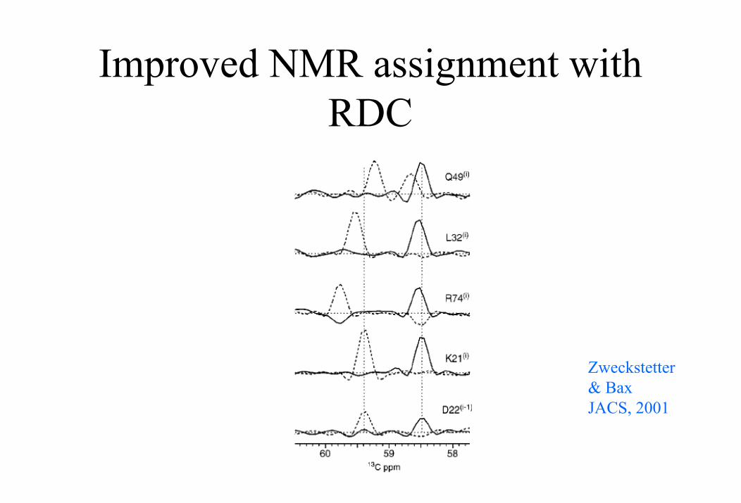
Improved NMR assignment with RDC
Zweckstetter& BaxJACS, 2001
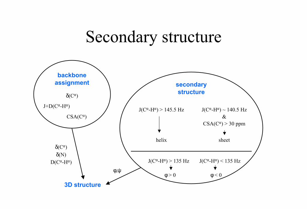
Secondary structure
secondary structure
backbone assignment
J+D(Cα-Hα)
D(Cα-Hα)
δ(Cα)
δ(Cα)δ(N)
J(Cα-Hα) > 145.5 Hz J(Cα-Hα) ~ 140.5 Hz
J(Cα-Hα) > 135 Hz J(Cα-Hα) < 135 Hz
CSA(Cα)CSA(Cα) > 30 ppm
φ> 0 φ< 0
sheethelix
&
φ,ψ
3D structure
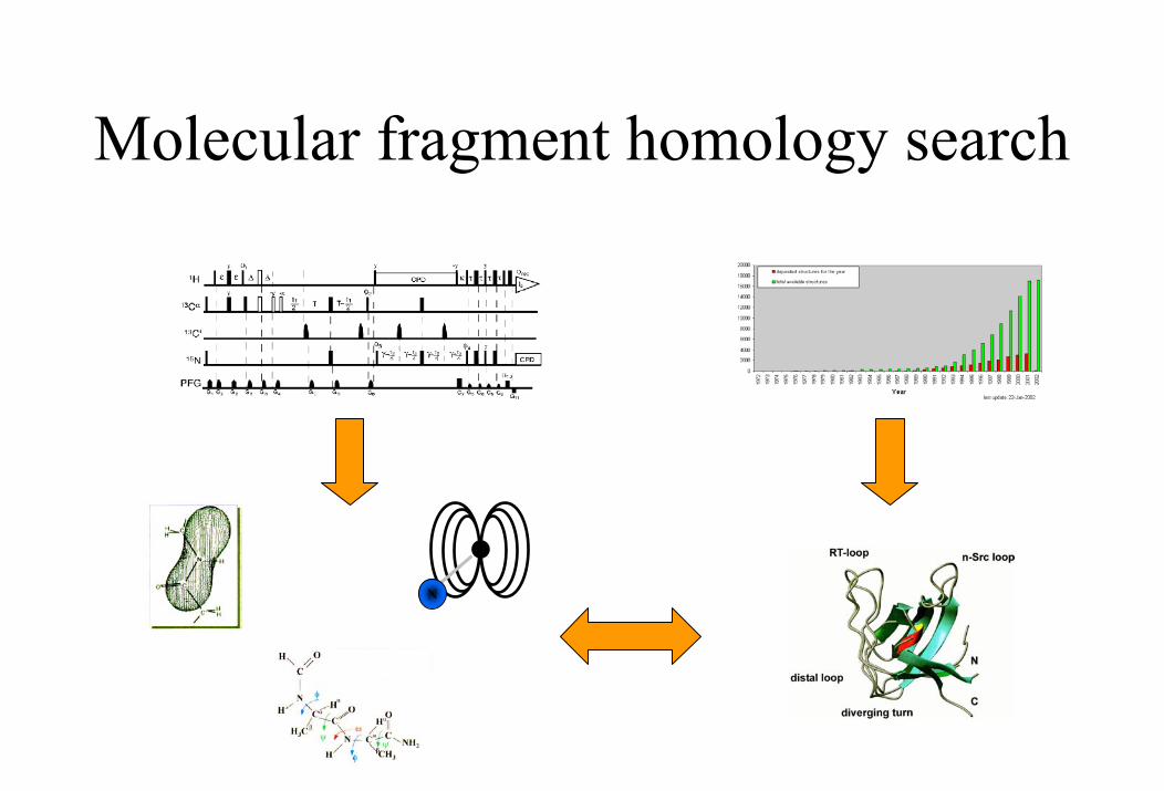
Molecular fragment homology search
N
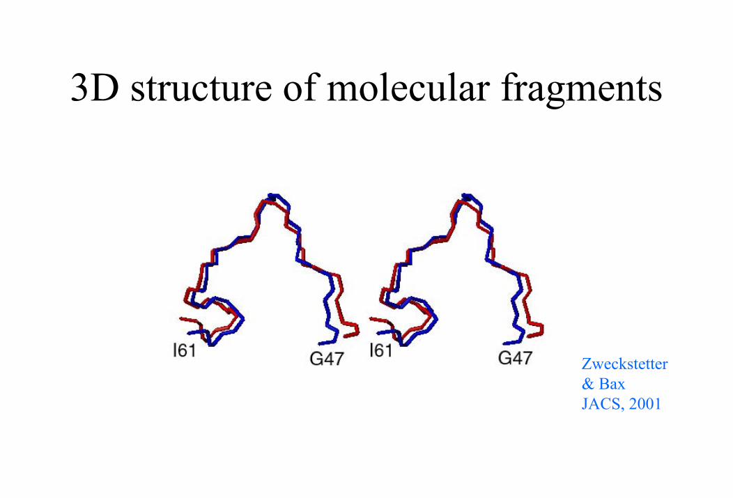
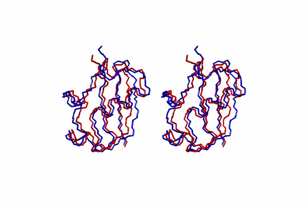
3D structure of molecular fragments
Zweckstetter& BaxJACS, 2001
References:
Tjandra, N. & Bax A., Science 278, 1111 (1997).Bax, A., Kontaxis, G. & Tjandra, N., Method Enzymol 339, 127 (2001).Prestegard, J.H., Al-Hashimi, H.M., & Tolman, J.R., Quart Rev Biophys 33,371 (2000).
Journal of American Chemical Society, Journal of Biomolecular NMR, Journal of Magnetic Resonance, ...


![[8] Dipolar Couplings in Macromolecular Structure ... · [8] DIPOLAR COUPLINGS AND MACROMOLECULAR STRUCTURE 127 [8] Dipolar Couplings in Macromolecular Structure Determination By](https://static.fdocuments.in/doc/165x107/605c24b70c5494344557be4f/8-dipolar-couplings-in-macromolecular-structure-8-dipolar-couplings-and.jpg)