RESEARCHARTICLE TheMIFAntagonistISO-1Attenuates ... MIF Antagonist ISO-1... · RESEARCHARTICLE...
17
RESEARCH ARTICLE The MIF Antagonist ISO-1 Attenuates Corticosteroid-Insensitive Inflammation and Airways Hyperresponsiveness in an Ozone- Induced Model of COPD Kirsty E. Russell 1 , Kian Fan Chung 1 , Colin J. Clarke 1 , Andrew L. Durham 1 , Patrick Mallia 2 , Joseph Footitt 2† , Sebastian L. Johnston 2 , Peter J. Barnes 1 , Simon R. Hall 3 , Karen D. Simpson 3 , Malcolm R. Starkey 4 , Philip M. Hansbro 4 , Ian M. Adcock 1 , Coen H. Wiegman 1 * 1 Airway Disease Section, National Heart & Lung Institute, NIHR Respiratory Biomedical Research Unit at the Royal Brompton NHS Foundation Trust and Imperial College London, London, United Kingdom, 2 Airway Disease Infection Section, National Heart & Lung Institute, Imperial College London, London, United Kingdom, 3 RRI DPU, GlaxoSmithKline, Stevenage, United Kingdom, 4 Priority Research Centre for Respiratory Diseases, Hunter Medical Research Institute and The University of Newcastle, Newcastle, Australia † Deceased. * [email protected] Abstract Introduction Macrophage migration inhibitory factor (MIF) is an inflammatory cytokine associated with acute and chronic inflammatory disorders and corticosteroid insensitivity. Its expression in the airways of patients with chronic obstructive pulmonary disease (COPD), a relatively ste- roid insensitive inflammatory disease is unclear, however. Methods Sputum, bronchoalveolar lavage (BAL) macrophages and serum were obtained from non- smokers, smokers and COPD patients. To mimic oxidative stress-induced COPD, mice were exposed to ozone for six-weeks and treated with ISO-1, a MIF inhibitor, and/or dexa- methasone before each exposure. BAL fluid and lung tissue were collected after the final exposure. Airway hyperresponsiveness (AHR) and lung function were measured using whole body plethysmography. HIF-1α binding to the Mif promoter was determined by Chro- matin Immunoprecipitation assays. Results MIF levels in sputum and BAL macrophages from COPD patients were higher than those from non-smokers, with healthy smokers having intermediate levels. MIF expression corre- lated with that of HIF-1α in all patients groups and in ozone-exposed mice. BAL cell counts, cytokine mRNA and protein expression in lungs and BAL, including MIF, were elevated in PLOS ONE | DOI:10.1371/journal.pone.0146102 January 11, 2016 1 / 17 OPEN ACCESS Citation: Russell KE, Chung KF, Clarke CJ, Durham AL, Mallia P, Footitt J, et al. (2016) The MIF Antagonist ISO-1 Attenuates Corticosteroid- Insensitive Inflammation and Airways Hyperresponsiveness in an Ozone-Induced Model of COPD. PLoS ONE 11(1): e0146102. doi:10.1371/ journal.pone.0146102 Editor: Bernhard Ryffel, French National Centre for Scientific Research, FRANCE Received: July 24, 2015 Accepted: December 14, 2015 Published: January 11, 2016 Copyright: © 2016 Russell et al. This is an open access article distributed under the terms of the Creative Commons Attribution License, which permits unrestricted use, distribution, and reproduction in any medium, provided the original author and source are credited. Data Availability Statement: Figures are available in the public repository http://figshare.com at the following DOIs: Fig 1 (10.6084/m9.figshare.2055990), Fig 2 (10.6084/m9.figshare.2055996), Fig 3 (10.6084/ m9.figshare.2055999), Fig 4 (10.6084/m9.figshare. 2056005), Fig 5 (10.6084/m9.figshare.2056008), Fig 6 (10.6084/m9.figshare.2056011), and Fig 7 (10. 6084/m9.figshare.2056014). Funding: KR is the recipient of a BBSRC CASE award with GSK. This work was also supported in part by the Medical Research Council Program Grant
Transcript of RESEARCHARTICLE TheMIFAntagonistISO-1Attenuates ... MIF Antagonist ISO-1... · RESEARCHARTICLE...

RESEARCH ARTICLE
The MIF Antagonist ISO-1 AttenuatesCorticosteroid-Insensitive Inflammation andAirways Hyperresponsiveness in an Ozone-Induced Model of COPDKirsty E. Russell1, Kian Fan Chung1, Colin J. Clarke1, Andrew L. Durham1, Patrick Mallia2,Joseph Footitt2†, Sebastian L. Johnston2, Peter J. Barnes1, Simon R. Hall3, KarenD. Simpson3, Malcolm R. Starkey4, Philip M. Hansbro4, Ian M. Adcock1, CoenH. Wiegman1*
1 Airway Disease Section, National Heart & Lung Institute, NIHR Respiratory Biomedical Research Unit atthe Royal Brompton NHS Foundation Trust and Imperial College London, London, United Kingdom, 2 AirwayDisease Infection Section, National Heart & Lung Institute, Imperial College London, London, UnitedKingdom, 3 RRI DPU, GlaxoSmithKline, Stevenage, United Kingdom, 4 Priority Research Centre forRespiratory Diseases, Hunter Medical Research Institute and The University of Newcastle, Newcastle,Australia
†Deceased.* [email protected]
Abstract
Introduction
Macrophage migration inhibitory factor (MIF) is an inflammatory cytokine associated with
acute and chronic inflammatory disorders and corticosteroid insensitivity. Its expression in
the airways of patients with chronic obstructive pulmonary disease (COPD), a relatively ste-
roid insensitive inflammatory disease is unclear, however.
Methods
Sputum, bronchoalveolar lavage (BAL) macrophages and serum were obtained from non-
smokers, smokers and COPD patients. To mimic oxidative stress-induced COPD, mice
were exposed to ozone for six-weeks and treated with ISO-1, a MIF inhibitor, and/or dexa-
methasone before each exposure. BAL fluid and lung tissue were collected after the final
exposure. Airway hyperresponsiveness (AHR) and lung function were measured using
whole body plethysmography. HIF-1α binding to theMif promoter was determined by Chro-
matin Immunoprecipitation assays.
Results
MIF levels in sputum and BAL macrophages from COPD patients were higher than those
from non-smokers, with healthy smokers having intermediate levels. MIF expression corre-
lated with that of HIF-1α in all patients groups and in ozone-exposed mice. BAL cell counts,
cytokine mRNA and protein expression in lungs and BAL, including MIF, were elevated in
PLOS ONE | DOI:10.1371/journal.pone.0146102 January 11, 2016 1 / 17
OPEN ACCESS
Citation: Russell KE, Chung KF, Clarke CJ, DurhamAL, Mallia P, Footitt J, et al. (2016) The MIFAntagonist ISO-1 Attenuates Corticosteroid-Insensitive Inflammation and AirwaysHyperresponsiveness in an Ozone-Induced Model ofCOPD. PLoS ONE 11(1): e0146102. doi:10.1371/journal.pone.0146102
Editor: Bernhard Ryffel, French National Centre forScientific Research, FRANCE
Received: July 24, 2015
Accepted: December 14, 2015
Published: January 11, 2016
Copyright: © 2016 Russell et al. This is an openaccess article distributed under the terms of theCreative Commons Attribution License, which permitsunrestricted use, distribution, and reproduction in anymedium, provided the original author and source arecredited.
Data Availability Statement: Figures are available inthe public repository http://figshare.com at thefollowing DOIs: Fig 1 (10.6084/m9.figshare.2055990),Fig 2 (10.6084/m9.figshare.2055996), Fig 3 (10.6084/m9.figshare.2055999), Fig 4 (10.6084/m9.figshare.2056005), Fig 5 (10.6084/m9.figshare.2056008), Fig6 (10.6084/m9.figshare.2056011), and Fig 7 (10.6084/m9.figshare.2056014).
Funding: KR is the recipient of a BBSRC CASEaward with GSK. This work was also supported inpart by the Medical Research Council Program Grant

ozone-exposed mice and had increased AHR. Dexamethasone had no effect on these
parameters in the mouse but ISO-1 attenuated cell recruitment, cytokine release and AHR.
Conclusion
MIF and HIF-1α levels are elevated in COPD BAL macrophages and inhibition of MIF func-
tion blocks corticosteroid-insensitive lung inflammation and AHR. Inhibition of MIF may pro-
vide a novel anti-inflammatory approach in COPD.
IntroductionMacrophage migration inhibitory factor (MIF) is an inflammatory cytokine originallydescribed as a T-cell mediated factor that suppressed the migration of macrophages and subse-quently as a factor regulating macrophage host-defence functions [1, 2]. Increased expressionand secretion of MIF has been reported in several acute and chronic inflammatory diseasessuch as sepsis [3], arthritis [4], asthma [5, 6] and lung cancer patients with COPD [7]. MIF isproduced by a variety of inflammatory and immune cells and its expression is regulated by sev-eral different stimuli; however, its precise mechanism of action is still unclear [1, 2].
Chronic obstructive pulmonary disease (COPD) is characterised by airflow limitation andtissue destruction as exemplified by the presence of emphysema [8]. No murine model canrecapitulate all the hallmark features of COPD but ozone-exposure and cigarette smoke-expo-sure can model aspects of COPD. Six-week ozone exposure of mice resulted in a COPD-likephenotype similar to that seen with more chronic 6 to 8 month cigarette smoke exposure. Thiswas associated with emphysema-like enlargement of the alveolar spaces, chronic lung inflamma-tion and enhanced levels of pro-inflammatory cytokines [9]. The inflammatory effects in the cig-arette smoke-induced COPDmodel can vary with exposure time and COPD-like features,however the rapid intense 8–12 week model exhibits major characteristics of COPD includingreduced lung function and emphysema-like lesions [10]. These models are also corticosteroid(CS)-insensitive, a main aspect of COPD and a critical issue with disease control [9, 10].
Under normoxic conditions, the continuous expression of the transcription factor, hypoxiainducible factor-1α (HIF-1α) is balanced by its degradation through the actions of prolyl-hydroxylases (PHD). However under hypoxic conditions, PHDs are inhibited and degradationreduced. This leads to HIF-1α stabilisation and subsequent nuclear translocation and tran-scription of target genes such as vascular endothelial growth factor (VEGF) [11, 12].
We hypothesised that MIF is involved in maintaining the chronic inflammatory process ofCOPD. We therefore investigated the role of MIF in the inflammation and pathophysiology ofCOPD by measuring MIF in patients with COPD and by studying the effect of a MIF inhibitor,(S,R)3-(4-hydroxyphenyl)-4,5-dihydro-5-isoxazole acetic acid methyl ester (ISO-1), in ourchronic ozone-exposed mouse model of COPD. ISO-1 inhibits MIF tautomerase activity in aconcentration-dependent manner with an IC50 of ~7μm [13], and has been previously shown toprevent airway hyperresponsiveness (AHR) in mouse ovalbumin (OVA)-challenge models [14].
Our study demonstrated enhanced MIF expression in the sputum and BAL macrophages ofpatients with COPD compared with control subjects. MIF expression correlated with that ofHIF-1α in patients and in an animal model of COPD and in mouse lung HIF-1α binding to theMif promoter was associated with enhanced MIF expression. ISO-1 attenuated ozone-inducedcell recruitment, cytokine release and AHR in the mouse but did not affect measures of emphy-sema. These data suggest that MIF may drive COPD inflammation but not emphysema butclinical trials using anti-MIF approaches are needed to confirm this.
MIF in COPD
PLOSONE | DOI:10.1371/journal.pone.0146102 January 11, 2016 2 / 17
G0600879 (KI, PJB, IMA and SLJ); British MedicalAssociation H. C. Roscoe Fellowships (JF and PM);British Lung Foundation/Severin Wunderman FamilyFoundation Lung Research Program Grant P00/2(SLJ); WellcomeTrust Grant 083567/Z/07/Z for theCentre for Respiratory Infection, Imperial College andthe National Institute for Health Research (NIHR)Biomedical Research Centre funding scheme and bythe NIHR respiratory Biomedical Research Unit at theRoyal Brompton and Harefield NHS Foundation andImperial College London. This work was alsosupported in part by a Wellcome Trust ProgrammeGrant (076472/2/05/Z) to PJB and IMA. IMA and PJBare also supported through the MRC-ABPICOPDMAP consortia (G1001367/1). PM is supportedby an Academy of Medical Sciences and WellcomeTrust Starter Grant award. PJB, SLJ and KFC areNIHR Senior Investigators. PH and MS are NIMRCFellows. CHW was a RCUK Fellow. Authors SRHand KDS are employed by GSK. GSK providedsupport in the form of salaries for authors (SRH,KDS) and were included in the study design and dataanalysis and preparation of the manuscript (SRH,KDS). The specific roles of these authors arearticulated in the “author contributions’ section. Theother funders had no role in study design, datacollection and analysis, decision to publish, orpreparation of the manuscript.
Competing Interests: SRH and KDS are employeesof GSK. This study was funded in part by GSK andby the MRC-ABPI COPDMAP consortia. There areno patents, products in development or marketedproducts to declare. This does not alter the authors'adherence to all the PLOS ONE policies on sharingdata and materials.

Materials and Methods
COPD SubjectsAged matched groups of non-smokers (NS) and smokers (S) with normal lung function andCOPD patients (GOLD stage II) were recruited. St Mary’s Hospital Local Ethics Committeeapproved the study (07\H0712\138). All subjects were aged 40–75 years; had no history ofasthma or allergic rhinitis and were not atopic on skin testing; had no current or previous his-tory of bronchiectasis, carcinoma of the bronchus or other significant respiratory disease(other than COPD); an absence of significant systemic disease; no COPD exacerbation orrespiratory tract infection within the previous eight weeks; had a serum antibody titre to rhino-virus 16<1.2 at screening and had not been treated with antibiotics, oral, inhaled or nasal topi-cal steroids, long-acting β-agonists or tiotropium in the previous three months. COPD patientshad an FEV1 50–79% predicted normal value and β2-agonist reversibility<12%; an FEV1/FVCratio<70% and were current or ex-smokers with at least 20 pack years cumulative smoking.Smokers had an FEV1�80% predicted normal value, FEV1/FVC ratio>70% and were currentor ex-smokers with at least 20 pack years cumulative smoking. In contrast, non-smokers hadan FEV1 �80% predicted normal value, FEV1/FVC ratio>70%.
All subjects gave written informed consent and none were using inhaled or oral CSs. Sub-jects were recruited to an experimental rhinovirus (RV) infection study and the samples usedwere those collected at baseline prior to inoculation [15, 16]; all subjects were free of respiratoryinfection for 8 weeks prior to the study Sputum was induced [15] and cytokines measured byELISA in sputum supernatant. Due to limited sample quantity not all patient samples wereassessed (11 NS, 8 S and 12 COPD patients). Bronchoalveolar lavage (BAL) was performed byfibreoptic bronchoscopy; macrophages were isolated from BAL [16] and whole cell proteinswere extracted and MIF was measured by ELISA (13 NS, 12 S and 12 COPD patients)(Table 1).
In a separate cohort, aged-matched non-smokers and smokers with normal lung functionand COPD subjects (GOLD stage I-III) were recruited. Cytokines in serum were measured byELISA. The Royal Brompton and Harefield Hospital Trust Ethics Committee approved thestudy (09\H0801\85) and all subjects gave written informed consent. No subjects were usinginhaled or oral CSs (Table 2).
Mice models of cigarette and ozone exposureThe cigarette smoke model was performed at The University of Newcastle (Australia); theinstitution animal ethics committee approved all experiments. Female BALB/c mice were
Table 1. Sputum and BAL participant details. FEV1: Forced expiratory volume in one second; FVC: forced vital capacity.
Non-smokers Smokers COPD
n 13 12 12
Gender (male/female) 6/7 7/5 8/4
Pack years 0 32(21–51)** 39(25–57)***
Age (years) 59(46–71) 54(41–66) 59(44–72)
FEV1 (%predicted) 106±4 99±3* 65±2***
FEV1/FVC (%) BAL cell count 79±1 1.98±0.04 77±2 2.44±0.15 58±2***2.28±0.18*
* p<0.05
** p<0.01
*** p<0.001 compared to healthy control, data expressed as median and percentiles or mean±SEM.
doi:10.1371/journal.pone.0146102.t001
MIF in COPD
PLOSONE | DOI:10.1371/journal.pone.0146102 January 11, 2016 3 / 17

exposed to cigarette smoke for 75 minutes, twice a day, 5 times week for 6, 8 or 12 weeks usingcustom-designed, purpose-built nose-only, directed-flow inhalation and smoke exposure sys-tems [10, 17]. Control groups were exposed to ambient air.
Ozone experiments were approved and performed under a British Home Office, UK ProjectLicense 70/7581 and approved by the Imperial College London institution animal ethics com-mittee. Male C57BL/6 mice (Harlan, UK) were exposed to 3ppm of ozone generated from anozoniser (model 500 Ozoniser, Sander, Germany) for 3 hours, twice a week for 6 weeks [18].Control groups were exposed to ambient air.
ISO-1 and dexamethasone treatmentsISO-1 (20mg/kg in 5% DMSO, Calbiochem, UK) and/or dexamethasone (2mg/kg, Sigma, UK)were given 1 hour (i.p.) before each ozone exposure. Air-exposed control mice received ISO-1and/or dexamethasone treatment at the same time points.
Pulmonary function analysisTwenty-four hours after the final ozone exposure, mice were anesthetised with ketamine(100 mg/kg, Ketaset, Fort Dodge, USA) and xylazine (10 mg/kg, Xylacare, Animal Care, UK)i.p., were tracheostomised and placed in a plethysmograph (eSpira™ Forced Manoeuvers Sys-tem, EMMS, UK) [18]. Functional residual capacity (FRC) was determined by Boyle’s law andlung compliance (Cchord) was measured from the quasi-static pressure-volume manoeuvre.Total lung capacity (TLC) and the forced expiratory volume in first 75 milliseconds of exhala-tion (FEV75) were recorded during fast-flow volume manoeuvre.
Measurement of airway hyperresponsivenessTracheostomised mice were ventilated (MiniVent, Hugo Sach Electronic, Germany) at 250breaths/minute and tidal volume of 250μl. Transpulmonary pressure was assessed via an oeso-phageal catheter (EMMS, Hants, UK). Pulmonary airway resistance (RL) was recorded for3-minutes after increasing concentrations (4-256mg/ml) of aerosolized acetylcholine (Sigma,UK). RL was expressed as percentage change from PBS baseline (Sigma, UK). The acetylcholineconcentration required to increase RL by 100% from baseline was calculated (PC100), -logPC100 was taken as a measure of AHR.
Table 2. Serum participant details. FEV1: Forced expiratory volume in one second; FVC: forced vitalcapacity.
Non-smokers Smokers COPD
N 14 22 25
Gender (male/female) 8/6 14/8 16/9
Pack years 0 28±3** 44±11***
Age (years) 51±2 59±2 70±2
FEV1 (%predicted) 105±4 86±3** 63±4***
FEV1/FVC (%) 98±3 84±3** 58±2***
* p<0.05
** p<0.01
*** p<0.001 compared to healthy control, data expressed as median and percentiles or mean±SEM.
doi:10.1371/journal.pone.0146102.t002
MIF in COPD
PLOSONE | DOI:10.1371/journal.pone.0146102 January 11, 2016 4 / 17
Bronchoalveolar lavage cellsAfter mice were sacrificed, BAL samples were obtained [9] and total cell counts calculated.Cytospin slides (Shandon Cytospin 4; Thermo Electron Corporation, USA) of BAL cells werestained using Diff-Quick kit (Reagena, Toivala, Finland) and differential cell counts performedin a blinded manner. In brief, following an overdose of aesthetic, mice were lavaged with one0.8-ml aliquot of PBS via a 1-mm diameter endotracheal tube, and bronchoalveolar lavage(BAL) fluid was retrieved. Total cell counts and differential cell counts from slide preparationsprepared by using a cytospin procedure and stained by Wright-Giemsa stain set (Sigma, UK)were determined under an optical microscope (Olympus BH2; Olympus Optical, Tokyo,Japan). At least 400 cells were counted per mouse and identified as macrophages, eosinophils,lymphocytes, and neutrophils according to standard morphology under ×400 magnification.
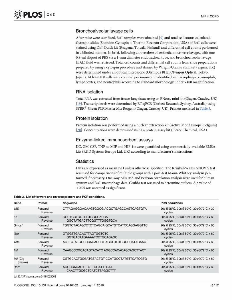
RNA isolationTotal RNAwas extracted from frozen lung tissue using an RNeasy mini kit (Qiagen, Crawley, UK)[19]. Transcript levels were determined by RT-qPCR (Corbett Research, Sydney, Australia) usingSYBR1Green PCRMaster Mix Reagent (Qiagen, Crawley, UK), Primers are listed in Table 3.
Protein isolationProtein isolation was performed using a nuclear extraction kit (Active Motif Europe, Belgium)[20]. Concentrations were determined using a protein assay kit (Pierce Chemical, USA).
Enzyme-linked immunosorbent assaysKC, GM-CSF, TNF-α, MIF and HIF-1α were quantified using commercially-available ELISAkits (R&D Systems Europe Ltd, UK) according to manufacturer’s instructions.
StatisticsData are expressed as mean±SD unless otherwise specified. The Kruskal-Wallis ANOVA testwas used for comparisons of multiple groups with a post-test Mann-Whitney analysis per-formed if necessary. One-way ANOVA and Pearson correlation analysis were used for humansputum and BAL macrophage data. Grubbs test was used to determine outliers. A p value of<0.05 was accepted as significant.
Table 3. List of forward and reverse primers and PCR conditions.
Gene Primer Sequence PCR conditions
18S ForwardReverse
CTTAGAGGGACAAGTGGCG ACGCTGAGCCAGTCAGTGTA 20s@95°C, 30s@60°C, 30s@72°C x 30cycles
Kc ForwardReverse
CGCTGCTGCTGCTGGCCACCAGGCTATGACTTCGGTTTGGGTGCA
20s@95°C, 30s@60°C, 30s@72°C x 60cycles
Gmcsf ForwardReverse
TGGTCTACAGCCTCTCAGCA GCATGTCATCCAGGAGGTTC 20s@95°C, 30s@60°C, 30s@72°C x 60cycles
Ifng ForwardReverse
GTGGTTGACACTTAGTGGTCTCGGTGACATGAAAATCCTGCAGAGC
20s@95°C, 30s@60°C, 30s@72°C x 60cycles
Tnfa ForwardReverse
AGTTCTATGGCCCAGACCCT AGGGTCTGGGCCATAGAACT 20s@95°C, 30s@62°C, 30s@72°C x 60cycles
Mif ForwardReverse
CAAGCCCGCACAGTACATC AGGCCACACAGCAGCTTACT 20s@95°C, 30s@60°C, 30s@72°C x 60cycles
Mif (CigSmoke)
ForwardReverse
CGTGCACTGCGATGTACTGT CCATGCCTATGTTCATCGTG 20s@95°C, 30s@60°C, 30s@72°C x 60cycles
Hprt ForwardReverse
AGGCCAGACTTTGTTGGATTTGAACAACTTGCGCTCATCTTAGGCTTT
20s@95°C, 30s@60°C, 30s@72°C x 60cycles
doi:10.1371/journal.pone.0146102.t003
MIF in COPD
PLOSONE | DOI:10.1371/journal.pone.0146102 January 11, 2016 5 / 17

Results
MIF protein expression is elevated in sputum from COPD patientsMIF protein levels were increased in sputum samples from COPD patients (7.9±2.4ng/ml,p<0.05, 95% CI[4.3, 11.5]) compared to non-smoking controls (3.8±1.6ng/ml, 95% CI[2.2, 5.4],Fig 1A). There was no significant difference in MIF expression between smokers (6.5±2.3ng/ml,95% CI [2.0, 11.0]) and COPD patients.
There were no significant differences in serumMIF concentrations between groups (Fig 1B).Intracellular/cytoplasmic MIF concentrations were significantly elevated in BALmacrophages iso-lated from smokers (67.0±6.8ng/ml, p<0.001, 95% CI [21.5, 110.0]) and COPD patients (96.7±6.8ng/ml, p<0.001, 95% CI [63.3, 206.6]) compared to non-smoking controls (10.8±2.1ng/ml,p<0.001, 95% CI [7.6, 15.3], Fig 1C). There were no outliers as determined by Grubbs test.
Correlation between MIF and HIF-1α protein expression in human BALmacrophagesHIF-1α protein expression was also elevated in BAL macrophages isolated from smokers (6.1±2.7pg/ml, p<0.001, 95% CI [4.3, 7.8]) and COPD patients (8.2±3.4pg/ml, p<0.001, 95% CI[6.5, 10.0]) compared to non-smokers (2.3±1.5ng/ml, 95% CI[1.5, 3.0] Fig 1D). HIF-1α and
Fig 1. MIF and HIF-1α expression levels in COPD.MIF protein was measured in sputum (A; RV cohort), serum (B) and isolated BAL macrophages (C; RVcohort). HIF-1α protein concentration was measured in isolated BAL macrophages (D; RV Cohort). Data are expressed as mean±SEM. *p<0.05 and***p<0.001 compared to non-smoking groups. Rhinovirus Infection (RV).
doi:10.1371/journal.pone.0146102.g001
MIF in COPD
PLOSONE | DOI:10.1371/journal.pone.0146102 January 11, 2016 6 / 17

MIF levels were strongly correlated in all groups with the strongest correlation seen in theCOPD group (Fig 2A–2C).
To test whether there was a mechanistic link driving these correlation we tested whetherHIF-1α protein bound to HIF-1α response elements (HREs) in the MIF promoter. Due to thelimited amounts of sample available from human BAL macrophages these experiments wereconducted in ozone-treated lung samples where there was also a good correlation betweenHIF-1α and MIF expression. Ozone exposure induced binding of HIF-1α protein to the HIF-1α response element (HRE)1 site (-702bp) in ozone-exposed animals 15-fold (p<0.05), but noton the HRE2 (-1483bp) site (Fig 2D). The binding to the HRE1 site in ozone-exposed mice wasassociated with elevatedMifmRNA expression in the lung.
Cigarette smoke-induced COPDmodel did not correlate with humanresultsTo investigate the potential function of MIF in COPD, we used the two different in vivomousemodels of COPD forMif gene expression and determined whether levels of MIF expression inthese models were similar to those seen in human COPD. After 6 and 8 weeks of cigarette
Fig 2. MIF and HIf-1α correlations in COPD and HIF-1 α regulation ofMif expression. Correlation analysis between HIF-1α and MIF proteinconcentrations in isolated BAL macrophages from non-smokers (A), smokers (B), and COPD patients (C). Chromatin immunoprecipitation analysis of HIF-1αbinding to HRE1 and HRE2 sites in theMif promoter in mouse lung tissue (D). Data are expressed as mean±SD for 6 animals per group. **p<0.01 comparedto air controls.
doi:10.1371/journal.pone.0146102.g002
MIF in COPD
PLOSONE | DOI:10.1371/journal.pone.0146102 January 11, 2016 7 / 17

smoke exposure,Mif expression in lung tissue was reduced compared to air control by 51%and 36% respectively (Table 4). At 12 weeks, there was no longer a significant difference inMifexpression between the smoking and air control groups.
In contrast, the 6-week ozone exposure model of COPD showed elevated levels ofMifexpression in lung tissue (0.5±0.1 vs 0.2±0.1, p<0.05, Table 4) correlating with the human datareported here. Hence, we opted to use the ozone-exposure model for examining the role ofMIF in COPD. There are some strain and gender differences in theMifmRNA levels reportedin the two animal models at baseline, however, in the context of this study the relative changesseen in the models and the relationship to the clinical samples are the most important.
ISO-1 but not dexamethasone attenuated ozone-induced BAL cellnumbers and cytokine releaseIn mice, ozone exposure led to an increase in total BAL cells (p<0.01, Fig 3A), reflected in anincrease in neutrophils (3.4-fold, p<0.05), macrophages (3.4-fold, p<0.01) and lymphocytes(2.9-fold, p<0.01) compared to air-exposed controls (Fig 3B–3D). Dexamethasone had noeffect on the number of BAL inflammatory cells in ozone-treated mice. In contrast, ISO-1treatment attenuated the number of total BAL cells (1.7-fold decrease, p<0.05), with a reduc-tion in macrophages (p<0.05) and lymphocytes (p<0.05) but not of neutrophils.
BAL KC (2.3-fold), GM-CSF (3.1-fold), TNF-α (2.3-fold), and MIF (2.8-fold) wereincreased in ozone-exposed mice (p<0.01, Fig 4A–4D). BAL KC and TNF-α levels werereduced 1.4-fold and 1.6-fold respectively by ISO-1 treatment when compared to ozone alone,but levels did not return to baseline values compared to the air and ISO-1 treated controlgroups (p<0.05, Fig 4A and 4C). ISO-1 had no effect on BAL GM-CSF levels (Fig 4B). Ozone-induced BAL MIF concentration was reduced by 1.5-fold with ISO-1 pre-treatment (p<0.05,Fig 4D). In contrast, dexamethasone pre-treatment had no effect on ozone-induced increase ofBAL KC, TNF-á or MIF levels (Fig 4A, 4C and 4D).
Ozone-induced cytokine expression in the lung is insensitive todexamethasone but sensitive to ISO-1 treatmentThe mRNA levels, normalised to the 18S housekeeping gene, of Kc (2.5-fold, p<0.01), Gmcsf(1.9-fold, p<0.05), Tnfα (2.3-fold, p<0.05) andMif (2.2-fold, p<0.05) were elevated afterozone exposure (Fig 5A, 5C, 5E and 5G). Dexamethasone treatment did not affect the ozone-induced increase in gene expression levels. In contrast, ISO-1 treatment reduced the mRNAlevels of each ozone-induced cytokine to basal levels (Fig 5A, 5C, 5E and 5G). Similarly, ozoneexposure enhanced the protein levels of these cytokines in the mouse lung and these were sup-pressed by ISO-1 but not dexamethasone treatment (Fig 5B, 5D, 5F and 5H).
Table 4. RelativeMifmRNA expression in lungs of mousemodels of COPD. Äct values ofMif expres-sion in lung tissue relative to Hprt (cigarette model) and 18s (ozone model) expression as measured by RT-qPCR. ND: not done.
Mif mRNA expression
Week 6 Week 8 Week 12
Cigarette smoke 0.6±0.1 0.8±0.2 1.0±0.1
Air control 1.2±0.4 1.7±0.4 1.1±0.3
Ozone 0.5±0.1 ND ND
Air control 0.2±0.1 ND ND
doi:10.1371/journal.pone.0146102.t004
MIF in COPD
PLOSONE | DOI:10.1371/journal.pone.0146102 January 11, 2016 8 / 17

ISO-1 attenuates ozone affected lung function and AHROzone alone increased RL, at the greatest concentration of acetylcholine, compared to air con-trol (226.5±29.8% versus 139.0±37.3%, p<0.05) and was significantly reversed by ISO-1 (170.3±20.7%, p<0.05 Fig 6A) but not by dexamethasone (206.9±40.3%, p<0.05, Fig 6A). -LogPC100
is the concentration of acetylcholine needed to increase the pulmonary resistance by 100%from baseline. -LogPC100 was decreased 1.6-fold in ozone-exposed mice (p<0.01, Fig 6B). ISO-1 treatment reduced ozone-induced -LogPC100 1.7±0.4mg/ml versus 1.3±0.1mg/ml, p<0.05)but this was not significantly affected by dexamethasone (1.5±0.1mg/ml; Fig 6B). In addition,FEV75 was decreased 1.2-fold in ozone-exposed mice compared to air controls (0.63±0.06mlversus 0.55±0.09ml, p<0.01, Fig 6C). This was significantly improved by ISO-1 but not bydexamethasone treatment (Fig 6C).
Fig 3. Effect of ISO-1 and dexamethasone on ozone-induced airway inflammatory cells. Total cell count (A), neutrophil (B), macrophage (C), andlymphocyte (D) counts in mouse BAL samples. Data are expressed as mean±SD for 6 animals per group. *p<0.05 and **p<0.01 compared to air controls,# p<0.05 compared to ozone exposed group.
doi:10.1371/journal.pone.0146102.g003
MIF in COPD
PLOSONE | DOI:10.1371/journal.pone.0146102 January 11, 2016 9 / 17

Cchord (Fig 6D), TLC and FRC (Fig 6E and 6F) were also significantly increased followingozone exposure (p<0.01). However, neither ISO-1 nor dexamethasone treatment had any sig-nificant effect on these parameters.
Combination of ISO-1 and dexamethasone has no effect on ozone-induced inflammationNo additive anti-inflammatory effect on mediator release or BAL cell number was seen with ISO-1 and dexamethasone in combination compared to dexamethasone or ISO-1 alone. Also, theozone-induced change in RL was not affected by treatment with ISO-1 and dexamethasone incombination. However, the attenuation of RL with ISO-1 alone was no longer evident in the pres-ence of dexamethasone (Fig 7). These changes may reflect the numbers used per group as thestudies were not powered to detect differences. Future studies should use more animals per group.
DiscussionWe demonstrate here that MIF protein expression is elevated in sputum samples, but notserum samples, from healthy smokers and COPD patients compared to healthy aged-matched
Fig 4. Effect of ISO-1 and dexamethasone on ozone-induced BAL inflammation. Cytokine protein levels in mouse BAL of ozone exposed and ISO-1- ordexamethasone-treated mice measured by ELISA. KC (A), GM-CSF (B), TNF-α (C) and MIF (D). Data are expressed as mean±SD for 6 animals per group.*p<0.05 and **p<0.01 compared to air controls, # p<0.05 compared to ozone exposed group.
doi:10.1371/journal.pone.0146102.g004
MIF in COPD
PLOSONE | DOI:10.1371/journal.pone.0146102 January 11, 2016 10 / 17

non-smoking controls. In addition, we demonstrate that MIF expression is greater in BALmacrophages from COPD patients compared to control subjects. MIF expression correlateswith HIF-1α which can enhance MIF expression by binding to specific regions within the MIFpromoter. We examined two models of COPD for MIF expression, a 6 week ozone-exposuremodel and a cigarette smoke model (6, 8 and 12 weeks) to see which model replicated thehuman COPD results. MIF inhibition using ISO-1 prevented ozone-induced BAL, lung inflam-mation and AHR. In contrast, dexamethasone did not affect these parameters, confirming the
Fig 6. Effect of ISO-1 and dexamethasone on ozone-induced changes in AHR and lung function.Mouse lung function measurements of pulmonaryresistance (RL; A), -logPC100 (B), FEV75 (C), lung compliance (Cchord; D), total lung capacity (TLC; E) and functional residual capacity (FRC; F). Data areexpressed as mean±SD for 6 animals per group. *p<0.05 and **p<0.01 compared to air controls, #p<0.05 compared to ozone-exposed group.
doi:10.1371/journal.pone.0146102.g006
Fig 5. Effect of ISO-1 and dexamethasone on ozone-induced lung inflammation. Cytokine mRNA (A, C, E & G) and protein (B, D, F & H) expressionlevels in the lung of ozone exposed and ISO-1- or dexamethasone-treated mice. KC (A&B), GM-CSF (C&D), TNF-α (E&F), and MIF (G&H). Data areexpressed as mean±SD for 6 animals per group. *p<0.05 and **p<0.01 compared to air controls, #p<0.05 compared to ozone exposed group.
doi:10.1371/journal.pone.0146102.g005
MIF in COPD
PLOSONE | DOI:10.1371/journal.pone.0146102 January 11, 2016 11 / 17

corticosteroid insensitive COPD model. Overall, these data indicate that MIF may play a rolein driving COPD inflammation and AHR but not emphysema.
In contrast to our results, MIF has been reported to be reduced in the blood of COPDpatients compared to healthy and smoking subjects [21, 22]. These authors also report reducedMIF levels in the lungs of mice exposed to cigarette smoke for 6 months or more, which wealso see in our more rapid cigarette smoke model, albeit at 12 weeks this reduction is no longer
Fig 7. Effect of ISO-1 and dexamethasone in combination on ozone-induced lung inflammation.Cytokine mRNA (A & C) and protein (B & D)expression levels in the lung of ozone exposed and the combination of ISO-1- plus dexamethasone-treated mice. KC (A&B) and MIF (C&D). Pulmonaryresistance (RL; E) was also measured. Data are expressed as mean±SD for 6 animals per group. *p<0.05 and **p<0.01 compared to air controls, #p<0.05compared to ozone exposed group.
doi:10.1371/journal.pone.0146102.g007
MIF in COPD
PLOSONE | DOI:10.1371/journal.pone.0146102 January 11, 2016 12 / 17
present. Moreover, these previous studies did not examine MIF levels in the lungs or airways ofCOPD patients or controls. Fallica and colleagues [21] report a decrease in serumMIF in COPDpatients as a whole compared to healthy smokers; this is mainly due to a marked reduction inMIF levels in GOLD IV patients whilst the GOLD II and III patients have similar serumMIF lev-els as healthy smokers. The 25 COPD patients examined in our study were generally GOLD IIpatients with eight GOLD III subjects. Whether reduced MIF expression occurs in the moresevere stages of COPD or in emphysematous patients will need to be further investigated.
In our ozone-induced model of COPD, we show increased MIF expression in the lungs at 6weeks. Sauler and colleagues [22] showed increased lung MIF levels after 6-months of smokeexposure in mice but levels were markedly reduced at later time points. Conversely, Fallicaet al., [21] demonstrate reduced MIF expression after 6 months of cigarette smoke exposure.Further comparisons between the smoking models and the ozone model are warranted particu-larly since the degree of inflammation and emphysema in both models is similar. Furthermore,how the data from these cigarette smoke models relate to the clinical results indicatingincreased MIF expression in sputum data also requires further experimentation. For this inves-tigation, the ozone model reflected the higher expression of MIF in COPD patients; thereforewe selected this model to study the inflammation role of MIF.
MIF and its receptors may also have a wider role in airways disease. Alveolar macrophagepolarisation is modulated by smoking and this has important implications for COPD patho-genesis [23]. The fact that MIF expression is altered in COPD and modulates macrophagenumbers in a murine model suggest that MIF may be implicated in smoking-induced macro-phage reprogramming although this needs to be formally studied. In addition, MIF and itsreceptor CD74 have been shown to be increased in pulmonary arterial hypertension (PAH), aknown COPD co-morbidity. Furthermore ISO-1 and anti-CD74 neutralizing antibodies par-tially reverse the development of PAH and inflammation in rats [24].
An anti-inflammatory effect of MIF inhibition has been previously reported in severalrodent models of disease. Anti-MIF antibodies have been shown to be protective against endo-toxemia [25], arthritis [26] sepsis [27] and OVA-induced allergic asthma [28]. In addition,Mifknockout mice also showed less inflammation in models of atopic dermatitis [29] and endotox-emia [30]. To our knowledge, this is the first time that MIF antagonism has been shown tohave an anti-inflammatory effect in a corticosteroid-resistant mouse model of COPD and thisalso provides evidence that targeting MIF may be a useful therapeutic approach for patientswith this disease. Chen et al. also reported on the effect of ISO-1 in the corticosteroid-sensitivemouse OVA-challenged model of airway inflammation [14].
COPD is characterised as neutrophilic and low doses of LPS in mice induces a neutrophil-rich lung inflammation [31], which is prevented by anti-MIF antibodies [32]. ISO-1 signifi-cantly suppressed ozone-induced total cellular recruitment, mostly as a result of reductions inBAL lymphocyte and macrophage but not neutrophil numbers. Magalhaes and colleagues alsoreported a reduction in AHR (Penh) in MIF knockout mice in response to metacholine in anOVA model of asthma [33]. Our data further supports the anti-inflammatory role of MIF inhi-bition, as we report a reduction in AHR (pulmonary resistance) however using a differentmodel and AHR measurement. In contrast, others did not show effect of ISO-1 on inflamma-tion in either OVA-challenged or LPS-treated mouse models [34].
Corticosteroid insensitivity is a major aspect of COPD and severe asthma [35]. The uniquefunction of MIF in counter-regulating the function of corticosteroids [25] has driven researchin many corticosteroid insensitive inflammatory diseases in an attempt to restore steroid-sensi-tivity, improve disease control, reduce exacerbations, disease progression and lower the dosesof oral corticosteroid prescribed. We therefore examined whether combined ISO-1 and dexa-methasone treatment would result in an enhanced anti-inflammatory effect in the lung.
MIF in COPD
PLOSONE | DOI:10.1371/journal.pone.0146102 January 11, 2016 13 / 17

Although Chen et al. found that ISO-1 and dexamethasone separately had a comparable anti-inflammatory effect in the OVA-induced AHR model, they did not examine the effect of com-bined treatments [14]. In contrast, we found that the effects of ISO-1 and dexamethasone werenot comparable and that ISO-1 pre-treatment had a more potent anti-inflammatory effect thandexamethasone in the ozone-induced model reflecting the corticosteroid-insensitive nature ofthis model. ISO-1 and dexamethasone treatment in combination showed no enhanced or addi-tive anti-inflammatory effects on any of the ozone-induced parameters measured, includingAHR or cytokines released in BAL fluid, indicating that MIF is unlikely to activate corticoste-roid-associated pathways in this model.
We have previously demonstrated that chronic ozone exposure resulted in the activation ofhypoxia-induced inflammatory pathways [20]. We now show that HIF-1α can bind to thenativeMif promoter at specific HREs and thereby provide a mechanism by whichMif expres-sion is enhanced in the lungs of ozone-exposed animals. Under hypoxic conditions, MIF inhib-ited dexamethasone-suppressed HIF-1α expression and also enhanced HIF-1α target geneexpression in a positive feedback loop [36]. In support of a positive feedback loop for HIF-1αand MIF, we found that the increased levels of both proteins correlated in BAL macrophagesfrom healthy (non-smokers), smokers and COPD patients. Furthermore in support of an arti-cle published by Gaber et al., we also observed a suppression of HIF-1α protein levels whenMIF was inhibited by ISO-1 [36].
A previous study Baugh JA et al. [37] demonstrated that HIF-1α, acting through an HRE at+25 in the 5'UTR of the MIF gene, is a potent inducer of MIF expression. They also reportedthat this effect is amplified by hypoxia-induced degradation of cAMP responsive element bind-ing protein (CREB). CREB expression is enhanced in COPD patients and a poor response tocorticosteroid therapy may be related to increase CREB-associated signalling [38]. How MIFinteracts with this pathway to modulate corticosteroid signalling in COPD patients requiresfurther investigation. The interaction of MIF with the NF-κB pathway and the modulation ofneutrophil apoptosis also requires further studies. NF-κB is activated in COPD patients [39]and this has been associated with a reduction in sputum neutrophils undergoing spontaneousapoptosis in COPD patients [40].
There are several limitations of the current study. We were unable to demonstrate a signifi-cant effect of ISO-1 on indicators of emphysema in the mouse model although AHR andinflammation were affected. This highlights the importance of COPD phenotyping in under-standing the disease process. It will be important to perform clinical studies with MIF-targetingagents in order to fully define a role for MIF in COPD inflammation. Furthermore, the resultsto date do not exclude a role for the D-dopachrome tautomerase (D-DT or MIF-2) in drivingCOPD inflammation. Finally, we only used female mice in this study and further studies usingmale mice are warranted as males make up more than 50% of COPD patients due to previoussmoking habits.
In conclusion, in our studies investigating the role of MIF in COPD inflammation and ininflammation and AHR in a mouse model of COPD we demonstrate a link between heightenedMIF expression and COPD inflammation. In the ozone-induced corticosteroid-insensitivemurine model of COPD, lung inflammation was attenuated by treatment with the MIF inhibi-tor, ISO-1. This supports a pro-inflammatory role for MIF in driving steroid-insensitive lunginflammation and cellular recruitment and infiltration to the lungs. However, ISO-1 treatmenthad no effect on suppressing ozone-induced neutrophilia and did not reverse corticosteroidinsensitivity or emphysema, suggesting that MIF is not the primary driver of neutrophilia, ste-roid insensitivity or emphysema in this COPD model. However, clinical trials in specific sub-sets of COPD patients will need to be conducted using anti-MIF agents will be required toconfirm this data.
MIF in COPD
PLOSONE | DOI:10.1371/journal.pone.0146102 January 11, 2016 14 / 17
AcknowledgmentsThis project was supported by the NIHR Respiratory Disease Biomedical Research Unit at theRoyal Brompton and Harefield NHS Foundation Trust and Imperial College London. Theviews expressed in this publication are those of the authors(s) and not necessarily those of theNHS, The National Institute for Health Research or the Department of Health.
Author ContributionsConceived and designed the experiments: KER KFC SLJ PJB SRH KDS IMA CHW. Performedthe experiments: KER CC ALD CHWMRS PMH JF PM. Analyzed the data: KER CHWMRSSRH KDS. Wrote the paper: KER KFC IMA CHW SLJ.
References1. Bloom BR, Bennett B. Mechanism of a reaction in vitro associated with delayed-type hypersensitivity.
Science. 1966; 153(3731):80–2. PMID: 5938421
2. David JR. Delayed hypersensitivity in vitro: its mediation by cell-free substances formed by lymphoidcell-antigen interaction. Proc Natl Acad Sci U S A. 1966; 56(1):72–7. PMID: 5229858
3. Calandra T, Echtenacher B, Roy DL, Pugin J, Metz CN, Hultner L, et al. Protection from septic shock byneutralization of macrophage migration inhibitory factor. Nat Med. 2000; 6(2):164–70. PMID: 10655104
4. Leech M, Metz C, Hall P, Hutchinson P, Gianis K, Smith M, et al. Macrophagemigration inhibitory factorin rheumatoid arthritis: evidence of proinflammatory function and regulation by glucocorticoids. Arthritisand rheumatism. 1999; 42(8):1601–8. PMID: 10446857
5. Rossi AG, Haslett C, Hirani N, Greening AP, Rahman I, Metz CN, et al. Human circulating eosinophilssecrete macrophage migration inhibitory factor (MIF). Potential role in asthma. The Journal of ClinicalInvestigation. 1998; 101(12):2869–74. PMID: 9637721
6. Mizue Y, Ghani S, Leng L, McDonald C, Kong P, Baugh J, et al. Role for macrophagemigration inhibi-tory factor in asthma. Proceedings of the National Academy of Sciences of the United States of Amer-ica. 2005; 102(40):14410–5. PMID: 16186482
7. SheWB, Liu XS, Ni W, Chen SX, Xu YJ. The expression of macrophagemigration inhibition factor inpulmonary tissues of smokers with or without chronic obstructive pulmonary disease. Zhonghua nei keza zhi. 2012; 51(11):863–6. PMID: 23291023
8. GOLD. Global Strategy for the Diagnosis, Management and Prevention of COPD, Global Initiative forChronic Obstructive Lung Disease (GOLD). Available from2013.
9. Triantaphyllopoulos K, Hussain F, Pinart M, Zhang M, Li F, Adcock I, et al. A model of chronic inflamma-tion and pulmonary emphysema after multiple ozone exposures in mice. American Journal of Physiol-ogy—Lung Cellular and Molecular Physiology. 2011; 300(5):L691–L700. doi: 10.1152/ajplung.00252.2010 PMID: 21355040
10. Beckett EL, Stevens RL, Jarnicki AG, Kim RY, Hanish I, Hansbro NG, et al. A new short-term mousemodel of chronic obstructive pulmonary disease identifies a role for mast cell tryptase in pathogenesis.Journal of Allergy and Clinical Immunology. 2013; 131(3):752–62.e7. doi: 10.1016/j.jaci.2012.11.053PMID: 23380220
11. Déry M-AC, Michaud MD, Richard DE. Hypoxia-inducible factor 1: regulation by hypoxic and non-hyp-oxic activators. The international journal of biochemistry & cell biology. 2005; 37(3):535–40.
12. Semenza GL. HIF-1: mediator of physiological and pathophysiological responses to hypoxia. J ApplPhysiol (1985). 2000; 88(4):1474–80. PMID: 10749844
13. Lubetsky JB, Dios A, Han J, Aljabari B, Ruzsicska B, Mitchell R, et al. The Tautomerase Active Site ofMacrophageMigration Inhibitory Factor Is a Potential Target for Discovery of Novel Anti-inflammatoryAgents. Journal of Biological Chemistry. 2002; 277(28):24976–82. PMID: 11997397
14. Chen PF, Luo YL, WangW, Wang JX, Lai WY, Hu SM, et al. ISO-1, a macrophage migration inhibitoryfactor antagonist, inhibits airway remodeling in a murine model of chronic asthma. Mol Med. 2010; 16(9–10):400–8. doi: 10.2119/molmed.2009.00128 PMID: 20485865
15. Mallia P, Footitt J, Sotero R, Jepson A, Contoli M, Trujillo-Torralbo MB, et al. Rhinovirus infectioninduces degradation of antimicrobial peptides and secondary bacterial infection in chronic obstructivepulmonary disease. Am J Respir Crit Care Med. 2012; 186(11):1117–24. doi: 10.1164/rccm.201205-0806OC PMID: 23024024
MIF in COPD
PLOSONE | DOI:10.1371/journal.pone.0146102 January 11, 2016 15 / 17

16. Mallia P, Message SD, Gielen V, Contoli M, Gray K, Kebadze T, et al. Experimental rhinovirus infectionas a humanmodel of chronic obstructive pulmonary disease exacerbation. Am J Respir Crit Care Med.2011; 183(6):734–42. doi: 10.1164/rccm.201006-0833OC PMID: 20889904
17. Hansbro PM, Hamilton MJ, Fricker M, Gellatly SL, Jarnicki AG, Zheng D, et al. Importance of Mast CellPrss31/Transmembrane Tryptase/Tryptase-γ in Lung Function and Experimental Chronic ObstructivePulmonary Disease and Colitis. Journal of Biological Chemistry. 2014; 289(26):18214–27. doi: 10.1074/jbc.M114.548594 PMID: 24821729
18. Li F, Wiegman C, Seiffert JM, Zhu J, Clarke C, Chang Y, et al. Effects of N-acetylcysteine in ozone-induced chronic obstructive pulmonary disease model. PLoS ONE. 2013; 8(11):e80782. doi: 10.1371/journal.pone.0080782 PMID: 24260479
19. Ito K, Ito M, Elliott WM, Cosio B, Caramori G, Kon OM, et al. Decreased Histone Deacetylase Activity inChronic Obstructive Pulmonary Disease. New England Journal of Medicine. 2005; 352(19):1967–76.
20. Wiegman CH, Li F, Clarke CJ, Jazrawi E, Kirkham P, Barnes PJ, et al. A comprehensive analysis of oxi-dative stress in the ozone-induced lung inflammation mouse model. Clinical science (London, England:1979). 2013.
21. Fallica J, Boyer L, Kim B, Serebreni L, Varela L, Hamdan O, et al. Macrophage Migration Inhibitory Fac-tor (MIF) is a Novel Determinant of Cigarette Smoke-induced Lung Damage. American Journal ofRespiratory Cell and Molecular Biology. 2014.
22. Sauler M, Leng L, Trentalange M, Haslip M, Shan P, Piecychna M, et al. Macrophage Migration Inhibi-tory Factor Deficiency in Chronic Obstructive Pulmonary Disease. American journal of physiology Lungcellular and molecular physiology. 2014.
23. Shaykhiev R1, Krause A, Salit J, Strulovici-Barel Y, Harvey BG, O'Connor TP, Crystal RG. Smoking-dependent reprogramming of alveolar macrophage polarization: implication for pathogenesis of chronicobstructive pulmonary disease. J Immunol. 2009 Aug 15; 183(4):2867–83. doi: 10.4049/jimmunol.0900473 PMID: 19635926
24. Le Hiress M, Tu L, Ricard N, Phan C, Thuillet R, Fadel E, Dorfmüller P, Montani D, de Man F, HumbertM, Huertas A, Guignabert C. Pro-inflammatory Signature of the Dysfunctional Endothelium in Pulmo-nary Hypertension: Role of MIF/CD74 Complex Am J Respir Crit Care Med. 2015 Jul 23.
25. Calandra T, Bernhagen J, Metz CN, Spiegel LA, Bacher M, Donnelly T, et al. MIF as a glucocorticoid-induced modulator of cytokine production. Nature. 1995; 377(6544):68–71. PMID: 7659164
26. Leech M, Metz C, Santos L, Peng T, Holdsworth SR, Bucala R, et al. Involvement of macrophagemigration inhibitory factor in the evolution of rat adjuvant arthritis. Arthritis and rheumatism. 1998; 41(5):910–7. PMID: 9588744
27. Al-Abed Y, Dabideen D, Aljabari B, Valster A, Messmer D, Ochani M, et al. ISO-1 Binding to the Tauto-merase Active Site of MIF Inhibits Its Pro-inflammatory Activity and Increases Survival in Severe Sep-sis. Journal of Biological Chemistry. 2005; 280(44):36541–4. PMID: 16115897
28. Kobayashi M, Nasuhara Y, Kamachi A, Tanino Y, Betsuyaku T, Yamaguchi E, et al. Role of macro-phagemigration inhibitory factor in ovalbumin-induced airway inflammation in rats. The European respi-ratory journal. 2006; 27(4):726–34. PMID: 16455830
29. Yoshihisa Y, Makino T, Matsunaga K, Honda A, Norisugi O, Abe R, et al. MacrophageMigration Inhibi-tory Factor Is Essential for Eosinophil Recruitment in Allergen-Induced Skin Inflammation. J Invest Der-matol. 2011; 131(4):925–31. doi: 10.1038/jid.2010.418 PMID: 21191413
30. Bozza M, Satoskar AR, Lin G, Lu B, Humbles AA, Gerard C, et al. Targeted disruption of migrationinhibitory factor gene reveals its critical role in sepsis. J Exp Med. 1999; 189(2):341–6. PMID: 9892616
31. Hirano S. Migratory responses of PMN after intraperitoneal and intratracheal administration of lipopoly-saccharide. The American journal of physiology. 1996; 270(5 Pt 1):L836–45. PMID: 8967519
32. Makita H, Nishimura M, Miyamoto K, Nakano T, Tanino Y, Hirokawa J, et al. Effect of Anti-MacrophageMigration Inhibitory Factor Antibody on Lipopolysaccharide-induced Pulmonary Neutrophil Accumula-tion. American Journal of Respiratory and Critical Care Medicine. 1998; 158(2):573–9. PMID: 9700137
33. Magalhães ES, Mourao-Sa DS, Vieira-de-Abreu A, Figueiredo RT, Pires AL, Farias-Filho FA, et al.Macrophagemigration inhibitory factor is essential for allergic asthma but not for Th2 differentiation.European Journal of Immunology. 2007; 37(4):1097–106. PMID: 17373669
34. Korsgren M, #228, llstr m L, Uller L, et al. Role of macrophage migration inhibitory factor (MIF) in allergicand endotoxin-induced airway inflammation in mice. Mediators of Inflammation. 2000; 9(1):15–23.PMID: 10877450
35. Adcock IM, Barnes PJ. Molecular mechanisms of corticosteroid resistance*. Chest. 2008; 134(2):394–401. doi: 10.1378/chest.08-0440 PMID: 18682458
36. Gaber T, Schellmann S, Erekul KB, Fangradt M, Tykwinska K, Hahne M, et al. Macrophage MigrationInhibitory Factor Counterregulates Dexamethasone-Mediated Suppression of Hypoxia-Inducible
MIF in COPD
PLOSONE | DOI:10.1371/journal.pone.0146102 January 11, 2016 16 / 17

Factor-1α Function and Differentially Influences Human CD4+ T Cell Proliferation under Hypoxia. TheJournal of Immunology. 2011; 186(2):764–74. doi: 10.4049/jimmunol.0903421 PMID: 21169549
37. Baugh JA, Gantier M, Li L, Byrne A, Buckley A, Donnelly SC. Dual regulation of macrophagemigrationinhibitory factor (MIF) expression in hypoxia by CREB and HIF-1. Biochem Biophys Res Commun.2006 Sep 8; 347(4):895–903. PMID: 16854377
38. Mroz RM, Holownia A, Chyczewska E, Drost EM, Braszko JJ, Noparlik J, Donaldson K, MacneeW.Cytoplasm-nuclear trafficking of CREB and CREB phosphorylation at Ser133 during therapy of chronicobstructive pulmonary disease. J Physiol Pharmacol. 2007 Nov; 58 Suppl 5(Pt 2: ):437–44.
39. Di Stefano A, Caramori G, Ricciardolo FL, Capelli A, Adcock IM, Donner CF. Cellular and molecularmechanisms in chronic obstructive pulmonary disease: an overview. Clin Exp Allergy. 2004 Aug; 34(8):1156–67. PMID: 15298554
40. Brown V, Elborn JS, Bradley J, Ennis M. Dysregulated apoptosis and NFkappaB expression in COPDsubjects. Respir Res. 2009 Mar 18; 10:24. doi: 10.1186/1465-9921-10-24 PMID: 19296848
MIF in COPD
PLOSONE | DOI:10.1371/journal.pone.0146102 January 11, 2016 17 / 17