RESEARCHARTICLE MASP-1InducedClotting TheFirstModel ... · RESEARCHARTICLE...
13
RESEARCH ARTICLE MASP-1 Induced Clotting – The First Model of Prothrombin Activation by MASP-1 Lorenz Jenny 1,2 , József Dobó 3 , Péter Gál 3 , Verena Schroeder 1,2 * 1 University Clinic of Haematology, Haemostasis Research Laboratory, University Hospital Bern, Bern, Switzerland, 2 Department of Clinical Research, University of Bern, Bern, Switzerland, 3 Institute of Enzymology, Research Centre for Natural Sciences, Hungarian Academy of Sciences, Budapest, Hungary * [email protected] Abstract Mannan-binding lectin-associated serine protease-1 (MASP-1), a protein of the comple- ment lectin pathway, resembles thrombin in terms of structural features and substrate speci- ficity. Due to its interplay with several coagulation factors, it has the ability to induce fibrin clot formation independent of the usual coagulation activation pathways. We have recently shown that MASP-1 activates prothrombin and identified arginine (R) 155, R271, and R393 as potential cleavage sites. FXa cleaves R320 instead of R393, and thrombin cleaves R155 and R284 in prothrombin. Here we have used three arginine-to-glutamine mutants of pro- thrombin, R271Q, R320Q, R393Q and the serine-to-alanine active site mutant S525A to investigate in detail the mechanism of MASP-1 mediated prothrombin activation. Prothrom- bin wildtype and mutants were digested with MASP-1 and the cleavage products were ana- lysed by SDS-PAGE and N-terminal sequencing. A functional clotting assay was performed by thrombelastography. We have found that MASP-1 activates prothrombin via two simulta- neous pathways, either cleaving at R271 or R393 first. Both pathways result in the formation of several active alternative thrombin species. Functional studies confirmed that both R393 and R320 are required for prothrombin activation by MASP-1, whereas R155 is not consid- ered to be an important cleavage site in this process. In conclusion, we have described for the first time a detailed model of prothrombin activation by MASP-1. Introduction The complement system is a central part of the innate immune system and has a crucial func- tion in the clearance of pathogens from the circulation. The lectin pathway is one of three pos- sible ways of the complement system to encounter threats due to infection. It recognises its targets by binding of mannan-binding lectin (MBL) or ficolins to specific patterns on foreign and/or altered surfaces [1]. This leads to the activation of the MBL-associated serine proteases (MASPs) MASP-1, MASP-2, and MASP-3 which then produce C3 convertase via C2 and C4 cleavage (reviewed in [2]). MASP-1 is now considered the central enzyme in the early lectin pathway as it is able to autoactivate, then activates both MASP-2 and MASP-3 [3]. Beside its central role in the lectin pathway, MASP-1 has been shown to interact with the coagulation system [4]. The serine protease domain of MASP-1 is more closely related to PLOS ONE | DOI:10.1371/journal.pone.0144633 December 8, 2015 1 / 13 OPEN ACCESS Citation: Jenny L, Dobó J, Gál P, Schroeder V (2015) MASP-1 Induced Clotting – The First Model of Prothrombin Activation by MASP-1. PLoS ONE 10 (12): e0144633. doi:10.1371/journal.pone.0144633 Editor: Suzan HM Rooijakkers, University Medical Center Utrecht, NETHERLANDS Received: September 10, 2015 Accepted: November 21, 2015 Published: December 8, 2015 Copyright: © 2015 Jenny et al. This is an open access article distributed under the terms of the Creative Commons Attribution License, which permits unrestricted use, distribution, and reproduction in any medium, provided the original author and source are credited. Data Availability Statement: All relevant data are within the paper. Funding: This work was supported by grants from the Swiss National Science Foundation (grant 310030_140925) to VS, OPO Foundation (Zurich, Switzerland) to VS, Hungarian Scientific Research Fund (OTKA grant NK100834) to JD and PG, and the János Bolyai Research Fellowship of the Hungarian Academy of Sciences to JD and PG. The funders had no role in study design, data collection and analysis, decision to publish, or preparation of the manuscript. Competing Interests: The authors have declared that no competing interests exist. source: https://doi.org/10.7892/boris.81888 | downloaded: 13.3.2017
-
Upload
hoangnguyet -
Category
Documents
-
view
219 -
download
0
Transcript of RESEARCHARTICLE MASP-1InducedClotting TheFirstModel ... · RESEARCHARTICLE...

RESEARCH ARTICLE
MASP-1 Induced Clotting – The First Modelof Prothrombin Activation by MASP-1Lorenz Jenny1,2, József Dobó3, Péter Gál3, Verena Schroeder1,2*
1 University Clinic of Haematology, Haemostasis Research Laboratory, University Hospital Bern, Bern,Switzerland, 2 Department of Clinical Research, University of Bern, Bern, Switzerland, 3 Institute ofEnzymology, Research Centre for Natural Sciences, Hungarian Academy of Sciences, Budapest, Hungary
AbstractMannan-binding lectin-associated serine protease-1 (MASP-1), a protein of the comple-
ment lectin pathway, resembles thrombin in terms of structural features and substrate speci-
ficity. Due to its interplay with several coagulation factors, it has the ability to induce fibrin
clot formation independent of the usual coagulation activation pathways. We have recently
shown that MASP-1 activates prothrombin and identified arginine (R) 155, R271, and R393
as potential cleavage sites. FXa cleaves R320 instead of R393, and thrombin cleaves R155
and R284 in prothrombin. Here we have used three arginine-to-glutamine mutants of pro-
thrombin, R271Q, R320Q, R393Q and the serine-to-alanine active site mutant S525A to
investigate in detail the mechanism of MASP-1 mediated prothrombin activation. Prothrom-
bin wildtype and mutants were digested with MASP-1 and the cleavage products were ana-
lysed by SDS-PAGE and N-terminal sequencing. A functional clotting assay was performed
by thrombelastography. We have found that MASP-1 activates prothrombin via two simulta-
neous pathways, either cleaving at R271 or R393 first. Both pathways result in the formation
of several active alternative thrombin species. Functional studies confirmed that both R393
and R320 are required for prothrombin activation by MASP-1, whereas R155 is not consid-
ered to be an important cleavage site in this process. In conclusion, we have described for
the first time a detailed model of prothrombin activation by MASP-1.
IntroductionThe complement system is a central part of the innate immune system and has a crucial func-tion in the clearance of pathogens from the circulation. The lectin pathway is one of three pos-sible ways of the complement system to encounter threats due to infection. It recognises itstargets by binding of mannan-binding lectin (MBL) or ficolins to specific patterns on foreignand/or altered surfaces [1]. This leads to the activation of the MBL-associated serine proteases(MASPs) MASP-1, MASP-2, and MASP-3 which then produce C3 convertase via C2 and C4cleavage (reviewed in [2]). MASP-1 is now considered the central enzyme in the early lectinpathway as it is able to autoactivate, then activates both MASP-2 and MASP-3 [3].
Beside its central role in the lectin pathway, MASP-1 has been shown to interact with thecoagulation system [4]. The serine protease domain of MASP-1 is more closely related to
PLOSONE | DOI:10.1371/journal.pone.0144633 December 8, 2015 1 / 13
OPEN ACCESS
Citation: Jenny L, Dobó J, Gál P, Schroeder V (2015)MASP-1 Induced Clotting – The First Model ofProthrombin Activation by MASP-1. PLoS ONE 10(12): e0144633. doi:10.1371/journal.pone.0144633
Editor: Suzan HM Rooijakkers, University MedicalCenter Utrecht, NETHERLANDS
Received: September 10, 2015
Accepted: November 21, 2015
Published: December 8, 2015
Copyright: © 2015 Jenny et al. This is an openaccess article distributed under the terms of theCreative Commons Attribution License, which permitsunrestricted use, distribution, and reproduction in anymedium, provided the original author and source arecredited.
Data Availability Statement: All relevant data arewithin the paper.
Funding: This work was supported by grants fromthe Swiss National Science Foundation (grant310030_140925) to VS, OPO Foundation (Zurich,Switzerland) to VS, Hungarian Scientific ResearchFund (OTKA grant NK100834) to JD and PG, and theJános Bolyai Research Fellowship of the HungarianAcademy of Sciences to JD and PG. The funders hadno role in study design, data collection and analysis,decision to publish, or preparation of the manuscript.
Competing Interests: The authors have declaredthat no competing interests exist.
source: https://doi.org/10.7892/boris.81888 | downloaded: 13.3.2017

thrombin and trypsin than to the rest of the C1r/C1s/MASP family of serine proteases [5]. Itswide substrate binding cleft and the consequential broad substrate specificity allows it to inter-act with several proteins of the coagulation system including coagulation factor XIII (FXIII),fibrinogen and prothrombin [6–8]. Furthermore MASP-1 is inhibited in the presence of gly-cosaminoglycans more efficiently by antithrombin than by C1-inhibitor [5,9].
In vivo evidence for a role of MASP-1 in coagulation came from MASP-1 and MBL knock-out mice. Upon tail tip excision the knockout mice showed prolonged bleeding time [10] and asignificant decrease in FeCl3-induced thrombogenesis [11].
We have shown recently that MASP-1 is able to trigger clot formation in whole blood andplatelet poor plasma, and that the effects of MASP-1 and thrombin are additive, although theeffect of MASP-1 is weaker and lags behind the effect of thrombin, and that MASP-1 inducedclotting depends on the presence of prothrombin [7,8]. Thus, we proposed that MASP-1induces clotting mainly via prothrombin activation and identified three potential cleavagesites: arginine 155 (R155), R271 and R393 [8].
In the course of FXa mediated prothrombin activation (Fig 1), various thrombin speciesoccur. Alpha-thrombin is the central enzyme in the coagulation cascade, and it is produced bycleavage at R271 and R320, whereby the site to be cleaved first depends on the absence/pres-ence of the prothrombinase complex [12]. Meizothrombin (mIIa) is the first intermediate to beproduced in presence of the prothrombinase complex and arises by cleavage of prothrombin atR320. In absence of the prothrombinase complex and in fluid phase, prothrombin is cleavedfirst at R271 to release prothrombin fragment F1.2 and prethrombin-2. Further (autolytic) deg-radation of α-thrombin by cleavage at R284, R383, and R393 leads to the formation of β-thrombin which has significantly reduced clotting activity [13].
MASP-2 was reported to cleave prothrombin in a similar fashion as FXa [14], whereasthrombin cleaves prothrombin at different sites (R155 and R284) than FXa [13,15]. In MASP-1mediated cleavage of fibrinogen and FXIII, however, MASP-1 does not cleave these substratesin the exact same way and/or efficiency as thrombin does [6].
It is interesting that MASP-1 mediated prothrombin activation seems to involve cleavagesites of both FXa and thrombin mediated proteolytic processes. This would imply that a newmodel, containing at least one new thrombin species, is necessary to explain clotting inducedby MASP-1. Therefore, the aim of our present study was to elucidate the mechanism and theorder of MASP-1 mediated prothrombin cleavage. We generated and expressed a novel pro-thrombin mutant (R393Q) which we used alongside with the prothrombin mutants R271Qand R320Q [12] in digestion and functional experiments.
Materials and Methods
2.1. MASP-1 proteinWe used a recombinant MASP-1 catalytic fragment (rMASP-1cf) in this study, since it is notyet possible to produce or purify sufficient amounts of pure and stable full-length MASP-1.This truncated form of MASP-1 consists of the CCP1-CCP2-SP domains while it lacks the N-terminal CUB1-EGF-CUB2 domains [16]. We have recently shown that rMASP-1cf and full-length MASP-1 show the same effects on clot formation [8].
2.2. Prothrombin mutantsIn the prothrombin mutants, the respective arginine (R) was replaced by glutamine (Q) inorder to abolish individual cleavage sites. The prothrombin mutants R271Q, R320Q andS525A were a kind gift from Prof. Sriram Krishnaswamy (University of Pennsylvania, Philadel-phia, USA). R271Q and R320Q are mutants of the FXa cleavage sites. S525A is a mutant of the
Prothrombin Activation by MASP-1
PLOS ONE | DOI:10.1371/journal.pone.0144633 December 8, 2015 2 / 13
Abbreviations: MBL, mannan-binding lectin; MASP,mannan-binding lectin-associated serine protease;FXIII, factor XIII; FX, factor X; rMASP-1cf, MASP-1catalytic fragment; MES, 2-(N-Morpholino)-ethanesulfonic acid; PVDF, polyvinylidene difluoride;FXa, activated factor X; CT, clotting time; kobs, kobserved; F1.2, prothrombin fragment 1+2; mIIa,meizothrombin; mIIR393, meizothrombin analoguecleaved at R393; ABEI/ABEII, anion binding exosite I/II.

active site of thrombin (it corresponds to S195A according to chymotrypsin numbering). Togenerate the prothrombin mutant R393Q a pcDNA 3.1 (+) plasmid holding the sequence forwildtype prothrombin [17], also a kind gift from Prof. Krishnaswamy, served as a template.The Arg393Gln mutation was introduced with mutagenic primers and the QuickChange muta-genesis kit (Stratagene, La Jolla, USA). Sequencing assured that the mutated prothrombin con-struct was correct.
HEK 293 cells (CRL-1573, directly obtained from ATCC, Manassas, USA) were cultured inD-MEM+/+ (containing 10% FCS, 1% Hepes, 1% non-essential amino acids, 1% sodium pyru-vate; Life Technologies, Carlsbad, USA). Transfection was performed with 7.5 μg of plasmidDNA and 10 μl of Lipofectamine 3000 per 5�105 cells for 24h in Opti-MEM (both Life Technolo-gies). Cells were then transferred into D-MEM+/+ and selected by geneticin (G418, 0.5mg/ml,Life Technologies). The amount of produced R393Q prothrombin by stable cell lines wasassessed by ELISA (Prothrombin (Factor II) human Elisa kit; Abcam, Cambridge, UK). Selectedcell lines were expanded in T150 flasks (Opti-MEM, supplemented with 10 μg/ml reduced vita-min K; Sigma-Aldrich, St. Louis, USA) and the conditioned medium was harvested daily for5 days and immediately treated with 5 mM benzamidine and stored at -20°C.
Fig 1. FXamediated prothrombin cleavage. Prothrombin activation by FXa. FXa cleaves prothrombin either at R271 or at R320. Intermediates gain theiractivity by cleavage at R320 and are then able to perform further cleavages at R155, R284, R383 and R393.
doi:10.1371/journal.pone.0144633.g001
Prothrombin Activation by MASP-1
PLOS ONE | DOI:10.1371/journal.pone.0144633 December 8, 2015 3 / 13

For the purification, the conditioned medium was thawed in a water bath and centrifuged for5 min at 1600 rpm. The supernatant was filtered through a 0.2 μm filter (Thermo Scientific, Wal-tham, USA) before applied to a Q-sepharose anion exchange column (HiTrap Q FF, GE Health-care, Chalfont St Giles, UK) equilibrated with 20 mMHepes and 1 mM benzamidine, pH 7.5.After washing with 20 mMHepes, pH 7.5, R393Q prothrombin was eluted with 20 mMHepes(pH 7.5) and a gradient of 0-1M NaCl, at a flow rate of 5 ml/min. Protein fractions were pooledand applied to a high-resolution Q sepharose anion exchange column (HiTrap Q HP, GEHealthcare) equilibrated with 20mMHepes, 0.5 M NaCl and 1 mM benzamidine, pH 7.5. Afterwashing with 20 mMHepes, pH 7.5, the protein was eluted with 20 mMHepes, 1 M NaCl, pH7.5, and prothrombin containing fractions were pooled and stored at -20°C. The benzamidinedid eluted before the prothrombin fraction and was therefore not present in the purified pro-thrombin samples. Both ion exchange chromatography steps were performed on an ÄKTA puri-fier 10 FPLC (fast protein liquid chromatography) device (GE Healthcare).
2.3. Thrombelastography experimentsThrombelastographic measurements were performed on a rotation thrombelastometry system(ROTEM1, Tem International, Munich, Germany). Wildtype or mutant prothrombin (finalconcentration of 100 μg/ml (1.4 μmol/l)) was added to fibrinogen (2 mg/ml (5.8 μmol/l),Hyphen BioMed) in TBS. Upon addition of rMASP-1cf (80 μg/ml (1.76 μmol/l)) or FXa as acontrol (5 μg/ml (114 nmol/l)), respectively, measurements were started and clotting times(CT), representing the lag time until the onset of clot formation, were recorded during 1 hour.
2.4. Prothrombin cleavage studiesCleavage studies were performed as described earlier [8]. Briefly, prothrombin wildtype (HyphenBioMed, Neuville-sur-Oise, France), or R271Q, R320Q, R393Q or S525Amutants (1.4 μmol/lcorresponding to 100 μg/ml) were incubated at 37°C for up to 90 min (up to 120 min to assessthe cleavage rate of full-length prothrombin) with rMASP-1cf (1.76 μmol/l corresponding to80 μg/ml) in a final volume of 50 μl Tris-buffered saline (TBS, 50 mmol/l Tris, 100 mmol/l NaCl,pH 7.4). After incubation, cleavage products were separated by SDS-PAGE and the gels werestained with Coomassie. For N-terminal sequencing, bands were transferred onto a polyvinyli-dene difluoride (PVDF) membrane byWestern blotting, excised from the membrane and under-went five cycles of Edman degradation and N-terminal sequencing.
2.5. Kinetic analysisWildtype and S525A mutant prothrombin cleavage reactions were prepared as described inparagraph 2.4. The gels were scanned with an Odyssey imaginer (Li-Cor, Lincoln, USA) andthe densitometric scans were quantified using Prism software (GraphPad Software, La Jolla,USA). The data was fitted by nonlinear regression using the equation Y = Y0+A�exp(-K�X),where X is the time and Y are the corresponding densitometric values. The calculated observedfirst order rate constant (k observed) is expressed by kobs/[E] where [E] stands for the totalenzyme concentration.
ResultsRecently, we have shown that MASP-1 is able to induce clot formation by an alternative pro-thrombin activation and identified R155, R271, and R393 as the putative cleavage sites [8]. Inthe present study we have used prothrombin mutants in which individual cleavage sites are
Prothrombin Activation by MASP-1
PLOS ONE | DOI:10.1371/journal.pone.0144633 December 8, 2015 4 / 13
abolished, and we are now able to elucidate the pathways of prothrombin activation by MASP-1 leading to active thrombin species.
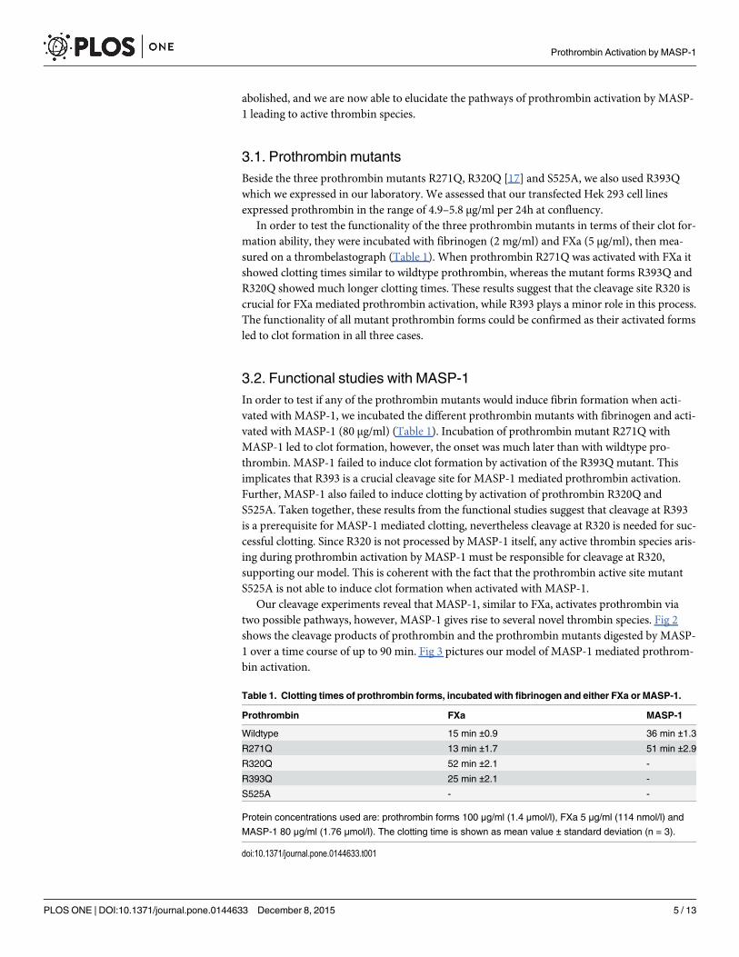
3.1. Prothrombin mutantsBeside the three prothrombin mutants R271Q, R320Q [17] and S525A, we also used R393Qwhich we expressed in our laboratory. We assessed that our transfected Hek 293 cell linesexpressed prothrombin in the range of 4.9–5.8 μg/ml per 24h at confluency.
In order to test the functionality of the three prothrombin mutants in terms of their clot for-mation ability, they were incubated with fibrinogen (2 mg/ml) and FXa (5 μg/ml), then mea-sured on a thrombelastograph (Table 1). When prothrombin R271Q was activated with FXa itshowed clotting times similar to wildtype prothrombin, whereas the mutant forms R393Q andR320Q showed much longer clotting times. These results suggest that the cleavage site R320 iscrucial for FXa mediated prothrombin activation, while R393 plays a minor role in this process.The functionality of all mutant prothrombin forms could be confirmed as their activated formsled to clot formation in all three cases.
3.2. Functional studies with MASP-1In order to test if any of the prothrombin mutants would induce fibrin formation when acti-vated with MASP-1, we incubated the different prothrombin mutants with fibrinogen and acti-vated with MASP-1 (80 μg/ml) (Table 1). Incubation of prothrombin mutant R271Q withMASP-1 led to clot formation, however, the onset was much later than with wildtype pro-thrombin. MASP-1 failed to induce clot formation by activation of the R393Q mutant. Thisimplicates that R393 is a crucial cleavage site for MASP-1 mediated prothrombin activation.Further, MASP-1 also failed to induce clotting by activation of prothrombin R320Q andS525A. Taken together, these results from the functional studies suggest that cleavage at R393is a prerequisite for MASP-1 mediated clotting, nevertheless cleavage at R320 is needed for suc-cessful clotting. Since R320 is not processed by MASP-1 itself, any active thrombin species aris-ing during prothrombin activation by MASP-1 must be responsible for cleavage at R320,supporting our model. This is coherent with the fact that the prothrombin active site mutantS525A is not able to induce clot formation when activated with MASP-1.
Our cleavage experiments reveal that MASP-1, similar to FXa, activates prothrombin viatwo possible pathways, however, MASP-1 gives rise to several novel thrombin species. Fig 2shows the cleavage products of prothrombin and the prothrombin mutants digested by MASP-1 over a time course of up to 90 min. Fig 3 pictures our model of MASP-1 mediated prothrom-bin activation.
Table 1. Clotting times of prothrombin forms, incubated with fibrinogen and either FXa or MASP-1.
Prothrombin FXa MASP-1
Wildtype 15 min ±0.9 36 min ±1.3
R271Q 13 min ±1.7 51 min ±2.9
R320Q 52 min ±2.1 -
R393Q 25 min ±2.1 -
S525A - -
Protein concentrations used are: prothrombin forms 100 μg/ml (1.4 μmol/l), FXa 5 μg/ml (114 nmol/l) and
MASP-1 80 μg/ml (1.76 μmol/l). The clotting time is shown as mean value ± standard deviation (n = 3).
doi:10.1371/journal.pone.0144633.t001
Prothrombin Activation by MASP-1
PLOS ONE | DOI:10.1371/journal.pone.0144633 December 8, 2015 5 / 13

Fig 2. Time-course of the digestion of prothrombin wildtype andmutants by MASP-1. rMASP-1cf andthe different prothrombin forms were incubated for up to 90 min. Bands were identified as a) uncleavedprothrombin, b) prethrombin-1, c) mIIR393 aa1-393, d) prethrombin-2, e) fragment F1.2 + 13 amino acids (upto R284), f) fragment F1.2, g) MASP-1 heavy chain, h) C-terminal part of thrombin heavy chain cleaved atR393, i) MASP-1 light chain, j) α-thrombinR393 light chain + heavy chain up to R393, k) fragment F1. Bandsnot indicated by letters are contaminations of the purification process or MASP-1 degradation products.
doi:10.1371/journal.pone.0144633.g002
Fig 3. Proposedmodel of MASP-1mediated prothrombin cleavage. There are two different pathways of prothrombin cleavage. MASP-1 cleaves first atR271, yielding prethrombin-2 which is subsequently cleaved at R393 to form the active species α-thrombinR393. When R393 is cleaved first, the activeintermediate mIIR393 results. This intermediate is cleaved by MASP-1 (at R271) and itself (at R320) resulting in β-thrombin’ and mIIR393’, respectively.Further active species cannot be excluded. The small letters correspond to the nomenclature of the bands in Fig 2.
doi:10.1371/journal.pone.0144633.g003
Prothrombin Activation by MASP-1
PLOS ONE | DOI:10.1371/journal.pone.0144633 December 8, 2015 6 / 13

3.3. Cleavage pathway via prethrombin-2In a first step, MASP-1 cleaves prothrombin at R271, yielding prethrombin-2 (Fig 2, band d)and prothrombin fragment F1.2 (Fig 2, band f). In a next step, prethrombin-2 is cleaved byMASP-1 at R393 which leads to the production of a novel species we termed α-thrombinR393(Fig 2, bands h and j). Incubation of the prothrombin mutant R271Q with MASP-1 showssome cleavage at R284 (a secondary cleavage site of α-thrombin) instead of the blocked R271Q,yielding a slightly larger fragment F1.2 (Fig 2, band e) and a slightly smaller prethrombin-2. AsMASP-1 shows a significantly lower cleavage rate at R284 in prothrombin than at R393 in pre-thrombin-2, there is no visible prethrombin-2 band in the R271Q digestion, instead this inter-mediate gets processed immediately by cleavage at R393 (Fig 2, band h; the correspondingtruncated form of band j could not be detected).
3.4. Cleavage pathway via meizothrombin analogue (mIIR393)The second pathway starts by cleavage of prothrombin at R393, yielding a novel species wecalled “meizothrombin analogue” (mIIR393) (Fig 2, bands c and h). In a next step, mIIR393 isprocessed at R271 which again yields α-thrombinR393 (Fig 2 bands h and j). This step can beperformed by either MASP-1 or mIIR393 itself as we have shown earlier by inhibition ofthrombin species with hirudin [8].
3.5. Further cleavage stepsThe novel thrombin species (α-thrombinR393 and mIIR393) are putatively active and partici-pate in further cleavage of prothrombin and intermediate cleavage products, therefore R271and R393 no longer remain the only sites that are cleaved during MASP-1 mediated prothrom-bin activation. Recently, we suggested the existence of such active species based on experimentswith hirudin which inhibits the activity of thrombin species but not MASP-1 [8]. Further evi-dence for the involvement of active thrombin species in this process now comes from the cleav-age experiments with the R320Q prothrombin mutant. It can be observed that mIIR393 andprethrombin-2 (Fig 2, bands c and d) are accumulating. This suggests that, in contrast toMASP-1, α-thrombinR393 and/or mIIR393 have indeed cleavage activity towards R320, ulti-mately leading to the production of alternative forms mIIR393’ and β-thrombin’ and possiblyeven α-thrombin. However, the presence of those fragments could not be verified on the gel asthey seem to be produced in small amounts and/or run along with fragments that are producedin significantly bigger amounts.
The cleavage at R155 (Fig 2, band b) which we had initially also identified as part of pro-thrombin activation by MASP-1, seems not to be a direct product of MASP-1 action. Thiscleavage product does not accumulate over time in presence of hirudin, thus it is more likely aproduct of prothrombin and mIIR393 auto degradation.
3.6. Involvement of active thrombin speciesIn FXa mediated prothrombin activation, several active thrombin species arise which them-selves participate in the cleavage cascade. We therefore used the prothrombin active sitemutant S525A to control for the contribution of active thrombin species in MASP-1 mediatedprothrombin cleavage.
Fig 4 shows the different cleavage pattern we obtained by comparing incubation of MASP-1with wildtype prothrombin and the mutant. The cleavage pattern of MASP-1 incubated withS525A shows a slower degradation of mIIR393 (Fig 4, band c) which is likely due to the fact,that this intermediate is mostly processed by itself at R271 and R320, whereas MASP-1 cleaves
Prothrombin Activation by MASP-1
PLOS ONE | DOI:10.1371/journal.pone.0144633 December 8, 2015 7 / 13

at R271 only. Also the intermediate F1.2 (Fig 4, band e) is much less prominent. The reason forthe smaller amount of F1.2 in the S525A mutant compared with wildtype prothrombin may bethat in the wildtype form the active species mIIR393, and not MASP-1, is mainly responsiblefor cleavage at R271. Additionally, mIIR393 may also exhibit a certain amount of back-cleavageon full-length prothrombin. Further it can be observed that the C-terminal part of the throm-bin heavy chain cleaved at R393 (Fig 4, band h) is less prominent. This suggests that someactive thrombin species cleave at R393 as well.
The decrease of the prothrombin band (Fig 4, band a) did not seem to differ between pro-thrombin wildtype and the active site mutant. To further evaluate this, we have calculatedpseudo first order rate constants to compare the kinetics of MASP-1 mediated decrease of wild-type prothrombin and the prothrombin active site mutant S525A (Fig 5).
The kobs/[E] (observed k) values for both prothrombin forms cleaved by MASP-1 were notsignificantly different (141.0�103M-1s-1 for the wildtype form and 136.9�103M-1s-1 for themutant). However, the fitted curves (Fig 5B) show that after 80 min the active site mutant pro-thrombin is processed more slowly than the wildtype form. This indicates that there is autoca-talytical cleavage of thrombin species involved in MASP-1 mediated prothrombin activation,nevertheless it seems to occur to a rather small extent and only in the late phase of the processwhen thrombin species become more abundant.
Taken together, all these results support the important role of active thrombin species inMASP-1 induces prothrombin activation.
DiscussionIn the last decade the interplay between the complement system and blood coagulation hasraised increasing interest and a growing number of interactions have been discovered [18].MASP-1 in particular has numerous interactions with the coagulation system [4]. Nevertheless,many of the underlying mechanisms of how MASP-1 influences the coagulation system andthe relevance of these interactions are not yet completely understood. In the present study we
Fig 4. Time-course of the digestion of prothrombin wildtype and active site mutant S525A by MASP-1. rMASP-1cf and the different prothrombin formswere incubated for up to 90 min. Bands were identified as a) uncleaved prothrombin, b) prethrombin-1, c) mIIR393 aa1-393, d) prethrombin-2, e) fragmentF1.2, f) MASP-1 heavy chain, g) degradation product of the MASP-1 heavy chain, h) C-terminal part of thrombin heavy chain cleaved at R393, i) MASP-1light chain, j) α-thrombinR393 light chain + heavy chain up to R393, k) fragment F1. Bands not indicated by letters are contaminations of the purificationprocess.
doi:10.1371/journal.pone.0144633.g004
Prothrombin Activation by MASP-1
PLOS ONE | DOI:10.1371/journal.pone.0144633 December 8, 2015 8 / 13

show for the first time a model of MASP-1 mediated prothrombin activation which was devel-oped by using prothrombin mutants R271Q, R320Q, and R393Q.
At first glance, prothrombin cleavage by MASP-1 strongly resembles FXa mediated pro-thrombin activation. Similar to FXa mediated prothrombin cleavage, there are two differentpathways, both ending up in the production of a common thrombin species [19]. Bothenzymes are able to cleave at position R271, however, they have different preferences regardingthe specific cleavage site that activates prothrombin between the heavy and the light chain: FXacleaves at R320 whereas MASP-1 cleaves 73 amino acids further upstream at R393. Neverthe-less, both cleavage sites are located within the interchain disulfide-bridge (C292-C438), there-fore cleavage at R393 does not lead to dissociation of the heavy and light chains.
In FXa mediated prothrombin cleavage, preference for the first cleavage site depends on itsenvironment: In the presence of an assembling prothrombinase complex (requires a membranesurface), R320 becomes the preferred cleavage site, which leads to the production of meizo-thrombin, an intermediate that acts anticoagulant. On the other hand, when FXa activates pro-thrombin in absence of a prothrombinase complex (typical for fluid phase activation), R271becomes the preferred site for the first cleavage, yielding prethrombin-2 [20,21]. In contrast,MASP-1 does not exhibit a clear preference for a specific first cleavage site in experiments per-formed in solution; R271 and R393 are selected at similar rates.
Meizothrombin (mIIa) and its MASP-1 cleaved analogue (mIIR393) are both active species.It is known that mIIa has an impaired ability to clot fibrinogen and even has anticoagulanteffects by binding thrombomodulin and activating protein C [22]. Its analogue mIIR393 is
Fig 5. Kinetic analysis of prothrombin cleavage by MASP-1. A, SDS-PAGE of prothrombin wildtype or the mutant form S525A (both 1.4μM) digested byMASP-1 (1.76 μM) over 120 min. B, curves fitted by non-linear regression.
doi:10.1371/journal.pone.0144633.g005
Prothrombin Activation by MASP-1
PLOS ONE | DOI:10.1371/journal.pone.0144633 December 8, 2015 9 / 13

likely to exhibit an impaired clotting ability as well. Evidence comes from the thrombelasto-graphic experiments with the prothrombin mutant R320Q. As mIIR393 accumulates, therewould be plenty of this intermediate available for clot formation, but clot formation does notoccur. This finding also implies that mIIR393 is able to cleave itself at R320 which then leads toproduction of the intermediate mIIR393’.
The finding that the cleavage site R320 is necessary for MASP-1 induced clotting suggeststhat the other novel thrombin species, α-thrombinR393, is not sufficiently capable to inducefibrin clotting either. Since prethrombin-2 also accumulates when R320 is abolished, it is possi-ble that either α-thrombinR393 or mIIR393 cleave prethrombin-2 at R320 producing α-throm-bin. However, since the α-thrombin heavy chain would run along with the vast amount of theMASP-1 heavy chain on the gel, it was not possible to isolate this band.
Our thrombelastographic experiments confirm that cleavage at R393 is required but not suf-ficient for MASP-1 mediated clotting, since not only blockage of R393 but also blockage ofR320 prevents clot formation. As R393 is cleaved earlier as R320, this suggests that the actionof mIIR393 or α-thrombinR393 is necessary for cleavage at R320 to yield the active species ulti-mately responsible for fibrin clotting. This is further supported by the absence of clot formationwhen the active site mutant S525A was used.The cleavage assay with the active site mutantS525A showed that the arising thrombin species are involved and have at least partly the samecleavage sites as MASP-1. The most prominent difference in the active site mutant is the accu-mulation of mIIR393 (Fig 4, band c), which also seems to be a crucial player in the MASP-1mediated prothrombin activation. mIIR393 does not only exert cleavage action on itself, itmost likely cleaves mIIR393’ and α-thrombinR393 as well. Since the C-terminal part of thethrombin heavy chain, cleaved at R393, is produced less when the mutant S525A is used, it canbe assumed that at least some thrombin species cleave at R393 as well.
It is further notable that the degradation product of the MASP-1 heavy chain (Fig 4, band g)is produced to a smaller extent during the digestion of the active site mutant prothrombin.This indicates that MASP-1 is a target for some active thrombin species. Assuming that throm-bin-degraded MASP-1 is inactive, this could explain why the cleavage rates of prothrombinwildtype and active site mutant do not differ significantly: while autocatalytic cleavage by activethrombin species does not occur in the active site mutant, there is at the same time more activeMASP-1 available.
It is also important to mention that the production of F1 (Fig 4, band k) is impaired in thedigestion of the active site mutant prothrombin. This confirms that R155 is mainly cleaved bythrombin species but is not a good cleavage site for MASP-1.
Prothrombin contains two electropositive exosites termed anion binding exosite I andanion binding exosite II (ABEI and ABEII). Both exosites are important players in the recogni-tion and the binding of specific substrates, effectors and inhibitors. ABEI is located next to theactive site cleft and is involved in the recognition and binding of fibrinogen, hirudin and FV[23, 24, 25]. The majority of the ABEI residues are located on the heavy chain of prothrombin.Cleavage at R155, R271, R320 or R393 individually does not disrupt this exosite. However,cleavage at both R320 and R393 (yielding β-thrombin, β-thrombin’ or mIIR393’) leads to a lossof five ABEI residues which are located between R382 and R393, the fragment lost in β-throm-bin production. The loss of this fragment results in a remarkable decrease of the efficiency ofthrombin species to process fibrinogen and therefore a reduced clotting activity [13]. Thisimplies that MASP-1 mediated clot formation is mainly driven by α-thrombinR393 andmIIR393, because β-thrombin’ and mIIR393’ lack a functional exosite I.
FXa binds to membranes in a calcium dependent matter to form the prothrombinase com-plex (in combination with FVa) which enhances the prothrombin activation significantly [26].Since MASP-1 also contains a Ca2+ dependent binding domain (EGF), it is possible that
Prothrombin Activation by MASP-1
PLOS ONE | DOI:10.1371/journal.pone.0144633 December 8, 2015 10 / 13

MASP-1 mediated prothrombin activation could be enhanced in the presence of phospholipidsand Ca2+. However, because our recombinant form of MASP-1 lacks the CUB-EGF-CUBdomains, it is not possible to test for an enhancing effect of phospholipids with our enzyme.
In summary, we have shown for the first time that MASP-1 activates prothrombin via twosimultaneous pathways which seem equally preferred in the fluid phase. Furthermore, we haveshown the existence of novel alternative thrombin forms we called α-thrombinR393, meizo-thrombin analogue (mIIR393), and β-thrombin’ which must be active in the process of MASP-1 mediated prothrombin cleavage. Even though the cleavage site R393 is necessary to induceclot formation, it is not sufficient by itself but it needs the support of the cleavage site R320which is the activation site of α-thrombin. It is important to mention that the experimentswere performed in purified systems that lack the effects of inhibitory and enhancing compo-nents that can be found in whole blood. However, we have recently shown that MASP-1 doesshow significant effects on clot formation in whole blood and plasma [8].
Our results do not suggest that MASP-1 competes with FXa for prothrombin activation.FXa as part of the prothrombinase complex is the activator of prothrombin in the coagulationcascade and it is of course much stronger and more efficient in activating prothrombin com-pared with MASP-1. However, we do believe that under certain pathophysiological conditionsactivated MASP-1 supports or even triggers clot formation and is able to sustain it beyond thenormal coagulation activation pathways. An example may be diabetes, both type 1 and type 2are recognised to have a strong inflammatory component. We have recently shown thatMASP-1 plasma levels are elevated in patients with type 1 diabetes [27]. In this inflammatoryenvironment, more MASP-1 may activate and lead to both activation of the complement lectinpathway as well as to low level prothrombin activation and fibrin formation. In this wayMASP-1 could contribute to thrombotic complications which are frequent in diabetes andother inflammatory diseases.
AcknowledgmentsThe authors would like to thank Prof. Johann Schaller and Urs Kämpfer (Department ofChemistry and Biochemistry, University of Bern) for their technical support and expertise inN-terminal protein sequencing and Prof. Dr. Sriram Krishnaswamy (University of Pennsylva-nia, Philadelphia, USA) for the kind donation of the prothrombin mutants R271Q R320Q andS525A and the plasmid carrying the prothrombin sequence.
L. Jenny designed and performed the experiments, analysed the data, and wrote the manu-script. J. Dobó and P. Gál produced the rMASP-1 fragment and revised the manuscript. V.Schroeder designed the study, analysed the data, and revised the manuscript.
Author ContributionsConceived and designed the experiments: LJ VS. Performed the experiments: LJ. Analyzed thedata: LJ VS. Contributed reagents/materials/analysis tools: JD PG. Wrote the paper: LJ JD PGVS.
References1. Ricklin D, Hajishengallis G, Yang K, Lambris JD. Complement: a key system for immune surveillance
and homeostasis. Nat Immunol. 2010; 11: 785–97. doi: 10.1038/ni.1923 PMID: 20720586
2. Yongqing T, Drentin N, Duncan RC, Wijeyewickrema LC, Pike RN. Mannose-binding lectin serine pro-teases and associated proteins of the lectin pathway of complement: two genes, five proteins andmany functions? Biochim Biophys Acta. 2012; 1824: 253–262. doi: 10.1016/j.bbapap.2011.05.021PMID: 21664989
Prothrombin Activation by MASP-1
PLOS ONE | DOI:10.1371/journal.pone.0144633 December 8, 2015 11 / 13
3. Megyeri M, Harmat V, Major B, Végh Á, Balczer J, Héja D, et al. Quantitative characterization of theactivation steps of mannan-binding lectin (MBL)-associated serine proteases (MASPs) points to thecentral role of MASP-1 in the initiation of the complement lectin pathway. J Biol Chem. 2013; 288:8922–8934. doi: 10.1074/jbc.M112.446500 PMID: 23386610
4. Dobó J, Schroeder V, Jenny L, Cervenak L, Závodszky P, Gál P. Multiple roles of complement MASP-1at the interface of innate immune response and coagulation. Mol Immunol. 2014; 61: 69–78. doi: 10.1016/j.molimm.2014.05.013 PMID: 24935208
5. Gál P, Dobó J, Závodszky P, Sim RB. Early complement proteases: C1r, C1s and MASPs. A structuralinsight into activation and functions. Mol Immunol. 2009; 46: 2745–2752. doi: 10.1016/j.molimm.2009.04.026 PMID: 19477526
6. Krarup A, Gulla KC, Gál P, Hajela K, Sim RB. The action of MBL-associated serine protease 1(MASP1) on factor XIII and fibrinogen. Biochim Biophys Acta. 2008; 1784: 1294–1300. doi: 10.1016/j.bbapap.2008.03.020 PMID: 18456010
7. Hess K, Ajjan R, Phoenix F, Dobó J, Gál P, Schroeder V. Effects of MASP-1 of the complement systemon activation of coagulation factors and plasma clot formation. PLoS One. 2012; 7: e35690. doi: 10.1371/journal.pone.0035690 PMID: 22536427
8. Jenny L, Dobó J, Gál P, Schroeder V. MASP-1 of the complement system promotes clotting via pro-thrombin activation. Mol Immunol. 2015; 65: 398–405. doi: 10.1016/j.molimm.2015.02.014 PMID:25745807
9. Parej K, Dobó J, Závodszky P, Gál P. The control of the complement lectin pathway activation revisited:both C1-inhibitor and antithrombin are likely physiological inhibitors, while α2-macroglobulin is not. MolImmunol. 2013; 54: 415–422. doi: 10.1016/j.molimm.2013.01.009 PMID: 23399388
10. Takahashi K, ChangWC, Takahashi M, Pavlov V, Ishida Y, La Bonte L, et al. Mannose-binding lectinand its associated proteases (MASPs) mediate coagulation and its deficiency is a risk factor in develop-ing complications from infection, including disseminated intravascular coagulation. Immunobiology.2011; 216: 96–102. doi: 10.1016/j.imbio.2010.02.005 PMID: 20399528
11. La Bonte LR, Pavlov VI, Tan YS, Takahashi K, Takahashi M, Banda NK, et al. Mannose-binding lectin-associated serine protease-1 is a significant contributor to coagulation in a murine model of occlusivethrombosis. J Immunol. 2012; 188: 885–891. doi: 10.4049/jimmunol.1102916 PMID: 22156595
12. Krishnaswamy S. The transition of prothrombin to thrombin. J Thromb Haemost. 2013; 11: Suppl. 1,265–276. doi: 10.1111/jth.12217 PMID: 23809130
13. Bovill EG, Tracy RP, Hayes TE, Jenny RJ, Bhushan FH &Mann KG. Evidence that meizothrombin isan intermediated product in the clotting of whole blood. Arterioscler Thromb Vasc Biol. 1995; 15: 754–758. PMID: 7773729
14. Krarup A, Wallis R, Presanis JS, Gál P, Sim RB. Simultaneous activation of complement and coagula-tion by MBL-associated serine protease 2. PLoS One. 2007; 2: e623. PMID: 17637839
15. Petrovan RJ, Govers-Riemslag JW, Novak G, Hemker HC, Tans G, Rosing J. Autocatalytic peptidebond cleaves in prothrombin and meizothrombin. Biochemistry. 1998; 37: 1185–1191. PMID: 9477942
16. Dobó J, Harmat V, Beinrohr L, Sebestyén E, Závodszky P, Gál P. MASP-1, a promiscuous complementprotease: structure of its catalytic region reveals the basis of its broad specificity. J Immunol. 2009; 183:1207–1214. doi: 10.4049/jimmunol.0901141 PMID: 19564340
17. Orcutt SJ, Krishnaswamy S. Binding of substrate in two conformations to human prothrombinase drivesconsecutive cleavage at two sites in prothrombin. J Biol Chem. 2004; 279: 54927–54936. PMID:15494418
18. Amara U, Flierl MA, Rittirsch D, Klos A, Chen H, Acker B, et al. Molecular intercommunication betweenthe complement and coagulation systems. J Immunol. 2010; 185: 5628–5636. doi: 10.4049/jimmunol.0903678 PMID: 20870944
19. Haynes LM, Bouchard BA, Tracy PB, Mann KG. Prothrombin activation by platelet-associated pro-thrombinase proceeds through the prethrombin-2 pathway via a concerted mechanism. J Biol Chem.2012; 287: 38647–38655. doi: 10.1074/jbc.M112.407791 PMID: 22989889
20. Krishnaswamy S, Mann KG, NesheimME. The prothrombinase-catalyzed activation of prothrombinproceeds through the intermediate meizothrombin in an ordered, sequential reaction. J Biol Chem.1986; 261: 8977–8984. PMID: 3755135
21. Mann KG, NesheimME, ChurchWR, Haley P, Krishnaswamy S. Surface-dependent reactions of thevitamin K-dependent enzyme complexes. Blood. 1999; 76: 1–16.
22. Bradford HN, Krishnaswamy S. Meizothrombin is an unexpectedly zymogen-like variant of thrombin. JBiol Chem. 2012; 287: 30414–30425. doi: 10.1074/jbc.M112.394809 PMID: 22815477
23. Higgins DL, Lewis SD, Shafer JA. Steady state kinetic parameters for the thrombin-catalyzed conver-sion of human fibrinogen to fibrin. J Biol Chem. 1983; 258: 9276–9282. PMID: 6409903
Prothrombin Activation by MASP-1
PLOS ONE | DOI:10.1371/journal.pone.0144633 December 8, 2015 12 / 13

24. Huntington JA. Molecular recognition mechanisms of thrombin. J Thromb Haemost. 2005; 3: 1861–1872. PMID: 16102053
25. Myles T, Yun TH, Hall SW, Leung LL. An extensive interaction interface between thrombin and factor Vis required for factor V activation. J Biol Chem. 2001; 276: 25143–25149. PMID: 11312264
26. Rosing J, Tang G, Govers-Riemslag JW, Zwaal RF, Hemker HC. The role of phospholipids and factorVa in the prothrombinase complex. J Biol Chem. 1980; 255: -283.
27. Jenny L, Ajjan R, King R, Thiel S, Schroeder V. Plasma levels of mannan-binding lectin-associated ser-ine proteases MASP-1 and MASP-2 are elevated in type 1 diabetes and correlate with glycaemic con-trol. Clin Exp Immunol. 2015; 180: 227–232. doi: 10.1111/cei.12574 PMID: 25533914
Prothrombin Activation by MASP-1
PLOS ONE | DOI:10.1371/journal.pone.0144633 December 8, 2015 13 / 13


![Effects of MASP-1 of the Complement System on Activation ... · However, MASP-1 has been suggested to act synergistically with MASP-2 to produce C3 convertase via C2 cleavage [11],](https://static.fdocuments.in/doc/165x107/5ff62654ce9bb6026f40b6de/effects-of-masp-1-of-the-complement-system-on-activation-however-masp-1-has.jpg)