RESEARCHARTICLE LateralSemicircularCanalAsymmetryin ... · RESEARCHARTICLE...
20
RESEARCH ARTICLE Lateral Semicircular Canal Asymmetry in Idiopathic Scoliosis: An Early Link between Biomechanical, Hormonal and Neurosensory Theories? Martin Hitier 1,2,3,4 *, Michèle Hamon 5 , Pierre Denise 4 , Julien Lacoudre 1 , Marie- Aude Thenint 5 , Jean-François Mallet 6 , Sylvain Moreau 1,2 , Gaëlle Quarck 4 1 Department of Otolaryngology—Head and Neck Surgery, CHU de Caen, Caen, F-14000, France, 2 Department of Anatomy, UNICAEN, Caen, 14032, France, 3 Department of Pharmacology and Toxicology; School of Medical Sciences and Brain Health Research Center, University of Otago, Dunedin, New Zealand, 4 U 1075 COMETE, INSERM, Caen, 14032, France, 5 Department of Neuroradiology, CHU de Caen, Caen, 14000, France, 6 Department of Paediatric surgery, CHU de Caen, Caen, 14000, France * [email protected] Abstract Introduction Despite its high incidence and severe morbidity, the physiopathogenesis of adolescent idio- pathic scoliosis (AIS) is still unknown. Here, we looked for early anomalies in AIS which are likely to be the cause of spinal deformity and could also be targeted by early treatments. We focused on the vestibular system, which is suspected of acting in AIS pathogenesis and which exhibits an end organ with size and shape fixed before birth. We hypothesize that, in adolescents with idiopathic scoliosis, vestibular morphological anomalies were already present at birth and could possibly have caused other abnormalities. Materials and Methods The vestibular organ of 18 adolescents with AIS and 9 controls were evaluated with MRI in a prospective case controlled study. We studied lateral semicircular canal orientation and the three semicircular canal positions relative to the midline. Lateral semicircular canal func- tion was also evaluated by vestibulonystagmography after bithermal caloric stimulation. Results The left lateral semicircular canal was more vertical and further from the midline in AIS (p = 0.01) and these two parameters were highly correlated (r = -0.6; p = 0.02). These mor- phological anomalies were associated with functional anomalies in AIS (lower excitability, higher canal paresis), but were not significantly different from controls (p>0.05). PLOS ONE | DOI:10.1371/journal.pone.0131120 July 17, 2015 1 / 20 OPEN ACCESS Citation: Hitier M, Hamon M, Denise P, Lacoudre J, Thenint M-A, Mallet J-F, et al. (2015) Lateral Semicircular Canal Asymmetry in Idiopathic Scoliosis: An Early Link between Biomechanical, Hormonal and Neurosensory Theories? PLoS ONE 10(7): e0131120. doi:10.1371/journal.pone.0131120 Editor: Manabu Sakakibara, Tokai University, JAPAN Received: January 4, 2015 Accepted: May 28, 2015 Published: July 17, 2015 Copyright: © 2015 Hitier et al. This is an open access article distributed under the terms of the Creative Commons Attribution License, which permits unrestricted use, distribution, and reproduction in any medium, provided the original author and source are credited. Data Availability Statement: All relevant data are within the paper and its Supporting Information file. Funding: This research was supported by a Marie Curie International Research Staff Exchange Scheme Fellowship within the 7th European Community Framework Program #918980, and Emergence Program (N° 11P03919 /11P03921) Basse- Normandie Région. The funders had no role in study design, data collection and analysis, decision to publish, or preparation of the manuscript. Competing Interests: The authors have declared that no competing interests exist.
Transcript of RESEARCHARTICLE LateralSemicircularCanalAsymmetryin ... · RESEARCHARTICLE...

RESEARCH ARTICLE
Lateral Semicircular Canal Asymmetry inIdiopathic Scoliosis: An Early Link betweenBiomechanical, Hormonal and NeurosensoryTheories?Martin Hitier1,2,3,4*, Michèle Hamon5, Pierre Denise4, Julien Lacoudre1, Marie-Aude Thenint5, Jean-François Mallet6, Sylvain Moreau1,2, Gaëlle Quarck4
1 Department of Otolaryngology—Head and Neck Surgery, CHU de Caen, Caen, F-14000, France,2 Department of Anatomy, UNICAEN, Caen, 14032, France, 3 Department of Pharmacology andToxicology; School of Medical Sciences and Brain Health Research Center, University of Otago, Dunedin,New Zealand, 4 U 1075 COMETE, INSERM, Caen, 14032, France, 5 Department of Neuroradiology, CHUde Caen, Caen, 14000, France, 6 Department of Paediatric surgery, CHU de Caen, Caen, 14000, France
Abstract
Introduction
Despite its high incidence and severe morbidity, the physiopathogenesis of adolescent idio-
pathic scoliosis (AIS) is still unknown. Here, we looked for early anomalies in AIS which are
likely to be the cause of spinal deformity and could also be targeted by early treatments. We
focused on the vestibular system, which is suspected of acting in AIS pathogenesis and
which exhibits an end organ with size and shape fixed before birth. We hypothesize that, in
adolescents with idiopathic scoliosis, vestibular morphological anomalies were already
present at birth and could possibly have caused other abnormalities.
Materials and Methods
The vestibular organ of 18 adolescents with AIS and 9 controls were evaluated with MRI in
a prospective case controlled study. We studied lateral semicircular canal orientation and
the three semicircular canal positions relative to the midline. Lateral semicircular canal func-
tion was also evaluated by vestibulonystagmography after bithermal caloric stimulation.
Results
The left lateral semicircular canal was more vertical and further from the midline in AIS
(p = 0.01) and these two parameters were highly correlated (r = -0.6; p = 0.02). These mor-
phological anomalies were associated with functional anomalies in AIS (lower excitability,
higher canal paresis), but were not significantly different from controls (p>0.05).
PLOS ONE | DOI:10.1371/journal.pone.0131120 July 17, 2015 1 / 20
OPEN ACCESS
Citation: Hitier M, Hamon M, Denise P, Lacoudre J,Thenint M-A, Mallet J-F, et al. (2015) LateralSemicircular Canal Asymmetry in Idiopathic Scoliosis:An Early Link between Biomechanical, Hormonal andNeurosensory Theories? PLoS ONE 10(7):e0131120. doi:10.1371/journal.pone.0131120
Editor: Manabu Sakakibara, Tokai University, JAPAN
Received: January 4, 2015
Accepted: May 28, 2015
Published: July 17, 2015
Copyright: © 2015 Hitier et al. This is an openaccess article distributed under the terms of theCreative Commons Attribution License, which permitsunrestricted use, distribution, and reproduction in anymedium, provided the original author and source arecredited.
Data Availability Statement: All relevant data arewithin the paper and its Supporting Information file.
Funding: This research was supported by a MarieCurie International Research Staff Exchange SchemeFellowship within the 7th European CommunityFramework Program #918980, and EmergenceProgram (N° 11P03919 /11P03921) Basse-Normandie Région. The funders had no role in studydesign, data collection and analysis, decision topublish, or preparation of the manuscript.
Competing Interests: The authors have declaredthat no competing interests exist.

Conclusion
Adolescents with idiopathic scoliosis exhibit morphological vestibular asymmetry, probably
determined well before birth. Since the vestibular system influences the vestibulospinal
pathway, the hypothalamus, and the cerebellum, this indicates that the vestibular system is
a possible cause of later morphological, hormonal and neurosensory anomalies observed
in AIS. Moreover, the simple lateral SCC MRI measurement demonstrated here could be
used for early detection of AIS, selection of children for close follow-up, and initiation of pre-
ventive treatment before spinal deformity occurs.
IntroductionAdolescent idiopathic scoliosis (AIS) is characterized by a spinal deformity of unknown originand affects 3% of children between the ages of 10 and 16 worldwide[1–4] [1,2], resulting inpain, poor self image with social consequences, and the possible burden of heavy treatmentwith a brace or spine surgery[3,5]. Even if the level of proof is currently weack[6], the spinaldeformity is often associated with other morphological (e.g. ribs, pelvis, arms, skull)[7–10],hormonal (e.g. leptin, melatonin signaling) [11–13], and neurosensory anomalies (e.g. vestibu-lar, neurosensory integration)[14–17]. Authors have identified genes[18–23] and suspectedenvironmental factors [24,25], and have elaborated theories to link these factors to the anoma-lies (for review see [26–28]). However, the pathogenesis of AIS remains unknown and is proba-bly multifactorial.[29]
One main difficulty is that some anomalies could be either the cause or the consequence ofothers. For example, the spinal deformity could be the cause or the consequence of limb asym-metry, or both could be the result of a common cause [27]. Spine deformity could also be theconsequence of brain anomalies [30,31] or its causes, with the brain trying to compensate forthe postural instability [32].
One strategy to be used to clarify the pathogenesis would be to establish the chronology ofAIS anomalies. This could help to identify causes of AIS, which should be those that appearearliest. Ultimately, this chronology may help to design new treatments early enough to stopthe disease at the earliest stage.
Here, we propose establishing a chronologic landmark in the vestibular system. Severalstudies argue for a vestibular impairment in AIS [14,15,33]. Additionally, vestibular lesion inguinea pigs or Xenopus have reproduced scoliosis, which indicates that the vestibule is a pos-sible cause of AIS [34,35]. The mechanism of vestibular induced scoliosis is more likely anasymmetry of the vestibulospinal pathway leading to an imbalance in paraspinal muscles.Most notably, the size and shape of the vestibular organ are fixed by the ossification of theotic capsule before birth, much as a fossil in rock [36–43]. Therefore, the shape of the adoles-cent vestibule (i.e. bony labyrinth) is thought to be similar to the shape exhibited at birth.We thus propose studying the AIS labyrinth to determine early malformation. More pre-cisely, we focused on the lateral semicircular canal (SCC) which can be both visualized byMRI and examined by a caloric test, allowing the evaluation of the right and left sides inde-pendently [44]. The lateral SCC is most frequently affected by malformation in the generalpopulation, probably because it is the last to be formed and ossified [45]. We hypothesizedthat in AIS, the lateral SCC could present early orientation troubles associated with func-tional impairment.
Lateral Semicircular Canal Asymmetry in Idiopathic Scoliosis
PLOS ONE | DOI:10.1371/journal.pone.0131120 July 17, 2015 2 / 20

Patients and MethodWe conducted a prospective case-control study including 18 cases of idiopathic scoliosis and 9controls approved by the Nord Ouest III Ethics Committee (approval no. 2008 A00598-47).Because all cases and controls were minor, informed written consents were obtained from theirparents, caretakers or guardians. One individual gave written informed consent (as outlined inPLoS consent form) to publish the image in Fig 1, Fig 2 and Fig 3 of this manuscript.
ParticipantsInclusion criteria for the scoliosis group included females and males 10–18 years of age pre-senting untreated idiopathic scoliosis with a Cobb angle between 10 and 50°. The types of scoli-osis were determined according to the definition of the Scoliosis Research Society: themaximum convexity (i.e. apex) gives the side (right or left) and the location of the scoliosis. An
Fig 1. Definition of planes and semicircular canal positionmeasurements. T2 3DMRI showing sagittal, parasagittal, and frontal planes. Distances ofthe semicircular canal are defined as: “lateral distance right” (LDR) for the right lateral canal, “lateral distance left” (LDL) for the left lateral canal, “posteriordistance right” for the right posterior canal (PDR), and “posterior distance left” (PDL) for the left posterior canal.
doi:10.1371/journal.pone.0131120.g001
Lateral Semicircular Canal Asymmetry in Idiopathic Scoliosis
PLOS ONE | DOI:10.1371/journal.pone.0131120 July 17, 2015 3 / 20

apex located between T2 vertebra and T11-T12 disc defined thoracic scoliosis, between T12and L1 thoracolumbar scoliosis and between L1-L2 discs and L4 lumbar scoliosis[46]. Controlsconsisted of adolescents recruited in the same school environment as scoliosis participants, butwith no particular medical history. Exclusion criteria for both groups were: non-idiopathic sco-liosis, medical history of orthopedic, endocrine, neurologic, or ear disease. Each participant(scoliosis or control) was assessed by an orthopedic surgeon (JFM), with evaluation of the scoli-osis location, orientation, and severity (i.e. Cobb angle on X-ray). An ENT specialist (JL) alsoperformed an otoscopy, a tympanometry, and tonal audiometry exams.
Fig 2. Measurement of lateral semicircular canal orientation in the parasagittal plane. T2 MRI image in the parasagittal plane showing the “VerticalLateral canal Sagittal angle Right” (VLSR) formed between the right lateral semicircular canal and the vertical (represented by the frontal plane).
doi:10.1371/journal.pone.0131120.g002
Fig 3. Measurement of the lateral semicircular canal in the frontal plane.MRI image in the frontal plane showing the “Intercanal Angle”(IA) formed by theaxis of the right and left lateral canals; the “Vertical Lateral canal Frontal angle Right”(VLFR) formed between the right lateral SCC and the vertical axis(defined by the sagittal plane); and the” Vertical Lateral canal Frontal angle Left” (VLFL) formed between the left lateral SCC and the vertical axis.
doi:10.1371/journal.pone.0131120.g003
Lateral Semicircular Canal Asymmetry in Idiopathic Scoliosis
PLOS ONE | DOI:10.1371/journal.pone.0131120 July 17, 2015 4 / 20

Radioanatomic measurementsWe used 1.5T MRI (General Electric) as a non irradiating imaging technique, with a T2 3DFastSpin Echo acquisition (scan time = 3 500 ms; echo time = 110 ms; and slice thickness =0.5 mm). We have used a field of view (i.e. FOV) measuring 180 mm X 180 mm with a slicethickness of 0.6 mm and a matrix of 288 X 288. The resulting voxel sized 0.6 X 0.6 x 0.6 mm3.Further interpolation allows one to reduce the voxel to 0.3 X 0.3 X 0.6 mm3
Orientation of the lateral SCC in the parasagittal and the frontal plane was analyzed by twodifferent neuroradiologists blinded to one another and to the study group.
Plane definitionThe sagittal plane was defined according to fixed neuroanatomic median landmarks (e.g. themesencephalic aqueduct, the 4th ventricle, the anterior median fissure). The right and left para-sagittal planes are the 2 planes parallel to the sagittal plane running through the right and leftlateral SCC, respectively. The frontal plane is defined as perpendicular to the sagittal and para-sagittal planes (Fig 1).
Analysis in the parasagittal planeWe evaluated the orientation of the lateral SCC in the parasagittal plane compared to the verti-cal axis. The vertical axis was defined as the frontal plane running through the most anteriorpart of the lateral SCC. We called the angle formed between the vertical and the lateral SCC theVertical Lateral canal Sagittal angle: VLSR for the right canal and VLSL for the left (Fig 2).
Analysis in the frontal planeWe evaluated the orientation of the lateral SCC in the frontal plane by means of both its anglewith the vertical axis and the angle between the 2 lateral SCC: the angle between the lateralSCC and the vertical axis (defined by the sagittal plane) was called the Vertical Lateral canalFrontal angle: VLFR for the right SCC, and VLFL for the left. The angle between the right lat-eral SCC and the left lateral SCC was called the Intercanal Angle (IA) (Fig 3).
Position of the three semi circular canalsWe assessed the position of the most lateral point of the lateral SCC (i.e. LCSl defined by [42])from the sagittal plane. We called this measure the Lateral Distance: LDR for the right lateralSCC, and LDL for the left SCC.
Similarly, the distance of the most posterior part of the posterior SCC from the sagittalplane was called the Posterior Distance: PDR for the right posterior SCC and PDL for the left.The the distance of the most superior part of the anterior SCC was called the Anterior Distance:ADR for the right anterior SCC, and ADL for the left. (Fig 1).
Morphologic asymmetryWe evaluated the asymmetry between the right and left vestibule by comparing the differencebetween measures from the right side and measures from the left side (e.g. VLFR-VLFL). Thisanalysis takes into account the side of the asymmetry. Right asymmetry will result in a positivevalue and left asymmetry results in negative values. Additionally, we evaluated the absoluteasymmetry, which is the asymmetry regardless of the side. The absolute asymmetry of a mor-phologic parameter is calculated by the absolute difference between the right and the leftparameters (e.g. [VLFR-VLFL]).
Lateral Semicircular Canal Asymmetry in Idiopathic Scoliosis
PLOS ONE | DOI:10.1371/journal.pone.0131120 July 17, 2015 5 / 20

Caloric testThe function of the lateral SCC was evaluated by vestibulonystagmography after alternate bin-aural bithermal caloric stimulation (250 mL warm water at 44°C for 30 s followed by 250 mLcold water at 30°C for 30 s) [47]. We used a VNG Ulmer-Synapsis device to evaluate the vestib-ular excitability, the directional preponderance, and the canal paresis index.
Among these three parameters, the canal paresis index is the only one which compares thefunction of the right and left lateral SCC (normal<15%) [48–50]. Additionally, the directionalpreponderance indicates stronger nystagmus beats in one side, independently of which lateralSCC is stimulated (normal<2°/s) [48–50].
Types of comparisonWe compared the morphologic parameters (angles or distances) and the functional parametersbetween scoliosis and control groups.
We also observed the scoliosis group for associations between the side of the scoliosis andasymmetry of morphologic or functional vestibular parameters. We then searched for correla-tions between the degree of scoliosis (i.e. Cobb angle) and morphologic (i.e. orientation andposition) or functional (i.e. caloric test) vestibular parameters.
Finally, we studied relations between morphologic and functional vestibular parameters.
Statistical analysisComparison between sex ratio and age in scoliosis and control groups were tested by Pearson’schi-square test and a t-test, respectively. Both radiologists' measurements were compared usinga t-test to check for non significant differences. Then, analysis was done using the means of thetwo radiologists' measurements. Outliers were identified using the Hoaglin method [51]. Wecompared scoliosis and control groups for morphologic and functional vestibular parameterswith a t-test or a Mann-Whitney U test depending on the distribution of each parameter. Effectsize was calculated with the Pearson product-moment correlation coefficient (r) after the t-test,or as: r = Z /
pN, after the Mann-Whitney U test. We used Fisher's exact test to compare the
normality of vestibular function because there was only 1 case (i.e.fewer than 5 cases) of abnor-mal vestibular function in the control group. We used one-way ANOVA to compare the 3types of scoliosis location for morphologic and functional vestibular parameters. The samemethod was used for the 3 types of scoliosis lateralization (right scoliosis, left scoliosis, doubleside scoliosis). Comparison combining both scoliosis location and side was done by two-waysANOVA. Comparison between side of scoliosis and side of morphologic or functional asym-metry was assessed with Pearson’s chi-square test. We analyzed correlations between vestibularmorphologic parameters and Cobb angle, or functional vestibular parameters using Pearsoncorrelation method or Spearman Rank correlation depending of the parameter distribution.We used two-way ANOVA to compare morphologic and functional vestibular associationsbetween scoliosis and control groups.
All analyses were carried out with SPSS software (IBM SPSS statistic 22.0), and p-values<0.05were considered statistically significant.
Results
Group characteristicsEighteen adolescents with AIS were included; one was considered as an outlier for morphologicvestibular parameters and was excluded from the study.
Lateral Semicircular Canal Asymmetry in Idiopathic Scoliosis
PLOS ONE | DOI:10.1371/journal.pone.0131120 July 17, 2015 6 / 20

Further analyses were thus realized with a scoliosis group including 17 patients, and a con-trol group including 9 participants. Both groups were similar in age and sex ratio and were freefrom otologic disease according to the ENT specialist (Table 1).
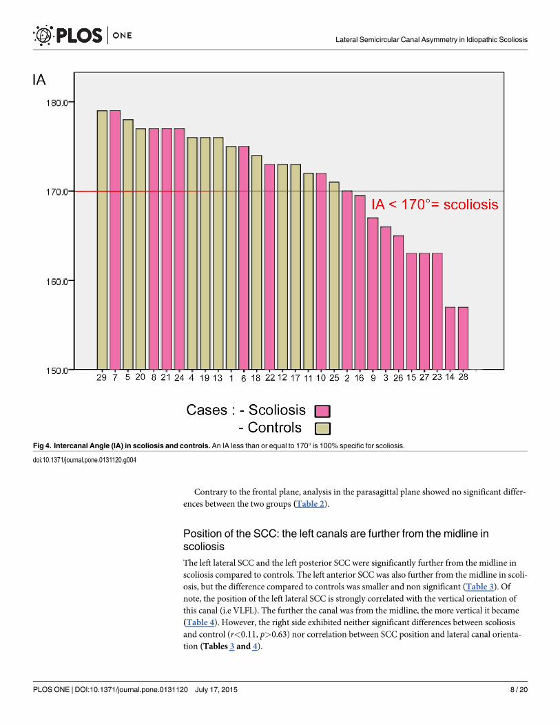
Orientation of the lateral SCC: The left lateral SCC is more vertical inscoliosisBoth lateral SCCs formed a smaller angle together in the scoliosis group as measured by the IA(p = 0.004) with a large difference compared to control (r = 0.46) (Table 2). Consequently, anIA of less than or equal to 170° was 100% specific for scoliosis, with a sensitivity of 59% (Fig 4).
The difference in orientation mostly concerned the left lateral SCC, which is closer to verti-cal as illustrated by a much smaller VLFL (p<0.001; r = 0.6).
This vertical orientation of the left lateral SCC is strongly correlated with the position of thevestibule in the scoliosis group (r = -0.6; p = 0.02). The more vertical the left SCC orientation is(i.e. smaller VLFL), the further the left vestibule is from the midline (i.e. longer LDL, ADL,PDL).
Conversely, the right lateral SCC showed no significant difference between the two groups.This asymmetry between VLFR and VLFL was mostly left sided (58%) in the scoliosis group(VLFR-VLFL>0), but right sided (75%) in the control group (VLFR-VLFL<0) (p = 0.15).
Table 1. Group characteristics.
Scoliosis Control p-value
Number 17 9
Sex Ratio (M/F) 4/13 3/6 0.60
Age +/- SD (year) 15.47 (+/- 1.84) 16.7 (+/- 1.5) 0.28
Cobb angle (°) mean +/- SD [min-max] 26.7 +/-8.3 [15–40] NA
Dorsal scoliosis 8 NA
Lumbar scoliosis 5 NA
Thoracolumbar scoliosis 4 NA
Right scoliosis 8 NA
Left scoliosis 6 NA
Right and left 3 NA
doi:10.1371/journal.pone.0131120.t001
Table 2. Left lateral SCC is more vertical in scoliosis.
Orientation of Lateral Semicircular Canal Scoliosis(1) Control (1) Significance(p) t(df) or U (2) Effect size « r »
AI 168.9 + /-7.0 175.1+ /-2.8 0.004** t(23) = -3.25 0.46
VLFR 85+ /-5.4 85.3+ /-3.9 0.86 t(22) = -0.18 0.03
VLFL 83.9+ /-3.5 88.6+ /-1.9 <0.001** t(23) = -4.4 0.60
Diff VLFR-VLFL 1.15+ /-6.0 -2.3+ /-3.3 0.15 t(23) = 1.5 0.30
AbsolDiffVLFR-VLFL 3.0 (13.6) 3.0 (11.8) 0.57 U = 58.0 0.11
VLSR 78.4+ /-6.6 77.7+ /-6.7 0.81 t(22) = -0.24 0.06
VLSL 79.0+ /-7.4 76.7+ /-5.3 0.47 t(21) = -0.73 0.16
(1) Mean (in degrees) +/- SD if T Test; or Median (in mm) (Mean Rank) if Mann Whitney U test.
(2) t value and degrees of freedom (df) if T Test; or U value if Mann Whitney U test.
**Significance < 0.01.
doi:10.1371/journal.pone.0131120.t002
Lateral Semicircular Canal Asymmetry in Idiopathic Scoliosis
PLOS ONE | DOI:10.1371/journal.pone.0131120 July 17, 2015 7 / 20
Contrary to the frontal plane, analysis in the parasagittal plane showed no significant differ-ences between the two groups (Table 2).
Position of the SCC: the left canals are further from the midline inscoliosisThe left lateral SCC and the left posterior SCC were significantly further from the midline inscoliosis compared to controls. The left anterior SCC was also further from the midline in scoli-osis, but the difference compared to controls was smaller and non significant (Table 3). Ofnote, the position of the left lateral SCC is strongly correlated with the vertical orientation ofthis canal (i.e VLFL). The further the canal was from the midline, the more vertical it became(Table 4). However, the right side exhibited neither significant differences between scoliosisand control (r<0.11, p>0.63) nor correlation between SCC position and lateral canal orienta-tion (Tables 3 and 4).
Fig 4. Intercanal Angle (IA) in scoliosis and controls. An IA less than or equal to 170° is 100% specific for scoliosis.
doi:10.1371/journal.pone.0131120.g004
Lateral Semicircular Canal Asymmetry in Idiopathic Scoliosis
PLOS ONE | DOI:10.1371/journal.pone.0131120 July 17, 2015 8 / 20

Function of the lateral SCCTwo adolescents with scoliosis exhibited a spontaneous nystagmus. The first was a permanenthorizontal left nystagmus associated with a dorsal right-lumbar left scoliosis. This patient wasexcluded from the caloric test analysis because the spontaneous and caloric nystagmus weredifficult to separate. The second patient exhibited an intermittent positional right nystagmusassociated with an abnormal right canal paresis (35%) and was included in the group analysis.In the scoliosis group, vestibular excitability was lower but the difference was small (r = 0.238)and non significant (p = 0.25) compared to controls. The directional preponderance wasabnormal (<2°/s) in half of the participants in both groups (p = 0.41).
Canal paresis was higher in the scoliosis group (mean = 18.25% +/- 16.37) compared to con-trols (mean = 10.33% +/- 5.87), but the difference was not significant according to the MannWhitney test (p = 0.52). Six of the 16 adolescents with scoliosis had abnormal (>15%) canalparesis compared to only 1 in the 9 controls (X(1) = 1.99; p = 0.158) (Table 5).
Table 3. Left lateral SCC and posterior SCC are further from themidline in scoliosis.
Distances of Semicircular Canal vertex from the Midline Scoliosis (1) Control (1) Significance(p) T(df) or U (2) Effect size « r »
LDR 42.9 + /-1.8 42.6+ /-1.3 0.63 t(20) = -0.49 0.11
LDL 43.9+ /-1.8 42.3+ /-3.9 0.01* t(20) = -2.16 0.44
Diff LDR-LDL -0.93+ /-1.5 0.29+ /-1.1 0.08 t(20) = -1.88 0.39
[Diff LDR-LDL] 1.0 (12.3) 1.0 (9.9) 0.40 U = 41.0 0.18
ADR 39.0+ /-0.5 40.0+ /-1.5 0.97 U = 52.0 0.01
ADL 40.2+ /-2.0 39.3+ /-1.1 0.19 t(20) = 1.12 0.24
Diff ADR-ADL -0.13+ /-1.4 0.42+ /-3.3 0.43 t(20) = -0.80 0.18
[Diff ADR-ADL] 1.0 (10.6) 2.0 (13.4) 0.30 U = 39.0 0.22
PDR 40.4+ /-1.7 40.2+ /-1.6 0.74 t(20) = 0.30 0.08
PDL 41.0 (13.3) 40.0 (7.7) 0.05* U = 26.0 0.41
Diff PDR-PDL -0.8+ /-1.6 0.29+ /-1.7 0.18 t(20) = -1.40 0.30
[Diff PDR-PDL] 1.0 (11.7) 2.0 (11.1) 0.82 U = 49.0 0.05
(1) Mean (in mm) +/- SD if T Test; or Median (in mm) (Mean Rank) if Mann Whitney U test.
(2) t value and degrees of freedom (df) if T Test; or U value if Mann Whitney U test.
*significant results with p-value < 0.05.
doi:10.1371/journal.pone.0131120.t003
Table 4. In scoliosis the orientation of the left lateral SCC is correlated with its position and other left SCC positions.
Parameter1 Parameter2 Coefficient of correlation p-value
VLFL LDL -0.60 0.02*
PDL -0.60 0.02*
ADL -0.50 0.04*
VLFR LDR -0.12 0.67
PDR 0.22 0.44
ADR 0.25 0.37
IA LDL -0.26 0.35
LDR -0.38 0.16
*Significant results with p-value < 0.05.
doi:10.1371/journal.pone.0131120.t004
Lateral Semicircular Canal Asymmetry in Idiopathic Scoliosis
PLOS ONE | DOI:10.1371/journal.pone.0131120 July 17, 2015 9 / 20

Analysis within the scoliosis groupAnalysis of location of the scoliosis. The IA was significantly different for the location of
the scoliosis group. In particular, thoracolumbar scoliosis (n = 4) showed a smaller IA with amean difference of 11.5° scoliosis compared to lumbar scoliosis (n = 5) (p = 0.033).
Thoracolumbar scoliosis also showed a more marked asymmetry in the location of lateralSCCs with a right canal located more laterally than the left compare with dorsal scoliosis(p<0.05) or lumbar scoliosis (p<0.03)
Two-way ANOVA combining location and side of scoliosis showed non significant maineffects in morphologic or functional vestibular parameters.
Analysis of side of the scoliosis. Right and left scoliosis were comparable for morphologicand functional vestibular parameters (p>0.05).
We found no significant association between the side of scoliosis and the side of asymme-tries for the position, orientation, or function of the lateral SCC.
Correlation of the Cobb angle with morphologic or functional vestibular parameters.No significant correlation was found between the Cobb angle and any morphological or func-tional parameters of the lateral SCC. (p>0.05).
We found no correlation between age and the Cobb angle nor with age and morphologicparameters of the lateral SCC.
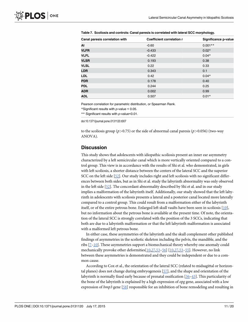
Relation between vestibular morphology and vestibular functionScoliosis and control participants with abnormal canal paresis (i.e.>15°) exhibited more verti-cal lateral SCC as shown by the smaller IA, VLFR, and VLFL (r>0.4, p<0.05) (Table 6). Canalparesis (for both groups) correlated with the IA, the VLFR, the VLFL, the LDL, and the ADL(Table 7). Nevertheless, no significant difference was demonstrated for these parameters linked
Table 5. Lateral SCC function.
Caloric Scoliosis Control Significance(p) T(df) or U (1) Effect size « r »
Vestibular excitability (°/s) 33.95+/-15.6 41.77+/-15.4 0.25 T(23) = -1.17 0.238
Directional Preponderance(°/s) 1.953+/-1.8 2.027+/-2.03 0.26 T(23) = -1.16 0.235
Directional preponderance >2°/s (n/total) 8/15 4/8 0.445
Canal paresis mean(%) 18.25 +/-4.09 11.6 +/-1.67
Canal paresis median (%) 13.2 11.2 0.52 U = 53.5 0.13
Canal paresis >15% (n/total) 6/16 1/9 0.218
(1) t value and degrees of freedom (df) if T Test; or U value if Mann Whitney U test; or χ value (degrees of freedom) if Chi square test.
doi:10.1371/journal.pone.0131120.t005
Table 6. Abnormal canal paresis is associated with more vertical lateral SCC.
Vestibular Morphology Paresis <15% (1) Paresis >15% (1) Significance (p-value) T(df) (2) Effect size « r »
IA 173.06+/-5.0 165.57+/-7.8 0.01* T(22) = 2.83 0.52
VLFR 86.35+/-4.1 82.29+/-4.7 0.046* T(22) = 2.11 0.41
VLFL 86.71+/-3.1 83.29+/-3.5 0.026* T(22) = 2.39 0.45
(1) Mean +/- SD in Scoliosis and control groups.
(2) t value (degrees of freedom) for T Test.
*Significant results with p-value < 0.05.
doi:10.1371/journal.pone.0131120.t006
Lateral Semicircular Canal Asymmetry in Idiopathic Scoliosis
PLOS ONE | DOI:10.1371/journal.pone.0131120 July 17, 2015 10 / 20
to the scoliosis group (p>0.75) or the side of abnormal canal paresis (p>0.056) (two-wayANOVA).
DiscussionThis study shows that adolescents with idiopathic scoliosis present an inner ear asymmetrycharacterized by a left semicircular canal which is more vertically oriented compared to a con-trol group. This view is in accordance with the results of Shi et al. who demonstrated, in girlswith left scoliosis, a shorter distance between the centers of the lateral SCC and the superiorSCC on the left side [52]. Our study includes right and left scoliosis with no significant differ-ences between both sides, but as in Shi et al. study the labyrinth abnormality was only observedin the left side [52]. The concordant abnormality described by Shi et al. and in our studyimplies a malformation of the labyrinth itself. Additionally, our study showed that the left laby-rinth in adolescents with scoliosis presents a lateral and a posterior canal located more laterallycompared to a control group. This could result from a malformation either of the labyrinthitself, or of the entire petrous bone. Enlarged left skull vaults have been seen in scoliosis [10],but no information about the petrous bone is available at the present time. Of note, the orienta-tion of the lateral SCC is strongly correlated with the position of the 3 SCCs, indicating thatboth are due to a labyrinth malformation or that the left labyrinth malformation is associatedwith a malformed left petrous bone.
In either case, these asymmetries of the labyrinth and the skull complement other publishedfindings of asymmetries in the scoliotic skeleton including the pelvis, the mandible, and theribs [7–10]. These asymmetries support a biomechanical theory whereby one anomaly couldmechanically provoke other deformities[10,27,53–56] [10,27,53–55]. However, no linkbetween these asymmetries is demonstrated and they could be independent or due to a com-mon cause.
According to Cox et al., the orientation of the lateral SCC (related to midsagittal or horizon-tal planes) does not change during embryogenesis [57], and the shape and orientation of thelabyrinth is normally fixed early because of prenatal ossification [36–43]. This particularity ofthe bone of the labyrinth is explained by a high expression of opg gene, associated with a lowexpression of bmp3 gene [58] responsible for an inhibition of bone remodeling and resulting in
Table 7. Scoliosis and controls: Canal paresis is correlated with lateral SCCmorphology.
Canal paresis correlation with Coefficient correlation r Significance p-value
AI -0.60 0.001**
VLFR -0.433 0.02*
VLFL -0.422 0.04*
VLSR 0.193 0.38
VLSL 0.22 0.33
LDR 0.343 0.1
LDL 0.42 0.04*
PDR 0.178 0.40
PDL 0.244 0.25
ADR 0.002 0.99
ADL 0.507 0.01*
Pearson correlation for parametric distribution, or Spearman Rank.
*Significant results with p-value < 0.05.
** Significant results with p-value<0.01.
doi:10.1371/journal.pone.0131120.t007
Lateral Semicircular Canal Asymmetry in Idiopathic Scoliosis
PLOS ONE | DOI:10.1371/journal.pone.0131120 July 17, 2015 11 / 20
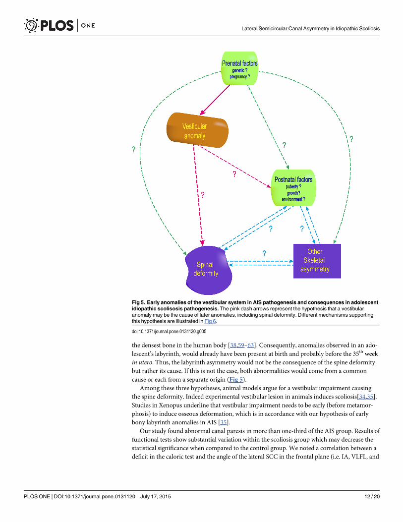
the densest bone in the human body [38,59–63]. Consequently, anomalies observed in an ado-lescent’s labyrinth, would already have been present at birth and probably before the 35th weekin utero. Thus, the labyrinth asymmetry would not be the consequence of the spine deformitybut rather its cause. If this is not the case, both abnormalities would come from a commoncause or each from a separate origin (Fig 5).
Among these three hypotheses, animal models argue for a vestibular impairment causingthe spine deformity. Indeed experimental vestibular lesion in animals induces scoliosis[34,35].Studies in Xenopus underline that vestibular impairment needs to be early (before metamor-phosis) to induce osseous deformation, which is in accordance with our hypothesis of earlybony labyrinth anomalies in AIS [35].
Our study found abnormal canal paresis in more than one-third of the AIS group. Results offunctional tests show substantial variation within the scoliosis group which may decrease thestatistical significance when compared to the control group. We noted a correlation between adeficit in the caloric test and the angle of the lateral SCC in the frontal plane (i.e. IA, VLFL, and
Fig 5. Early anomalies of the vestibular system in AIS pathogenesis and consequences in adolescentidiopathic scolisosis pathogenesis. The pink dash arrows represent the hypothesis that a vestibularanomaly may be the cause of later anomalies, including spinal deformity. Different mechanisms supportingthis hypothesis are illustrated in Fig 6.
doi:10.1371/journal.pone.0131120.g005
Lateral Semicircular Canal Asymmetry in Idiopathic Scoliosis
PLOS ONE | DOI:10.1371/journal.pone.0131120 July 17, 2015 12 / 20

VLFR). This could suggest that the verticalization of the lateral SCC either reduces the conduc-tion of the water temperature during the test, or is associated with a functional impairement.
In a previous study with caloric testing in AIS, Sahlstrand and al. found a significantly lowervestibular response located on the side of the spine concavity in AIS [33]. But they found nosignificant difference in a control group [33], and neither did Wiener et al.[14]. Additionally,Sahlstrand and al. observed occulomotor anomalies in more than half of their AIS group thatwere either spontaneous (15%) or positional (36%) nystagmus (without describing the orienta-tion). Spontaneous nystagmus can signify a SCC impairment, and in our study the two hori-zontal nystagmus more specifically indicated a lateral SCC. Positional nystagmus is interpretedby Sahlstrand et al. to originate from the brainstem [33].
In addition to the lateral SCC, the otolith function could also be impaired in AIS since theutricle is known to be roughly parallel to the lateral SCC [64]. The function of otolith sensors(i.e. utricle and saccule) have been studied using off-vertical-axis rotation in patients with scoli-osis. Sixty-seven percent exhibited a significantly greater value of directional preponderance,arguing for an impairment of the vestibulospinal function [14].
Additionally, AIS patient show worse sway (relative to controls) when vestibular inputs arethe only available [15]. Taken together these studies suggest a vestibular system involved inAIS. Yet anomalies of the vestibular organ alone are not sufficient to explain long-term posturalimpairment leading to spinal deformity.
Complete vestibular asymmetry (e.g. unilateral labyrinthectomy or vestibular neurectomy)results in postural symptoms which quickly disappear due to central compensation [65,66]. Inaddition, in normal human, the brain manages physiological asymmetry because of vestibularpredominance in the side of handedness [67,68].
AIS symptoms could thus result from vestibular organ abnormalities associated with brainimpairments which can be detected by MRI studies. White matter in the corpus callosum andleft corticothalamic tract shows changes [30]. Cortical areas, including vestibular cortices, arethinner and there are changes in the cortical network [31,69,70]. The ventral brainstem is alsoasymmetric and the cerebellum is thicker in the right lobules VIIIa, VIIIb, and the bilateral lob-ules X [32,71].Of note, the cerebellum (including lobule VIII) is connected to the vestibular sys-tem either directly from the vestibular organ or through the vestibular nuclei complex [72–78](Fig 6). Moreover, the cerebellum is particularly rich in melatonin receptor, a hormone sus-pected of acting in scoliosis pathogenesis [12,55,79]. Melatonin reciprocally interacts with thevestibular system by influencing the firing of the medial vestibular nuclei [80], the vestibulo-sympathetic system [81], and balance [82] (Fig 6).
Anomalies of the central nervous system in AIS may constitute an adaptation to compensatefor the instability induced by the spine deformity [32]. They also could result from early abnor-mal vestibular input occurring at a critical period for the central nervous system. The lack ofappropriate inputs during critical periods induce permanent brain abnormalities, as demon-strated for the visual [83], auditory [84] and tactile [85] sensory systems. Critical periods aredemonstrated for the vestibular system [86,87] and for multisensory integration as well. There-fore, brain anomalies and multisensory impairments demonstrated in AIS could result fromearly vestibular abnormalities appearing before the critical period [15,16,88,89]. This point isreinforced by animal models where scoliosis appears only if vestibular lesion is early enough[35].
Finally, in addition to early modification of the central nervous system and the vestibulosp-inal pathway, the vestibular system could also contribute to spine deformity by influencing theneuroendocrine or the vestibulosympathetic system (Fig 6). In fact, the lateral vestibular nucleiand the vestibular cortex (e.g. the insula) project out to the lateral hypothalamus which is richin leptin receptors [90–93]. Leptin is one of the main hormones suspected of being involved in
Lateral Semicircular Canal Asymmetry in Idiopathic Scoliosis
PLOS ONE | DOI:10.1371/journal.pone.0131120 July 17, 2015 13 / 20

scoliosis [27]. The lateral hypothalamus, the cerebellum, and the rostro-ventro-lateral medul-lary structur also influence the vestibulosympathetic system [94–96] which regulates bone
Fig 6. Influence of the vestibular system on key elements involved in AIS pathogenesis theories, including hormonal and neurosensory theories.
doi:10.1371/journal.pone.0131120.g006
Lateral Semicircular Canal Asymmetry in Idiopathic Scoliosis
PLOS ONE | DOI:10.1371/journal.pone.0131120 July 17, 2015 14 / 20
remodeling [97,98]. Therefore, the vestibular sympathetic system may contribute to bonedeformity and to the bone demineralization demonstrated in adolescent scoliosis [99–102].
Altogether, these results are in accordance with: 1) in the biomechanical theory of AIS, earlyasymmetry exists before the spine deformity and may therefore be its cause rather than its con-sequence; 2) in the endocrine theory, the vestibular anomalies could interact with hormonesearly before puberty; 3) in the neurosensory theory, vestibular impairment may start earlyenough to influence brain maturation and might precede the biomechanical or hormonalphenomena.
The multiple consequences of an early vestibular impairment, including some compensa-tory mechanisms, may explain why the lateral SCC parameters are not directly correlatedwith the Cobb angle. Another reason may be that scoliosis is a dynamic process and etiologicfactor(s) may correlate to the progression of the process rather than the degree of spine defor-mity in one measurement. Furthermore, the mild spine deformities of our participants (Cobb’sangle< 40°) may decrease the chance of demonstrating statistical correlation. The influence ofthese last two factors was demonstrated in sway in AIS where participants exhibit moreimpairment in case of deformities> 40° or progression> 10°/ year. [103]
Additionally, other factors (e.g. sex, body mass index, internal organ asymmetry) may alsoexplain the lack of correlation, for example between the left lateral SCC anomalies and the sideof the scoliosis [27,104–107]. The involvement of late factors appearing during childhood orpuberty is also likely to explain why the spine deformity appears only in adolescence despitethe early vestibular anomaly. Lastly, vestibular anomaly may also act as an onset factor of scoli-osis without further role in progression.[108]
Finally our results lead to another question: What is the origin of the lateral SCC anomaly?The estimate of early occurrence of the malformation focuses the answer on genetic factorsand/or environmental factors during pregnancy. This underscores that lateral SCC malforma-tion and spine deformity could also be independent from each other but induced by a commoncause. One hypothesis of common causes could be genes involved in growth accelerationoccurring during SCC embryogenesis [42] and puberty.
ConclusionWe have used the labyrinth of adolescents as a “living fossil” to explore the chronology of AISand to show that anomalies of the vestibular system start before birth. As the earliest anomalydemonstrated so far, the vestibule may be the cause of the spinal deformity. If not, both anoma-lies might still result from a common cause which appears before birth according to the timingof the vestibular malformation. In either case, findings encourage the search during pregnancyfor causative environmental factors which could lead to prenatal preventive treatment. More-over, a simple MRI measurement of the lateral SCC, as demonstrated here, could be used topredict AIS and initiate preventive treatment during childhood. However, the best treatmentswill certainly need a complete understanding of AIS chronology which requires multiple land-marks. The lateral semicircular canal constitutes a first milestone which can now open the roadto longitudinal studies.
Supporting InformationS1 Table. Data set of each participant.(XLSX)
Lateral Semicircular Canal Asymmetry in Idiopathic Scoliosis
PLOS ONE | DOI:10.1371/journal.pone.0131120 July 17, 2015 15 / 20

AcknowledgmentsWe thank Dr. Vincent Patron, Dr. Stéphane Besnard and two anonymous reviewers for theircomments on the manuscript.
Author ContributionsConceived and designed the experiments: PD GQ JFMM. Hitier M. Hamon. Performed theexperiments: JL JFMM. Hamon MAT SM. Analyzed the data: JL M. Hitier. Contributedreagents/materials/analysis tools: SM. Wrote the paper: M. Hitier PD.
References1. KaneWJ. Scoliosis prevalence: a call for a statement of terms. Clin Orthop. 1977; 126: 43–46. PMID:
598138
2. Reamy BV, Slakey JB. Adolescent idiopathic scoliosis: review and current concepts. Am Fam Physi-cian. 2001; 64: 111–116. PMID: 11456428
3. Weinstein SL, Dolan LA, Cheng JCY, Danielsson A, Morcuende JA. Adolescent idiopathic scoliosis.The Lancet. 2008; 371: 1527–1537.
4. Altaf F, Gibson A, Dannawi Z, Noordeen H. Adolescent idiopathic scoliosis. BMJ. 2013; 346: f2508.doi: 10.1136/bmj.f2508 PMID: 23633006
5. Asher MA, Burton DC. Adolescent idiopathic scoliosis: natural history and long term treatment effects.Scoliosis. 2006; 1: 2. doi: 10.1186/1748-7161-1-2 PMID: 16759428
6. Schlösser TPC, van der Heijden GJMG, Versteeg AL, Castelein RM. How “Idiopathic” Is AdolescentIdiopathic Scoliosis? A Systematic Review on Associated Abnormalities. PLoS ONE. 2014; 9:e97461. doi: 10.1371/journal.pone.0097461 PMID: 24820478
7. Burwell RG, Aujla RK, Freeman BJC, Dangerfield PH, Cole AA, Kirby AS, et al. Patterns of extra-spi-nal left-right skeletal asymmetries in adolescent girls with lower spine scoliosis: relative lengthening ofthe ilium on the curve concavity & of right lower limb segments. Stud Health Technol Inform. 2006;123: 57–65. PMID: 17108404
8. Normelli H, Sevastik J, Akrivos J. The length and ash weight of the ribs of normal and scoliotic per-sons. Spine. 1985; 10: 590–592. PMID: 4081873
9. Burwell RG, Freeman BJC, Dangerfield PH, Aujla RK, Cole AA, Kirby AS, et al. Left-right upper armlength asymmetry associated with apical vertebral rotation in subjects with thoracic scoliosis: anomalyof bilateral symmetry affecting vertebral, costal and upper arm physes? Stud Health Technol Inform.2006; 123: 66–71. PMID: 17108405
10. Shi L, Heng PA, Wong T-T, ChuWCW, Yeung BHY, Cheng JCY. Morphometric analysis for patholog-ical abnormality detection in the skull vaults of adolescent idiopathic scoliosis girls. Med Image Com-put Comput Assist Interv. 2006; 9: 175–182. PMID: 17354888
11. Qiu Y, Sun X, Qiu X, Li W, Zhu Z, Zhu F, et al. Decreased circulating leptin level and its associationwith body and bone mass in girls with adolescent idiopathic scoliosis. Spine. 2007; 32: 2703–2710.doi: 10.1097/BRS.0b013e31815a59e5 PMID: 18007248
12. Moreau A, Forget S, Azeddine B, Angeloni D, Fraschini F, Labelle H, et al. Melatonin signaling dys-function in adolescent idiopathic scoliosis. Spine. 2004; 29: 1772–1781. PMID: 15303021
13. Girardo M, Bettini N, Dema E, Cervellati S. The role of melatonin in the pathogenesis of adolescent idi-opathic scoliosis (AIS). Eur Spine J. 2011; 20 Suppl 1: S68–74. doi: 10.1007/s00586-011-1750-5PMID: 21416282
14. Wiener-Vacher SR, Mazda K. Asymmetric otolith vestibulo-ocular responses in children with idio-pathic scoliosis. J Pediatr. 1998; 132: 1028–1032. PMID: 9627598
15. Simoneau M, Lamothe V, Hutin É, Mercier P, Teasdale N, Blouin J. Evidence for cognitive vestibularintegration impairment in idiopathic scoliosis patients. BMC Neurosci. 2009; 10: 102. doi: 10.1186/1471-2202-10-102 PMID: 19706173
16. Assaiante C, Mallau S, Jouve J-L, Bollini G, Vaugoyeau M. Do Adolescent Idiopathic Scoliosis (AIS)Neglect Proprioceptive Information in Sensory Integration of Postural Control? PLoS ONE. 2012; 7:e40646. doi: 10.1371/journal.pone.0040646 PMID: 22815779
17. Simoneau M, Mercier P, Blouin J, Allard P, Teasdale N. Altered sensory-weighting mechanisms isobserved in adolescents with idiopathic scoliosis. BMC Neurosci. 2006; 7: 68. doi: 10.1186/1471-2202-7-68 PMID: 17052338
Lateral Semicircular Canal Asymmetry in Idiopathic Scoliosis
PLOS ONE | DOI:10.1371/journal.pone.0131120 July 17, 2015 16 / 20

18. Takahashi Y, Kou I, Takahashi A, Johnson TA, Kono K, Kawakami N, et al. A genome-wide associa-tion study identifies common variants near LBX1 associated with adolescent idiopathic scoliosis. NatGenet. 2011; 43: 1237–1240. doi: 10.1038/ng.974 PMID: 22019779
19. Kou I, Takahashi Y, Johnson TA, Takahashi A, Guo L, Dai J, et al. Genetic variants in GPR126 areassociated with adolescent idiopathic scoliosis. Nat Genet. 2013; 45: 676–679. doi: 10.1038/ng.2639PMID: 23666238
20. Londono D, Kou I, Johnson TA, Sharma S, Ogura Y, Tsunoda T, et al. A meta-analysis identifies ado-lescent idiopathic scoliosis association with LBX1 locus in multiple ethnic groups. J Med Genet. 2014;51: 401–406. doi: 10.1136/jmedgenet-2013-102067 PMID: 24721834
21. Chettier R, Nelson L, Ogilvie JW, Albertsen HM, Ward K. Haplotypes at LBX1 Have Distinct Inheri-tance Patterns with Opposite Effects in Adolescent Idiopathic Scoliosis. PloS One. 2015; 10:e0117708. doi: 10.1371/journal.pone.0117708 PMID: 25675428
22. Sharma S, Londono D, Eckalbar WL, Gao X, Zhang D, Mauldin K, et al. A PAX1 enhancer locus isassociated with susceptibility to idiopathic scoliosis in females. Nat Commun. 2015; 6: 6452. doi: 10.1038/ncomms7452 PMID: 25784220
23. Miyake A, Kou I, Takahashi Y, Johnson TA, Ogura Y, Dai J, et al. Identification of a SusceptibilityLocus for Severe Adolescent Idiopathic Scoliosis on Chromosome 17q24.3. PLoS ONE. 2013; 8:e72802. doi: 10.1371/journal.pone.0072802 PMID: 24023777
24. Worthington V, Shambaugh P. Nutrition as an environmental factor in the etiology of idiopathic scolio-sis. J Manipulative Physiol Ther. 1993; 16: 169. PMID: 8492060
25. De George FV, Fisher RL. Idiopathic scoliosis: genetic and environmental aspects. J Med Genet.1967; 4: 251. PMID: 6082901
26. Kouwenhoven J-WM, Castelein RM. The pathogenesis of adolescent idiopathic scoliosis: review ofthe literature. Spine. 2008; 33: 2898–2908. doi: 10.1097/BRS.0b013e3181891751 PMID: 19092622
27. Burwell RG, Aujla RK, Grevitt MP, Dangerfield PH, Moulton A, Randell TL, et al. Pathogenesis of ado-lescent idiopathic scoliosis in girls—a double neuro-osseous theory involving disharmony betweentwo nervous systems, somatic and autonomic expressed in the spine and trunk: possible dependencyon sympathetic nervous system and hormones with implications for medical therapy. Scoliosis. 2009;4: 24. doi: 10.1186/1748-7161-4-24 PMID: 19878575
28. WangWJ, Yeung HY, ChuWC-W, Tang NL-S, Lee KM, Qiu Y, et al. Top theories for the etiopatho-genesis of adolescent idiopathic scoliosis. J Pediatr Orthop. 2011; 31: S14–27. doi: 10.1097/BPO.0b013e3181f73c12 PMID: 21173615
29. Lowe TG, Edgar M, Margulies JY, Miller NH, Raso VJ, Reinker KA, et al. Etiology of Idiopathic Scolio-sis: Current Trends in Research*. J Bone Jt Surg. 2000; 82: 1157–1157.
30. Shi L, Wang D, ChuWCW, Burwell RG, Freeman BJC, Heng PA, et al. Volume-based morphometryof brain MR images in adolescent idiopathic scoliosis and healthy control subjects. AJNR Am J Neu-roradiol. 2009; 30: 1302–1307. doi: 10.3174/ajnr.A1577 PMID: 19386729
31. Wang D, Shi L, ChuWC, Burwell RG, Cheng JC, Ahuja AT. Abnormal cerebral cortical thinning pat-tern in adolescent girls with idiopathic scoliosis. Neuroimage. 2012; 59: 935–942. doi: 10.1016/j.neuroimage.2011.07.097 PMID: 21872666
32. Shi L, Wang D, Hui SCN, Tong MCF, Cheng JCY, ChuWCW. Volumetric changes in cerebellarregions in adolescent idiopathic scoliosis compared with healthy controls. Spine J. 2013; 13: 1904–1911. doi: 10.1016/j.spinee.2013.06.045 PMID: 23988458
33. Sahlstrand T, Petruson B. A Study of Labyrinthine Function in Patients with Adolescent IdiopathicScoliosis I. An Electro-Nystagmographic Study. Acta Orthop. 1979; 50: 759–769.
34. DeWaele C, Graf W, Josset P, Vidal PP. A radiological analysis of the postural syndromes followinghemilabyrinthectomy and selective canal and otolith lesions in the guinea pig. Exp Brain Res. 1989;77: 166–182. PMID: 2792260
35. Lambert FM, Malinvaud D, Glaunès J, Bergot C, Straka H, Vidal PP. Vestibular asymmetry as thecause of idiopathic scoliosis: a possible answer from Xenopus. J Neurosci. 2009; 29: 12477–12483.doi: 10.1523/JNEUROSCI.2583-09.2009 PMID: 19812323
36. Sato T. Vergleichende Untersuchungen über die Bogengänge des Labyrinthes beim neugeborenenund beim erwachsenen Menschen. Wiesbaden: Bergmann; 1902.
37. Bast TH. Ossification of the otic capsule in human fetuses. Contributions to Embryology. Washington:Carnegie Institution of Washington; 1930.
38. Bast TH, Anson BJ. The temporal bone and the ear. Springfield, IL: CC Thomas; 1949.
39. Hublin J-J, Spoor F, Braun M, Zonneveld F, Condemi S. A late Neanderthal associated with UpperPalaeolithic artefacts. Nature. 1996; 381: 224–226. PMID: 8622762
Lateral Semicircular Canal Asymmetry in Idiopathic Scoliosis
PLOS ONE | DOI:10.1371/journal.pone.0131120 July 17, 2015 17 / 20

40. Spoor F, Esteves F, Tecelão Silva F, Pacheco Dias R. The bony labyrinth of Lagar Velho 1. TheLapedo Child, a Gravettian Human Skeleton from the Abrigo Do Lagar Velho. Zilhao J, Trinkhaus E,Duarte C. Lisbon: Instituto Portugus de Arqueologia; 2002. pp. 287–292.
41. Spoor F, Hublin J-J, Kondo O. The bony labyrinth of the Dederiyeh child. Neanderthal Burials Excava-tions of the Dederiyeh Cave, Afrin, Syria. Tokyo: The Tokyo University Press; 2002. pp. 215–220.
42. Jeffery N, Spoor F. Prenatal growth and development of the modern human labyrinth. J Anat. 2004;204: 71–92. doi: 10.1111/j.1469-7580.2004.00250.x PMID: 15032915
43. DahmMC, Shepherd RK, Clark GM. The Postnatal Growth of the Temporal Bone and its Implicationsfor Cochlear Implantation in Children. Acta Otolaryngol (Stockh). 2009; 113: 4–39.
44. Curthoys IS. The interpretation of clinical tests of peripheral vestibular function. The Laryngoscope.2012; 122: 1342–1352. doi: 10.1002/lary.23258 PMID: 22460150
45. NemzekWR, Brodie HA, Chong BW, Babcook CJ, Hecht ST, Salamat S, et al. Imaging findings of thedeveloping temporal bone in fetal specimens. AJNR Am J Neuroradiol. 1996; 17: 1467–1477. PMID:8883642
46. Richards BS, Sucato DJ, Konigsberg DE, Ouellet JA. Comparison of reliability between the Lenkeand King classification systems for adolescent idiopathic scoliosis using radiographs that were notpremeasured. Spine. 2003; 28: 1148–1156; discussion 1156–1157. doi: 10.1097/01.BRS.0000067265.52473.C3 PMID: 12782983
47. British Society of Audiology. Recommended procedure. The caloric test. London: BSA; 2010.
48. Hallpike CS. The caloric tests. J Laryngol Otol. 1956; 70: 15–28. PMID: 13278645
49. Jonkees LB, Maas JP, Philopszoon AJ. Clinical nystagmography. A detailed study of electro-nystag-mography in 341 patients with vertigo. Pract Otorhinolaryngol (Basel). 1962; 24: 65–93.
50. Pietkiewicz P, Pepaś R, Sułkowski WJ, Zielińska-Bliźniewska H, Olszewski J. Electronystagmographyversus videonystagmography in diagnosis of vertigo. Int J Occup Med Environ Health. 2012; 25: 59–65. doi: 10.2478/s13382-012-0002-1 PMID: 22219058
51. Hoaglin DC, Iglewicz B. Fine-tuning some resistant rules for outlier labeling. J Am Stat Assoc. 1987;82: 1147–1149.
52. Shi L, Wang D, Chu CW, others. Automatic MRI Segmentation and morphoanatomy of the vestibularsystem in adolescent idiopathic scoliosis. Neuroimage. 2011; 54 Suppl 1: 180–188. doi:10.1016
53. Zhou S, Yan J, Da H, Yang Y, Wang N, WangW, et al. A correlational study of scoliosis and trunk bal-ance in adult patients with mandibular deviation. PLoS ONE. 2013; 8: e59929. doi: 10.1371/journal.pone.0059929 PMID: 23555836
54. Dalleau G, Leroyer P, Beaulieu M, Verkindt C, Rivard C-H, Allard P. Pelvis morphology, trunk postureand standing imbalance and their relations to the Cobb angle in moderate and severe untreated AIS.PLoS ONE. 2012; 7: e36755. doi: 10.1371/journal.pone.0036755 PMID: 22792155
55. Burwell RG, Dangerfield PH, Freeman BJC. Concepts on the pathogenesis of adolescent idiopathicscoliosis. Bone growth and mass, vertebral column, spinal cord, brain, skull, extra-spinal left-rightskeletal length asymmetries, disproportions and molecular pathogenesis. Stud Health TechnolInform. 2008; 135: 3–52. PMID: 18401079
56. Kouwenhoven J-WM, Smit TH, van der Veen AJ, Kingma I, van Dieën JH, Castelein RM. Effects ofdorsal versus ventral shear loads on the rotational stability of the thoracic spine: a biomechanical por-cine and human cadaveric study. Spine. 2007; 32: 2545–2550. doi: 10.1097/BRS.0b013e318158cd86 PMID: 17978652
57. Cox PG, Jeffery N. Morphology of the mammalian vestibulo-ocular reflex: The spatial arrangement ofthe human fetal semicircular canals and extraocular muscles. J Morphol. 2007; 268: 878–890. doi: 10.1002/jmor.10559 PMID: 17659532
58. Stankovic KM, Adachi O, Tsuji K, Kristiansen AG, Adams JC, Rosen V, et al. Differences in geneexpression between the otic capsule and other bones. Hear Res. 2010; 265: 83–89. doi: 10.1016/j.heares.2010.02.006 PMID: 20146935
59. Sørensen MS, Bretlau P, Jørgensen MB. Quantum type bone remodeling in the otic capsule of thepig. Acta Otolaryngol (Stockh). 1990; 110: 217–223.
60. Sørensen MS, Bretlau P, Jørgensen MB. Human perilabyrinthine bone dynamics. A functionalapproach to temporal bone histology. Acta Oto-Laryngol Suppl. 1992; 496: 1–27.
61. Frisch T, Sørensen MS, Overgaard S, Lind M, Bretlau P. Volume-referent bone turnover estimatedfrom the interlabel area fraction after sequential labeling. Bone. 1998; 22: 677–682. PMID: 9626408
62. Frisch T, Overgaard S, Sørensen MS, Bretlau P. Estimation of volume referent bone turnover in theotic capsule after sequential point labeling. Ann Otol Rhinol Laryngol. 2000; 109: 33–39. PMID:10651409
Lateral Semicircular Canal Asymmetry in Idiopathic Scoliosis
PLOS ONE | DOI:10.1371/journal.pone.0131120 July 17, 2015 18 / 20

63. Frisch T Bloch SL Sørensen MS. Prevalence, size and distribution of microdamage in the human oticcapsule. Acta Otolaryngol (Stockh). 2015; 1–5. doi: 10.3109/00016489.2015.1035400
64. Naganuma H, Tokumasu K, Okamoto M, Hashimoto S, Yamashina S. Three-dimensional analysis ofmorphological aspects of the human utricular macula. Ann Otol Rhinol Laryngol. 2003; 112: 419.PMID: 12784980
65. Curthoys IS. Vestibular compensation and substitution. Curr Opin Neurol. 2000; 13: 27–30. PMID:10719646
66. Smith PF, Curthoys IS. Mechanisms of recovery following unilateral labyrinthectomy: a review. BrainRes Rev. 1989; 14: 155–180. PMID: 2665890
67. Dieterich M, Bense S, Lutz S, Drzezga A, Stephan T, Bartenstein P, et al. Dominance for vestibularcortical function in the non-dominant hemisphere. Cereb Cortex. 2003; 13: 994–1007. PMID:12902399
68. Best C, Lange E, Buchholz H-G, Schreckenberger M, Reuss S, Dieterich M. Left hemispheric domi-nance of vestibular processing indicates lateralization of cortical functions in rats. Brain Struct Funct.2013; 219: 2141–2158. doi: 10.1007/s00429-013-0628-1 PMID: 23979449
69. Liu T, ChuWCW, Young G, Li K, Yeung BHY, Guo L, et al. MR analysis of regional brain volume inadolescent idiopathic scoliosis: neurological manifestation of a systemic disease. J Magn ResonImaging JMRI. 2008; 27: 732–736. doi: 10.1002/jmri.21321
70. Wang D, Shi L, Liu S, Hui SCN, Wang Y, Cheng JCY, et al. Altered Topological Organization of Corti-cal Network in Adolescent Girls with Idiopathic Scoliosis. PLoS ONE. 2013; 8: e83767. doi: 10.1371/journal.pone.0083767 PMID: 24376742
71. Geissele AE, Kransdorf MJ, Geyer CA, Jelinek JS, Van Dam BE. Magnetic resonance imaging of thebrain stem in adolescent idiopathic scoliosis. Spine. 1991; 16: 761–763. PMID: 1925751
72. Korte GE, Mugnaini E. The cerebellar projection of the vestibular nerve in the cat. J Comp Neurol.1979; 184: 265–277. doi: 10.1002/cne.901840204 PMID: 762284
73. Angelaki DE, Yakusheva TA, Green AM, Dickman JD, Blazquez PM. Computation of egomotion inthe macaque cerebellar vermis. The Cerebellum. 2010; 9: 174–182. doi: 10.1007/s12311-009-0147-zPMID: 20012388
74. Brodal A, Brodal P. Observations on the secondary vestibulocerebellar projections in the macaquemonkey. Exp Brain Res. 1985; 58: 62–74. PMID: 3987852
75. Blanks RHI, Precht W, Torigoe Y. Afferent projections to the cerebellar flocculus in the pigmented ratdemonstrated by retrograde transport of horseradish peroxidase. Exp Brain Res. 1983; 52: 293–306.PMID: 6641889
76. Carleton SC, Carpenter MB. Distribution of primary vestibular fibers in the brainstem and cerebellumof the monkey. Brain Res. 1984; 294: 281–298. PMID: 6200186
77. Walberg F, Dietrichs E. The interconnection between the vestibular nuclei and the nodulus: a study ofreciprocity. Brain Res. 1988; 449: 47–53. PMID: 2456133
78. Kotchabhakdi N, Walberg F. Cerebellar afferent projections from the vestibular nuclei in the cat: anexperimental study with the method of retrograde axonal transport of horseradish peroxidase. ExpBrain Res. 1978; 31: 591–604. PMID: 350598
79. Mazzucchelli C, Pannacci M, Nonno R, Lucini V, Fraschini F, Michaylov Stankov B. The melatoninreceptor in the human brain: cloning experiments and distribution studies. Mol Brain Res. 1996; 39:117–126. PMID: 8804720
80. Podda MV, Deriu F, Giaconi E, Milia M, Tolu E. Melatonin inhibits rat medial vestibular nucleus neuronactivity in vitro. Neurosci Lett. 2003; 341: 209–212. PMID: 12697285
81. Cook JS, Ray CA. Melatonin attenuates the vestibulosympathetic but not vestibulocollic reflexes inhumans: selective impairment of the utricles. J Appl Physiol. 2010; 109: 1697–1701. doi: 10.1152/japplphysiol.00698.2010 PMID: 20829497
82. Fraschini F, Cesarani A, Alpini D, Esposti D, Stankov BM. Melatonin Influences Human Balance. Neu-rosignals. 1999; 8: 111–119. doi: 10.1159/000014578
83. Hensch TK. Critical period mechanisms in developing visual cortex. Curr Top Dev Biol. 2005; 69:215–237. PMID: 16243601
84. Keuroghlian AS, Knudsen EI. Adaptive auditory plasticity in developing and adult animals. Prog Neu-robiol. 2007; 82: 109–121. PMID: 17493738
85. Simons DJ, Land PW. Early experience of tactile stimulation influences organization of somatic sen-sory cortex. Nature. 1987; 326: 694–697. doi: 10.1038/326694a0 PMID: 3561512
Lateral Semicircular Canal Asymmetry in Idiopathic Scoliosis
PLOS ONE | DOI:10.1371/journal.pone.0131120 July 17, 2015 19 / 20

86. Eugène D, Deforges S, Vibert N, Vidal P-P. Vestibular critical period, maturation of central vestibularneurons, and locomotor control. Ann N Y Acad Sci. 2009; 1164: 180–187. doi: 10.1111/j.1749-6632.2008.03727.x PMID: 19645897
87. Moorman SJ, Cordova R, Davies SA. A critical period for functional vestibular development in zebra-fish. Dev Dyn. 2002; 223: 285–291. PMID: 11836792
88. Simoneau M, Mercier P, Blouin J, Allard P, Teasdale N. Altered sensory-weighting mechanisms isobserved in adolescents with idiopathic scoliosis. BMC Neurosci. 2006; 7: 68. PMID: 17052338
89. Wallace MT, Stein BE. Early experience determines how the senses will interact. J Neurophysiol.2007; 97: 921–926. PMID: 16914616
90. Risold PY, Thompson RH, Swanson LW. The structural organization of connections between hypo-thalamus and cerebral cortex. Brain Res Rev. 1997; 24: 197–254. doi: 10.1016/S0165-0173(97)00007-6 PMID: 9385455
91. Katafuchi T, Puthuraya KP, Yoshimatsu H, Oomura Y. Responses of rat lateral hypothalamic neuronactivity to vestibular nuclei stimulation. Brain Res. 1987; 400: 62–69. PMID: 3815070
92. Mercer JG, Hoggard N, Williams LM, Lawrence CB, Hannah LT, Trayhurn P. Localization of leptinreceptor mRNA and the long form splice variant (Ob-Rb) in mouse hypothalamus and adjacent brainregions by in situ hybridization. FEBS Lett. 1996; 387: 113–116. PMID: 8674530
93. Bouret SG, Draper SJ, Simerly RB. Trophic action of leptin on hypothalamic neurons that regulatefeeding. Sci Signal. 2004; 304: 108.
94. Yates BJ. Vestibular influences on the sympathetic nervous system. Brain Res Rev. 1992; 17: 51–59.PMID: 1638275
95. Yates BJ. Vestibular influences on the autonomic nervous system. Ann N Y Acad Sci. 1996; 781:458–473. PMID: 8694435
96. Yates BJ, Bronstein AM. The effects of vestibular system lesions on autonomic regulation: observa-tions, mechanisms, and clinical implications. J Vestib Res. 2005; 15: 119–129. PMID: 16179761
97. Vignaux G Besnard S Ndong J Philoxène B Denise P Elefteriou F. Bone remodeling is regulated byinner ear vestibular signals. J Bone Miner Res Off J Am Soc Bone Miner Res. 2013; doi: 10.1002/jbmr.1940
98. Denise P, Besnard S, Vignaux G, Sabatier JP, Edy E, Hitier M, et al. Sympathetic B antagonist pre-vents bone mineral density decrease induced by labyrinthectomy. Aviakosmicheskaia Ekol MeditsinaAerosp Environ Med. 2009; 43: 36–38.
99. Cheng JC, Guo X, Sher AH. Persistent Osteopenia in Adolescent Idiopathic Scoliosis: A LongitudinalFollow-Up Study. Spine. 1999; 24: 1218–1222. PMID: 10382248
100. Cheng JC, Guo X. Osteopenia in adolescent idiopathic scoliosis: a primary problem or secondary tothe spinal deformity? Spine. 1997; 22: 1716–1721. PMID: 9259781
101. Cheng JCY, Qin L, Cheung CSK, Sher AHL, Lee KM, Ng SWE, et al. Generalized low areal and volu-metric bone mineral density in adolescent idiopathic scoliosis. J Bone Miner Res. 2000; 15: 1587–1595. PMID: 10934658
102. Cheng JC, Tang SP, Guo X, Chan CW, Qin L. Osteopenia in adolescent idiopathic scoliosis: a histo-morphometric study. Spine. 2001; 26: C1–C5.
103. Byl NN, Gray JM. Complex balance reactions in different sensory conditions: adolescents with andwithout idiopathic scoliosis. J Orthop Res. 1993; 11: 215–227. PMID: 8483034
104. Kouwenhoven J-WM, Vincken KL, Bartels LW, Castelein RM. Analysis of preexistent vertebral rota-tion in the normal spine. Spine. 2006; 31: 1467–1472. PMID: 16741456
105. Burwell RG, Freeman BJC, Dangerfield PH, Aujla RK, Cole AA, Kirby AS, et al. Etiologic theories ofidiopathic scoliosis: neurodevelopmental concept of maturational delay of the CNS body schema(“body-in-the-brain”). Stud Health Technol Inform. 2006; 123: 72–79. PMID: 17108406
106. Jansen M. Physiological Scoliosis. Br Med J. 1912; 2: 1372–1373.
107. Janssen MMA, Kouwenhoven J-WM, Schlösser TPC, Viergever MA, Bartels LW, Castelein RM, et al.Analysis of preexistent vertebral rotation in the normal infantile, juvenile, and adolescent spine. Spine.2011; 36: E486–491. doi: 10.1097/BRS.0b013e3181f468cc PMID: 21240053
108. Goldberg CJ, Dowling FE, Fogarty EE. Adolescent idiopathic scoliosis: is rising growth rate the trig-gering factor in progression? Eur Spine J. 1993; 2: 29–36. PMID: 20058445
Lateral Semicircular Canal Asymmetry in Idiopathic Scoliosis
PLOS ONE | DOI:10.1371/journal.pone.0131120 July 17, 2015 20 / 20