RESEARCHARTICLE AbnormalProteinGlycosylationand ... · RESEARCHARTICLE...
19
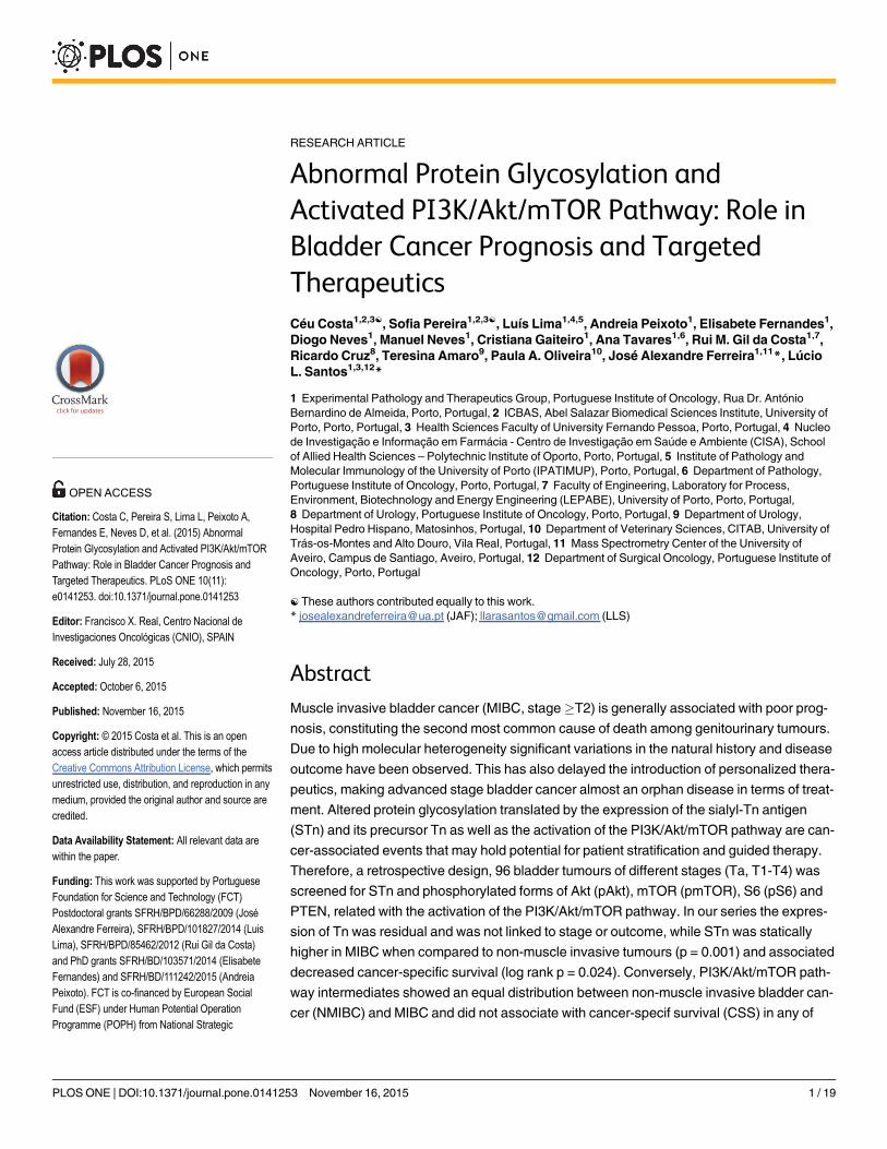
RESEARCH ARTICLE Abnormal Protein Glycosylation and Activated PI3K/Akt/mTOR Pathway: Role in Bladder Cancer Prognosis and Targeted Therapeutics Céu Costa 1,2,3☯ , Sofia Pereira 1,2,3☯ , Luís Lima 1,4,5 , Andreia Peixoto 1 , Elisabete Fernandes 1 , Diogo Neves 1 , Manuel Neves 1 , Cristiana Gaiteiro 1 , Ana Tavares 1,6 , Rui M. Gil da Costa 1,7 , Ricardo Cruz 8 , Teresina Amaro 9 , Paula A. Oliveira 10 , José Alexandre Ferreira 1,11 *, Lúcio L. Santos 1,3,12 * 1 Experimental Pathology and Therapeutics Group, Portuguese Institute of Oncology, Rua Dr. António Bernardino de Almeida, Porto, Portugal, 2 ICBAS, Abel Salazar Biomedical Sciences Institute, University of Porto, Porto, Portugal, 3 Health Sciences Faculty of University Fernando Pessoa, Porto, Portugal, 4 Nucleo de Investigação e Informação em Farmácia - Centro de Investigação em Saúde e Ambiente (CISA), School of Allied Health Sciences – Polytechnic Institute of Oporto, Porto, Portugal, 5 Institute of Pathology and Molecular Immunology of the University of Porto (IPATIMUP), Porto, Portugal, 6 Department of Pathology, Portuguese Institute of Oncology, Porto, Portugal, 7 Faculty of Engineering, Laboratory for Process, Environment, Biotechnology and Energy Engineering (LEPABE), University of Porto, Porto, Portugal, 8 Department of Urology, Portuguese Institute of Oncology, Porto, Portugal, 9 Department of Urology, Hospital Pedro Hispano, Matosinhos, Portugal, 10 Department of Veterinary Sciences, CITAB, University of Trás-os-Montes and Alto Douro, Vila Real, Portugal, 11 Mass Spectrometry Center of the University of Aveiro, Campus de Santiago, Aveiro, Portugal, 12 Department of Surgical Oncology, Portuguese Institute of Oncology, Porto, Portugal ☯ These authors contributed equally to this work. * [email protected] (JAF); [email protected] (LLS) Abstract Muscle invasive bladder cancer (MIBC, stage T2) is generally associated with poor prog- nosis, constituting the second most common cause of death among genitourinary tumours. Due to high molecular heterogeneity significant variations in the natural history and disease outcome have been observed. This has also delayed the introduction of personalized thera- peutics, making advanced stage bladder cancer almost an orphan disease in terms of treat- ment. Altered protein glycosylation translated by the expression of the sialyl-Tn antigen (STn) and its precursor Tn as well as the activation of the PI3K/Akt/mTOR pathway are can- cer-associated events that may hold potential for patient stratification and guided therapy. Therefore, a retrospective design, 96 bladder tumours of different stages (Ta, T1-T4) was screened for STn and phosphorylated forms of Akt (pAkt), mTOR (pmTOR), S6 (pS6) and PTEN, related with the activation of the PI3K/Akt/mTOR pathway. In our series the expres- sion of Tn was residual and was not linked to stage or outcome, while STn was statically higher in MIBC when compared to non-muscle invasive tumours (p = 0.001) and associated decreased cancer-specific survival (log rank p = 0.024). Conversely, PI3K/Akt/mTOR path- way intermediates showed an equal distribution between non-muscle invasive bladder can- cer (NMIBC) and MIBC and did not associate with cancer-specif survival (CSS) in any of PLOS ONE | DOI:10.1371/journal.pone.0141253 November 16, 2015 1 / 19 a11111 OPEN ACCESS Citation: Costa C, Pereira S, Lima L, Peixoto A, Fernandes E, Neves D, et al. (2015) Abnormal Protein Glycosylation and Activated PI3K/Akt/mTOR Pathway: Role in Bladder Cancer Prognosis and Targeted Therapeutics. PLoS ONE 10(11): e0141253. doi:10.1371/journal.pone.0141253 Editor: Francisco X. Real, Centro Nacional de Investigaciones Oncológicas (CNIO), SPAIN Received: July 28, 2015 Accepted: October 6, 2015 Published: November 16, 2015 Copyright: © 2015 Costa et al. This is an open access article distributed under the terms of the Creative Commons Attribution License, which permits unrestricted use, distribution, and reproduction in any medium, provided the original author and source are credited. Data Availability Statement: All relevant data are within the paper. Funding: This work was supported by Portuguese Foundation for Science and Technology (FCT) Postdoctoral grants SFRH/BPD/66288/2009 (José Alexandre Ferreira), SFRH/BPD/101827/2014 (Luis Lima), SFRH/BPD/85462/2012 (Rui Gil da Costa) and PhD grants SFRH/BD/103571/2014 (Elisabete Fernandes) and SFRH/BD/111242/2015 (Andreia Peixoto). FCT is co-financed by European Social Fund (ESF) under Human Potential Operation Programme (POPH) from National Strategic
Transcript of RESEARCHARTICLE AbnormalProteinGlycosylationand ... · RESEARCHARTICLE...
RESEARCH ARTICLE
Abnormal Protein Glycosylation andActivated PI3K/Akt/mTOR Pathway: Role inBladder Cancer Prognosis and TargetedTherapeuticsCéu Costa1,2,3☯, Sofia Pereira1,2,3☯, Luís Lima1,4,5, Andreia Peixoto1, Elisabete Fernandes1,Diogo Neves1, Manuel Neves1, Cristiana Gaiteiro1, Ana Tavares1,6, Rui M. Gil da Costa1,7,Ricardo Cruz8, Teresina Amaro9, Paula A. Oliveira10, José Alexandre Ferreira1,11*, LúcioL. Santos1,3,12*
1 Experimental Pathology and Therapeutics Group, Portuguese Institute of Oncology, Rua Dr. AntónioBernardino de Almeida, Porto, Portugal, 2 ICBAS, Abel Salazar Biomedical Sciences Institute, University ofPorto, Porto, Portugal, 3 Health Sciences Faculty of University Fernando Pessoa, Porto, Portugal, 4 Nucleode Investigação e Informação em Farmácia - Centro de Investigação em Saúde e Ambiente (CISA), Schoolof Allied Health Sciences – Polytechnic Institute of Oporto, Porto, Portugal, 5 Institute of Pathology andMolecular Immunology of the University of Porto (IPATIMUP), Porto, Portugal, 6 Department of Pathology,Portuguese Institute of Oncology, Porto, Portugal, 7 Faculty of Engineering, Laboratory for Process,Environment, Biotechnology and Energy Engineering (LEPABE), University of Porto, Porto, Portugal,8 Department of Urology, Portuguese Institute of Oncology, Porto, Portugal, 9 Department of Urology,Hospital Pedro Hispano, Matosinhos, Portugal, 10 Department of Veterinary Sciences, CITAB, University ofTrás-os-Montes and Alto Douro, Vila Real, Portugal, 11 Mass Spectrometry Center of the University ofAveiro, Campus de Santiago, Aveiro, Portugal, 12 Department of Surgical Oncology, Portuguese Institute ofOncology, Porto, Portugal
☯ These authors contributed equally to this work.* [email protected] (JAF); [email protected] (LLS)
AbstractMuscle invasive bladder cancer (MIBC, stage�T2) is generally associated with poor prog-
nosis, constituting the second most common cause of death among genitourinary tumours.
Due to high molecular heterogeneity significant variations in the natural history and disease
outcome have been observed. This has also delayed the introduction of personalized thera-
peutics, making advanced stage bladder cancer almost an orphan disease in terms of treat-
ment. Altered protein glycosylation translated by the expression of the sialyl-Tn antigen
(STn) and its precursor Tn as well as the activation of the PI3K/Akt/mTOR pathway are can-
cer-associated events that may hold potential for patient stratification and guided therapy.
Therefore, a retrospective design, 96 bladder tumours of different stages (Ta, T1-T4) was
screened for STn and phosphorylated forms of Akt (pAkt), mTOR (pmTOR), S6 (pS6) and
PTEN, related with the activation of the PI3K/Akt/mTOR pathway. In our series the expres-
sion of Tn was residual and was not linked to stage or outcome, while STn was statically
higher in MIBC when compared to non-muscle invasive tumours (p = 0.001) and associated
decreased cancer-specific survival (log rank p = 0.024). Conversely, PI3K/Akt/mTOR path-
way intermediates showed an equal distribution between non-muscle invasive bladder can-
cer (NMIBC) and MIBC and did not associate with cancer-specif survival (CSS) in any of
PLOS ONE | DOI:10.1371/journal.pone.0141253 November 16, 2015 1 / 19
a11111
OPEN ACCESS
Citation: Costa C, Pereira S, Lima L, Peixoto A,Fernandes E, Neves D, et al. (2015) AbnormalProtein Glycosylation and Activated PI3K/Akt/mTORPathway: Role in Bladder Cancer Prognosis andTargeted Therapeutics. PLoS ONE 10(11):e0141253. doi:10.1371/journal.pone.0141253
Editor: Francisco X. Real, Centro Nacional deInvestigaciones Oncológicas (CNIO), SPAIN
Received: July 28, 2015
Accepted: October 6, 2015
Published: November 16, 2015
Copyright: © 2015 Costa et al. This is an openaccess article distributed under the terms of theCreative Commons Attribution License, which permitsunrestricted use, distribution, and reproduction in anymedium, provided the original author and source arecredited.
Data Availability Statement: All relevant data arewithin the paper.
Funding: This work was supported by PortugueseFoundation for Science and Technology (FCT)Postdoctoral grants SFRH/BPD/66288/2009 (JoséAlexandre Ferreira), SFRH/BPD/101827/2014 (LuisLima), SFRH/BPD/85462/2012 (Rui Gil da Costa)and PhD grants SFRH/BD/103571/2014 (ElisabeteFernandes) and SFRH/BD/111242/2015 (AndreiaPeixoto). FCT is co-financed by European SocialFund (ESF) under Human Potential OperationProgramme (POPH) from National Strategic
these groups. However, the overexpression of pAKT, pmTOR and/or pS6 allowed discrimi-
nating STn-positive advanced stage bladder tumours facing worst CSS (p = 0.027). Further-
more, multivariate Cox regression analysis revealed that overexpression of PI3K/Akt/
mTOR pathway proteins in STn+ MIBC was independently associated with approximately
6-fold risk of death by cancer (p = 0.039). Mice bearing advanced stage chemically-induced
bladder tumours mimicking the histological and molecular nature of human tumours were
then administrated with mTOR-pathway inhibitor sirolimus (rapamycin). This decreased the
number of invasive lesions and, concomitantly, the expression of STn and also pS6, the
downstream effector of the PI3K/Akt/mTOR pathway. In conclusion, STn was found to be
marker of poor prognosis in bladder cancer and, in combination with PI3K/Akt/mTOR path-
way evaluation, holds potential to improve the stratification of stage disease. Animal experi-
ments suggest that mTOR pathway inhibition could be a potential therapeutic approach for
this specific subtype of MIBC.
IntroductionBladder cancer is the second most deadly genitourinary tumour and presents significantlyworse prognosis uponmuscularis propria invasion [1]. Approximately 20–30% of the newlydiagnosed cases are muscle invasive bladder cancers (MIBC; T2-T4 stages), while 50% are non-muscle invasive bladder tumours (NMIBC) with high potential to progress to invasion. MIBCtreatment includes cystectomy and (neo)adjuvant cisplatin-based chemotherapy regimens [2].However, significant variations in the natural history of the disease and responses to treatmentcan be observed between tumours with identical histological features, reflecting their highmolecular heterogeneity [3]. Furthermore, approximately 50% of cases develop metastasiswithin 5 years, urging the identification of biomarkers to assist prognostication and the devel-opment of more effective targeted therapeutics [4].
To meet this need, we have recently addressed the expression of the cancer-associated sia-lyl-Tn antigen (STn) on a small prospective series of unselected bladder cancer patients [5].STn is an abnormal post-translational modification that results from a premature stop in cell-membrane proteins O-glycosylation by sialylation of the Tn antigen (Fig 1A). In bladdertumours, STn it was mainly present in advanced stage cases, while absent from most low-gradeNMIBC [5]. Moreover, it was not expressed by the normal urothelium, denoting a cancer-spe-cific nature [5]. Studies in vitro showed that STn expression endowed bladder cancer cells withhigh invasion capability [5] and an immunotolerogenic phenotype, potentially favoring diseasedissemination [6]. Alterations in cell-surface protein glycosylation have be implicated in theactivation of intracellular oncogenic signalling pathways [7], including the phosphoinositide-3kinase (PI3K)/Akt signalling pathway [8] which is thought to play a critical role in bladder can-cer development. These preliminary observations support the hypothesis that STn expressionmay play a key role in disease outcome, which warrants a deeper investigation. Several studiesalso suggest that Tn antigen, which is a precursor of STn, may be also implicated in oncogenicevents [7]; however nothing is known about the expression of this glycan in bladder tumours.
The phosphatidylinositol-3-kinase (PI3K)/Akt and the mammalian target of rapamycin(mTOR) pathways are interconnected signaling cascades essential for bladder cell growth andsurvival (Fig 1B). The PI3K/Akt/mTOR or mTOR pathway integrates a multiplicity of extracel-lular signals to regulate downstream signaling and protein synthesis, which ultimately leads to
Glycosylation and PI3K/Akt/mTOR Pathway in Bladder Cancer
PLOS ONE | DOI:10.1371/journal.pone.0141253 November 16, 2015 2 / 19
Reference Framework (NSRF). The authors alsoacknowledge financial support from ICBAS-UP (CéuCosta and Sofia Pereira) and the PortugueseAssociation of Urology/Pfizer prize 2013. The fundershad no role in study design, data collection andanalysis, decision to publish, or preparation of themanuscript.
Competing Interests: The authors have declaredthat no competing interests exist.
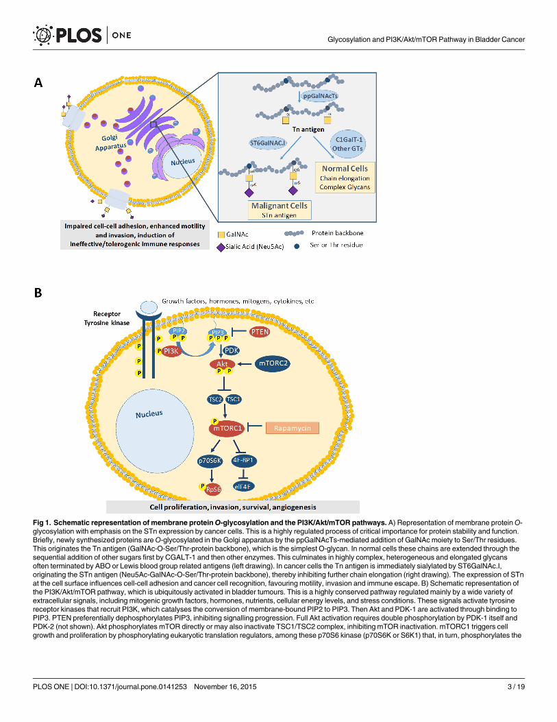
Fig 1. Schematic representation of membrane proteinO-glycosylation and the PI3K/Akt/mTOR pathways. A) Representation of membrane proteinO-glycosylation with emphasis on the STn expression by cancer cells. This is a highly regulated process of critical importance for protein stability and function.Briefly, newly synthesized proteins areO-glycosylated in the Golgi apparatus by the ppGalNAcTs-mediated addition of GalNAc moiety to Ser/Thr residues.This originates the Tn antigen (GalNAc-O-Ser/Thr-protein backbone), which is the simplest O-glycan. In normal cells these chains are extended through thesequential addition of other sugars first by CGALT-1 and then other enzymes. This culminates in highly complex, heterogeneous and elongated glycansoften terminated by ABO or Lewis blood group related antigens (left drawing). In cancer cells the Tn antigen is immediately sialylated by ST6GalNAc.I,originating the STn antigen (Neu5Ac-GalNAc-O-Ser/Thr-protein backbone), thereby inhibiting further chain elongation (right drawing). The expression of STnat the cell surface influences cell-cell adhesion and cancer cell recognition, favouring motility, invasion and immune escape. B) Schematic representation ofthe PI3K/Akt/mTOR pathway, which is ubiquitously activated in bladder tumours. This is a highly conserved pathway regulated mainly by a wide variety ofextracellular signals, including mitogenic growth factors, hormones, nutrients, cellular energy levels, and stress conditions. These signals activate tyrosinereceptor kinases that recruit PI3K, which catalyses the conversion of membrane-bound PIP2 to PIP3. Then Akt and PDK-1 are activated through binding toPIP3. PTEN preferentially dephosphorylates PIP3, inhibiting signalling progression. Full Akt activation requires double phosphorylation by PDK-1 itself andPDK-2 (not shown). Akt phosphorylates mTOR directly or may also inactivate TSC1/TSC2 complex, inhibiting mTOR inactivation. mTORC1 triggers cellgrowth and proliferation by phosphorylating eukaryotic translation regulators, among these p70S6 kinase (p70S6K or S6K1) that, in turn, phosphorylates the
Glycosylation and PI3K/Akt/mTOR Pathway in Bladder Cancer
PLOS ONE | DOI:10.1371/journal.pone.0141253 November 16, 2015 3 / 19
a competitive growth advantage, metastatic competence, angiogenesis, and therapy resistance[9]. The signaling cascade begins with PI3K activation in the cell membrane followed by ser-ine/threonine kinase Akt cell membrane translocation and activation. The best studied down-stream substrate of Akt is the serine/threonine kinase mTOR, whose downstream effector is S6kinase-1 (S6K1). In particular, a subset of mTOR pathway alterations have been shown tooccur in bladder cancer, such as mutations in PIK3CA gene, which culminates with increasedmTOR signaling and bladder cancer cells resistance to apoptosis [10]. Moreover, the pharma-cological or biochemical inhibition of the PI3K pathway drastically reduced the invasive capac-ity of bladder cancer cell lines. Furthermore, over half of primary human bladder tumourspresent high Akt phosphorylation and the aberrant activation of this pathway has been sug-gested to contribute to invasion [11]. Another event influencing mTOR pathway activation inbladder tumours involves the loss of tumor suppressor PTEN (phosphatase and tensin homo-log deleted on chromosome ten) function [12]. PTEN normally suppresses activation of thePI3K/Akt/mTOR pathway antagonizing PI3K and preventing activation of Akt and PDK-1.PTEN also functions to regulate chemotaxis and cell motility, thereby promoting tumor inva-sion [13]. In summary, there are evidences that a comprehensive evaluation of PI3K/Akt/mTOR pathway associated proteins may hold significant potential for value for patient stratifi-cation. Moreover, many preclinical and clinical studies support that mTOR inhibitors, such assirolimus (rapamycin) and their derivatives may improve cancer treatment [13,14].
Based on these observations we hypothesize that Tn and/or STn may act synergistically withthe mTOR pathway to drive bladder cancer progression. As such, we have devoted to evaluat-ing the expression of STn and proteins associated with the activation of the PI3K/Akt/mTORpathway activation in bladder tumours at different stages. We anticipate that the combinationof extracellular and intracellular oncogenic events may improve patient stratification and pro-vide insights for novel therapeutics. Furthermore we have estimated the impact of sirolimus inchemically-induced urothelial tumours in mice, envisaging the creation of a rationale for moreeffective bladder cancer therapeutics.
Materials and Methods
Ethics StatementThis work involves experiences in tumour samples of patients diagnosed with bladder cancerin the Portuguese Insitute of Oncology of Porto. All procedures were performed after patient’swritten informed consent and approved by the Ethics Committee of Portuguese Institute ofOncology—Porto. All clinicopathological information was obtained from patients’ clinicalrecords.
It also involves animal experiments. All procedures involving animals were performed inaccordance with the European Directive 2010/63/EU. During the course of this study, the ani-mals were fed ad libitum with standardized food (Tecklad Global Diet, Harlan, Spain). The fol-lowing protocol was approved by the Portuguese Ethics Committee for AnimalExperimentation (Direção Geral de Veterinária, Approval no. 520/000/000/2003). All miceused in the experiment were acclimatized for one week under routine laboratory conditionsbefore starting the experiments. They were housed randomly in groups of 4–5 in plastic cages,with hard wood chips for bedding. The animals were maintained in a room with a controlled
ribosomal protein S6 (pS6), and the eukaryotic translation initiation factor 4E–binding protein 1 (4E-BP1). For the protein mTOR to activate its signallingcascade, it must form the rapamycin-sensitive ternary complex mTORC1. Key PI3K/Akt/mTOR-pathway proteins pAkt, pmTOR and pS6 explored in thisstudied are highlighted by orange circles.
doi:10.1371/journal.pone.0141253.g001
Glycosylation and PI3K/Akt/mTOR Pathway in Bladder Cancer
PLOS ONE | DOI:10.1371/journal.pone.0141253 November 16, 2015 4 / 19
temperature of 23±2°C, a 12-hour light/dark cycle and 55±5% humidity. The animals' drinkingsolutions were changed once a week or earlier if necessary, and the volume drunk was recorded.Weekly food intake was also noted. All mice were monitored throughout the experiment forsigns of distress and loss of body weight. The animals were sacrificed with 0.4% sodium pento-barbital (1 ml/Kg, intraperitoneal).
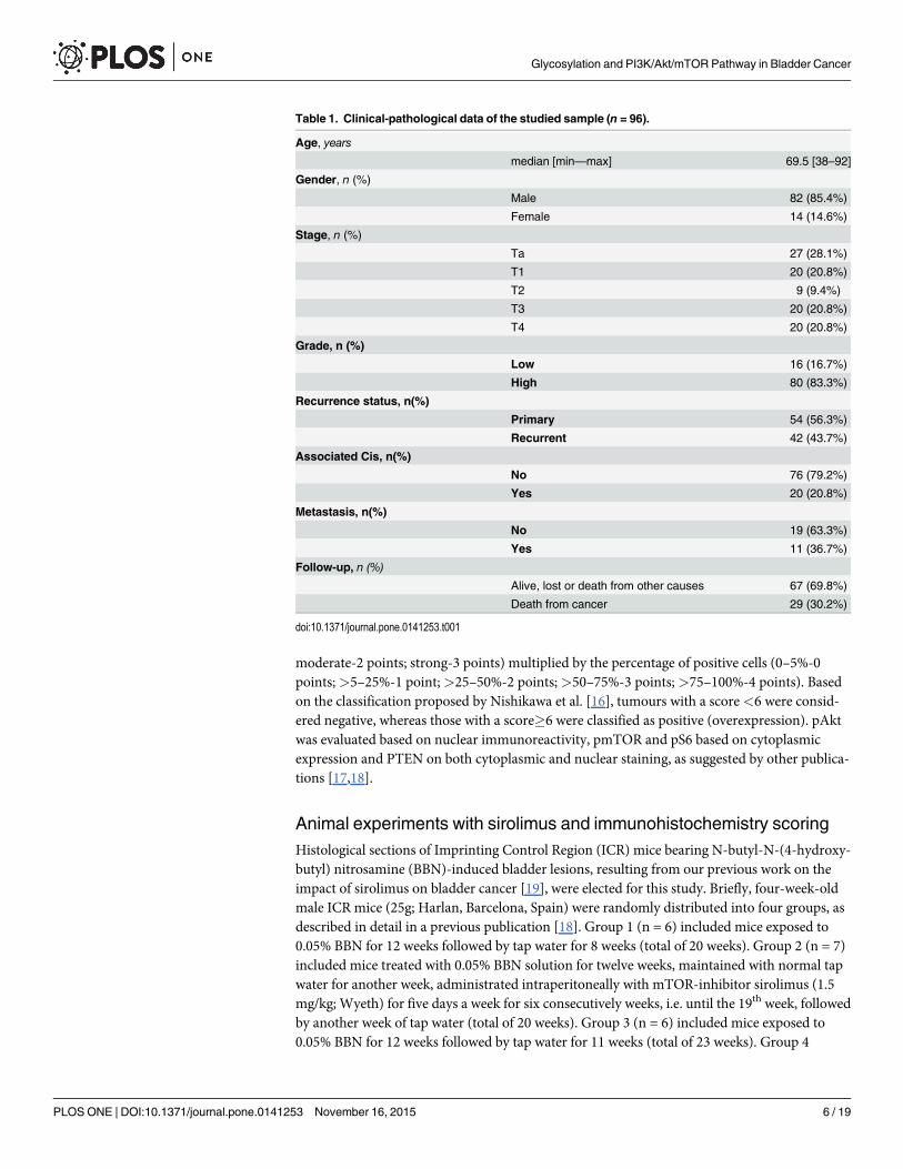
PopulationThis study was performed in a retrospective series of 96 formalin-fixed paraffin-embeddedbladder tumours obtained from archived paraffin blocks at the Portuguese Institute of Oncol-ogy—Porto (IPOP), Portugal. Bladder tumours were extracted from 82 men and 14 women,ranging in age from 38 to 92 years (median of 69.5 years), admitted and treated at the IPOPbetween 2005 and 2007. Forty seven of the examined tumours were histologically classified asNMBIC (Ta and T1) and 49 as invasive lesions (T2-T4). Sixteen were low grade and 80 werehigh grade tumours, according to the 2004 WHO grading criteria. Furthermore, carcinoma insitu (CIS) was found concomitantly in 20.8% of the patients. The average follow up time periodwas 45 months (1–134 months). Cystectomy was performed in 64 patients (66.7%) while theother 32 (33.3%) were submitted to transurethral resection. Lymphadenectomy was performedin approximately 47% of the patients and from those 37% presented metastasis. Fifty four(56.3%) tumours were primary and 42 (43.7%) were recurrent tumors. From the recurrenttumours, 38% had no prior treatment, 27% were treated with Mitomicin C, 11% with BCG and19% were submitted to both treatments. Moreover 5% of these patients were treated withneoadjuvant chemotherapy prior to the cystectomy. Table 1 summarizes the clinicopathologi-cal information.
Cancer-specific survival (CSS) was defined as the period between the tumour removal bysurgery and either patient death by cancer or the last follow-up information. All procedureswere performed after patient’s informed consent and approved by the Ethics Committee ofIPO-Porto. All clinicopathological information was obtained from patients’ clinical records.
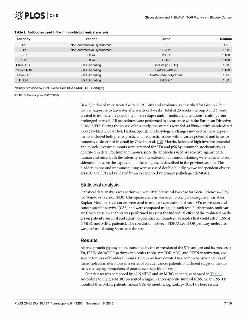
ImmunohistochemistryThe expressions of STn antigen, its precursor Tn, and phosphorylated forms of Akt (pAkt),mTOR (pmTOR), S6 (pS6) and PTEN in bladder tumours were accessed by immunohis-tochemistry using the avidin/streptavidin peroxidase method, as described by Ferreira et al.[5]. Information on the primary antibodies and dilutions used in this study are summarized inTable 2. Immunoreactivity was revealed using diaminobenzidine (DAB, Thermo ScientificLabVision) as chromogen and sections were counterstained with Harris’s hematoxylin. Nega-tive controls were performed by replacing the primary antibody with 5% bovine serum albu-min (BSA). Positive controls were known positive tissues for the antigens under study.
Immunohistochemistry scoring of human tumoursThe immunostained sections were assessed double-blindly by light microscopy by two inde-pendent observers (CC and SP) and validated by an experienced pathologist (TA). Disaccord-ing readings were re-analyzed using a double-headed microscope (Olympus BX46; OlympusCorporation), and consensus was reached. A semi-quantitative approach was established toscore the immunohistochemical labeling based on the extent and intensity of the staining.
Given the absence of Tn and STn in the healthy urothelium [5], tumours were classified aspositive for these antigens when membrane and/or cytoplasmic immunoreactivity wereobserved in more than 5% of the tumour, as described by Ferreira et al. [5,15]. pAkt, pmTOR,pS6 and PTEN expressions were scored according to the staining intensity (weak-1 point;
Glycosylation and PI3K/Akt/mTOR Pathway in Bladder Cancer
PLOS ONE | DOI:10.1371/journal.pone.0141253 November 16, 2015 5 / 19
moderate-2 points; strong-3 points) multiplied by the percentage of positive cells (0–5%-0points;>5–25%-1 point;>25–50%-2 points;>50–75%-3 points;>75–100%-4 points). Basedon the classification proposed by Nishikawa et al. [16], tumours with a score<6 were consid-ered negative, whereas those with a score�6 were classified as positive (overexpression). pAktwas evaluated based on nuclear immunoreactivity, pmTOR and pS6 based on cytoplasmicexpression and PTEN on both cytoplasmic and nuclear staining, as suggested by other publica-tions [17,18].
Animal experiments with sirolimus and immunohistochemistry scoringHistological sections of Imprinting Control Region (ICR) mice bearing N-butyl-N-(4-hydroxy-butyl) nitrosamine (BBN)-induced bladder lesions, resulting from our previous work on theimpact of sirolimus on bladder cancer [19], were elected for this study. Briefly, four-week-oldmale ICR mice (25g; Harlan, Barcelona, Spain) were randomly distributed into four groups, asdescribed in detail in a previous publication [18]. Group 1 (n = 6) included mice exposed to0.05% BBN for 12 weeks followed by tap water for 8 weeks (total of 20 weeks). Group 2 (n = 7)included mice treated with 0.05% BBN solution for twelve weeks, maintained with normal tapwater for another week, administrated intraperitoneally with mTOR-inhibitor sirolimus (1.5mg/kg; Wyeth) for five days a week for six consecutively weeks, i.e. until the 19th week, followedby another week of tap water (total of 20 weeks). Group 3 (n = 6) included mice exposed to0.05% BBN for 12 weeks followed by tap water for 11 weeks (total of 23 weeks). Group 4
Table 1. Clinical-pathological data of the studied sample (n = 96).
Age, years
median [min—max] 69.5 [38–92]
Gender, n (%)
Male 82 (85.4%)
Female 14 (14.6%)
Stage, n (%)
Ta 27 (28.1%)
T1 20 (20.8%)
T2 9 (9.4%)
T3 20 (20.8%)
T4 20 (20.8%)
Grade, n (%)
Low 16 (16.7%)
High 80 (83.3%)
Recurrence status, n(%)
Primary 54 (56.3%)
Recurrent 42 (43.7%)
Associated Cis, n(%)
No 76 (79.2%)
Yes 20 (20.8%)
Metastasis, n(%)
No 19 (63.3%)
Yes 11 (36.7%)
Follow-up, n (%)
Alive, lost or death from other causes 67 (69.8%)
Death from cancer 29 (30.2%)
doi:10.1371/journal.pone.0141253.t001
Glycosylation and PI3K/Akt/mTOR Pathway in Bladder Cancer
PLOS ONE | DOI:10.1371/journal.pone.0141253 November 16, 2015 6 / 19
(n = 7) included mice treated with 0.05% BBN and sirolimus, as described for Group 2, butwith an exposure to tap water afterwards of 3 weeks (total of 23 weeks). Group 3 and 4 werecreated to estimate the possibility of late relapse and/or molecular alterations resulting fromprolonged survival. All procedures were performed in accordance with the European Directive2010/63/EU. During the course of this study, the animals were fed ad libitum with standardizedfood (Tecklad Global Diet, Harlan, Spain). The histological changes induced by these experi-ments included both preneoplastic and neoplastic lesions with invasive potential and invasivetumours, as described in detail by Oliveira et al. [18]. Herein, lesions of high invasive potentialand muscle invasive tumours were screened for STn and pS6 by immunohistochemistry, asdescribed in detail for human tumours, since the antibodies used are reactive against bothhuman and mice. Both the intensity and the extension of immunostaining were taken into con-sideration to score the expression of the antigens, as described in the previous section. Thebladder lesions and immunostaining were assessed double-blindly by two independent observ-ers (CC and SP) and validated by an experienced veterinary pathologist (RMGC).
Statistical analysisStatistical data analysis was performed with IBM Statistical Package for Social Sciences—SPSSfor Windows (version 20.0). Chi-square analysis was used to compare categorical variables.Kaplan-Meier survival curves were used to evaluate correlation between STn expression andcancer-specific survival (CSS) and were compared using log-rank test. Furthermore, multivari-ate Cox regression analysis was performed to assess the individual effect of the evaluated mark-ers on patient’s survival and adjust to potential confounders (variables that could affect CSS ofNMIBC and MIBC patients). The correlation between PI3K/Akt/mTOR pathway moleculeswas performed using Spearman rho test.
ResultsAltered protein glycosylation, translated by the expression of the STn antigen and its precursorTn, PI3K/Akt/mTOR pathway molecules (pAkt, pmTOR, pS6), and PTEN inactivation, aresalient features of bladder tumours. Herein we have devoted to a comprehensive analysis ofthese molecular alterations in a series of bladder cancer patients at different stages of the dis-ease, envisaging biomarkers of poor cancer-specific survival.
Our dataset was composed by 47 NMIBC and 49 MIBC patients, as showed in Table 1.According to Fig 2, NMIBC presented a higher cancer-specific survival (CSS; mean CSS: 119months) than MIBC patients (mean CSS: 43 months; log rank, p<0.001). These results
Table 2. Antibodies used in the immunohistochemical analysis.
Antibody Vendor Clone Dilution
Tn Non-commercial Hybridoma* IE3 1:5
STn Non-commercial Hybridoma* TKH2 1:20
Ki-67 Dako MIB-1 1:100
p53 Dako DO-7 1:100
Phos-AKT Cell Signaling Ser473 (736E11) 1:50
Phos-mTOR Cell Signaling Ser2448(49F9) 1:100
Phos-S6 Cell Signaling Ser240/244 polyclonal 1:75
PTEN Cell Signaling D4.3 XP 1:50
*Kindly provided by Prof. Celso Reis (IPATIMUP, UP, Portugal)
doi:10.1371/journal.pone.0141253.t002
Glycosylation and PI3K/Akt/mTOR Pathway in Bladder Cancer
PLOS ONE | DOI:10.1371/journal.pone.0141253 November 16, 2015 7 / 19
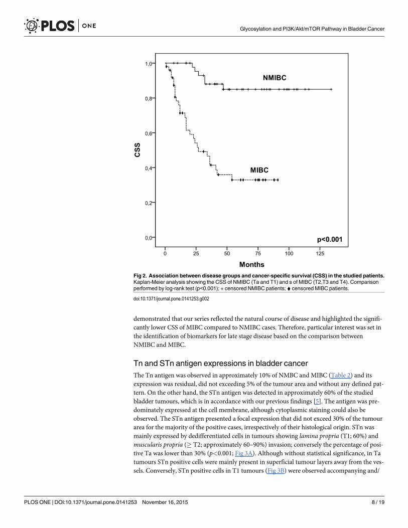
demonstrated that our series reflected the natural course of disease and highlighted the signifi-cantly lower CSS of MIBC compared to NMIBC cases. Therefore, particular interest was set inthe identification of biomarkers for late stage disease based on the comparison betweenNMIBC and MIBC.
Tn and STn antigen expressions in bladder cancerThe Tn antigen was observed in approximately 10% of NMBC and MIBC (Table 2) and itsexpression was residual, did not exceeding 5% of the tumour area and without any defined pat-tern. On the other hand, the STn antigen was detected in approximately 60% of the studiedbladder tumours, which is in accordance with our previous findings [5]. The antigen was pre-dominately expressed at the cell membrane, although cytoplasmic staining could also beobserved. The STn antigen presented a focal expression that did not exceed 30% of the tumourarea for the majority of the positive cases, irrespectively of their histological origin. STn wasmainly expressed by dedifferentiated cells in tumours showing lamina propria (T1; 60%) andmuscularis propria (� T2; approximately 60–90%) invasion; conversely the percentage of posi-tive Ta was lower than 30% (p<0.001; Fig 3A). Although without statistical significance, in Tatumours STn positive cells were mainly present in superficial tumour layers away from the ves-sels. Conversely, STn positive cells in T1 tumours (Fig 3B) were observed accompanying and/
Fig 2. Association between disease groups and cancer-specific survival (CSS) in the studied patients.Kaplan-Meier analysis showing the CSS of NMIBC (Ta and T1) and s of MIBC (T2,T3 and T4). Comparisonperformed by log-rank test (p<0.001); + censored NMIBC patients; ^ censored MIBC patients.
doi:10.1371/journal.pone.0141253.g002
Glycosylation and PI3K/Akt/mTOR Pathway in Bladder Cancer
PLOS ONE | DOI:10.1371/journal.pone.0141253 November 16, 2015 8 / 19
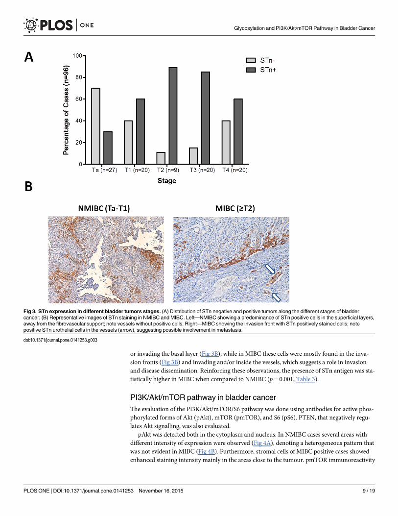
or invading the basal layer (Fig 3B), while in MIBC these cells were mostly found in the inva-sion fronts (Fig 3B) and invading and/or inside the vessels, which suggests a role in invasionand disease dissemination. Reinforcing these observations, the presence of STn antigen was sta-tistically higher in MIBC when compared to NMIBC (p = 0.001, Table 3).
PI3K/Akt/mTOR pathway in bladder cancerThe evaluation of the PI3K/Akt/mTOR/S6 pathway was done using antibodies for active phos-phorylated forms of Akt (pAkt), mTOR (pmTOR), and S6 (pS6). PTEN, that negatively regu-lates Akt signalling, was also evaluated.
pAkt was detected both in the cytoplasm and nucleus. In NMIBC cases several areas withdifferent intensity of expression were observed (Fig 4A), denoting a heterogeneous pattern thatwas not evident in MIBC (Fig 4B). Furthermore, stromal cells of MIBC positive cases showedenhanced staining intensity mainly in the areas close to the tumour. pmTOR immunoreactivity
Fig 3. STn expression in different bladder tumors stages. (A) Distribution of STn negative and positive tumors along the different stages of bladdercancer; (B) Representative images of STn staining in NMIBC and MIBC. Left—NMIBC showing a predominance of STn positive cells in the superficial layers,away from the fibrovascular support; note vessels without positive cells. Right—MIBC showing the invasion front with STn positively stained cells; notepositive STn urothelial cells in the vessels (arrow), suggesting possible involvement in metastasis.
doi:10.1371/journal.pone.0141253.g003
Glycosylation and PI3K/Akt/mTOR Pathway in Bladder Cancer
PLOS ONE | DOI:10.1371/journal.pone.0141253 November 16, 2015 9 / 19
was cytoplasmic and, in occasional cases, nuclear. In urothelium with apparent normal histol-ogy pmTOR expression was restricted to superficial cell layers. In NMIBC pmTOR expressionwas evenly distributed across the several layers of urothelial cells, although there was a moreintense staining in the superficial layers (Fig 4C). Moreover, several areas with variable stainingintensity were observed, denoting a heterogeneous expression. In MIBC positives cases,pmTOR expression was focal and heterogeneous (Fig 4D). pS6 immunoreactivity was predom-inantly cytoplasmic. In NMIBC pS6 expression was noted in all the superficial layers, both inumbrella and differentiated cells (Fig 4E). The immunoreactivity of pS6 varied across thetumour cells. In MIBC pS6 presented a diffuse expression throughout the tumour, being morepresent in basal and mitotic cells (Fig 4F). Several positive cases presented increased pS6 stain-ing intensity in the invasion front as well as pS6 expression in tumour infiltrating lymphocytesand endothelial cells.
Taking into account the extension of staining and its intensity, 62/94 (66%), 33/96 (34%)and 45/95 (47%) of the bladder tumours were considered positive for pAkt, pmTOR and pS6,respectively. A Spearman rho test showed that pAkt, pmTOR, pS6 expressions were signifi-cantly correlated (P<0.05) irrespectively of the tumour stage, thus in accordance with a fullyactive pathway. Furthermore, despite histological differences, these markers presented an equaldistribution among the NMIBC and MIBC and could not be associated with muscle invasion(Table 3).
On the other hand, 37/92 (40%) of the tumours were considered positive for PTEN. PTENwas expressed in the cytoplasm and nucleus of the same cells, however with lower extension ofexpression in MIBC (33%, Fig 4G) compared to NMIBC (83%; Fig 4H). Moreover, the PTEN-negative phenotype was significantly associated with muscle invasion (Ta and T1; p<0.001,Table 3), which may contribute to maintain an active PI3K/Akt/mTOR/S6 pathway in thesecases.
Table 3. Association between the evaluatedmarkers and the stage of disease.
Bladder Cancer
NMIBC MIBC Pn (%) n (%)
Tn
Negative 41 (87.2) 45 (91.8)
Positive 6 (12.8) 4 (8.2) 0.461
STn
Negative 27 (57.4) 12 (24.5)
Positive 20 (42.6) 37 (75.5) 0.001
pAKT
Negative 13 (28.9) 19 (38.8)
Positive 32 (71.1) 30 (61.2) 0.312
pmTor
Negative 30 (63.8) 33 (67.3)
Positive 17 (36.2) 16 (32.7) 0.717
pS6
Negative 22 (47.8) 28 (57.1)
Positive 24 (52.2) 21 (42.9) 0.183
PTEN
Negative 18 (38.3) 37 (82.2)
Positive 29 (61.7) 8 (17.8) <0.001
doi:10.1371/journal.pone.0141253.t003
Glycosylation and PI3K/Akt/mTOR Pathway in Bladder Cancer
PLOS ONE | DOI:10.1371/journal.pone.0141253 November 16, 2015 10 / 19
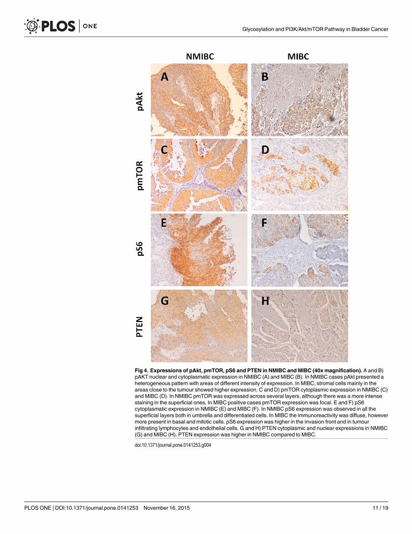
Fig 4. Expressions of pAkt, pmTOR, pS6 and PTEN in NMIBC and MIBC (40xmagnification). A and B)pAKT nuclear and cytoplasmatic expression in NMIBC (A) and MIBC (B). In NMIBC cases pAkt presented aheterogeneous pattern with areas of different intensity of expression. In MIBC, stromal cells mainly in theareas close to the tumour showed higher expression. C and D) pmTOR cytoplasmic expression in NMIBC (C)and MIBC (D). In NMIBC pmTORwas expressed across several layers, although there was a more intensestaining in the superficial ones. In MIBC positive cases pmTOR expression was focal. E and F) pS6cytoplasmatic expression in NMIBC (E) and MIBC (F). In NMIBC pS6 expression was observed in all thesuperficial layers both in umbrella and differentiated cells. In MIBC the immunoreactivity was diffuse, howevermore present in basal and mitotic cells. pS6 expression was higher in the invasion front and in tumourinfiltrating lymphocytes and endothelial cells. G and H) PTEN cytoplasmic and nuclear expressions in NMIBC(G) and MIBC (H). PTEN expression was higher in NMIBC compared to MIBC.
doi:10.1371/journal.pone.0141253.g004
Glycosylation and PI3K/Akt/mTOR Pathway in Bladder Cancer
PLOS ONE | DOI:10.1371/journal.pone.0141253 November 16, 2015 11 / 19
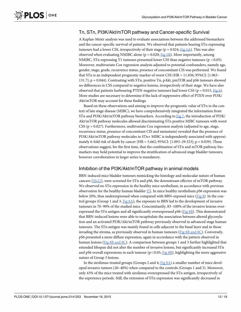
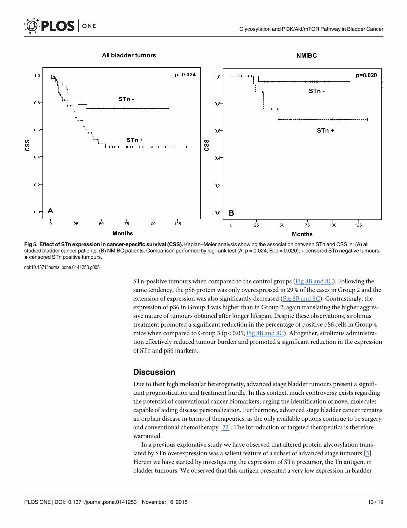
Tn, STn, PI3K/Akt/mTOR pathway and Cancer-specific SurvivalA Kaplan-Meier analysis was used to evaluate associations between the addressed biomarkersand the cancer-specific survival of patients. We observed that patients bearing STn expressingtumours had a lower CSS, irrespectively of their stage (p = 0.024; Fig 5A). This was alsoobserved when evaluating NMIBC alone (p = 0.020; Fig 5B). More importantly, amongNMIBC, STn expressing T1 tumours presented lower CSS than negative tumours (p<0.05).Moreover, multivariate Cox regression analysis adjusted to potential confounders, namely age,gender, stage, grade, recurrence status, presence of concomitant CIS was performed. We foundthat STn is an independent prognostic marker of worst CSS (HR = 11.836; 95%CI: [1.063–131.7]; p = 0.044). Contrasting with STn, positive Tn, pAkt, pmTOR and pS6 tumours showedno differences in CSS compared to negative lesions, irrespectively of their stage. We have alsoobserved that patients harbouring PTEN-negative tumours had lower CSS (p = 0.015, Fig 6).More studies are necessary to determine if the lack of suppressive effect of PTEN over PI3K/Akt/mTOR may account for these findings.
Based on these observations and aiming to improve the prognostic value of STn in the con-text of late stage disease (MIBC), we have comprehensively integrated the information fromSTn and PI3K/Akt/mTOR pathway biomarkers. According to Fig 7, the introduction of PI3K/Akt/mTOR pathway molecules allowed discriminating STn positive MIBC tumours with worstCSS (p = 0.027). Furthermore, multivariate Cox regression analysis (adjusted to age, stage,recurrence status, presence of concomitant CIS and metastasis) revealed that the presence ofPI3K/Akt/mTOR pathway molecules in STn+ MIBC is independently associated with approxi-mately 6-fold risk of death by cancer (HR = 5.662; 95%CI: [1.093–29.323]; p = 0.039). Theseobservations suggest, for the first time, that the combination of STn and mTOR pathway bio-markers may hold potential to improve the stratification of advanced stage bladder tumours;however corroboration in larger series is mandatory.
Inhibition of the PI3K/Akt/mTOR pathway in animal modelsBBN-induced mice bladder tumours mimicking the histology and molecular nature of humancancers [20,21], were screened for STn and pS6, the downstream effector of mTOR pathway.We observed no STn expression in the healthy mice urothelium, in accordance with previousobservation for the healthy human bladder [5]. In mice healthy urothelium pS6 expression wasbelow 20%, thus underexpressed when compared with BBN-exposed mice (Fig 8). In the con-trol groups (Group 1 and 3, Fig 8A), the exposure to BBN led to the development of invasivetumours in 70–90% of the studied mice. Concomitantly, 83–100% of the invasive lesions over-expressed the STn antigen and all significantly overexpressed pS6 (Fig 8B). This demonstratedthat BBN-induced lesions were able to recapitulate the association between altered glycosyla-tion and an activated PI3K/Akt/mTOR pathway previously observed in advanced stage humantumours. The STn antigen was mainly found in cells adjacent to the basal layer and in thoseinvading the stroma, as previously observed in human tumours (Fig 8B and 8C). Conversely,pS6 presented a more diffuse expression, again in accordance with the pattern observed inhuman lesions (Fig 8B and 8C). A comparison between groups 1 and 3 further highlighted thatextended lifespan did not alter the number of invasive lesions, but significantly increased STnand pS6 overall expressions in each tumour (p<0.05; Fig 8B), highlighting the more aggressivenature of Group 3 lesions.
In the sirolimus-treated groups (Groups 2 and 4; Fig 8A) a smaller number of mice devel-oped invasive tumors (20–40%) when compared to the controls (Groups 1 and 3). Moreover,only 43% of the mice treated with sirolimus overexpressed the STn antigen, irrespectively ofthe experience periods. Still, the extension of STn expression was significantly decreased in
Glycosylation and PI3K/Akt/mTOR Pathway in Bladder Cancer
PLOS ONE | DOI:10.1371/journal.pone.0141253 November 16, 2015 12 / 19
STn-positive tumours when compared to the control groups (Fig 8B and 8C). Following thesame tendency, the pS6 protein was only overexpressed in 29% of the cases in Group 2 and theextension of expression was also significantly decreased (Fig 8B and 8C). Contrastingly, theexpression of pS6 in Group 4 was higher than in Group 2, again translating the higher aggres-sive nature of tumours obtained after longer lifespan. Despite these observations, sirolimustreatment promoted a significant reduction in the percentage of positive pS6 cells in Group 4mice when compared to Group 3 (p<0.05; Fig 8B and 8C). Altogether, sirolimus administra-tion effectively reduced tumour burden and promoted a significant reduction in the expressionof STn and pS6 markers.
DiscussionDue to their high molecular heterogeneity, advanced stage bladder tumours present a signifi-cant prognostication and treatment hurdle. In this context, much controversy exists regardingthe potential of conventional cancer biomarkers, urging the identification of novel moleculescapable of aiding disease personalization. Furthermore, advanced stage bladder cancer remainsan orphan disease in terms of therapeutics, as the only available options continue to be surgeryand conventional chemotherapy [22]. The introduction of targeted therapeutics is thereforewarranted.
In a previous explorative study we have observed that altered protein glycosylation trans-lated by STn overexpression was a salient feature of a subset of advanced stage tumours [5].Herein we have started by investigating the expression of STn precursor, the Tn antigen, inbladder tumours. We observed that this antigen presented a very low expression in bladder
Fig 5. Effect of STn expression in cancer-specific survival (CSS). Kaplan–Meier analysis showing the association between STn and CSS in: (A) allstudied bladder cancer patients; (B) NMIBC patients. Comparison performed by log-rank test (A: p = 0.024; B: p = 0.020); + censored STn negative tumours;^ censored STn positive tumours.
doi:10.1371/journal.pone.0141253.g005
Glycosylation and PI3K/Akt/mTOR Pathway in Bladder Cancer
PLOS ONE | DOI:10.1371/journal.pone.0141253 November 16, 2015 13 / 19
tumours and was not associated with any particular stage of the disease. These findings suggestthat the Tn antigen is rapidly sialylated or capped with more extended glycans in bladdertumours. Moreover, we have confirmed that STn expression is more associated with muscleinvasive than non-muscle invasive disease in a larger patient set, suggesting that sialylationplays a key role in stopping protein glycosylation in advanced stage bladder tumours. Further-more, we have provided new insights regarding its correlation with decreased survival, as previ-ously observed for digestive track tumours [23–25]. Accordingly, we and other authors haveshown that STn expression is responsible by the modulation of cell surface glycoprotein func-tions in ways that favour malignant phenotypes in gastric [26], breast [27] and bladder [5] can-cers. Namely, STn expression altered the adhesive properties of cancer cells, possibly byimpairing integrin function [26,27]. Furthermore, it enhanced cell motility, invasion [26,27]and epithelial-to-mesenchymal transition, a key event leading to metastasis [28]. We have alsodemonstrated that STn expression protects bladder cancer cells from adverse host immuneresponses [6]. Namely, it impaired dendritic cell maturation inducing a tolerogenic phenotypeand limiting their capacity to trigger protective anti-tumour T-cell responses [6]. In resume, asignificant amount of data supports a key role of STn in disease progression and dissemination,making of STn antigen, and in particular STn-glycoproteins, potential anti-cancer targets. Nev-ertheless, there is scarce information about the molecular nature of this subset of STn-express-ing aggressive tumours and consequently about the best therapeutic options.
Fig 6. Effect of PTEN expression and cancer-specific survival (CSS) in the studied patients. Kaplan-Meier analysis showing the effect in CSS of PTEN expression in all studied bladder cancer patients.Comparison performed by log-rank test (p = 0.013); + censored PTEN negative tumours; ^ censored PTENpositive tumours.
doi:10.1371/journal.pone.0141253.g006
Glycosylation and PI3K/Akt/mTOR Pathway in Bladder Cancer
PLOS ONE | DOI:10.1371/journal.pone.0141253 November 16, 2015 14 / 19
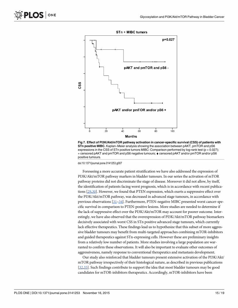
Foreseeing a more accurate patient stratification we have also addressed the expression ofPI3K/Akt/mTOR pathway markers in bladder tumours. In our series the activation of mTORpathway proteins did not discriminate the stage of disease. Moreover it did not allow, by itself,the identification of patients facing worst prognosis, which is in accordance with recent publica-tions [29,30]. However, we found that PTEN expression, which exerts a suppressive effect overthe PI3K/Akt/mTOR pathway, was decreased in advanced stage tumours, in accordance withprevious observations [31–34]. Furthermore, PTEN-negative MIBC presented worst cancer-spe-cific survival in comparison to PTEN-positive lesions. More studies are needed to determine ifthe lack of suppressive effect over the PI3K/Akt/mTORmay account for poorer outcome. Inter-estingly, we have also observed that the overexpression of PI3K/Akt/mTOR pathway biomarkersdecisively associated with worst CSS in STn positive advanced stage tumours, which currentlylack effective therapeutics. These findings lead us to hypothesize that this subset of more aggres-sive bladder tumours may benefit frommulti-targeted approaches combining mTOR-inhibitorsand guided therapeutics against STn-expressing cells. However these are preliminary insightsfrom a relatively low number of patients. More studies involving a large population are war-ranted to confirm these observations. It will also be important to evaluate other outcomes ofaggressiveness, namely response to conventional therapeutics and metastasis development.
Our study also reinforced that bladder tumours present extensive activation of the PI3K/Akt/mTOR pathway irrespectively of their histological nature, as described in previous publications[32,35]. Such findings contribute to support the idea that most bladder tumours may be goodcandidates for mTOR-inhibitors therapeutics. Accordingly, mTOR-inhibitors have been
Fig 7. Effect of PI3K/Akt/mTOR pathway activation in cancer-specific survival (CSS) of patients withSTn positive MIBC. Kaplan–Meier analysis showing the association between pAKT, pmTOR and pS6expressions in the CSS of STn positive tumors MIBC: Comparison performed by log-rank test (p = 0.027);+ censored pAKT and pmTOR and pS6 negative tumours; ^ censored pAKT and/or pmTOR and/or pS6positive tumours.
doi:10.1371/journal.pone.0141253.g007
Glycosylation and PI3K/Akt/mTOR Pathway in Bladder Cancer
PLOS ONE | DOI:10.1371/journal.pone.0141253 November 16, 2015 15 / 19
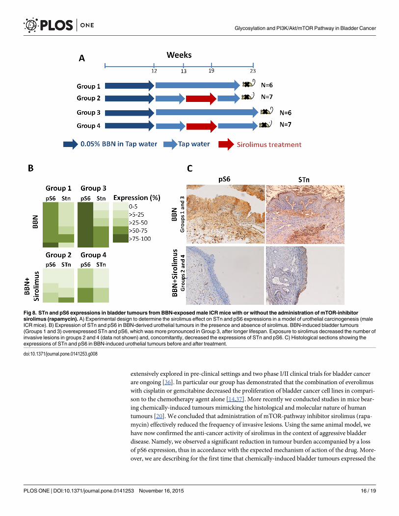
extensively explored in pre-clinical settings and two phase I/II clinical trials for bladder cancerare ongoing [36]. In particular our group has demonstrated that the combination of everolimuswith cisplatin or gemcitabine decreased the proliferation of bladder cancer cell lines in compari-son to the chemotherapy agent alone [14,37]. More recently we conducted studies in mice bear-ing chemically-induced tumours mimicking the histological and molecular nature of humantumours [20]. We concluded that administration of mTOR-pathway inhibitor sirolimus (rapa-mycin) effectively reduced the frequency of invasive lesions. Using the same animal model, wehave now confirmed the anti-cancer activity of sirolimus in the context of aggressive bladderdisease. Namely, we observed a significant reduction in tumour burden accompanied by a lossof pS6 expression, thus in accordance with the expected mechanism of action of the drug. More-over, we are describing for the first time that chemically-induced bladder tumours expressed the
Fig 8. STn and pS6 expressions in bladder tumours from BBN-exposedmale ICRmice with or without the administration of mTOR-inhibitorsirolimus (rapamycin). A) Experimental design to determine the sirolimus effect on STn and pS6 expressions in a model of urothelial carcinogenesis (maleICRmice). B) Expression of STn and pS6 in BBN-derived urothelial tumours in the presence and absence of sirolimus. BBN-induced bladder tumours(Groups 1 and 3) overexpressed STn and pS6, which was more pronounced in Group 3, after longer lifespan. Exposure to sirolimus decreased the number ofinvasive lesions in groups 2 and 4 (data not shown) and, concomitantly, decreased the expressions of STn and pS6. C) Histological sections showing theexpressions of STn and pS6 in BBN-induced urothelial tumours before and after treatment.
doi:10.1371/journal.pone.0141253.g008
Glycosylation and PI3K/Akt/mTOR Pathway in Bladder Cancer
PLOS ONE | DOI:10.1371/journal.pone.0141253 November 16, 2015 16 / 19
STn antigen, thereby mimicking the glycosylation pattern of human cancers. These observationsare of particular importance due the lack of accurate models to access the biological role of thisantigen. In fact most established cancer cell lines express residual amounts of this antigen,denoting a dependence on the tumours microenvironment. We believe that BBN-inducedtumours may now constitute key models to develop successful therapeutics against STn positivebladder lesions. Moreover importantly, we have concluded that the administration of sirolimuscontributed to reduce the number of STn positive cells. These observations reinforce a possibleassociation between STn and an active PI3K/Akt/mTOR pathway in invasive tumours, as sug-gested upon the evaluation of human cancers. It also points out that sirolimus may constitute avaluable approach to manage STn and PI3K/Akt/mTOR-positive, which face worst OS. Still,these preliminary evidences and more in depth studies are needed before progressing to clinicalphases. Namely, it will be important to support these findings in other models such as patient-derived xenografts and compare the effect of sirolimus with conventional chemotherapeuticsfor bladder cancer (cisplatin/gemcitabine-based regimens).
In resume, we have demonstrated that the STn antigen is a biomarker of poor prognosis,particularly in MIBC. We also suggest the existence of potentially more aggressive subgroup ofSTn positive MIBC characterized by an active mTOR-pathway. Such observations also providethe first link between these two apparently unrelated events in bladder cancer (altered glycosyl-ation and the PI3K/Akt/mTOR-pathway activation). Using animal models we have also con-cluded that the administration of mTOR-pathway inhibitor sirolimus offers potential againstthese highly malignant tumours. More validation studies are now warranted to set the pace forclinical trials. Taking into consideration its cell-surface nature and key role played by STnmalignancy, specific antibody-based therapeutics can also be envisaged [22,38]. The combina-tion of these approaches may provide novel ways to improve MIBC management, whichremains an orphan disease in terms of innovative treatments [22].
AcknowledgmentsThis work was supported by Portuguese Foundation for Science and Technology (FCT) Post-doctoral grants SFRH/BPD/66288/2009 (José Alexandre Ferreira), SFRH/BPD/101827/2014(Luis Lima), SFRH/BPD/85462/2012 (Rui Gil da Costa) and PhD grants SFRH/BD/103571/2014 (Elisabete Fernandes) and SFRH/BD/111242/2015 (Andreia Peixoto). FCT is co-financedby European Social Fund (ESF) under Human Potential Operation Programme (POPH) fromNational Strategic Reference Framework (NSRF). The authors also acknowledge financial sup-port from ICBAS-UP (Céu Costa and Sofia Pereira) and the Portuguese Association of Urol-ogy/Pfizer prize 2013. The authors also thank Professor Celso Reis (IPATIMUP, UP, Portugal)by kindly providing the anti-STn TKH2 and the anti-Tn IE3 monoclonal antibodies used inthis work.
Author ContributionsConceived and designed the experiments: CC SP LL PO JAF LLS. Performed the experiments:CC SP AP EF AT DNMN CG RMGC PO. Analyzed the data: CC SP LL RMGC RC TA POJAF LLS. Contributed reagents/materials/analysis tools: RC PO JAF LLS. Wrote the paper: LLJAF LLS PO.
References1. Pasin E, Josephson DY, Mitra AP, Cote RJ, Stein JP. Superficial bladder cancer: an update on etiology,
molecular development, classification, and natural history. Rev Urol. 2008; 10: 31–43. PMID:18470273
Glycosylation and PI3K/Akt/mTOR Pathway in Bladder Cancer
PLOS ONE | DOI:10.1371/journal.pone.0141253 November 16, 2015 17 / 19
2. Babjuk M, Burger M, Zigeuner R, Shariat SF, van Rhijn BWG, Compérat E, et al. EAU guidelines onnon-muscle-invasive urothelial carcinoma of the bladder: update 2013. Eur Urol. 2013; 64: 639–53. doi:10.1016/j.eururo.2013.06.003 PMID: 23827737
3. Miakhil I, Parker SG, Kommu SS, Nethercliffe J. A review of molecular biomarkers for bladder cancer.Int J Med Biomed Res. 2013; 2: 186–194.
4. Dovedi SJ, Davies BR. Emerging targeted therapies for bladder cancer: a disease waiting for a drug.Cancer Metastasis Rev. 2009; 28: 355–67. doi: 10.1007/s10555-009-9192-9 PMID: 19997963
5. Ferreira JA, Videira PA, Lima L, Pereira S, Silva M, Carrascal M, et al. Overexpression of tumour-asso-ciated carbohydrate antigen sialyl-Tn in advanced bladder tumours. Mol Oncol. 2013; 7: 719–31. doi:10.1016/j.molonc.2013.03.001 PMID: 23567325
6. Carrascal MA, Severino PF, Guadalupe Cabral M, Silva M, Ferreira JA, Calais F, et al.Sialyl Tn-expressing bladder cancer cells induce a tolerogenic phenotype in innate and adaptive immune cells.Mol Oncol. 2014; 8: 753–65. doi: 10.1016/j.molonc.2014.02.008 PMID: 24656965
7. Dall’Olio F, Malagolini N, Trinchera M, Chiricolo M. Mechanisms of cancer-associated glycosylationchanges. Front Biosci (Landmark Ed). 2012; 17: 670–99.
8. Ma H, Zhou H, Song X, Shi S, Zhang J, Jia L. Modification of sialylation is associated with multidrugresistance in human acute myeloid leukemia. Oncogene. 2015; 34: 726–40. doi: 10.1038/onc.2014.7PMID: 24531716
9. Porta C, Paglino C, Mosca A. Targeting PI3K/Akt/mTOR Signaling in Cancer. Front Oncol. 2014; 4: 1–11.
10. Ching CB, Hansel DE. Expanding therapeutic targets in bladder cancer: the PI3K/Akt/mTOR pathway.Lab Invest. Nature Publishing Group; 2010; 90: 1406–1414.
11. Wu X, Obata T, Khan Q, Highshaw RA, De VereWhite R, Sweeney C. The phosphatidylinositol-3kinase pathway regulates bladder cancer cell invasion. BJU Int. 2004; 93: 143–150. PMID: 14678387
12. Keniry M, Parsons R. The role of PTEN signaling perturbations in cancer and in targeted therapy. Onco-gene. 2008; 27: 5477–5485. doi: 10.1038/onc.2008.248 PMID: 18794882
13. Tamura M, Gu J, Tran H, Yamada KM. PTEN gene and integrin signaling in cancer. J Natl Cancer Inst.1999; 91: 1820–1828. PMID: 10547389
14. Pinto-Leite R, Arantes-Rodrigues R, Palmeira C, Gaivão I, Cardoso ML, Colaço A, et al. Everolimusenhances gemcitabine-induced cytotoxicity in bladder-cancer cell lines. J Toxicol Env Heal A. 2012;75: 788–99.
15. Munari E, Fujita K, Faraj S, Chaux A, Gonzalez-Roibon N, Hicks J, et al. Dysregulation of mammaliantarget of rapamycin pathway in upper tract urothelial carcinoma. Hum Pathol. Elsevier Inc.; 2013; 44:2668–2676.
16. Nishikawa M, Miyake H, Behnsawy HM, Fujisawa M. Significance of 4E-binding protein 1 as a thera-peutic target for invasive urothelial carcinoma of the bladder. Urol Oncol. 2015; 33: 166.e9–15.
17. Chaux A, Compérat E, Varinot J, Hicks J, Lecksell K, Solus J, et al. High levels of phosphatase and ten-sin homolog expression are associated with tumor progression, tumor recurrence, and systemic metas-tases in pt1 urothelial carcinoma of the bladder: A tissue microarray study of 156 patients treated bytransurethral resect. Urology. 2013; 81: 116–122. doi: 10.1016/j.urology.2012.09.007 PMID: 23273076
18. Oliveira PA, Arantes-Rodrigues R, Sousa-Diniz C, Colaço A, Lourenço L, De La Cruz P LF, et al. Theeffects of sirolimus on urothelial lesions chemically induced in ICRmice by BBN. Anticancer Res. 2009;29: 3221–3226. PMID: 19661338
19. Lima L, Severino PF, Silva M, Miranda A, Tavares A, Pereira S et al. Response of high-risk of recur-rence/progression bladder tumours expressing sialyl-Tn and sialyl-6-T to BCG immunotherapy. Br JCancer. 2013; 109: 2106–14. doi: 10.1038/bjc.2013.571 PMID: 24064971
20. Vasconcelos-Nóbrega C, Colaço A, Lopes C, Oliveira PA. BBN as an urothelial carcinogen. In Vivo(Brooklyn). 2012; 26: 727–739.
21. Palmeira C, Oliveira PA, Lameiras C, Amaro T, Silva VM, Lopes C, et al. Biological similarities betweenmurine chemical-induced and natural human bladder carcinogenesis. Oncol Lett. 2010; 1: 373–377.PMID: 22966311
22. Azevedo R, Ferreira JA, Peixoto A, Neves M, Sousa N, Lima A et al. Emerging antibody-based thera-peutic strategies for bladder cancer: A systematic review. J Control Release. 2015;
23. Flucke U, Zirbes T, Schröder W, Mönig S, Koch V, Schmitz K, et al. Expression of mucin-associatedcarbohydrate core antigens in esophageal squamous cell carcinomas. Anticancer Res. 2001; 21:2189–2193. PMID: 11501845
24. Victorzon M, Nordling S, Nilsson O, Roberts P, Haglund C. Sialyl Tn antigen is an independent predic-tor of outcome in patients with gastric cancer. Int J Cancer. 1996; 65: 295–300. PMID: 8575847
Glycosylation and PI3K/Akt/mTOR Pathway in Bladder Cancer
PLOS ONE | DOI:10.1371/journal.pone.0141253 November 16, 2015 18 / 19
25. Tsuchiya A, Kikuchi Y, Ando Y, Abe R. Correlation between expression of sialosyl-T antigen and sur-vival in patients with gastric cancer. Br J Surg. 1995; 82: 960–962. PMID: 7648120
26. Pinho S, Marcos NT, Ferreira B, Carvalho AS, Oliveira MJ, Santos-Silva F, et al. Biological significanceof cancer-associated sialyl-Tn antigen: modulation of malignant phenotype in gastric carcinoma cells.Cancer Lett. 2007; 249: 157–70. PMID: 16965854
27. Julien S, Adriaenssens E, Ottenberg K, Furlan A, Courtand G, Vercoutter-Edouart AS, et al. ST6Gal-NAc I expression in MDA-MB-231 breast cancer cells greatly modifies their O-glycosylation pattern andenhances their tumourigenicity. Glycobiology. 2006; 16: 54–64. PMID: 16135558
28. Lin J-C, Liao S-K, Lee E-H, Hung M-S, Sayion Y, Chen H-C, et al. Molecular events associated with epi-thelial to mesenchymal transition of nasopharyngeal carcinoma cells in the absence of Epstein-Barrvirus genome. J Biomed Sci. 2009; 16: 105. doi: 10.1186/1423-0127-16-105 PMID: 19930697
29. Korkolopoulou P, Levidou G, Trigka EA, Prekete N, Karlou M, Thymara I, et al. A comprehensive immu-nohistochemical and molecular approach to the PI3K/AKT/mTOR (phosphoinositide 3-kinase/v-aktmurine thymoma viral oncogene/mammalian target of rapamycin) pathway in bladder urothelial carci-noma. BJU Int. 2012; 110: 1237–1248.
30. Fahmy M, Mansure JJ, Brimo F, Yafi FA, Segal R, Althunayan A, et al. Relevance of the mammaliantarget of rapamycin pathway in the prognosis of patients with high-risk non–muscle invasive bladdercancer. Hum Pathol. Elsevier Inc.; 2013; 44: 1766–1772.
31. Sun CH, Chang YH, Pan CC. Activation of the PI3K/Akt/mTOR pathway correlates with tumour pro-gression and reduced survival in patients with urothelial carcinoma of the urinary bladder. Histopathol-ogy. 2011; 58: 1054–1063. doi: 10.1111/j.1365-2559.2011.03856.x PMID: 21707707
32. Saal LH, Johansson P, Holm K, Gruvberger-Saal SK, She Q-B, Maurer M, et al. Poor prognosis in carci-noma is associated with a gene expression signature of aberrant PTEN tumor suppressor pathwayactivity. Proc Natl Acad Sci U S A. 2007; 104: 7564–7569. PMID: 17452630
33. Harris L, De La Cerda J, Tuziak T, Rosen D, Xiao L, Shen Y, et al. Analysis of the Expression of Bio-markers in Urinary Bladder Cancer Using a Tissue Microarray. Mol Carcinog. 2008; 47: 678–685. doi:10.1002/mc.20420 PMID: 18288642
34. Calderaro J, Rebouissou S, de Koning L, Masmoudi A, Hérault A, Dubois T, et al. PI3K/AKT pathwayactivation in bladder carcinogenesis. Int J cancer. 2014; 134: 1776–84. doi: 10.1002/ijc.28518 PMID:24122582
35. Ferreira J, Bernardo C, Amaro T, Costa C, Lopes P, Silva V, et al. Patient-derived sialyl-Tn positiveinvasive bladder cancer xenografts in nude mice: An exploratory model study. Eur Urol Suppl. Euro-pean Association of Urology; 2014; 13: e518.
36. Massari F, Ciccarese C, Santoni M, Brunelli M, Conti A, Modena A, Montironi R, Santini D, Cheng L,Martignoni G, Cascinu S TG. The route to personalized medicine in bladder cancer: where do westand? Target Oncol. 2015;
37. Kjeldsen T, Clausen H, Hirohashi S, Ogawa T, Iijima H, Hakomori S. Preparation and characterizationof monoclonal antibodies directed to the tumor-associated O-linked sialosyl-2——6 alpha-N-acetylga-lactosaminyl (sialosyl-Tn) epitope. Cancer Res. 1988; 48: 2214–2220. PMID: 2450649
38. Fernandes E and Ferreira JA, Andreia P, Luís L, Barroso S, Sarmento B, et al. New trends in guidednanotherapies for digestive cancers: A systematic review. J Control Release. 2015; 209: 288–307. doi:10.1016/j.jconrel.2015.05.003 PMID: 25957905
Glycosylation and PI3K/Akt/mTOR Pathway in Bladder Cancer
PLOS ONE | DOI:10.1371/journal.pone.0141253 November 16, 2015 19 / 19