RESEARCH REPORT 155 - HSE: Information about health … · HSE Health & Safety Executive Dose...
69
HSE Health & Safety Executive Dose constraints for comforters and carers Prepared by Royal Hallamshire Hospital for the Health and Safety Executive 2003 RESEARCH REPORT 155
Transcript of RESEARCH REPORT 155 - HSE: Information about health … · HSE Health & Safety Executive Dose...

HSE Health & Safety
Executive
Dose constraints for comforters and carers
Prepared by Royal Hallamshire Hospital for the Health and Safety Executive 2003
RESEARCH REPORT 155

HSE Health & Safety
Executive
Dose constraints for comforters and carers
Mark Singleton, Cathy Griffiths, Giles Morrison and Tracy Soanes
Department of Medical Physics Royal Hallamshire Hospital
Glossop Road Sheffield
South Yorkshire S10 2JF
This report has been prepared to enable guidance to be developed for employers, to assist them in meeting relevant legislative requirements for the exposure of persons who offer support and care to patients undergoing procedures involving ionising radiation where this would not be considered part of their occupation. The report identifies relevant legislation and guidance, discusses its interpretation, identifies circumstances in which these persons are exposed and presents information relating to the extent of these exposures, including results of dose measurements. Significant use of published information has been made, that has been supplemented wherever possible, with contributions from healthcare professionals. Our sincere thanks go to all persons who have contributed information or comments during the production of this report. Persons who have provided dose or related information included in this report are identified in Appendix B.
This report and the work it describes was funded by the Health and Safety Executive, the Health and Safety Executive Northern Ireland and the Sheffield Teaching Hospitals NHS Trust. Its contents, including any opinions and/or conclusions expressed, are those of the authors alone and do not necessarily reflect HSE policy.
HSE BOOKS

© Crown copyright 2003
First published 2003
ISBN 0 7176 2748 9
All rights reserved. No part of this publication may bereproduced, stored in a retrieval system, or transmitted inany form or by any means (electronic, mechanical,photocopying, recording or otherwise) without the priorwritten permission of the copyright owner.
Applications for reproduction should be made in writing to: Licensing Division, Her Majesty's Stationery Office, St Clements House, 2-16 Colegate, Norwich NR3 1BQ or by e-mail to [email protected]
ii

CONTENTS
1. INTRODUCTION 1.1 European legislative background 1.2 UK legislation and guidance
2. INTERPRETATION AND IMPLEMENTATION OF LEGISLATION 2.1 Dose constraints 2.2 Use of the term ‘Comforter and Carer’
3. PROJECT METHOD
4. DIAGNOSTIC RADIOLOGY 4.1 Identification of exposed persons and exposure scenarios 4.2 Professional body guidance4.3 Results of dose measurements, risk assessments and other relevant information 4.4 Summary of results4.5 Conclusion
5. BEAM THERAPY 5.1 Identification of exposed persons and exposure scenarios 5.2 Professional body guidance5.3 Information relating to the exposure of persons supporting or caring for beam
therapy patients 5.4 Conclusion
6. DIAGNOSTIC NUCLEAR MEDICINE 6.1 Identification of exposed persons and exposure scenarios 6.2 Professional body guidance6.3 The exposure of partners, close friends, parents or guardians supporting/caring for
diagnostic nuclear medicine patients 6.4 Summary of results 6.5 Conclusion
7. THERAPEUTIC NUCLEAR MEDICINE 7.1 Identification of relevant nuclear medicine procedures, exposed persons and exposure
scenarios 7.2 Professional body guidance and best practice 7.3 Exposure of partners and parents supporting/caring for nuclear medicine therapy
patients 7.4 Summary of results 7.5 Conclusion
8. BRACHYTHERAPY 8.1 Identification of exposed persons, exposure scenarios and professional body guidance 8.2 The exposure of supporters/carers of brachytherapy patients 8.3 Summary of results 8.4 Conclusion
9. FINAL CONCLUSION
iii

APPENDIX A REFERENCES Legislation and guidance Published reports, articles and abstracts
APPENDIX B Healthcare professionals who provided information which has been included in the text of this report.
iv

1. INTRODUCTION
Regulation 8 of the Ionising Radiations Regulations 1999 (IRR99) (1) requires that employers restrict the exposure to ionising radiations of persons affected by their undertaking to a level which is as low as reasonably practicable. Regulation 8(3) and paragraph 126 of the Approved Code of Practice to IRR99 (L121) (1) indicate that dose constraints should be used to achieve restriction of exposure for comforters and carers.
This project has been undertaken to collate information from a spectrum of medical diagnostic and therapeutic techniques that lead to exposure of persons other than employees. This includes identification of medical procedures and circumstances under which persons are exposed and, where information is available, providing data on the levels of dose typically received and number of people exposed. Where possible, additional information and opinions have been collated concerning the implementation of measures used to restrict exposure.
1.1 EUROPEAN LEGISLATIVE BACKGROUND
European Directives The Basic Safety Standards Directive 1996 96/29 Article 6 (2) states that all exposures to ionising radiation as a result of ‘practices’ shall be Justified and Optimised (to a level which is ‘As low as reasonably achievable’). With three exceptions specified in Article 6(4), the sum of all exposures to an individual from practices are subject to a dose limit. The excepted groups of persons are:
· Patients undergoing medical diagnosis or treatment. · Volunteers participating in medical research. · The exposure of individuals ‘knowingly and willingly’ helping (other than as part of
their occupation) in the support and comfort of patients undergoing medical diagnosis or treatment.
The Ionising Radiations Regulations 1999 (IRR99) (1) refers to the latter group of people as ‘comforters and carers’.
The Basic Safety Standards Directive 1996 96/29 (2) is supplemented by the MedicalExposure Directive 1997 97/43 (3). Article 3(2) of this Directive states that the exposure ofindividuals ‘knowingly and willingly’ helping the patient (other than as part of theiroccupation) shall show a sufficient net benefit. In determining the balance between benefitsand the possible radiation detriment, the Article requires that the direct health benefits to thepatient and or the benefits to the supporting/caring individual should be taken into account.
Gill [19] observes that the Medical Exposure Directive (3), unlike ICRP Publication 60 (4),does not label the exposure of comforters and carers as a medical exposure. Article 4(a and b) of this Directive requires Member States to ensure that dose constraints areestablished for the exposure of comforters and carers. Article 4(c) requires where appropriate, written instructions to be issued to the patient or legal guardian so as to restrict,as far as reasonably achievable, the exposure of persons who may come into contact with thatpatient and to provide information on the risks of ionising radiation.
European Commission guidance (5) (Radiation Protection 97 - Radiation protection followingIodine-131 therapy), states that Article 4 of the Medical Exposure Directive (3) applies todiagnostic radiology, radiotherapy and diagnostic and therapeutic nuclear medicine.‘Knowing and willing’ individuals are identified as those living under the same roof as thepatient and those who visit the patient in the hospital or at his/her home.
1

1.2 UK LEGISLATION AND GUIDANCE
Implementation of the Basic Safety Standards Directive (2) and Medical Exposure Directive (3) with respect to comforters and carers is legislated by the Ionising Radiations Regulations 1999 (IRR99) (1). Requirements of the Medical Exposure Directive with respect to the provision of information to the patient or legal guardian are transposed into the Ionising Radiation (Medical Exposure) Regulations 2000 (IRMER) (reg. 7 (5 and 6)) (6).
Regulation 2 of IRR99 (1) defines a comforter and carer as an individual who (other than as part of his/her occupation) ‘knowingly and willingly’ incurs an exposure to ionising radiation resulting from the support and comfort of another person who is undergoing a medical exposure.
For persons regarded as comforters and carers, IRR99 Regulation 11 disapplies the schedule 4 “other person” dose limit of 1 mSv in a calendar year or 5 mSv in 5 consecutive calendar years.
The 5mSv in 5 consecutive year limit may be applied to persons exposed to ionising radiation from the medical exposure of another (other than comforters and carers). The associated guidance states that this limit should enable a hospital to make sensible arrangements for the release of radioactive patients in situations where a person may be significantly exposed due to their close proximity to a patient, but who cannot be treated as a comforter and carer. Such persons are stated as including children who are unable to give their consent to the exposure and other relatives or friends who will not have the opportunity to give consent unless they visit the hospital.
Paragraph 126 of the Approved Code of Practice to IRR99 (L121) (1), states that it should always be appropriate to use dose constraints for comforters and carers. Paragraph 132 of L121 (1) quotes a National Radiological Protection Board (NRPB) derived constraint for comforters and carers of 5 mSv from their involvement in one series or course of treatment. It states that in most circumstances, it should be possible to design control measures that result in doses less than this and suggests that advice from professional bodies should be used to select appropriate constraints. Paragraph 128 (L121) (1) states that comforters and carers will normally be relatives or friends of the patient and are likely to receive 1 mSv or more in a year resulting from direct radiation or contamination during the comfort and support they offer.
Paragraph 42 of L121 (1) states that although a risk assessment doesn’t apply to the protection of those undergoing medical examination or treatment, it does apply to the protection of comforters and carers. There is also a general requirement in Regulation 3 of the Management of Health and Safety at Work regulations 1999 (7), for an employer to complete an assessment of risks to the health and safety of persons not in his employment arising out of, or in connection with, the conduct by him of his undertaking.
The definition of a dose constraint in the Basic Safety Standards Directive, Medical Exposure Directive, Ionising Radiations Regulations 1999 and Ionising Radiation (Medical Exposure) Regulations 2000 (1, 2, 3, 6) is essentially the same: ‘A restriction on the prospective doses to individuals which may result from a defined source, for use at the planning stage in radiation protection whenever optimisation is involved.’
European Commission guidance (5) in relation to the assessment of dose constraints for treatment with Iodine-131 and on the drafting of instructions to limit the exposure of family, proposes the following dose constraints (Table 1.1):
2

Table 1.1 Proposed EC dose constraints for family and close friends per treatment with Iodine-131
Group of persons Dose constraints Children (including unborn children*) 1 mSv Adults up to about 60 years old 3 mSv** Adults 60+ years old 15 mSv
* Unborn children includes an embryo or foetus. ** These levels are not expected to be applied to family and close friends comforting very ill in-patients, such as mothers taking care of hospitalised children.
The higher dose constraint of 15 mSv suggested for adults over 60 years old is based on the principle that the lifetime radiation detriment to 60+ and 65+ year olds is between 3 and 10 and 5 and 10 times less respectively than for the general population.
3

2. INTERPRETATION AND IMPLEMENTATION OF LEGISLATION
2.1 DOSE CONSTRAINTS
A dose constraint is an upper threshold on the optimisation process. It is not a limit and practical circumstances may result in optimised doses being higher than the specified constraint. L121 (1) paragraph 137 states that if relevant information is available, a dose constraint for an occupational exposure could be chosen that is representative of a well managed operation. Thomson and Harding [9] distinguish constraints used to protect members of the patient’s family from constraints used to protect persons exposed as a result of their employment. They consider the former to be medical exposures which in general should not be exceeded. For the purposes of deriving constraints for these exposures, they believe that extreme outlying maximum values should not be used. This report [9] was written prior to the issue of the Medical Exposure Directive which, as observed by Gill [19], differs from ICRP Publication 60 (4) in that it does not label the exposure of knowing and willing persons supporting/caring for patients as medical exposure.
With respect to nuclear medicine procedures, Mountford and O’Doherty [10] state that restrictions to control the exposure of members of the public must be based on measurements of the dose to the critical group when exposed to patients with a known level of retained radioactivity. They believe these measurements should be used to determine a level of activity which will produce a dose just less than the appropriate dose limit for the critical group in question (not necessarily ALARP). In considering what measurements should be used to derive restrictions, they state that mean or median values will result in an unacceptably high probability of persons exceeding the dose limit. They suggest use of 95th percentile values as a means of allowing extreme values to be excluded while at the same time giving an acceptable level of probability of an individual in the critical group remaining within the relevant dose limit.
For the purposes of this report, 95th percentile values will be presented where information is available. Where information is limited, efforts will be made to identify extreme outlying values.
2.2 USE OF THE TERM ‘COMFORTER AND CARER’
A significant number of health professionals contacted in the course of collating data for this report, were asked for details or opinions concerning use of the term comforter and carer. The general opinion emerging from these discussions was that exposed persons should only be regarded as comforters and carers where the dose they incur, having complied with appropriate dose control measures, may exceed the “other persons” dose limit (1mSv per year). This view will have been formed to a large extent from paragraph 128 of L121 (1), which states that comforters and carers are likely to receive 1 mSv or more in a year during the support and comfort they offer.
Revised Medical and Dental Guidance Notes (MDGNs) (8) have recently been issued which address the issue of the exposure of persons supporting the patient (other than as part of their employment). It should be noted that they are a good practice guide and although supported by relevant regulatory authorities, they do not repeat or interpret legal requirements or advice contained in IRR99, the ACOP (L121) (1) or other legislation.
4

Radiology departments seem more likely to describe all persons supporting the patient (other than as part of their employment) as comforters and carers regardless of the extent of the potential exposure. This difference in approach is reflected in the MDGNs (8), chapter 3 of which identifies parents or other persons supporting children in radiology as comforters and carers. With respect to nuclear medicine procedures, chapter 15 recommends a dose constraint for comforters and carers of 5 mSv per procedure and a dose constraint for other members of the household (who are not ‘knowingly & willingly’ exposed) of 1 mSv per procedure.
Persons contacted generally agreed that a non occupational supporter/carer should, where practicable, be given information and an explanation of the resulting risks commensurate with their maximum likely exposure, regardless of whether the exposed person is regarded as a comforter and carer. However, it was identified that provision of information can raise unnecessary concern in the exposed person or patient. Loss of patient or carer co-operation as a result of anxiety may result in compromised patient care, an increase in staff dose and possibly an increase in patient dose, if examinations subsequently need to be repeated.
Article 3(2) of the Medical Exposure Directive (3) states that the justification of the exposure of individuals ‘knowingly and willingly’ helping and supporting patients should take into account also the direct health benefits to the patient, the benefits to the exposed supporter/carer and the detriment that this exposure might cause. i.e. the non occupational supporter/carer can be exposed (within the usual framework of protection) if they receive a benefit from being with the patient i.e. parents of children . However, if the patient doesn’t actually benefit from the presence or close proximity of the non occupational supporter/carer, then s/he would need to be made aware that her/his exposure is unnecessary. In these circumstances, and where higher doses are possible, it is likely that where practicable to do so, intervention to prevent the possibility of significant exposure will be appropriate, e.g. prevent access to a beam therapy room, prevent prolonged close contact with a radioactive inpatient. If higher doses will be received (e.g. outside the hospital), it may be appropriate to organise a case conference involving hospital management, RPA, the patient and the potentially exposed person to discuss the issues, including radiation risks. Although not stated in legislation or guidance, it would seem appropriate in these circumstances that a comprehensive record detailing the magnitude of the anticipated radiation exposure should be signed by the comforter and carer and retained by the employer, confirming that the exposure will be received ‘knowingly and willingly’.
Within the hospital, it is expected that the exposure of non occupational supporters/carers can be controlled to within the “other person” dose limit. However, it is possible that a close friend or relative may insist on giving increased levels of support, comfort or care to a patient contrary to the advice issued by the department to ensure dose minimisation. If the risks are too high, the department may have little choice but to refuse treatment. If the treatment is to proceed, the department has a duty to ensure all possible engineering controls have been put in place to restrict exposure, and to give frequent ongoing advice to the exposed person concerning the doses received and the implications of these exposures. All possible measures must be taken to ensure doses are ALARP {centre A}.
With respect to nuclear medicine patients, there is an issue regarding the identification of close friends or family who may be exposed outside the hospital to an extent that requires them legally to be treated as a comforter and carer. The employer can only do what is reasonable, both practically and within the bounds of patient confidentiality, to identify this comforter and carer, and then communicate with him/her so they are knowing and willing. It seems reasonable that the extent of the comforter and carer exposure should determine how the employer and ultimately the clinicians, radiographers and dentists comply with the
5

‘knowingly and willingly’ requirement. L121 paragraphs 196 and 197 (1) state that it is appropriate to use a dose limit of 5mSv per 5 consecutive years for persons who may be significantly exposed due to their close proximity with a patient but who cannot be treated as a comforter and carer. Persons who cannot be treated as a comforter and carer are stated as including children who are unable to give their consent to the exposure and other relatives or friends who don’t have the opportunity to give consent unless they visit the hospital.
Classes of persons who fulfil the IRR99 definition of a comforter and carer will in the majority of circumstances be adult close friends or relatives of the patient. Religious clerics are unpaid in respect of exposure to radiation and they offer spiritual care and support to patients, even if they have no personal knowledge of the patient until they are called to attend. Certain ceremonies of significant importance to the patient and friends or family, may result in exposure of the Rabbi or Minister to ionising radiation to an extent which would require them to be treated as a comforter and carer if it was not an occupational exposure. The HSE consider this exposure to be incurred as a result of a cleric’s work and therefore these persons should not be treated as comforters and carers.
In the case of patients administered with radioactive materials, the significant proportion of the exposure of a comforter and carer will usually occur following departure of the patient from hospital due to patient retained radioactivity. However, close friends and relatives are occasionally exposed to ionising radiation in hospital due to contact with a dying patient who retains radioactivity. This exposure is subject to controls which, in most circumstances, are expected to keep doses well within relevant limits. However, a dying patient who has recently undergone a therapeutic Iodine-131 procedure may present significant problems in terms of the management of doses to close friends and relatives who want to visit and remain with the patient in his/her remaining days. Some of these exposed persons may have to be treated as comforters and carers. Doses received by these persons in this situation could vary significantly depending on how long the patient lives. As part of the process of ensuring doses are received ‘knowingly and willingly’, it would be prudent for these comforters and carers to be aware of the circumstances which may affect their exposure, and for them to be involved in discussions concerning options for dose limitation.
The decision to treat a young person as a comforter and carer must depend on the potential doses that could be received and whether the individual will understand the information presented to them so that the exposure is incurred ‘knowingly and willingly’. Barrington et al [17] feel that it is inappropriate to apply the term ‘knowingly and willingly’ to children as young as 11 years. This view is supported by the MDGNs (chapter 1) (8), which state that comforters and carers are normally adults over the age of 18 years of age who have the mental capacity to understand and accept the risks associated with the exposure. For the purpose of this report, it is considered that children who have sufficient understanding to be treated as comforters and carers will be able to follow precautions issued to minimise their exposure. As a result, the parent and child (comforter and carer) contact pattern is expected to be similar to that of a spouse but with lower levels of close contact at night.
The MDGNs (chapter 1) (8) state that pregnant women should not in general be comforters and carers but should be subject to procedures to ensure that the dose to the foetus does not exceed 1 mSv. With respect to the situation of a radioactive patient being cared for by a pregnant close friend or relative, the MDGNs (chapter 15) (8) state that the risks she and her unborn child may incur must be explained to her. In formulating advice for the pregnant mother, they advise use of a 0.3 mSv constraint per procedure. The MDGNs (8) recognise that situations might occur which make strict adherence to a 1 mSv foetus dose limit extremely impractical. Chapter 1 recommends that such situations should be handled sensitively and a level of control maintained involving active review of the accrued dose. In
6
these circumstances, where reasonably practicable, it would be appropriate to obtain the consent of the father prior to the exposure, as he has rights with respect to the well being of his unborn child.
The MDGNs (chapter 15) (8) recommend that the patient or representative should sign that they understand the instructions issued aimed at minimising the exposure of the patient and his/her contacts. Given the extent of doses that may be received by a comforter and carer and the legal requirement for doses to be received knowingly and willingly, it would seem appropriate to obtain the signature of the person acting in this capacity and where appropriate and reasonably practicable, the signature of the father of an exposed unborn child, against a record of the issued advice.
7

3. PROJECT METHOD
The project was conducted in five stages between 16th July 2001 and March 31st 2003.
Stage 1 - Based on existing knowledge, a list of scenarios was developed for Diagnostic Radiology, Nuclear Medicine (Diagnostic and Therapy), Beam Therapy and Brachytherapy procedures, in which members of the public could be exposed to ionising radiation whilst providing support and comfort to a patient. Literature searches were completed for all medical procedures and situations where non occupational supporters/carers of patients were exposed to ionising radiations.
Stage 2 - Requests for information were made directly to more than 51 health care professionals. Contact was usually made by an initial telephone call, which was followed by an email similar to the transcript in table 3.1. To extend the opportunity to contribute to the project to a wider audience of radiation protection practitioners, an email open question was placed on the Joint Information System Committee (JISC) and The Society for Radiological Protection (SRP) mailbases.
The response to mailbase ‘open requests’ was disappointing. The response to requests made directly to health care professionals was generally encouraging, although contributions often consisted of opinions concerning interpretation of legislation and use of the term comforter and carer rather than the provision of dose information. It is particularly note worthy that very little information was found or supplied concerning doses to close friends and family of brachytherapy patients.
Stage 3 – All information received from participants or from literature searches was reviewed.Where necessary, participants were contacted for clarification, explanation or additionalinformation. Data was collated, exposure scenarios identified for which information had not been obtainedand health care professionals were contacted to determine possible sources.
Stage 4 - Progress reports were written and discussed during three meetings with the HSE Project Officer. The HSE suggested contacts for relevant information, assisted with interpretation of legislation and provided guidance with respect to the structure and aim of the final report.
Stage 5 – All information obtained from literature searches or supplied by participants was reviewed to determine its importance with respect to the project objectives and where sufficient information had been provided, its reliability and limitations. Where directly relevant information was scarce, related data was developed and interpreted to support existing knowledge. This applies in particular to diagnostic nuclear medicine where the majority of dose information presented in this report has been estimated from published doserate information using assumptions concerning contact patterns between the patients and the non-occupational supporter/carer.
It should be noted that exposure of non occupational supporters/carers could occur outside an incorrectly shielded diagnostic X ray or beam therapy room (fault condition). However, doses arising from fault conditions should not be considered in the derivation of dose constraints and were not considered in the project. All suitable information obtained within the contract period was presented in a final report. The report was subjected to section QA checks and submitted to the HSE project officer for further comment.
8

Table 3.1 Transcript of email used as the basis of email requests sent to potential project participants.
We are conducting a project on behalf of the HSE, investigating the practical issues related to the implementation of requirements for ‘Dose Constraints for Comforters and Carers’. We are seeking as wide a range of dose information as possible for supporters and carers of patients undergoing the full range of diagnostic and therapeutic procedures involving ionising radiations to quantify the range of doses experienced, and the frequency with which such occasions arise.
The HSE will use this information as a basis to provide further guidance in meeting the requirements of IRR99 in respect of this issue.
We would be very grateful if anybody with relevant information would forward it to us in order to provide the HSE with the fullest possible report.
Ideally the information should include the following:
* Circumstances in which ‘Comforters and Carers’ might be exposed and the advice that was issued to restrict exposure;
* Doses received and method used to assess them, or details of risk assessments used to demonstrate the doses experienced fall below any other relevant dose limit;
* Numbers of persons exposed per year and the number who receive repeat exposures (Age and sex if available).
Exposure scenarios for which information is desired includes:
* Friends and close family of nuclear medicine diagnostic or therapy patients;
* Visitors to patients undergoing nuclear medicine therapy or brachytherapy;
* Persons who remain in diagnostic X ray rooms during examinations;
* Persons who remain in beam therapy rooms during treatment.
Any information which is available, relating to any of these situations will significantly benefit the project.
If you have any relevant unpublished information which you are willing to provide for use in the project, please contact me directly by email – (email address). If you’re not certain if the data you have is appropriate to this project, please call me on - (telephone number) to discuss it. All information will be gratefully received. The sources of all information included in the project report will be acknowledged unless the supplier requests otherwise.
Thank you for your time and willingness to participate in this project.
Mark Singleton.Radiation Protection Adviser, Radiation Protection ServicesSheffield Teachings Hospitals NHS Trust
9

4. DIAGNOSTIC RADIOLOGY
4.1 IDENTIFICATION OF EXPOSED PERSONS AND EXPOSURE SCENARIOS
Potentially any adult (Parent/Guardian) supporting/calming a child or incapable adult undergoing a plain film X ray, fluoroscopy investigation or CT could be a comforter and carer. The common justification for parental exposure as a supporter is by the presence of the parent calming the patient sufficiently to achieve the images required without repeats, allowing compliance with the ALARP principle, while also minimising distress to the patient. This may be equally true in some cases with an incapable adult.
The following must be taken into account: · The adult female could be pregnant, particularly in the case of paediatric patients. · Some hospitals have policies whereby children are not to be separated from their
parent/guardian. · Males of certain ethnicities may be reluctant to leave their wife/female children while
she/they are undergoing medical exposure, possibly resulting in a greater frequency of exposure for persons in this group.
Supporter doses may be avoided or reduced if the patient can be adequately sedated (for which a relative risk assessment is needed in Paediatrics) or preferably by physical methods, used to support the patient, e.g. metal restraining stands for infants or sand bags and Velcro bands for larger children or adults.
However, in diagnostic radiology, the general principle usually applied is that relatives etc. are not allowed into Controlled Areas for any procedure, but held especially true for high dose procedures, such as CT and interventional radiology etc., though they may observe from behind a control panel or from within any other suitably shielded area. If their presence in the Controlled Area is necessary, they will be required to wear a lead apron and possibly a thyroid collar, as described in the relevant Local Rules. Dosemeters are not routinely supplied to individual supporters because of the relative insensitivity of common monitoring techniques (i.e. Film badge or TLD) and the expense of direct reading digital dosimeters, combined with the likelihood that the supporter may leave the x-ray department without returning the dosimeter.
4.2 PROFESSIONAL BODY GUIDANCE
The MDGNs (chapter 3) (8) make the following recommendations with respect to radiography.
· If children or weak or anaesthetised patients require support, mechanical devices should be used whenever possible.
· The employer should ensure that arrangements are in place for any person who supports a patient to be given adequate instruction and be informed of the level of risk involved.
· If appropriate the person should wear protective clothing. · A record should be kept of those holding the patient and, where possible, a suitable
dosemeter used to measure dose to the relevant part of the body. Alternatively a record should be kept of those holding patients, including their positioning and details of the examination, so that a retrospective dose can be calculated.
10
4.3 RESULTS OF DOSE MEASUREMENTS, RISK ASSESSMENTS AND OTHER RELEVANT INFORMATION
Project participants have provided the majority of information presented in this chapter. The contributed information has been supported by relevant published information concerning occupational exposures.
The information consists of:
· 3 contributions of dose measurements made by dosemeters worn by more than one supporter;
· 1 set of published data using digital personal dosemeters; and · 2 contributions of estimated doses based on assumptions concerning film size, focus
to film distance and percentage scatter.
The contributed dose measurements are not accompanied by accurate information concerning the number of times a dosemeter was worn, therefore these results can only be used to give an indication of the maximum likely dose received by a supporter during one procedure. However, the dose measurements performed are supported by dose estimates performed separately, which together allow satisfactory conclusions to be made concerning the level of exposure of these supporters. This is further supported by occupational measurements routinely performed on radiology staff working in interventional/high exposure areas, where experience has shown that radiology staff (other than the radiologist/cardiologist nearest the patient) do not routinely receive measurable whole body doses.
The paucity of published information may be accounted for by the general opinion of RPAs that the very low level of doses expected to these supporters is outweighed by its value to the patient, in the reduction of the number of repeat films required. In addition, most Medical Physics departments will undertake scatter distribution measurements around anthropomorphic phantoms in CT and Interventional x-ray rooms, which will be unique to any given installation. A further factor is that increasing activity in optimisation of x-ray systems, primarily to reduce patient doses according to the ALARP principle, will by definition reduce any potential occupational or supporter exposure.
It may be a fair criticism that, because the occupational doses recorded for radiology staff working in general radiology are usually below the measurement threshold for the type of dosemeters in routine use, there has been little perceived urgency to either use more sensitive dosemeters or undertake any action to further reduce occupational exposure in this area. Only with the relatively recent rapid growth in the number of interventional radiology procedures have occupational doses in radiology become a significant issue.
11

4.3.1 Measurements in Plain Film Diagnostic Radiology Table 4.1 presents a summary of approximately 6000 whole body and skin (body and hand) doses to persons supporting patients undergoing various types of X ray diagnostic procedures in the period 1996 to 2001 {centre B}.
Table 4.1 Results of measurements of dose to persons supporting patients
during X ray radiography {centre B}
Body Hand
Hp (10) Hp (0.07) Hp (0.07)
Dose Range Number of incidences recorded
0 – < 0.1 mSv 5954 5586 2928
0.1 - < 0.2 mSv 705 1016 2393
0.2 - < 0.3 mSv 81 120 602
0.3 – < 0.4 mSv 28 40 197
0.4 - < 0.5 mSv 11 11 100
0.5 - < 0.6 mSv 1 4 60
0.6 - < 1.0 mSv 5 8 63
1.0 - < 3.0 mSv 1 1 26
3.0 - < 4.0 mSv 1 1 5
4.0 - < 5.0 mSv 0 0 1
Total number of measurements 6787 6787 6375
Average / Incidence mSv 0.016 0.023 0.09
Maximum mSv 3.1 3 4.5
95th Percentile mSv 0.1 0.1 0.3
99th Percentile mSv 0.2 0.2 0.6
The above data relates to dosemeters supplied to installations/rooms for a one (or occasionally two) month period for the monitoring of persons supporting patients. The significant majority of results are associated with plain film procedures. Within the monitoring period, a dosemeter would be issued serially to all persons who remained in the room with a patient. The results do include an unknown component from occupational exposures, where staff held
12

patients. However, the inclusion of a small component of occupational dose as a result of the use of these dosemeters by members of staff while supporting a patient during X ray radiography does not undermine the validity of these results, as they remain representative of the exposures received by supporters.
The departments kept records of the wearers, the exposures performed and details of the total number of exposures for which the badges were issued. These were sent to the dosimetry service supplying the badge for processing / reading. All whole body doses exceeding 1 mSv and extremity doses exceeding 10 mSv per monitoring period were investigated. The supplier of this information believes that the higher results were retained in the record because they were thought to have been due to the dosemeters being worn for large numbers of procedures, rather than as a result of high exposures. This could not be confirmed without going back through the records held by the relevant departments. The information provided included the user department, X-ray room and date of measurement. There is no clear correlation between higher results and certain types of procedures or the date of measurement.
4.3.2 Measurements From Fluoroscopic Examinations
Nursing staff who remained close to patients during lengthier fluoroscopy examinations were monitored by film badge for two 8 week periods {centre D}. All doses (both below and above lead apron) were below the 0.05 mSv threshold of badge detection. These results are presented in Table 4.2.
Table 4.2 Results of film badge measurements of doses to nurses required to support
patients during X ray fluoroscopy procedures {centre D}.
Exposure (mSv) Workload
Period Body Eye Hand Date Procedure Screening time (Seconds)
4.11.94 Ba meal 62 3.11.94 – 29.12.94 <0.05 <0.05 <0.05
9.11.94 15.11.94
Ba enema Ba swallow
245 96
17.11.94 Ba enema 155 28.1.95 Venogram 77
29.12.94 -23.2.95 <0.05 <0.05 <0.05
14.2.95 11.3.95
Ba enema Venogram
176 200
11.3.95 Venogram 302
The risk assessment produced by this department states that projected doses are less than 100 mSv for typical exposures of 80kV, 50mAs provided that the helper is positioned > 0.25 m away from the primary beam.
4.3.3 Measurements From Computed Tomography Examinations
The cumulative dose recorded by monthly issue film badge worn by supporters/carers in a heavily used CT scanner room for the whole of 2001 was 1 mSv, with the greatest monthly dose being 0.3 mSv {centre C}. It is estimated that a badge will be worn approximately 30 times a month, so the average dose per monthly issue is expected to be in the range 3 to 10 mSv. This centre reports that measurements by integrating dosemeter for persons remaining in CT rooms are well below 1 mSv.
13

4.3.4 Measurements From Angiography/Interventional Radiology
A published study [35] used digital personal dosemeters during 46 angiography/interventional radiology procedures to monitor staff doses. The mean screening time for these procedures was 12 minutes (range 0.5 – 55 minutes). The electronic dosemeter readings of equivalent dose at a depth of 10 mm tissue [Hp(10)] were recorded. The mean equivalent dose recorded by the operator, outside the lead apron, was 30.3 mSv (range 0 – 172 mSv).
Trunk and neck doses were recorded using TLDs. The mean doses recorded by this method were:
Trunk Dose < 0.1 mSv per month Neck Dose < 0.1 mSv per month
This study also found that neither screening time nor DAP value correlated sufficiently well with operator dose to be used as a predictor of occupational exposure. Therefore it would suggest that this method would not be suitable for supporter estimates of equivalent dose.
4.3.5 Scattered Dose Estimates in Plain Film Radiology
Table 4.3 presents scattered dose estimates for various distances from a patient undergoing plain film radiography at the most commonly used range of kilovoltages {centre E}.
These estimates are based on the following assumptions: focus to film distance = 100 cm; field size = 43 x 35 cm at film; a constant percentage of radiation is scattered from the patient (approximately 0.12% for a 400 cm2 square field).
It is stated that this value will vary with angle of scatter and radiation field but will generally be less than 0.2%/400 cm2. The scatter is calculated per 100 mAs of exposure
Table 4.3 Estimated scatter doses at various distances from
a patient undergoing X ray radiography {centre E}
kV
Approximate tube output at 1m -
(mGy/mAs)
Dose at Specified Distance from Patient - (mGy/100mAs) 43 x 35 cm film size
0.3 m 0.5 m 1.0 m 2.0 m 4.0 m 60 20 79 28 7 2 0.4 70 35 188 68 17 4 1.1 80 50 351 127 32 8 2.0 90 65 578 208 52 13 3.3 100 90 988 356 89 22 5.6 110 105 1394 502 126 31 7.8 120 120 1896 683 171 43 10.7
The dose at 30 cm from a patient undergoing an x-ray with e.g. 80kV, 43 x 35 cm film size and a focus to skin distance of about 70 cm could be about 350 mGy for an exposure of 100 mAs. Hence, for an exposure of 20 mAs, the scattered radiation dose would be (20/100 x 350) mGy or 70 mGy. Since the supporter should be wearing a lead apron (e.g. 0.25 mm lead equivalence), which would transmit approximately 5% of the incident scattered radiation, the dose under the lead apron would be estimated as 18 mGy/100 mAs or 3.5 mGy for 20 mAs, accordingly. Assuming whole body exposure to scatter (WT=1), then the effective dose would be 3.5 mSv.
14

4.3.6 Scattered Dose Estimates in Paediatric Plain Film Radiology
Scatter doses have been calculated for a series of radiographic examinations where a parent held the patient {centre D}. It was assumed that the patient/child distance was 0.25 m and the tube output at 1m was 70 mGy/mAs at 80kV. Results are presented in Table 4.4.
Table 4.4 Estimated scatter doses to a parent holding a child undergoing
various radiographic examinations {centre D}
1
Examination
Skull
mAs
7
kV
84
Field size (cm)
X Y 20 24
Distance (m)
0.25
Scatter
(mGy) 9.08
Total scatter (mGy)
7 82 20 24 0.25 8.49 6 75 22 22 0.25 5.72 23.3
2 Skull 8 77 19 23 0.25 7.34 7 75 20 23 0.25 6.31
3.6 74 23 19.5 0.25 3.04 16.7 3 Skull 7 76 21 20 0.25 5.93
7 76 17 19 0.25 4.44 6.5 73 23 20 0.25 5.43 15.8
4 Skull 6.8 80 22.5 20 0.25 7.17 6.8 78 22 20 0.25 6.52 6.8 75 22 20 0.25 5.84 19.5
5 Skull 7.5 80 19 24 0.25 8.02 6.5 80 24 23 0.25 8.58 6 75 22 23 0.25 6.01 16.6
6 Skull 7.5 82 19 23.5 0.25 8.40 7.5 80 18 28.5 0.25 9.13 7 70 23.5 25 0.25 6.81 24.3
7 Forearm 2 58 21 6.5 0.25 0.23 2 58 16 6 0.25 0.16 0.4
8 Foot and Ankle 2 52 8 15 0.25 0.15 2 52 6.5 13 0.25 0.10
2.2 55 12.5 7 0.25 0.13 2.2 55 7.5 13.5 0.25 0.16 0.5
9 Wrist 4 60 20 11 0.25 0.86 4 60 19 9 0.25 0.65 3 58 10 19 0.25 0.50 3 58 9.5 19.5 0.25 0.48 2.5
10 Foot and Ankle 3 60 17.5 9 0.25 0.45 3 60 17.5 9 0.25 0.45 0.9
11 Chest 2.3 72 16.5 15.5 0.25 0.97 1.0 12 Chest 2 70 24 25.5 0.25 2.03 2.0
Average 10.3 Maximum 24.3
95th Percentile 23.8 It can be seen that the average scattered dose to the supporter, once attenuated by a lead apron (est. 5% transmission) would be of the order of 0.5 mSv rising to approximately 1.25 mSv as a maximum, equivalent to approximately 5 hours natural background radiation, for a complete series of radiographs.
15

Looking at the series of skull radiographs, the total mAs for a series is approximately 20 mAs in each case, giving rise to an average total scatter of 19.4 mGy. This would lead to a supporter (wearing a 0.25 mm Pb apron) exposure of about 0.97 mSv for whole body exposure. This is somewhat lower than the 3.5 mSv estimated from table 4.2. This can be accounted for by the area exposed, which is approximately 400cm2 for the paediatric skull projections, compared with 1505 cm2 for the scatter calculated in table 4.2. This correction would reduce the estimated scatter dose from table 4.2 to 0.93 mSv. A slight difference would be expected as a result of slightly varying mAs between series, but this demonstrates the reliability of the published scatter factors, applied by RPAs from multiple centres.
4.4 SUMMARY OF RESULTS
Dose measurements were obtained for supporters of patients undergoing a wide range of plain film, CT and fluoroscopy procedures and dose estimates were obtained from general and paediatric plain film examinations. Methods used relied primarily upon the serial issue of film badge monitors over a period of one or two months, or estimates based on exposure factors and published data on scatter factors. Results are presented here to enable comparison between dose measurements and dose estimates for supporters of patients undergoing similar procedures.
4.4.1 Plain Film Procedures (Information provided by centres B, D and E)
Dosemeters were worn by more than one supporter (mSv) {centre B} Average 0.02 99th Percentile 0.2
Maximum 3.1 95th Percentile 0.1
Whole body dose estimates for supporter of paediatric patients undergoing multiple exposure plain film examinations (mSv) {centre D}
Average 0.01 Maximum 0.02 95th Percentile 0.02
Whole body ‘upper’ estimates of doses outside the lead apron, to supporters of patients undergoing a diagnostic X ray exposure in the described circumstances. {centre E}. 20 mAs, 80 kv 50 mGy/mAs at 0.3 m – 0.07 mSv 100 mAs, 80 kv 50 mGy/mAs at 0.3 m – 0.35 mSv
4.4.2 CT Procedures (Information provided by centre C)
Film badge measurements Average dose per month 0.08 mSv Maximum dose per month 0.3 mSv Estimated average likely supporter dose per examination 0.003 mSv Estimated maximum likely supporter dose per examination 0.01 mSv
4.4.3 Fluoroscopy Examinations (Information provided by centre D)
Film badges worn by supporting nursing staff for two, 2 month issue periods failed to measure a dose greater than the 0.05 mSv threshold of badge detection. Each badge was worn during 4 fluoroscopy procedures {centre D}.
16

4.4.4 Angiographic/Interventional Radiology (Published information [35])
Mean Personal Digital dosemeter 30.3 mSv per procedure Maximum 172 mSv
Mean TLD Dose Trunk <0.1 mSv/month Neck <0.1 mSv/month
4.5 CONCLUSION
The maximum exposure to a supporter arising from this range of diagnostic radiology procedures would be equivalent to approximately 12 hours natural background radiation, but were generally found to be significantly lower than this. Even at the maximum demonstrated level of exposure, a supporter, regularly supporting a particularly ill child or relative in routine diagnostic radiological examinations would have to do so for several hundred exposures before the 1mSv ‘Other persons’ annual Dose Limit was reached. Even if the NRPB recommended constraint of 0.3 mSv is applied, the supporter would have to provide support for in excess of 100 examinations within a year to approach this figure.
Hence, while there may be areas for consideration where further dose reduction to the patient (and hence staff/supporters etc.) might be achieved, the following have been demonstrated for diagnostic radiology:
1. The likely doses received by a supporter can be estimated with an acceptable degree of accuracy, for the purposes of risk assessment, using data already available;
2. The doses recorded by direct monitoring support the conclusion that the level to which supporters are exposed remains very low;
3. Measurements of occupational exposure, where available, can be used as an indicator for supporter exposure;
4. Screening time/DAP meter results do not correlate to the occupational doses recorded. The implication is that good radiation protection practice has a significant role in achieving dose reduction when support is provided to the patient. Hence the quality of information/instruction provided to the supporter and how clearly the relevant advice is provided (not covered here) may have a more significant impact on exposure than patient dose reduction alone.
5. There is a need for a wider assessment of exposures to supporters routinely encountered within diagnostic radiology.
17

5. BEAM THERAPY
5.1 IDENTIFICATION OF EXPOSED PERSONS AND EXPOSURE SCENARIOS
The doses to persons supporting patients undergoing beam therapy will vary significantly with relatively small changes in position in relation to the primary beam. Due to the extent of doses that may be received, it must be strongly recommended that no person should remain with the patient during beam therapy.
The presence of a person in the room, to remain with the patient during treatment may be desired or requested in the following circumstances.
· An anxious child who requires calming or the support of an adult (parent/guardian) to enable treatment to proceed.
· A male reluctant to leave his wife for cultural/religious reasons. · An incapable adult who requires calming or the support of a competent adult.
In the case of children or incapable adults undergoing treatment, non occupational supporter/carer doses can be avoided or reduced if the patient can be adequately sedated or supported by mechanical means e.g. stand, sand bags, Velcro supports. ‘Vac Fix’ bags (moulded to patient shape by evacuating air from pellet filled bag).
Consideration should be given to the possibility that a supporting female could be pregnant.
5.2 PROFESSIONAL BODY GUIDANCE
The MDGNs advise that only in exceptional circumstances (‘compelling clinical reasons’) should a person other than the patient be in the room during kilo-voltage treatment. No person other than the patient should be present in the room during mega-voltage treatments. The RPA should be consulted before any treatment involving a person remaining in the room with the patient and a written system of work specified for the individual circumstances (chapter 8). For treatment up to 50kV, a protective apron should be worn and, if the hands are likely to be close to the radiation beam, protective gloves. Use should also be made of protective panels.
5.3 INFORMATION RELATING TO THE EXPOSURE OF PERSONS SUPPORTING OR CARING FOR BEAM THERAPY PATIENTS
One incident report has been provided and a reference to an incident has been found in the Journal of Radiological Protection. It seems probable from details in the report and article that they relate to the same event, which concerned a father restraining his distressed 4 year old son who was undergoing therapy on a linac. The child had already undergone brain surgery and the anaesthetist was unwilling to risk a general anaesthetic. During simulation the patient was supported by his father who wore a full lead apron. Because it would not be possible for such support to be given during treatment, the parents were provided with a shell to practice with at home in an attempt to acclimatise the child to wearing the shell and to being left alone for a short time, although it was understood he would be sedated at the time of the treatment. At the time of presenting for treatment, the parents did not actually allow sedation and the radiographers could not ensure the high degree of positional accuracy required for the treatment of a small cranial field very close to the eyes. Restraints were considered likely to exacerbate the patient’s adverse reaction to lying still and there was a significant risk that the restless child could fall from the high and narrow couch. After many attempts to calm the child, the radiographers decided to allow the father to hold him during the treatment. The
18

father was supplied with a lead apron and instructed how to hold the patient while remaining as far from the beam as possible. The father was also instructed not to move towards the linac head under any circumstances. Proceeding with the treatment in this manner contravened the local rules and the RPA was not contacted until after the exposure was completed. Dosimetry was not worn by the father.
A reconstruction using the same treatment parameters resulted in an estimated dose to the father of 0.3 mSv {centre F}[19].
5.4 CONCLUSION
The general lack of dose information for supporters of beam therapy patients will be due to the rarity of these exposures. Relevant persons contacted during the course of the project viewed the presence of a person other than the patient within the treatment room as exceptional, and with the exception of the incident described earlier, they were not aware of any events of this nature.
Discussions also indicate the large range of doses that could be received particularly to extremities, if exposed in the beam.
Requests for a supporter/carer to remain with the patient during beam therapy should be subjected to a realistic assessment of the risks. The assessment should involve a Medical Physics Expert, a senior radiographer, the medical consultant and RPA. In the unlikely event that the assessment concludes that the exposure of the comforter and carer can take place, the results of the assessment must be explained to him/her as part of a formal process of knowingly and willingly obtaining consent. It would also seem appropriate for the comforter and carer to sign a disclaimer given the extreme doses that could be received as a result of his/her failure to comply with issued precautions.
The MDGN (chapter 8) (8) advise use of lead aprons for supporters of patients undergoing therapy up to 50kV. At energies above this, some dose reduction will be gained from shielding however, shielding effectiveness will reduce rapidly with increasing beam energy.
While it is of interest to have dose information for these events, given the range and potential severity of doses that could be received, its value with respect to the derivation of dose constraints is limited.
19

6. DIAGNOSTIC NUCLEAR MEDICINE
6.1 IDENTIFICATION OF EXPOSED PERSONS AND EXPOSURE SCENARIOS
The following exposure scenarios were identified during stage one of the project and are considered to represent the majority of circumstances in which persons can be exposed to patients administered ARSAC (11) listed radiopharmaceuticals in a typical nuclear medicine department.
· A friend/relative accompanying a patient to hospital: Exposure will occur in the waiting room or outside the hospital, in the period between injection and scan (e.g. bone scans) and during the journey home.
· A friend or relative caring for or assisting a child or incapable adult: Exposure will occur during the administration and subsequent care of the patient whilst s/he remains radioactive. Doses may be reduced or avoided if the patient can be adequately sedated but this is not always clinically desirable or acceptable especially in adults. Provided basic precautions are followed, exposure to contamination should not be significant.
· A friend or relative living with a radioactive patient. · A partner sleeping with a radioactive patient. · Unpaid volunteers e.g. persons who provide transport, hospital portering or patient
visiting services.
It should be noted that in all these circumstances, a supporting female could be pregnant.
6.2 PROFESSIONAL BODY GUIDANCE
Relevant advice in MDGNs (chapter 15) (8) is based partly on a report of the Working Party of the Radiation Protection Committee of the British Institute of Radiology (BIR) [3]. The report states that with the exception of the administration of > 30 MBq Iodine-131, in general, no precautions or restrictions on travel or contact with other people are necessary for a patient in whom the radioactivity is within the normal range of quantities administered for diagnostic procedures. The BIR report references the results of an integral dose study for adult patients and family [1] and a study of doses to passengers on public transport [32]. The MDGNs (8) repeat the conclusion of the BIR Radiation Protection Committee but in addition consider the results of adult patient doserate studies to identify procedures and activities for which precautions are necessary if the radioactive adult provides the majority of close care to a baby [4, 5, 6, 7]. The MDGNs also advise that restrictions may be necessary after administration of positron emitting radionuclides.
Specific precautions for radioactive Iodine-131 patients are discussed in chapter 7.
The BIR report [3] was published prior to the coming into force of IRR99 and does not give guidance with respect to paragraph 135 L121 (Regulation 8) (1) concerning the use of constraints for members of the public who are exposed but not as comforters and carers.
With respect to patients leaving hospital after the administration of radioactive substances, the MDGNs (8) recommend the following dose constraints per procedure (Table 6.1):
20

Table 6.1 Medical and Dental Guidance Notes recommended dose constraints
per nuclear medicine procedure
Group of persons Dose constraint Comforters and Carers (Non pregnant) 5 mSv Non pregnant members of the household 1 mSv Pregnant members of the household 0.3 mSv* Other members of the general public 0.3 mSv
* If someone caring for the patient is pregnant, the risks she and her unborn child may incur must be explained to her. It would be prudent to use a dose constraint of 0.3 mSv per procedure in formulating advice to her. It is recommended that where possible, the dose to the unborn child be kept below 1 mSv over the term of the pregnancy.
These constraints are recommended for all radioactive patients leaving hospital regardless of the difference in potential radiation exposure that exists between diagnostic and therapeutic procedures. However, the radiation doses to members of the patient’s household and to other members of the public that result from the discharge of a radioactive patient from hospital must be kept ALARP and MDGNs advise that in order to achieve this, appropriate advice should be developed for each clinical procedure.
In the absence of prescriptive guidance, it may be the case that some administering departments will continue to use the Ionising Radiations Regulations 1985 MBqMeV Guidance Note (9) restrictions on the basis that it is not in the interests of ALARP to cease use of a low cost method of dose reduction if it appears to be accepted by patients and their contacts.
6.3 THE EXPOSURE OF PARTNERS, CLOSE FRIENDS, PARENTS OR GUARDIANS SUPPORTING/CARING FOR DIAGNOSTIC NUCLEAR MEDICINE PATIENTS
The majority of information presented in this chapter has been derived from the results of published doserate studies. In comparison with nuclear medicine therapy, few integral dose studies have been completed for nuclear medicine diagnostic procedures. Possible reasons for this include the lower supporter/carer doses associated with these procedures, the relative simplicity of taking doserate measurements compared with the issuing, wearing and collection of dosemeters, and the adaptability of the resulting doserate information which can be applied to a variety of exposure scenarios. This latter quality is of particular value when studying doses that might be received by children who come into close contact with radioactive adults.
The information presented in this chapter consists of: · 2 integral dose studies considering spouses, partners or close friends of patients (1
published study and one contribution of a draft submission abstract) · 1 published doserate and integral dose combined study · 1 integral dose study considering parents/guardians of nuclear medicine paediatric
patients (information provided for the project) · 6 published doserate studies that have been used to estimate doses to partners/spouses
and parents/guardians using freely available spreadsheets. The main author of one of these studies provided raw data to enable calculation of doses for different exposure scenarios.
21

6.3.1 The Exposure of Partners or Close Friends (Published integral dose studies)
Thomson et al [1] used digital personal dosemeters to measure doses to relatives of outpatients, mainly spouses, exposed to patients undergoing 99mTc, 111In and 131I procedures. The measurements were used to calculate cumulative doses for total excretion. Guidance was given concerning avoidance of non-essential close contact and sleeping separately. Patients were left to decide whether or not to comply with the guidance concerning sleeping arrangements. Most of the data relates to persons who continued to sleep in the same bed as their partner {centre I}.
Table 6.2 Doses to relatives (mainly spouses) of diagnostic nuclear medicine out-patients
Procedure No. of measurements
Monitoring period (days)
Activity administered
(MBq)
Maximum cumulative dose (mSv)
99mTc MDP
26 1 550 0.019
99mTc Blood pool
Not specified 1 800 0.12
111In Oncoscint
3 2 150 0.5 Maximum components:
Day - 0.17 n=3 Night (From sharing
double bed) 0.35 n=2
6.3.2 The Exposure of Partners or Close Friends (An integral dose study - contributed information)
Benatar et al {centre L} have recently completed a study of the radiation doses received by persons who remain with fluorodeoxyglucose (18F FDG) positron emission tomography (PET) patients in a waiting room during the uptake period. Doses were measured with a personal dosemeter worn by the accompanying person who sat at the side or at the foot end of the supine patient. Results are given in Table 6.3.
Table 6.3 Radiation doses received by persons remaining with 18F FDG PET
patients during the uptake period
Supporter/ Administered Mean time Number of Doses to Supine patient activity (MBq) spent in measurements accompanying
Distance and contact during person position uptake period
(mSv) 0.5 m from side of
supine patient 90 29 + 9
2 m from side of supine patient
350 + 16 1.1 + 0.1 hours 64 11 + 4
0.5 m from feet of supine patient 25 8 + 3
22

Patients did not remain perfectly still during the uptake period. A few accompanying persons, despite being requested to stay in position, were observed wandering around the room before returning to their chair. Forty three of the 179 patients received frusemide at the time of the tracer injection and therefore made several visits to the toilet during the uptake period. None of the measurements to which these observations relate were excluded from the study, as they were considered representative of the actual waiting room situation.
The dosemeter used was an ALOKA electronic (semiconductor) personal dosemeter calibrated against a national standard and checked against a caesium-137 source.
Using the result for the dose at 0.5 m from the side of the patient, considering physical decay only, the dose to person remaining in this position (0.5 m) for 10 hours would be 83 mSv.
6.3.3 The Exposure of Partners or Close Friends (Published doserate studies)
The results of published doserate studies [4, 5, 7, 8, 11] enable cumulative doses to be estimated using a valid expression for reduction of doserate with time and an appropriate exposure model. The limitations of this were discussed by Thomson and Harding [9] and Mountford and O’Doherty [10], and include wide variations in doserates due to patient size and radiopharmaceutical distribution, the accuracy of measurements particularly close to the patient, difficulties in determining an appropriate exposure model and the use of models which consist of an unrealistic constant repetitive sequence of exposures at certain distances from the patient. Methods of estimating doses have been devised by Mountford and Mountford et al [11, 5] and developed by Cormack and Shearer [12].
Table 6.4 quotes doserate per unit activity factors from 3 studies [5, 7 and 8] and presents estimated cumulative doses to a partner exposed each day for 3 hours at 1m in the morning and evening followed by 8 hours at 0.1m at night. Factors for Cardiac 1 day, MUGA heart, Indium-111 Octreotide and 18F FDG studies are 95th percentile. All other factors are
111Inmaximum values. The 95th percentile factors for cardiac 1 day, MUGA heart and Octreotide were calculated from the study data of Greaves et al consisting of doserate measurements made at 0.1 m, 0.5 m and 1 m from the skin surface at the level of the thyroid, chest and bladder {centre M}.
All doses with the exception of those for 18F FDG are calculated for a 12:00 noon administration at the ARSAC DRL. Calculations for all radiopharmaceuticals with the exception of 18F FDG are completed using personal computer spreadsheets that implement the Cormack and Shearer methodology for estimating cumulative doses [12] {centre Q}. 18F FDG doses are obtained from the relevant report [8] and are for an administration at 90% of the ARSAC (11) DRL.
Mountford and O’Doherty [10] state that where serial doserate measurements cannot be made to determine the decrease in doserate with time, it may be acceptable to use the physical halflife for shorter lived radionuclides.
It is expected that the use of physical half-life for 99mTc and 18F and the consideration of administered activities at the ARSAC (11) DRL will generally contribute to an overestimate of cumulative doses. All the doserate per unit administered activity factors quoted in Tables 6.4, 6.6 and 6.7 are derived from anterior dose rate measurements. Mountford and O’Doherty [10] state that the equivalent values at the same distance from other sides of the patient may differ depending on the radiopharmaceutical distribution and patient size. They reference the work of Gunasekera et al [33] which shows that 30 minutes after administration of Iodine– 131 for treatment of thyrotoxicosis, the anterior dose rates measured at 0.1 m, 0.5 m, and 1.0
23

m from 99 patients were 30 – 61% (average values) greater than the corresponding values measured at these distances from the posterior, right and left lateral surfaces of the patient.
Use of mean rather than maximum or 95th percentile doserates will result in a factorial reduction in doserates of approximately 0.7 for 99mTc, 0.45 for 201Tl, 0.43 for 111In Octreotide and 0.7 for 18F FDG. The mean doserate for 111In leucocyte procedures reported by Greaves et al [7] is a factor of approximately 0.6 less than that reported by Mountford et al [5] at 0.1 m distance. However, the number of patients involved in both studies is small at 5 and 3 respectively and the range of activities administered is large.
Table 6.4 Doses to the spouses of diagnostic nuclear medicine out-patients estimated from doserate per unit
administered activity factors
Dos
erat
e/H
alf
life
data
re
fere
nce
Proc
edur
e (S
tudi
es
mea
sure
d)
Rad
ionu
clid
e
Tim
e fr
om a
dmin
. to
mea
sure
men
t (h
rs) Maximum departure
doserate per unit activity
(mSv/MBq)
ARSAC DRL MBq
Half-life – hours
(Administered fraction if not
100%)
Estimated maximum dose to an
adult. - (mSv)0.1
m 0.5 m
1.0 m
[5] Bone (37)
99mTc 3.2 0.2 0.06 0.01 600
Physical
0.2 800
SPECT 0.3
Thyroid (2)
99mTc 0.5 0.3 0.06 0.02 900 * 0.5
Marrow (3)
99mTc 1.3 0.4 0.07 0.02 400 0.3
[7] Cardiac 1 day (14)
99mTc 5 0.31 0.05 0.02 800 0.5 1000 SPECT
0.6 MUGA heart (7)
99mTc 0.5 0.42 0.08 0.03 800 0.6
[7]{13} /[13]
Octreotide (6) **
111In 1 0.72 0.15 0.06 110 2.9 65.2
(0.74) (0.26) 0.7
220 SPECT
1.4 [5] / [11]
Thyroid (2)
123I 0.5 0.6 0.1 0.03 20 4.2 12.4 (0.4) (0.6) 0.04
[5] / [5] Cardiac. (10)
201Tl 4.6 0.2 0.07 0.02 80 50.9 65.9 (0.63)(0.37) 0.5
[5] / [11]
Leucocyte (3)
111In 28 1.6 0.2 0.06 20 67.3 1.0
[8] Fluorodeox yglucose
(75) 18F 2 0.4 0.2 0.08 400 Physical 0.1
* In the absence of doserate per unit administered activity factors for Parathyroid 99mTc MIBI imaging, information for standard thyroid imaging is used. ** Greaves and Tindale [7] state that where possible, doserate measurements for Indium Octreotide studies were made at 1, 27, and 52 hours following injection. Where measurements were not possible they interpolated results assuming bi-exponential decay using the half-life data reported by Kurturan et al (1997) [13]. Four of the 1 hour doserates were interpolated in this manner.
24

Use of a more realistic night time exposure pattern of 4 hours at 0.1 m followed by 4 hours at 0.5 m reduces the 111In Octreotide (SPECT) result from 1.4 mSv to 0.9 mSv and the 111In Leucocyte result from 1.0 mSv to 0.6mSv. Mountford et al [5,6 and 10] made reference to the work of Hesslewood, Thornback and Brameld [39] who reported that infection can be detected with an 111In leucocyte scan using an administered activity as low as 4 MBq.
The time of administration is an important factor affecting the doses received by the spouse. For an 800 MBq 99mTc MDP administration for a bone scan, the dose to a spouse is estimated as 0.4 mSv for a 3 pm administration and 0.2 mSv for an 8 am administration.
6.3.4 The Exposure of Partners or Close Friends (A published combined doserate/integral dose study)
An American combined doserate and patient integral dose study was completed by Benedetto et al [18]. In this study, patients wore a TLD on their skin at the navel for 24 hours following administration. Skin contact doserate measurements at the navel were also taken at various times during this period and the patient total body and skin dose were calculated using the absorbed fraction dosimetry model. Measurements and calculations were made for the following radiopharmaceuticals and administered activities: 111In Chloride or Oxine (37 MBq), 201Tl Chloride (74 MBq), 99mTc Sulphur colloid (148 MBq), 99mTc DTPA (444 MBq), 99mTc Etidronate (555 MBq), 99mTc red blood cells (925 MBq). They found good agreement between the results which were used to estimate doses to family members. with a line source of radioactivity located along the central longitudinal axis. It was assumed that the patient could be represented by a 12 cm radius right circular cylinder and spent the first 8 hours after administration at 2 m from the partner and the next 16 hours at 1 m from the partner. Based on these assumptions, they calculated an average dose to family members of 20 mSv per procedure. The authors consider the exposure scenario for all members of the family including the spouse to be reasonably conservative, noting that most workers will be absent from home 10-12 hours per day. This statement may reflect differences between American and British methods for calculating doses to exposed members of the family, the latter generally giving greater consideration to worse case exposure scenarios e.g. O’Doherty et al [15], a partner exposed each day for 3 hours at 1 m in the morning and evening followed by 8 hours at 0.1 m at night.
6.3.5 Exposure of Parents Supporting/Caring for Paediatric Nuclear Medicine Patients (An integral dose study – contributed information)
Doses to parents and other close relatives of young children undergoing Iodine–131 mIBG therapy for neuroblastoma were investigated {centre G} (section 7.3.7). Doses to parents as a result of their child’s diagnostic scan were included in the investigation. Cumulative doses were assessed by TLD worn at chest or waist level for 1 week starting at the commencement of the diagnostic tracer study. The children are described as ‘young’. The age range is expected to be 2- 5 years although older children have undergone Iodine-131 mIBG therapy. Precautions to minimise exposure relied on parents following instructions and using common sense.
25

Table 6.5 Results of measurements of dose to parents of paediatric patients
undergoing Iodine-123 and Iodine-131 mIBG diagnostic scans
Procedure No. of Mean Range measurements (mSv) (mSv)
Iodine – 123 mIBG scan 9 0.28 <0.1 – 0.51 Iodine – 131 mIBG
74 MBq scan 10 0.35 0.1 – 0.65
6.3.6 Exposure of Parents Supporting/Caring for Paediatric Nuclear Medicine Patients (Published doserate studies)
Mountford et al [2] published the results of a multicentre study of paediatric doserates following 99mTc administrations for a variety of procedures. The maximum doserates per unit administered activity obtained from this study are given in Table 6.6 along with estimated doses to a parent looking after: · An infant requiring high levels of contact administered 30% of the adult dose. · A 2-5 year old child administered 50% of the adult dose. · A 5-11 year old child administered 80% of the adult dose.
The dose estimates assume an administration at 10:00 am and are based on contact patternsidentified or used previously by Rose et al [14] and O’Doherty et al [15]. · Infant requiring high levels of contact: 15 periods of contact per day of duration 35
minutes each at 0.1 m from the child. (Fretful or sick child). · Child 2-5 years old: A daily pattern of contact of 2 hours at 0.1 m and 8 hours at 1 m. · Child 5-11 years old: A daily pattern of contact of 2 hours at 0.1 m and 4 hours at 1 m. The estimates are made using spreadsheets that implement the Cormack and Shearer dose estimation methodology [12] {centre Q}. The reduction in doserate with time is due to physical decay only. (Only procedures which are significant in terms of high administered activity or high maximum doserate are considered).
26

Table 6.6 Paediatric maximum doserates per unit administered 99mTc activity
and estimated cumulative doses to a parent Pr
oced
ure
Maximum departure doserate
per unit activity (mSv/MBq)
Adult ARSAC
DRL (MBq)
Estimated maximum dose to a parent -(mSv) according to the child’s age and
fraction of adult dose administered.
0.1 m
0.5 m
1.0 m
Infant 2-5 year old 5-11 year old 0.3x adult
dose administered
0.5x adult dose
administered
0.8x adult dose
administered
Static renal 0.3 0.2 0.1 80 0.03 0.03 0.02
Dynamic renal 0.5 0.2 0.09 300 0.19 0.15 0.08
Bone 0.3 0.1 0.02 600 0.22 0.12 0.08
800 SPECT 0.3 0.16 0.1 Meckels 0.4 0.05 0.02 400 0.2 0.1 0.07 Biliary
(colloid liver )
0.3 0.07 0.06 80 0.03 0.02 0.01
200 SPECT 0.07 0.06 0.04
Erythrocytes 0.1 0.02 0.01 400 0.05 0.03 0.02
These results are consistent with the conclusion of Mountford et al [2], that a young fretful infant would require an administered activity of 500 MBq 99mTc for the dose to a parent to exceed 1 mSv, based on external exposure. It should be noted that a small component of the total dose to the parent may be due to inadvertent ingestion or inhalation of radioactivity.
6.3.7 Parent/Child Contact Patterns (Published information)
The contact patterns described above and the use of administered activities which are likely to be the maximum for a child in the specified age groups are expected to contribute to an overestimate of parent doses. However, Greaves at al [16] observed unexpected contact patterns between adults and children which might increase doses to parents. In addition there may be circumstances where children/dependant young adults are administered activities of radiopharmaceutical which are typical of those given to adults which would also increase dose to the parent. Greaves et al [16], questioned nuclear medicine patients concerning their contact with children, ability to avoid close contact with children and their anxiety concerning close contact (described to the patient as spending time within a distance of 1 m from the child). In a 4 month period, 593 patients were questioned, 99 stated that they had contact with children and 90 agreed to answer questions concerning contact periods. Sixty-one of these patients were outpatients and 29 in-patients. The survey identified 8 patients who spent greater than 20 hours per day in close contact with a child. Two 5-9 year olds spent 20 - 24 hours/day and one 13 year old spent 15-20 hours/day within a distance of 1 m from their parent. One of these 5-9 year old children liked to lie on his father watching the television all evening and the father felt that avoidance of close contact would be difficult. This study highlighted the importance of not making assumptions concerning contact with a child based on the age of the child. It is likely that this conclusion will also be valid for the situation of a
27

parent caring for a paediatric nuclear medicine patient. Following a nuclear medicine procedure, a parent may be inclined to offer support and care to his/her child resulting in greater than normal contact times. Considering the situation of the son lying on his father while watching television, a daily contact pattern of 3 hours at 1 m and 3.5 hours at 0.1 m in the evening, results in a dose to the father of 0.2 mSv following an administration of 360 MBq 99mTc (0.6 x 600 MBq) to the child at 12:00 noon. Contact times could also be higher than normal where a single parent cares for a young dependant infant due to the absence of another person to assist with the care of the child. Mountford and O’Doherty [10] note that Hall [40] has shown variations in social contact distances between ethnic groups. Mountford and O’Doherty [10] state that the greatest variation is likely to be in the contact times and distances between children or children and adults, and identified a need for more data concerning social behaviour.
6.3.8 Exposure of Parents Supporting/Caring for a Handicapped Child (Published doserate studies)
Table 6.7 presents doses estimated using the Cormack and Shearer methodology for estimating cumulative doses for a parent caring for a handicapped 15 year old child weighing 80 kg [12] {centre Q}. It is assumed that the adult dose is administered and doserate per unit administered activity factors are similar to those measured for adult patients. The estimate is based on the following daily contact pattern. · In the hour following administration (12:00 noon), the parent spends 45 minutes at 0.1 m
from the patient. · Every other hour of the day, 10 minutes at 0.1 m · Every 4th hour commencing at 09:00 and ending at 21:30, 30 minutes at 0.5 m · Every 4th hour commencing at 11:00 and ending at 23:30, 30 minutes at 1 m
All procedures considered involve the administration of 99mTc radiopharmaceuticals and the doserate reduces with time due to physical decay only.
In situations where the handicapped child is incontinent, a small component of the total dose to the parent may be due to inadvertent ingestion or inhalation of radioactivity.
Table 6.7 Cumulative dose estimates for a parent caring for a handicapped 15 year old child weighing 80
kg who has undergone a nuclear medicine 99mTc procedure
Maximum departure ARSAC Estimated
Tim
e fr
om doserate per unit DRL maximum dose to
(Stu
dies
(hrs
) activity the parent.
Dos
erat
e re
fere
nce.
Proc
edur
e
mea
sure
d)
adm
inis
tratio
n to
mea
sure
men
t
(mSv/MBq) (MBq) - (mSv) 0.1 m 0.5 m 1.0 m
[5] Bone (37) 3.2 0.2 0.06 0.01 600 0.2
800 SPECT 0.3 Dynamic renal (5) 0.5 0.3 0.08 0.02 300 0.2
Brain (9) 1.8 0.2 0.06 0.01 500 0.2
800 SPECT 0.3
28

6.3.9 Doses to Family Members Due to the Ingestion or Inhalation of Radioactivity
Thomson and Harding [9] review available information regarding doses incurred due to the ingestion or inhalation of radioactive contamination from patients undergoing diagnostic and therapeutic nuclear medicine procedures. With respect to diagnostic nuclear medicine, they reference Ho and Shearer [41] and agree with their conclusion that there is essentially no problem of contamination, particularly for 99mTc.
6.4 SUMMARY OF RESULTS
The majority of information presented in this chapter has been derived from the results of published doserate studies using a published methodology [12] for calculating doses from a repeated contact pattern. The advantages and disadvantages of dose estimates calculated in this manner have been described within the text of this report. It is generally believed that they will lead to an overestimate of doses received by supporters and carers. This view is confirmed by results of the only significant integrated dose study found during literature searches, which reported doses to the partner/spouse which are on average approximately 40% of doses estimated for similar procedures presented in this report.
6.4.1 Measurements of dose to a spouse, partner or close friend Radiopharmaceutical
99mTc (General) 111In (Oncoscint) 18F (FDG) Maximum dose 0.02 - 0.12 0.5 0.08 or Dose range (mSv)
6.4.2 Estimates of dose to a spouse or partner Radiopharmaceutical
111In, 201Tl, 123I 99mTc (General) 18F (FDG) (General)
Average (Range) (mSv) 0.4 (0.2 – 0.6) 0.7 (0.04 – 1.4) 0.1
6.4.3 Measurements of dose to a parent/guardian Radiopharmaceutical
131I mIBG 123I mIBG
Average (Range) (mSv) 0.4 (0.1 – 0.7) 0.3 (< 0.1 – 0.5)
6.4.4 Estimates of dose to a parent/guardian Radiopharmaceutical - 99mTc (General)
Age of Handicapped Infant 2-5 year old 5-11 year old All children child 15 year old Average 0.1 0.08 0.05 0.2 0.1(Range) (0.03 – 0.3) (0.02 – 0.2) (0.01 – 0.1) (0.2 – 0.3) (0.01 – 0.3) (mSv)
It should be noted that dose estimates were only completed for procedures that were considered more likely to result in significant exposure of a supporters/carer of a diagnostic nuclear medicine patient. Therefore the average doses and ranges presented in these tables is likely to be higher than the true average and range of doses received by these persons.
29

6.5 CONCLUSION
The few published integral dose studies sourced as part of this work, plus data submitted, combined with dose estimates completed for this project, show that the majority of supporters/carers of diagnostic nuclear medicine patients do not have to restrict their normal contact with the patient to keep their dose to within the 1 mSv/year ‘other person’ dose limit. Dose estimates based on doserate measurements and a fixed contact pattern show that the spouse/partner of patients administered 111In radiopharmaceuticals could exceed 1mSv. However, there are good reasons for regarding dose estimates based on dose rate measurements and a repeated contact pattern as overestimates of the actual dose received. With regard to paediatric nuclear medicine patients, dose measurements and dose estimates based on doserate measurements show that it is extremely unlikely that parents/guardians of children undergoing nuclear medicine diagnostic procedures will exceed the 1 mSv ‘other person’ dose limit. This conclusion takes into account the significant variations that can exist in contact patterns between parents/guardians and children. It has been shown that doses to supporters/cares of patients undergoing Fluorine 18 FDG procedures will not exceed 1 mSv and are unlikely to exceed a constraint of 0.3 mSv.
The completion of further integral dose studies for a wider range of nuclear medicine procedures and administered activities would benefit the understanding of the extent at which doses might approach and exceed 0.3 mSv. However, dose estimates based on dose rate measurements and a repeated contact pattern are adequate for the purposes of risk assessment and if necessary, for providing relevant advice to minimise exposure. Administering departments should identify circumstances where it would be appropriate to complete an individual risk assessment to determine whether precautions are necessary. Such circumstances include the administration of activities significantly greater than the ARSAC DRL or the attendance of a patient for multiple nuclear medicine procedures.
The dose constraints per procedure for pregnant and non-pregnant supporters/carers recommended in the non-statutory Medical and Dental Guidance Notes (8) are set at a level which is less than half the annual average dose in the UK from natural background radiation. Taking into consideration the practicalities of the issue and implementation of precautions, these constraints will be appropriate for the majority of circumstances in which supporters/carers are exposed to diagnostic nuclear medicine patients. The notes give due regard to the increased risk, greater concern and possibly greater willingness of pregnant supporters/carers to implement restrictions, by recommending use of a 0.3 mSv constraint where possible. It has been shown that this constraint can be exceeded and it would be in the interest of administering departments to derive suitable precautionary advice for situations where this could occur, and to discuss it with the patient. Such action will ensure doses remain ALARP. In most cases, these precautions, even taking into account the inherent pessimism of dose estimates based on dose rate measurements, will be simple and easy to follow. It can be concluded that a supporter/carer of a patient undergoing an ARSAC listed procedure in a typical nuclear medicine department will not have to be treated as a comforter and carer.
30

7. THERAPEUTIC NUCLEAR MEDICINE
7.1 IDENTIFICATION OF RELEVANT NUCLEAR MEDICINE PROCEDURES, EXPOSED PERSONS AND EXPOSURE SCENARIOS
The majority of exposure scenarios and procedures were identified during stage one of the project. Further exposure scenarios and procedures were identified during literature searches and discussion with healthcare professionals.
Relevant procedures: · Beta/Gamma: Iodine-131 Thyroid ablation, Iodine-131 Thyrotoxicosis, Iodine-131
mIBG, Erbium-169 colloid, Samarium-153 (bone metastases), Rhenium-186 and Dysprosium-165 ferric hydroxide intra-articular, and Indium 111 Octreotide.
· Pure Beta: 32P Phosphate, Yttrium-90 silicate intra-articular and intracavitary, Strontium89 Chloride.
· Labelled antibodies and peptides for treatment of lung, breast, colorectal and prostate cancer.
Persons exposed will include: · A friend/relative who could be young, living with or caring for a patient. Higher doses
may be received by elderly adults caring for a Nuclear Medicine therapy patient, although the dose constraint of 15 mSv for a 60 year old which is considered reasonable in EC Guidance (5), has not been adopted in the UK. Higher doses to the carer are more likely if the patient is expected to die. The patient may want to die at home raising issues concerning contamination control and adequate control of doses.
· A friend/relative visiting a patient in hospital. Higher doses are more likely if imminent death of the patient is expected but the patient survives for longer than anticipated.
· An adult (parent/guardian) supporting a child. Higher doses are more likely if the child’s medical condition deteriorates requiring more intensive nursing care. Doses are likely to be lower or avoided if the patient can be sedated, although this is not common practice and would probably not be clinically supported.
· A friend or relative travelling with a patient from hospital. · Unpaid volunteers e.g. persons who provide transport, hospital portering or patient
visiting services.
It should be noted that in many of these circumstances, a supporting female could be pregnant.
7.2 PROFESSIONAL BODY GUIDANCE AND BEST PRACTICE
The MDGNs (8) recommend that generic and individual risk assessments are used to control doses to constraints given in Table 6.1. As stated in the previous chapter, in recommending dose constraints for persons exposed following the departure of a radioactive patient from hospital, they make no distinction between therapeutic and diagnostic procedures.
The advice for the release of patients is more flexible compared with the IRR85 MDGNs (9), recognising that patients and chemical form of isotope affect the doserate and retention of radionuclides. The IRR85 MDGNs (9) allowed release of patients when the product of activity and total gamma energy per disintegration was less than 150 MBq. MeV (800 MBq Iodine-131).
31

Iodine-131 – Thyrotoxicosis therapy The MDGNs (chapter 15) (8) give advice based on the 1999 report of the BIR Working Party [3] and Barrington et al [17]. The Working Party report [3] itself references the work of Thomson et al [1] and a study of doses to passengers on public transport [32]. The notes specify restrictions for different retained Iodine-131 activities but state that contact patterns and domestic circumstances must be determined to ensure assumptions made in deriving the specified advice are relevant. The Working Party report [3] states that it is established practice that retained Iodine-131 activity is less than 800 MBq prior to release of a patient. The MDGNs (8) state that patients retaining greater than 800 MBq of Iodine-131 can be discharged providing an individual risk assessment is completed that takes into account home circumstances, non occupational supporters/carers are able and willing to comply with restrictions and that they are willing to be exposed. The MDGNs (8) reference freely available software which can be used to estimate doses to exposed individuals following departure of the patient from hospital.
Iodine-131 – Carcinoma therapy No specific advice is given in the MDGNs (8). Chapter 15 notes the differing time variation of external doserate for the different stages of treatment and states that for all stages, doserates decrease quicker than for thyrotoxicosis therapy. The work of Barrington et al [20] is referenced from which precautions can be derived but notes that an integral dose study to verify the results has yet to be completed.
Other Gamma emitters The MDGNs advise that the effective half-life and retained activity should be determined as part of an individual patient risk assessment. (The effective half-life can usually be obtained from ICRP 53.) A generic risk assessment can be used if sufficient reliable information is available concerning doserate, effective half-life and contact patterns with other persons. Caution is advised with respect to the use of the equivalent Iodine-131 formula quoted in the BIR report [3] for radiopharmaceuticals with an effective half-life different to Iodine-131. An alternative formula for estimating periods of non-close contact based on published restrictions for hyperthyroid iodine therapy patients has been derived by M.L. Waller [21B]. The formula has been verified by its author using two worked examples for therapy treatments with different effective half-lives and energies.
Pure Beta emitters The MDGNs (8) and BIR working party [3] state that up to 200 MBq of 32P, 90Y, 89Sr can be administered without the need for precautions providing the risk of contamination has been assessed. Both documents advise that the RPA should be contacted if larger activities or different radionuclides are to be administered. (Administration of greater than 200 MBq of 32P is not common. Higher activities of 90Y are occasionally administered for therapy, but use of this isotope is more common for radiation synovectomy where typically 185 – 300 MBq is administered. Strontium (89Sr) chloride is occasionally administered for the palliation of metastatic bone pain. The recommended 89Sr activity to be administered is 150 MBq (Nycomed Amersham).)
7.3 EXPOSURE OF PARTNERS AND PARENTS SUPPORTING/CARING FOR NUCLEAR MEDICINE THERAPY PATIENTS
A long standing interest in the radiation protection issues associated with the exposure of relatives to nuclear medicine therapy patients has resulted in the publication of a number of important integral external dose studies for frequently completed Iodine-131 procedures. Published data has also been found for less frequent paediatric Iodine-131 mIBG therapies, Iodine-131 anti-B1 antibody therapy and 165Dy-feric hydroxide radiation synovectomy
32

procedures. Earlier published studies often provide important information concerning the extent of doses that can be received where little action is taken by the patient or members of their family and friends to minimise exposure and more recent studies show the effect of various dose reduction measures. Published information concerning doses to parents of paediatric Iodine-131 mIBG patients has been supplemented by contributions from project participants. Data has also been provided for thyrotoxicosis and thyroid carcinoma Iodine –131 therapies. Repeat treatment data for Iodine-131 thyrotoxicosis and thyroid carcinoma therapies has been provided and published information was sourced that detailed the exposure of members of the household to Iodine-131 contamination and exposure of persons during transport.
Table 7.1 presents a summary of the sources of information in the order in which they are presented in this chapter.
Table 7.1 A summary of the sources of information presented in this chapter
Therapeutic procedure Number of published studies
Number of contributed studies
Iodine-131 thyrotoxicosis 6 dose studies 1 (single measurement) Iodine-131 thyroid
carcinoma 4 dose studies 1 (2 parent dose measurements)
Iodine-131 anti B1 antibody therapy 1 dose study 0
Paediatric Iodine-131 mIBG 2 dose studies 2 165Dy-ferric hydroxide
synovectomy A published doserate study 0
Therapy with pure beta emitters
Derivation from theory of specific Bremsstrahlung
constant 0
Iodine-131 Inhalation/Ingestion doses
2 reviews 4 studies 0
Iodine-131 – Thyrotoxicosis and thyroid carcinoma 0 2 repeat treatment rates
Iodine-131 – Doses received during transport 2 0
General – Doses to volunteers 1 review 0
7.3.1 Iodine-131 Thyrotoxicosis Therapy (Published integral dose studies)
7.3.1.1 Buchan and Brindle [25] measured doses to members of the family of outpatients treated with Iodine-131 for thyrotoxicosis. Each member of the family was given a TLD mounted in a perspex locket which was worn round the neck for a period of 21 days following administration. Thomson and Harding [9] correct these results for the total time under the time –activity curve and normalise for an administration of 200 MBq. The authors state that no instructions were given to minimise radiation hazards except where there was the possibility of transferring radioiodine to very young children.
33

Cumulative dose to the spouse (administration of 200 MBq) – Mean: 1.10 mSv, Median: 0.96 mSv, Maximum: 3.2 mSv, 95thpercentile: 2.4 mSv
7.3.1.2 Jacobson et al [26] issued wristband dosemeters to the members of families of patients undergoing treatment with Iodine-131 for thyrotoxicosis (6 patients) and cancer of the thyroid (1 patient). The dosemeters measured whole body and skin doses and were worn for approximately 2 months following Iodine-131 administration. Thomson and Harding [9] identify that the measurements taken by Buchan and Brindle [25] are essentially skin doses and for comparison use the skin dose of Jacobson et al, normalised for a dose of 200 MBq. Three spouses received doses of: 0.06, 0.62, and 13.6 mSv (200 MBq Iodine-131). The latter result is associated with close/prolonged contact during a car journey as the family went on a 2 week holiday following treatment. The patients were advised to avoid close contact with young children but there is no mention of restrictions to minimise exposure of the spouse. It is not known whether the 0.06 mSv result is due to the use of separate sleeping accommodation.
7.3.1.3 Thomson et al [1] carried out direct measurements of the radiation doses to relatives. Details of this study were published by Thomson and Harding [9]. Digital dosemeters were carried by relatives at all times except at night when the meter was placed under their pillow. Readings were noted morning and night. In addition, TLDs were taped to the chest and back of the patient’s spouse at night only. During the day, the TLDs were placed in a lead pot. Measurements were made up to 6 days, and the average Iodine-131 excretion curve of Hilditch et al [28] was used to correct the measurements to total excretion. Results are normalised for an administration of 200 MBq Iodine-131. For the night time figures of the spouse (n=27), the digital dose meters indicated a median of 1.1 mSv and maximum of 4.1 mSv. However, only 4 of the 27 values exceeded 2 mSv. The geometric mean of the anterior and posterior TLD figures (n=18) gave a median of 1 mSv and maximum of 2 mSv. It was thought that the few high figures observed with the digital dosemeters were due to placement of the meter under the pillow such that it was inadvertently too close to the patient’s thyroid, giving a falsely high value. Measurements for single beds gave a maximum of 0.25 mSv and for separate rooms the maximum was 0.02 mSv. For an administration of 200 MBq of Iodine-131, the sum of the day and night values for the spouse was: Median - 1.2 mSv Maximum (using TLD figures) - 2.6 mSv. (Day – 0.2 Night – 1.0 mSv) (Day – 0.6 Night – 2.0 mSv)
7.3.1.4 Monsieurs et al [29] assessed doses to relatives of thyroid cancer and thyrotoxicosis patients undergoing treatment with Iodine-131 in Belgian hospitals. Measurements were made of doses to 94 relatives which included 9 partners, 5 parents and 5 children of thyroid cancer patients, and 47 partners, 2 parents and 26 children of thyrotoxicosis patients. All relatives were asked to wear a wrist TLD for a period of 7 days starting from when the patient returned home. Sixty one family members also agreed to wear another wrist TLD for an additional 7 days. The measured doses to relatives of thyrotoxic patients were adjusted for an exposure to infinity using an effective half-life of 5.1 days. The 7 and 14 day TLD results were placed in one of three groups determined by the period the participating hospital advised patients to sleep alone. However, this grouping is not maintained for individual relative groups following adjustment of the 7 and 14 day results for total excretion.
34
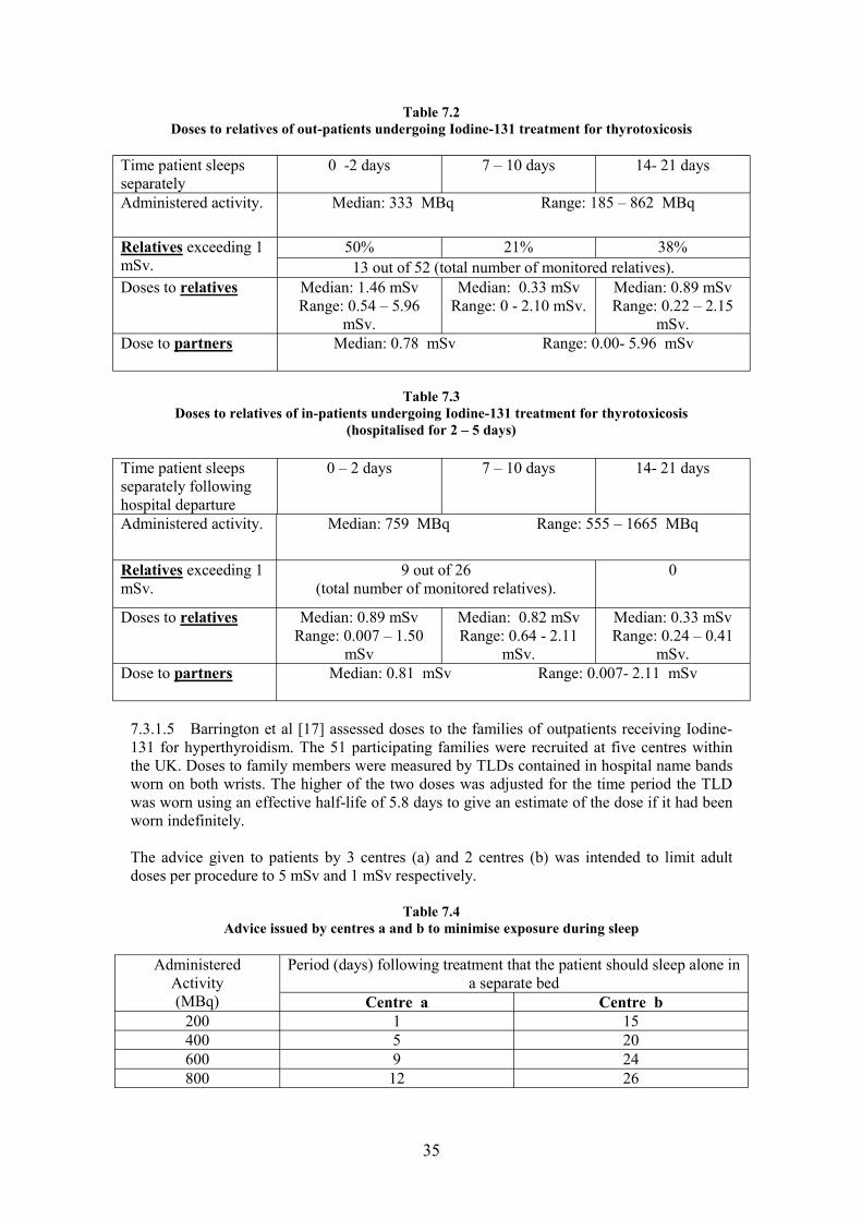
Table 7.2 Doses to relatives of out-patients undergoing Iodine-131 treatment for thyrotoxicosis
Time patient sleeps separately
0 -2 days 7 – 10 days 14- 21 days
Administered activity. Median: 333 MBq Range: 185 – 862 MBq
Relatives exceeding 1 50% 21% 38% mSv. 13 out of 52 (total number of monitored relatives). Doses to relatives Median: 1.46 mSv
Range: 0.54 – 5.96 Median: 0.33 mSv
Range: 0 - 2.10 mSv. Median: 0.89 mSv Range: 0.22 – 2.15
mSv. mSv. Dose to partners Median: 0.78 mSv Range: 0.00- 5.96 mSv
Table 7.3 Doses to relatives of in-patients undergoing Iodine-131 treatment for thyrotoxicosis
(hospitalised for 2 – 5 days)
Time patient sleeps 0 – 2 days 7 – 10 days 14- 21 days separately following hospital departure Administered activity. Median: 759 MBq Range: 555 – 1665 MBq
Relatives exceeding 1 9 out of 26 0 mSv. (total number of monitored relatives).
Doses to relatives Median: 0.89 mSv Range: 0.007 – 1.50
Median: 0.82 mSv Range: 0.64 - 2.11
Median: 0.33 mSv Range: 0.24 – 0.41
mSv mSv. mSv. Dose to partners Median: 0.81 mSv Range: 0.007- 2.11 mSv
7.3.1.5 Barrington et al [17] assessed doses to the families of outpatients receiving Iodine131 for hyperthyroidism. The 51 participating families were recruited at five centres within the UK. Doses to family members were measured by TLDs contained in hospital name bands worn on both wrists. The higher of the two doses was adjusted for the time period the TLD was worn using an effective half-life of 5.8 days to give an estimate of the dose if it had been worn indefinitely.
The advice given to patients by 3 centres (a) and 2 centres (b) was intended to limit adult doses per procedure to 5 mSv and 1 mSv respectively.
Table 7.4 Advice issued by centres a and b to minimise exposure during sleep
Administered Activity
Period (days) following treatment that the patient should sleep alone in a separate bed
(MBq) Centre a Centre b 200 1 15 400 5 20 600 9 24 800 12 26
35

Table 7.5 Doses received by adult family members (n=31) related to the Iodine-131
activity administered for the treatment of hyperthyroidism (Barrington et al [17])
Administered Activity (MBq)
Dose (mSv)
Minimum 200 0.2 (minimum recordable dose)
Maximum 608 5.8
Median 388 0.5
Adult family members following the advice provided by centre b received only 32% of the dose of adults following the advice provided by centre a. The authors state that 97% of comforters and carers received doses of less than 3 mSv. They went on to state that patients administered 200 MBq, 400 MBq, 600 MBq or 800 MBq should be advised to sleep alone for 1, 5, 9, or 12 days respectively from their partners to comply with their imposed 3 mSv dose constraint.
7.3.1.6 Mathieu et al [30] measured doses to family members of patients treated with Iodine131 for thyroid cancer (22 patients) and hyperthyroidism (18 patients). The patients and families were personally instructed about the aim and design of the study and standard advice was given: 1) To keep close contact to a minimum for a few days (usually 5) 2) To observe reasonable standards of personal hygiene and cleanliness 3) To avoid urine and saliva contamination.
Dosemeters were permanently worn on the chest for 2 to 3 weeks by each individual living in the patient’s house. All hyperthyroid patients were treated as out-patients and received 200 MBq (n=4), 300 MBq (n=2), 400 MBq (n=7), or 600 MBq (n=5) Iodine-131. Of the 16 couples involved in the study, only 5 adopted separate sleeping arrangements. The median of the measured doses received over a period of 3 weeks by the partners who adopted separate sleeping arrangements was 1.07 mSv (range 0.22 – 1.27) (n=5). The median of measured doses of partners who did not sleep separately was 1.01 mSv (range 0.05 – 5.23) (n=11). Mathieu et al [30] note that the partners of two patients with retained activities less than 440 MBq and who did not adopt separate sleeping arrangements or current advice, received doses measured over 21 days of 4.3 mSv and 5.2 mSv
7.3.2 Iodine-131 Thyrotoxicosis Therapy (Contributed dose measurement)
A TLD issued to the spouse of an Iodine-131 thyrotoxicosis therapy patient in 1999 recorded a dose of 0.85 mSv. The patient slept separately from their spouse for 5 days following treatment. Factors affecting the measured dose include the period for which the dosemeter was worn (approximately 2 – 3 weeks), and the placement of the TLD on the bed head representative of the average body position after separate sleeping arrangements ended {centre A}.
36
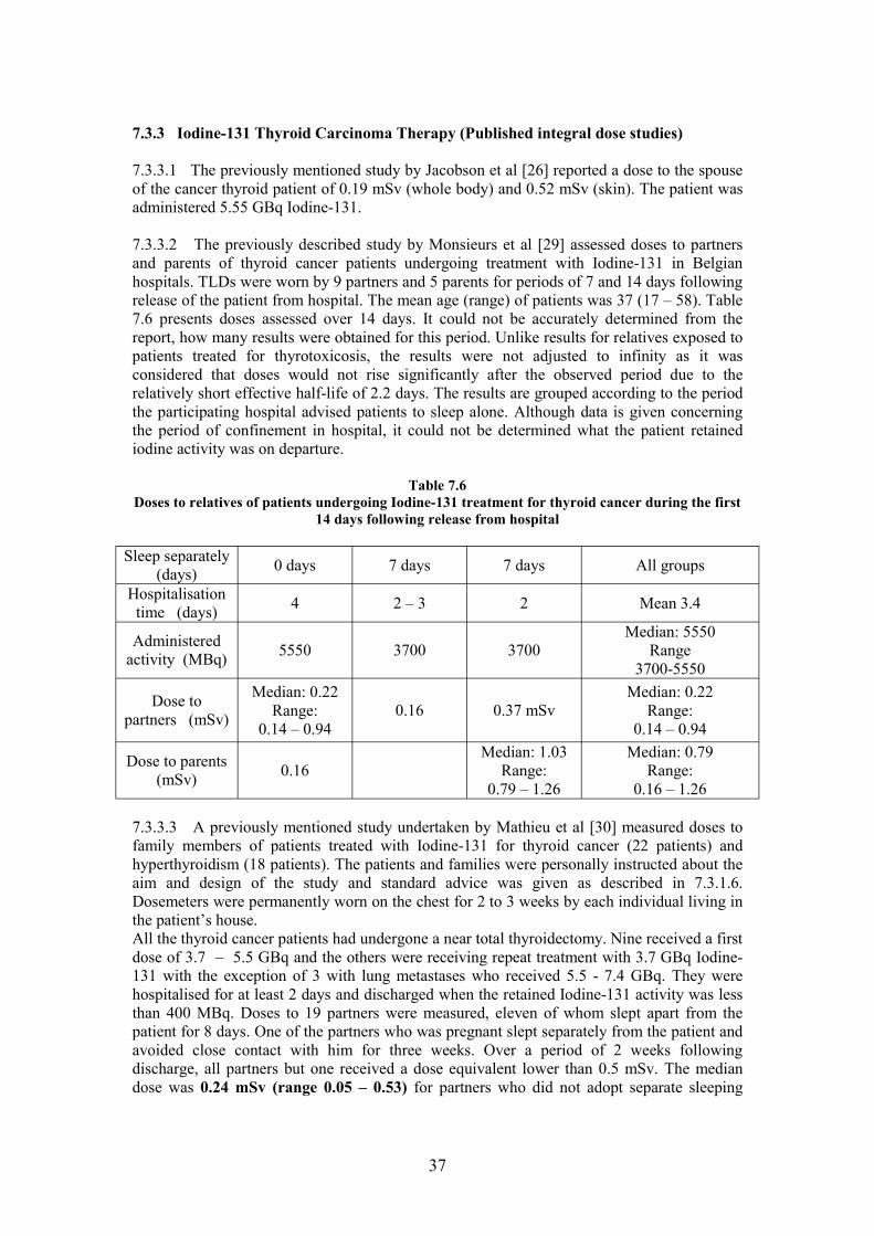
7.3.3 Iodine-131 Thyroid Carcinoma Therapy (Published integral dose studies)
7.3.3.1 The previously mentioned study by Jacobson et al [26] reported a dose to the spouse of the cancer thyroid patient of 0.19 mSv (whole body) and 0.52 mSv (skin). The patient was administered 5.55 GBq Iodine-131.
7.3.3.2 The previously described study by Monsieurs et al [29] assessed doses to partners and parents of thyroid cancer patients undergoing treatment with Iodine-131 in Belgian hospitals. TLDs were worn by 9 partners and 5 parents for periods of 7 and 14 days following release of the patient from hospital. The mean age (range) of patients was 37 (17 – 58). Table 7.6 presents doses assessed over 14 days. It could not be accurately determined from the report, how many results were obtained for this period. Unlike results for relatives exposed to patients treated for thyrotoxicosis, the results were not adjusted to infinity as it was considered that doses would not rise significantly after the observed period due to the relatively short effective half-life of 2.2 days. The results are grouped according to the period the participating hospital advised patients to sleep alone. Although data is given concerning the period of confinement in hospital, it could not be determined what the patient retained iodine activity was on departure.
Table 7.6 Doses to relatives of patients undergoing Iodine-131 treatment for thyroid cancer during the first
14 days following release from hospital
Sleep separately (days) 0 days 7 days 7 days All groups
Hospitalisation time (days) 4 2 – 3 2 Mean 3.4
Administered activity (MBq) 5550 3700 3700
Median: 5550 Range
3700-5550
Dose to partners (mSv)
Median: 0.22 Range:
0.14 – 0.94 0.16 0.37 mSv
Median: 0.22 Range:
0.14 – 0.94
Dose to parents (mSv) 0.16
Median: 1.03 Range:
0.79 – 1.26
Median: 0.79 Range:
0.16 – 1.26
7.3.3.3 A previously mentioned study undertaken by Mathieu et al [30] measured doses to family members of patients treated with Iodine-131 for thyroid cancer (22 patients) and hyperthyroidism (18 patients). The patients and families were personally instructed about the aim and design of the study and standard advice was given as described in 7.3.1.6. Dosemeters were permanently worn on the chest for 2 to 3 weeks by each individual living in the patient’s house. All the thyroid cancer patients had undergone a near total thyroidectomy. Nine received a first dose of 3.7 – 5.5 GBq and the others were receiving repeat treatment with 3.7 GBq Iodine131 with the exception of 3 with lung metastases who received 5.5 - 7.4 GBq. They were hospitalised for at least 2 days and discharged when the retained Iodine-131 activity was less than 400 MBq. Doses to 19 partners were measured, eleven of whom slept apart from the patient for 8 days. One of the partners who was pregnant slept separately from the patient and avoided close contact with him for three weeks. Over a period of 2 weeks following discharge, all partners but one received a dose equivalent lower than 0.5 mSv. The median dose was 0.24 mSv (range 0.05 – 0.53) for partners who did not adopt separate sleeping
37

arrangements (n=8), and 0.17 mSv (range 0.02 – 0.49) for those partners who did (n=11). The pregnant partner received a dose over a period of 3 weeks of 0.11 mSv.
7.3.3.4 Thomson et al [9] reference a study by Harbert and Wells [42] who used film badges to measure doses in the home to relatives of patients receiving Iodine-131 for thyroid ablation (n=7, 825-1340 MBq) and thyroid cancer (n=4, 7770-11,507 MBq). The thyroid cancer patients were hospitalised until retained activity was in the range 680-1570 MBq. They reported median (maximum) doses to the family of cancer patients of 0.3 (0.8) mSv and for thyroid ablation patients 0.2 (0.5) mSv.
7.3.4 Iodine-131 Thyroid Carcinoma Therapy (Contributed dose measurements)
Doses to the mother and father of a 10 year old boy were measured with personal electronicdosimeters during the inpatient phase of his treatment for thyroid cancer with Iodine-131{centre K}. Following administration of 3700 MBq liquid Iodine-131, the hospital allowedthe parents unrestricted access to the child’s room but were advised to avoid close contact. Alarge radiation shield designed for Cs-137 was provided for them to sit behind and the timespent by each parent in the room was documented. The patient coped well with the restrictions and confinement, however, the mother was reported to be very possessive and demanding on ward staff. The patient was given 3700 MBq for treatment of metastases following an earlier dose of1850 MBq for thyroid remnant ablation. The patient’s body level was monitored with a doserate meter and he was discharged late the following day when his Iodine-131 retention wasabout 400 MBq, mostly within the GI tract and bladder with about 3 MBq in thethyroid region (determined from a scan). The dose recorded on the electronic personal dosemeters worn by his parents were as follows:
Mother: First day: 22 mSv over 4.5 hours (Stayed 1 hr immediately after drink and returnedfor 3.5 hrs in evening). Second day: 5 mSv over 1.5 hours. Father: First day: 22 mSv over 3.5 hours in evening.It was suggested that the mother on the second day and the father during his one visit,probably did not make good use of the shield.
7.3.5 Iodine-131 anti-B1 Antibody Therapy (A published integral dose study)
Rutar et al [24] issued dosemeters to members of the family of patients treated in America with Iodine-131 Anti-B1 Antibody. Results are summarised in Table 7.7 along with maximum, minimum, average and 95th percentile values which have been calculated for this report from these results. Family members received film badges, TLDs, OSLDs (optically stimulated luminescent dosemeter) or electronic digital dosimeters. In most cases, the family member was given more than a single type of device and instructions were given concerning how they were to be worn. They were asked to log their activities/behaviour.
To meet the predetermined conditions for release from hospital, the patient had to agree to sleep alone for 3 nights, refrain from travelling on a long automobile trip (> 6 hours) for 2 days, refrain from travelling by aeroplane or mass transportation for 4 days, avoid prolonged contact with children and pregnant women for 10 days, maintain a prudent distance (> 9 feet) from others for 4 days. The effective half-life measured from tracer studies was combined with the maximum dosemeter reading and the period worn to calculate the total cumulative dose to the family member.
38

Table 7.7 Estimates of doses to family members (caregivers) of patients
undergoing Iodine-131 anti-B1 antibody therapy
Patient number.
Admin. Activity (GBq)
Intravenous
Effective T1/2
(hours)
Initial doserate mSv/hr (1 m)
Doserate factor (mSv/hr/ MBq)
Cumulative dose -(mSv)
1 3.85 54.8 0.14 0.04 0.72 2 1.67 72.1 Not measured. 0.70 3 1.48 85.2 1.76 4 1.54 63.6 0.09 0.06 1.74 5 1.14 71 0.06 0.05 0.52 6 1.26 64 0.04 0.03 1.11 7 3.06 75.6 0.14 0.05 2.33 8 4.25 56.3 0.14 0.03 2.69 9 2.15 62.9 0.05 0.02 0.96 10 2.7 52.9 0.08 0.03 0.27 11 0.94 80.3 0.03 0.03 1.71 12 1.74 71.3 0.09 0.05 1.45 13 2.27 62.5 0.09 0.04 0.71 16 3.57 59.4 0.18 0.05 1.74 17 2.48 62 0.11 0.04 0.60 18 3.92 67 0.12 0.03 1.53 19 3.86 45.5 0.1 0.03 1.49 20 1.76 77.4 0.08 0.05 2.31 21 3.58 73 0.13 0.04 2.27 22 4.77 67.9 0.16 0.03 1.49
Number of measurements
20 20 18 18 20
Maximum 4.77 85.20 0.18 0.06 2.69 Minimum 0.94 45.50 0.03 0.02 0.27 Average 2.60 66.24 0.10 0.04 1.41
95th percentile 2.35 Results are taken from Table 2 of the referenced report excluding two results for a dosemeter which was shared (patients 14 and 15).
All but one of the patients were released immediately after administration. Patient 22 was retained in hospital for 1 hour due to a combination of the measured doserate and administered activity exceeding release parameters. The report does not clearly specify the relationship between the patient and the monitored family member however, they are described as ‘caregivers’ which implies a certain degree of close contact under normal circumstances.
Patient number 11 was to receive conventional chemotherapy shortly after the therapeutic administration and in the short time that the dosemeters were worn (3 days) the patient and caregiver spent more time together than usual.
39

7.3.6 Paediatric Iodine-131 mIBG Therapy (Published integral dose studies)
7.3.6.1 Van der Steen et al [23] measured doses to parents of paediatric patients during the in-patient phase of their mIBG Iodine-131 therapy. This study involved 3 patients aged 4, 5 and 7 (patients 1,2 and 3). Patient 1 had 1 treatment (2.2 GBq) and patients 2 and 3 each had 3 treatments (1.5, 3.8, 3.7 GBq and 1.7, 2.7, 1.7 GBq respectively). The presence of one parent in the nursing room was arranged during the treatment. The parent was requested to keep at a distance whenever possible, to leave the room when the patient was asleep and to wear a pocket dosemeter to record external dose. Basic instructions were given for minimising ingestion hazards both during the period of stay in hospital and for one week after departure. The parents thyroid was blocked commencing 1 day before treatment and continuing during the stay in hospital.
Parent doses measured with a pocket dosemeter were recorded for each treatment. Total external accrued doses in the hospital were 0.67 mSv, 1.89 mSv and 2.98 mSv respectively for parents 1, 2 and 3. Doses for each treatment are separately reported. The doses following departure from hospital were extrapolated by multiplying the external dose from the inpatient phase by the ratio of the Iodine-131 burden at departure and the administered activity. The results are included in Table 7.8 along with other published and provided information for paediatric Iodine-131 mIBG therapy.
7.3.6.2 Tristam [38] describes a worst case scenario of the exposure of a parent at the bedside of a child who has been administered 10 GBq Iodine-131 mIBG. An initial patient doserate at 0.5 m from a ‘reference child’ (25kg) of 2 mSv/hour is combined with a biexponential expression for reduction of dose rate with time to estimate a dose of 30 mSv if the parent remains at the bedside and takes no precautions. Doses are significantly lower than this when parents are advised to reduce time spent close to their children, to keep as large a distance as possible from the patient and to use portable bed shields. The summed dose to a mother and father of an 8 year old of 0.34 mSv measured by direct read-out dosemeters is described as typical. A dose of 1.5 mSv to a parent during the whole course of therapy of a 5 year old is reported as the highest in their experience. It is believed that the child in this case had 3 courses of treatment {centre R}.
7.3.7 Paediatric Iodine-131 mIBG Therapy (Contributed integral dose measurements)
7.3.7.1 Cumulative dose information (measured by TLD) for parents of paediatric patients undergoing mIBG Iodine-131 therapy has been provided {centres G and H}. Reported doses per procedure are in the range <0.1 – 1.2 mSv for the in-patient phase and 0.1 – 2.7 mSv for the out-patient phase. The mean dose to persons monitored during both phases was 1.29 mSv. This does not include a dose due to contact with the patient following an associated diagnostic procedure. Administered activities were in the range 2 GBq to 12 GBq of Iodine-131. Measures to restrict exposure during the in-patient phase are listed as sedation and catheterisation of the child, the use of mobile lead shielding, restricting time at certain distances, the use of intercoms/CCTV and the setting of daily dose limits. The degree of use or compliance with these control measures is not known. It was stated that generally parents co-operated well with the wearing and return of dosemeters, although some were known to deliberately not wear dosemeters or turn the accumulating dosemeter off if they thought the daily limit was going to be exceeded. During the outpatient phase, the control of exposure of parents is described as being more difficult and relied on parents following instructions and using common sense. Doses to visitors during the in-patient phase are in the range 10 – 400 mSv (mean – 111 mSv). Persons visiting the patient once or twice generally received doses of < 100 mSv.
40

7.3.7.2 Measurements of doses to a mother and father were made by personal direct reading dosemeters during the first 5 – 6 days of the Iodine-131 mIBG treatments of their child in 1999 {centre J}. The father received doses of 101 mSv and 241 mSv, and the mother received 23 and 70 mSv for the first and second treatments of the child. The child had 2 more Iodine131 mIBG treatments but further records of doses to the parents were not found. The child was administered doses in the range 5.5 – 5.9 GBq Iodine-131. Lead shields were provided and the parents sat outside the room whenever they were not attending to the child.
Table 7.8 Doses to parents of children undergoing Iodine-131 mIBG therapy
(Published and provided information)
Information source Doses to parents per procedure – Range and Mean (in brackets) (mSv)
(Number of measurements of parent dose)
In-patient phase Out-patient phase TOTAL
Van der Steen et al 0.3 - 1.25 0.01 - 0.22 * 0.31 - 1.34 (7) (0.8) (0.1) (0.9) **
Tristam 0.34 – 1.5 Not measured
Centres G and H (32 in patient) (11 out patient)
0.1 - 1.2 (0.5)
0.1 - 2.7 (0.5)
0.9 - 3.8 (1.5)***
Centre J (4)
0.023 – 0.241 (0.109) Not measured.
* The outpatient doses were extrapolated from inpatient doses as described in the text. ** The total dose to two parents whose children received multiple treatments were 2.01 and 3.4 mSv. *** These values include doses received by parents from their child’s 74 MBq Iodine-131 mIBG scan. The mean dose for parents monitored during the treatment phases only was 1.29 mSv.
7.3.8 Radiation Synovectomy using 165Dy-ferric hydroxide (A published doserate study)
Havlik et al [27] measured the doserates from joints of patients treated in Austria with approximately 10GBq of 165Dy-ferric hydroxide. (165Dy: Physical half-life – 2.33 hours, main beta transitions - 0.3 (1.7%), 1.2 (15%), 1.3 (83%) MeV, main gamma transitions – 95 (4.0%), 361 (0.8%) keV, total gammas ~ 8% per disintegration.) Measurements were taken at distances of 5 cm, 0.5 m, 1 m and 2 m up to 200 minutes following administration. They assumed the effective half-life equalled the physical half-life due to lack of activity distribution and excretion and calculated the doses that could be received. Column 2 of Table 7.9 presents the maximum cumulative dose that could be received at the specified distance. Column 3 presents the cumulative dose received at 1 m assuming contact starts 5 hours following administration.
41

Table 7.9 Cumulative doses to persons exposed to patients undergoing
radiation synovectomy using 165Dy-ferric hydroxide Distance (m) Maximum dose that could be
received (mSv) Cumulative dose at 1 m commencing 5 hours after administration (mSv)
0.05 18,400
880.5 650 1 390 2 57
7.3.9 Therapy with Pure Beta Emitters (Published information)
Work completed in New York (Zanzonico et al [22]) demonstrated that the patient release criteria for external doses, as published in 1970 by the American National Council on Radiation Protection and Measurement (NCRP), can be met immediately following administration of beta decay therapy radiopharmaceuticals. This conclusion is drawn from the derivation of a ‘specific bremsstrahlung constant’ for beta emitters in bone and soft tissue. This constant is then used to calculate the patient retained activity which results in a cumulative effective dose of 5 mSv at 1 m with an exposure factor of 0.25. As an example, the authors state that at least 145,000 MBq of 89Sr Strontium Chloride would need to be administered before medical confinement was necessary. It should be noted that the report does not consider the potential contamination problems that could be associated with this quantity of retained activity or the exposure associated with close contact dose due to beta radiations.
7.3.10 Doses to Family Members Due to the Ingestion or Inhalation of Radioactivity (Published information)
Thomson and Harding [9] review available information regarding doses incurred due to the ingestion or inhalation of radioactive contamination from patients undergoing diagnostic and therapeutic nuclear medicine procedures. With respect to nuclear medicine therapy, they reference the work of Buchan and Brindle [44] who made a single measurement of Iodine131 uptake for 39 subjects associated with patients treated for hyperthyroidism approximately one week after treatment. They measured a maximum of 1.11 kBq Iodine-131 in the thyroid of a patient’s partner. They stated that Iodine-131 uptake was most likely to have arisen from several small amounts of ingestion. They found no link between iodine uptake and patient administered activity and no difference between groups who observed strict hygienic precautions and those who behaved normally. For the maximum value of 1.11 kBq, Thomson and Harding [9] estimate a dose to an adult’s thyroid of 1.6 mGy, assuming an uptake of 30 % following a single intake. For the same level of ingestion of Iodine-131 by a pregnant mother, they estimate the dose to the foetal thyroid as 15 mGy. Calculation of effective dose from the dose to a single organ can only be regarded as an approximation, as no account is taken of the dose to organs during the metabolism and excretion of Iodine-131 and the dose to target organs due to penetrating radiation from thyroid retained Iodine-131. Completing such calculations for the maximum thyroid doses presented above results in an effective dose of 0.08 mSv and 0.75 mSv to an adult and foetus respectively.
In the previously described study by Jacobson et al [26], twin sodium iodide detectors were used to analyse Iodine-131 transfer from patients to their families. They demonstrated a multi-transfer pattern of iodine intake and stated a link between spouse thyroid activity and intimacy. The mean thyroid dose to the husbands of 5 patients was assessed as 0.4 mGy (range 0.07 – 1.24 mGy). The associated effective doses are 0.02 mSv (range 0.004 – 0.062 mSv).
42

In the previously described study undertaken by Van der Steen et al [23], internal doses to 3 parents of paediatric Iodine-131 mIBG therapy patients were assessed by daily analysis of urine. The parents were given basic instructions for minimising ingestion hazards both during the period of stay in hospital and for one week after departure. The parents’ thyroid was blocked commencing 1 day before treatment and continuing during the stay in hospital. Intakes were estimated to be 0.09, 164.2 and 4.57 kBq respectively for patients 1,2 and 3. Results are reported for each treatment and body dose equivalent estimated taking into account the blocked thyroid.
Reiners et al [31] in a review of published studies of doses to persons exposed to hyperthyroid patients undergoing Iodine-131 therapy summarise results of Lassman et al [36] and Wellner et al [37]. Maximum Iodine-131 intakes of family members measured by whole body counter and thyroid probe were 4 kBq and 0.2 kBq respectively. All patients were hospitalised for at least 48 hours. The application of iodine kinetic model calculations by Wellner et al to their data showed that a maximum effective dose of more than 6 mSv might be reached when patients are treated as out patients. Reiners et al [32] state that this high value is based on model assumptions, and it is concluded that an additional risk of exposure to family members by incorporation is possible and may increase when patients are treated as outpatients.
A significant multicentre study assessing the radiation contamination risk posed by iodine therapy patients is currently being undertaken in the UK. Published results are expected in the near future.
7.3.11 Iodine-131 Therapy Repeat Treatment Rates
Iodine-131 thyroid cancer repeat treatment data is presented in Table 7.10. It should be noted that this data may slightly underestimate the repeat treatment rate due to patients receiving treatment either before or after the specified period.
Table 7.10 Iodine-131 thyroid cancer repeat treatment rates
Total No. of Administrations
Number of patients –
{centre N – data for 1988
to 2002}
Number of patients –
{centre N – data for 1992
to 2002}
Number of patients (Number of patient receiving multiple
treatments within a 5 year period) {centre A – data for 1993 to 2002}
1 140 111 119 2 26 21 12 (11) 3 15 11 4 (4) 4 2 0 2 (2) 5 0 0 0 6 2 0 0 7 2 0 0 8 1 0 0
Total patients 188 143 137 Total number of administrations
279 186 163
Percentage receiving 2 or
more treatments
26 % 22 % 12 %
43

Iodine-131 thyrotoxicosis repeat treatment data is presented in Table 7.11.
Table 7.11 Iodine-131 thyrotoxicosis repeat treatment rates
Total No. of Administrations
Number of patients (hospital 1) {centre A –data for 1997 to 2002}
Number of patients (hospital 2) {centre A –data for 1992 to 2002}
1 419 1541 2 27 144 3 3 7 4 0 2
Total patients 449 1694 Total number of administrations
482 1858
Percentage receiving 2 or
more treatments
7 % 9 %
All hospital 1 treatments were carried out within a 5 year period with 17 of 30 re-treatmentscompleted within a 12 month period. Three hospital 2 re-treatments were carried out over a period greater than 5 years.
7.3.12 Doses received from exposure to Iodine-131 therapy patients during transport
A BNMS conference abstract, Gunasekera et al [32], considers doses to persons exposed on public transport. A standard bus seat was obtained and patients asked to sit beside a water filled phantom for 20 minutes within 30 minutes of an Iodine–131 administration for thyrotoxicosis. Digital dosemeters were placed on the lateral chest wall adjacent to and opposite the patient, and a digital dosemeter was placed in a waterproof case in the middle of the phantom. Results of 23 sets of measurements normalised for an activity of 200 MBq are given in Table 7.12.
Table 7.12 Doses recorded by digital dosemeters placed on and inside a water filled phantom exposed to
thyrotoxicosis therapy patients retaining 200 MBq Iodine-131
Dosemeter position Doses recorded by digital dosemeter (mSv/hour --- 200MBq retained Iodine-131)
Adjacent Median 115 (range 29 - 229)
Middle Median 20 (range 9 - 40)
Opposite Median 4 (range 2 - 6)
Observing the variation in dose values, Gunasekera et al conclude that assumptions based on lateral doses at 0.1 m are pessimistic and that the effective dose is likely to be half of the adjacent value.
Barrington et al [17] assessed doses to relatives accompanying patients returning home following treatment with Iodine-131 for thyrotoxicosis. The measured doses were scaled to take account of the position of the patient and accompanying person in the car and converted from a surface entry dose to a mean whole body dose using an attenuation factor determined experimentally. Data was obtained for 37 passengers. Thirty-three travelled in private cars,
44

one by taxi and one by train. The median activity administered to patients whose relatives accompanied them home was 400 MBq (range; 195 - 800 MBq). The median journey time was 30 minutes (range: 6 – 125 minutes). The median whole body dose recorded was 0.03 mSv during the first hour of the journey (range: 0.002 – 0.52 mSv). The maximum dose received was recorded by a passenger in a car where the maximum distance between seating was only 71 cm, whereas a typical maximum distance in a modern car is 115 cm. The next highest dose recorded was 0.25 mSv.
7.3.13 Doses received by unpaid volunteers
Unpaid volunteers e.g. Persons who provide transport, hospital portering or patient visiting services may be exposed to ionising radiation as a result of the support, and in some cases, the care that they provide to patients who are radioactive. Administering departments will need to identify persons who provide close support or care to patients so that appropriate dose control measures can be implemented if necessary. Although the level of very close contact (0.1 m) between a volunteer and patient is expected to be less than that between a partner/spouse and patient, the volunteer/patient contact times at distances of 0.5 m and 1 m may be sufficient to result in a significant dose. Using the Cormack and Shearer methodology [12], the dose to a volunteer carer of a patient administered 800 MBq of Iodine-131 for thyrotoxicosis is estimated as approximately 1 mSv. This is based upon a repeated daily contact pattern of 1 hour at 0.5 m and 5 hours at 1 m commencing on the day of administration. The circumstances of each exposure are likely to vary significantly and need to be considered in a specific risk assessment. The possibility of repeat exposures also needs to be considered. It may very occasionally be necessary to treat a volunteer carer as a comforter and carer. In these situations, administering centres will need to choose constraints carefully given that doses may be incurred through the care of patients treated at more than one centre with associated difficulties in assessing and controlling doses. In these circumstances it would be appropriate to specify a constraint of 1 mSv per year.
Mountford and O’Doherty [10] reference the work of Harding et al [43], who estimated from dose rate measurements at 0.5 m from the patient that the dose to a porter will be about 0.1 mSv per month. This assumes the journey time between the ward and department is greater than average and that the same porter accompanies every in-patient making this journey. It is expected that contact times between volunteer porters and patients will be less than the contact times considered in this estimate.
45

7.4 SUMMARY OF RESULTS
The results of a number of important integral dose studies have been presented which have been supplemented by valuable contributions from project participants, particularly for paediatric Iodine-131 mIBG therapy. Limited information has been found concerning the exposure of supporters/carers to patients undergoing therapy with pure beta emitters.
The following tables summarise data presented in this chapter. Information from different studies have not been combined as the activities administered, precautions followed by exposed persons and the presentation of results varies appreciably between the studies and needs to be read with the dose information to assist an understanding of it.
Table 7.13 Doses to the spouse/partner following Iodine 131 thyrotoxicosis therapy
(Rounded to nearest 0.1 mSv. Published and provided dose information.)
Administ- Precautions Measured doses (mSv) Comments/ ration followed by Published or provided
information and (MBq) exposed Mean Range, supporter/
carer or
median maximum or
95th percentile reference
200 None 1.1 2.4 Published - [25]
200 Not known 4.8 0.1 – 13.6 Only 3 measurements Published - [26]
200 None 1.2 2.6 Published - [1, 9] Majority
continued to sleep with the patient.
Median 333 Out-patient 0.8 0 - 6.0 Range treatment.
(185 – 862) See comments. Slept separately for 0 – 21 days following
departure from hospital. Median 759 Various, 0.8 0.1 – 2.1 Range following Published - [29]
(555 – 1665) hospitalisation for 2-5 days. - See
comments. Median 388 Slept separately. 0.5 0.2 – 5.8 Published - [17] Range 1 – 26 days. (200 – 608) Precautions do
not include hospitalisation.
Mean 400 None (11 out of 1.0 0.1 – 5.2 Published - [30] Range 16 couples)
(200 – 600) 5/16 couples slept 1.1 0.2 – 1.3 separately (period
not specified). 444 Slept separately 0.9 Single measurement.
for 5 days Provided {centre A}
46

Table 7.14 Doses to the spouse/partner and parents following Iodine 131 thyroid carcinoma therapy
(Rounded to nearest 0.1 mSv. Published and provided dose information.)
Child or Adult Precautions Measured doses (mSv) Comments/ Administration followed by Published or provided
information and (MBq) exposed Mean or Range, supporter/ carer.
median maximum or 95th
reference.
percentile Adult - Period of Partner Single measurement 5550 hospitalisation 0.5 Published – [26]
not specified.
Adult Hospitalised for 0.2 0.1 – 0.5 Published - [30] at least 2 days.
3700 – 7400 (8/19 couples) Hospitalised for 0.2 0.1 – 0.5 at least 2 days. Slept separately
for further 8 days.
(11/19 couples) Thyroid ablation 825 – 1340 and Thyroid cancer 7770-11,507
Cancer patients hospitalised
until retained activity fell to
680 – 1570
Ablation: 0.2
Cancer: 0.3
Ablation: 0.5
Cancer: 0.8
Age not known Published [42]
MBq.
Age range – Hospitalisation Partners Partners Published - [29] 17-58 years. for 2 – 5 days 0.2 0.1 – 0.9 (Partners and then patients parents were slept separately Parents Parents
exposed.) for 0 – 7 days. 0.8 0.2 – 1.3 3700 - 5550
Child Advised to Doses to Parent dose measured 3700 avoid close
contact. Shield provided.
mother and father
both < 0.1
during in-patient phase. Provided {centre K}
Table 7.15 Doses to the spouse/partner (caregiver) following Iodine-131 anti-B1 antibody therapy
(Rounded to nearest 0.1 mSv. Published dose information.)
Administr- Precautions followed by Measured doses (mSv) Comments ation (GBq)
exposed supporter/ carer Mean 95th
percentile Mean 2.6 Slept separately for 3 nights 1.4 2.4 Published
Range Outpatient treatment - 1 of 22 [24] (0.9 – 4.8) patients retained in hospital for 1
hour.
47

Table 7.16 Doses to parents following paediatric Iodine 131 mIBG therapy
(Rounded to nearest 0.1 mSv. Published and provided dose information.)
Administration (GBq)
Precautions followed by
exposed supporter/
Measured doses/procedure
(mSv)
Comments/ Published or provided
information and reference
carer Mean or
median
Range, maximum
or 95th
percentile 2 – 12 1.5 0.9 – 3.8 Includes dose received from a
74 MBq Iodine-131 thyroid scan.
Provided {centres G and H} 1.5 – 3.8 Various
precautions followed during in-patient phase.
0.9 0.3 – 1.3 Dose during out-patient phase extrapolated from in-patient
dose measurement. Published [23]
5.5 – 5.9 0.1 <0.1 – 0.2 In-patient phase only. Dose to mother and father measured following two
treatments. Provided {centre J}
Various Parents advised to maximise distance,
minimise close contact and use
bed shield.
0.2 0.5 In-patient phase only. Maximum dose - 1.5mSv from
3 treatments . Mean dose is half the summed
parent dose of 0.34 mSv. Published and provided
[38]{centre R}
Table 7.17 Doses to members of the family following Iodine 131 therapy.
Transport and incorporation of Iodine-131. (Published and provided information.)
Circumstances of exposure
Administration (MBq)
Measured doses/procedure
(mSv)
Comments/ Published or provided
information and reference Mean
or median
Range, maximum
or 95th
percentile Transport Median:
400 Range:
(195 – 800)
0.03 0.002 – 0.52 Median journey time - 30 minutes (range: 6 – 125
minutes). Maximum dose recorded by a
passenger in small car. Published [17]
Incorporation Various 0.02 6 Maximum dose based on exposure modelling.
Published [26, 31, 36, 37]
48

Table 7.18 Cumulative doses to persons exposed to patients undergoing
radiation synovectomy using 165Dy-ferric hydroxide
Cumulative dose at 1 m commencing 5 hours after administration (mSv)
88
Table 7.19 Iodine-131 Therapy Repeat Treatment Rates
Therapeutic procedure
Percentage receiving 2 or more treatments
Comments/ Centre contributing information
Iodine-131 26 % centre N – data for 1988 to 2002 thyroid ca 22 % centre N – data for 1992 to 2002 treatment 12 % centre A – data for 1993 to 2002
Iodine-131 7 % centre A – data for 1997 to 2002 thyrotoxicosis
treatment 9 % centre A– data for 1992 to 2002
Table 7.20 Doses received by volunteer carers and porters
Carer maximum dose/procedure (mSv) assuming daily contact pattern of 1 hour at 0.5 m and 5 hours at 1 m commencing on the day 1 Dose estimate using Cormack
and Shearer methodology [12] of administration of 800 MBq Iodine-131.
Expected to be an Porter maximum dose (mSv/month) 0.1 overestimate of doses received
by volunteer porters.
7.5 CONCLUSION
It has been shown that supporters/carers of nuclear medicine therapy patients can receive doses significantly greater than the “other person” dose limit of 1 mSv in a calendar year or 5 mSv in 5 consecutive calendar years, unless precautions are implemented to restrict close contact.
Although doses from transport and incorporation of radionuclides may contribute to the total supporter/carer effective dose, it is the external dose due to close contact with the patient that will predominate in the majority of circumstances. This applies particularly to Iodine-131 thyrotoxicosis therapy, where a combination of the photon emissions of this radionuclide, high uptake and long effective half-life can create practical radiation protection problems in comparison with other nuclear medicine therapeutic procedures. For supporters/carers of these patients, a period of sleeping separately is a more appropriate control measure than a period of stay in hospital, taking into account economic, social factors and achievable dose reduction. Where there is rapid excretion of activity in a short period after administration, such as for Iodine-131 thyroid cancer/ablation therapy and mIBG therapy, a period of stay in hospital is an effective control measure.
49

For all modalities for which information has been presented, supporter/carer doses can, in the majority of circumstances, be restricted to 1 mSv/procedure by following appropriate advice as specified in guidance such as the MDGNs (8) or derived from published integral dose studies. Doserate studies have also been published from which restrictions can be derived if necessary [20]. For reasons given in 6.3.3, doses estimated using this method will in general be greater than the actual dose. For radiopharmaceuticals for which no information has been published, supporter/carer doses and restrictions can be derived from measurements of doserate and knowledge of the effective half-life, assuming a repeated contact pattern using a published methodology such as that published by Cormack and Shearer [12]. Chapter 15 of the MDGN (8) also identifies a method for estimating restrictions based on an equivalent activity of Iodine-131. Caution is advised in the use of this method, which may not always be applicable [21]. The dose constraints/procedure recommended in chapter 15 of the MDGNs (8) will be appropriate for the majority of nuclear medicine therapy procedures conducted in the UK.
Doses to supporters/carers during transport are unlikely to contribute significantly to the total dose providing reasonable measures can be taken to minimise close contact.
The information presented in this report indicates that in the majority of circumstances, doses due to the inhalation or ingestion of radionuclides will not form a significant component of the total supporter/carer dose. Published information suggests that high doses may be received, however, the true extent of these doses and the circumstances in which they occur are not certain. The UK multicentre study currently in progress to assess the radiation contamination risk posed by Iodine-131 will provide useful information in this area.
There will be situations where domestic circumstances, clinical circumstances, patient age and/or well being, result in supporter/carer doses exceeding 1 mSv/procedure. The most likely domestic circumstance will involve the inability or unwillingness of the supporter/carer or patient to comply with sleeping restrictions. A likely clinical circumstance could involve the unusual uptake or retention of an administered radionuclide making generic restrictions inadequate. These situations need to be identified and acted upon appropriately taking into account the risks to the supporter/carer. It may be appropriate, where the supporter/carer is not pregnant, to apply a higher constraint of 3 mSv/procedure and to derive associated restrictions. If these restrictions will not or cannot practicably be complied with to keep doses within a constraint of 3 mSv, consideration should be given to the treatment of the supporter/carer as a comforter and carer. This course of action would be particularly appropriate where the supporter/carer is 60 years of age or over.
It has been shown that repeat Iodine-131 thyrotoxicosis and ablation/cancer treatments are unlikely to lead to supporter/carer doses exceeding 5mSv in a 5 year period. However, modern computerised patient booking systems should be used to assist identification of repeat treatments and prompt the completion of a risk assessment. Appropriate action will be dependant on the doses that have been received previously and the risks to the exposed supporter/carer. If there is a possibility that doses could exceed 5 mSv in 5 consecutive years, consideration should be given to the treatment of the supporter/carer as a comforter and carer and/or the strengthening of control measures to achieve greater dose reduction. The possibility of the need for further repeat treatment should also be considered.
50

8. BRACHYTHERAPY
8.1 IDENTIFICATION OF EXPOSED PERSONS, EXPOSURE SCENARIOS AND PROFESSIONAL BODY GUIDANCE
The MDGNs (chapter 15) (8) advise that patients undergoing therapy with sealed source temporary implants or surface moulds should not be allowed to leave the hospital until the sources have been removed. Friends and relatives will be exposed when visiting these patients unless use of a remote afterloading technique allows the source to be temporarily removed or retracted.
The MDGNs (chapter 15) (8) advise that patients receiving permanent implants should remain in hospital long enough to ascertain that the sources are not likely to migrate from the implant site and be expelled from the body while radioactive. Friends and relatives may be exposed to these patients during transport from the hospital and subsequently at home when they are in the vicinity of the patient. The MDGNs (8) also advise that patients receiving 125I seed implants should avoid prolonged periods of very close contact (< 0.1 m) with pregnant women and children for 2 months after treatment
With respect to the exposure of supporters/carers in hospital, the MDGNs (chapter 9) (8) give advice concerning the use of bed shields, the arrangement of beds and the inclusion of written arrangements in local rules for restricting access to patients.
Sources implanted: Iridium-192 wire/hairpins. Ruthenium-106 eye plaques.
Caesium-137 appliances. Exposure is not expected to occur if remotely operated afterloading equipment is used.
Strontium-90 Appliances. Gold-198 grains Iodine-125 seeds. Cobalt-60 plaques.
The patient could be young in which case parents or guardians may receive a higher than normal exposure. Female supporters could be pregnant however, local procedures may restrict access to inpatients of visiting children and pregnant women.
8.2 THE EXPOSURE OF SUPPORTERS/CARERS OF BRACHYTHERAPY PATIENTS
8.2.1 Sources of information
The majority of information presented in this chapter consists of contributions from project participants. The main reason for the lack of directly relevant published information is likely to be the increased use of remote afterloading techniques that do not involve exposure of visitors during patient treatment. A secondary reason will be the circumstances in which the majority of exposures will occur. Exposures generally occur in the controlled environment of a specialised ward, where generic risk assessments can be used to specify simple control measures that take into account hospital specific parameters such as ward layout and clinical management of the patient.
51
The most common procedures that could involve exposure of the patient’s friends and family outside of the hospital are prostate cancer treatment using Iodine-125 seeds and the treatment of various tumours using Gold-198 grain implants. The information presented in this chapter consists of:
· A risk assessment for persons exposed in hospital to brachytherapy patients. · Doserate measurements at 1 m from patients with Iridium-192 and Gold-198 grain
implants and estimates of doses to patient contacts in hospital and following departure from hospital (gold grain permanent implants).
· Doserate measurements taken at various distances from a patient undergoing treatment with Iridium-192 implants.
· Measurements of the dose to parents of a paediatric patient undergoing treatment with Iridium-192 wire.
· Results of a generic risk assessment for patients with Iodine-125 seed implants for the treatment of prostate cancer. The assessment is based on ‘in-house’ and published patient doserates.
To supplement the information provided, doserate measurements for Iodine-125 and Gold198 permanent implants have been used to estimate doses to a spouse due to contact with the patient during the in-patient and out-patient phases. Doses during the out patient phase were calculated assuming a repeated contact pattern [12].
8.2.2 A risk assessment for intracavitary and interstitial brachytherapy based on air kerma rates
A risk assessment has been provided for visitors to patients undergoing intracavitary brachytherapy for gynaecological disease and interstitial brachytherapy for anal disease {centre D}. The assessment is based on the following exposure parameters:
· Nursing staff to discourage visiting during treatment, but when allowed, visits are restricted to a maximum of half an hour per day.
· Median treatment times: Intracavitary - 55 hours and interstitial 48 hours. Longest conceivable treatment time - Two 60 hour long sorbo applications in the same patient. Therefore visiting is expected to occur during 2 days of treatment but could occur during 6 days of treatment.
· The average of the 1 m Air Kerma Rate constants (AKR) from the use of sources in 1998, for calculating the representative dose.
· The maximum 1 m AKR of sources used in 1998, for calculating the highest conceivable dose.
52
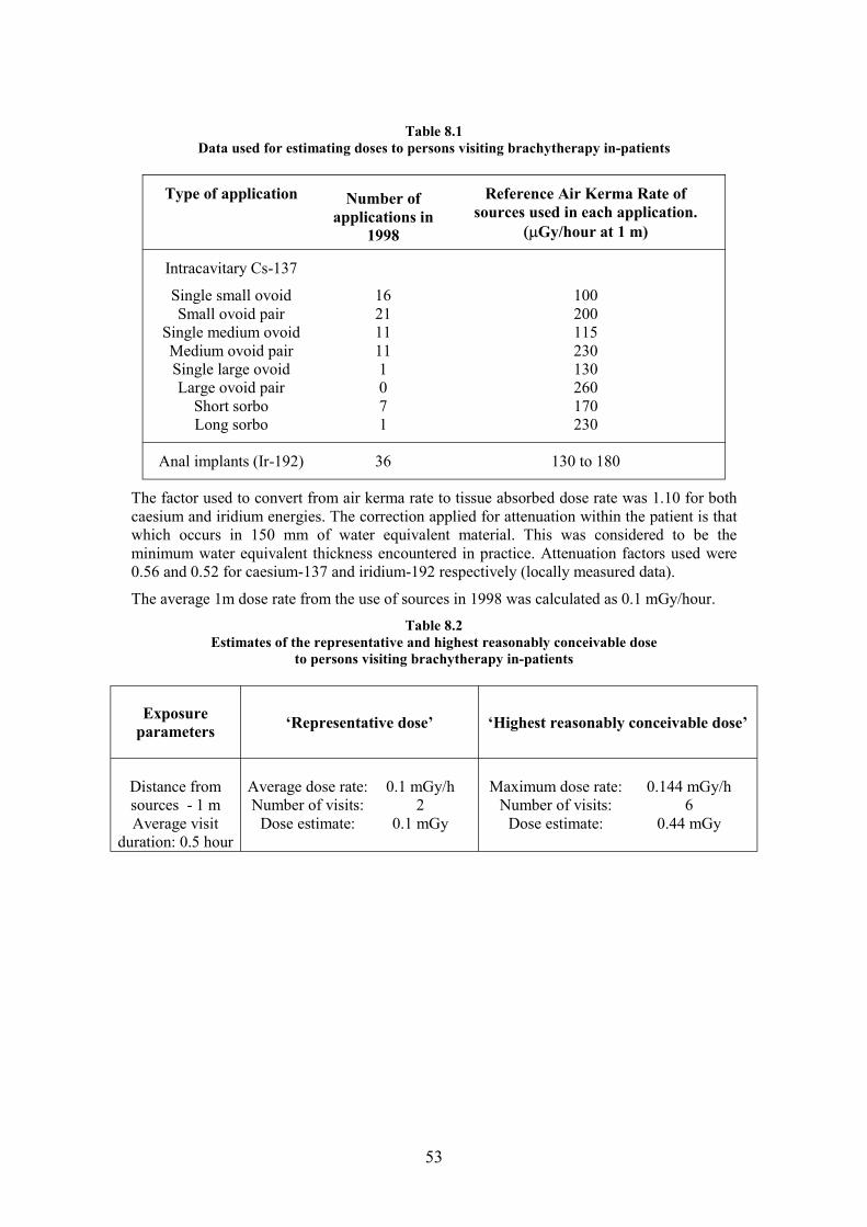
Table 8.1 Data used for estimating doses to persons visiting brachytherapy in-patients
Type of application Number of applications in
1998
Reference Air Kerma Rate of sources used in each application.
(mGy/hour at 1 m)
Intracavitary Cs-137
Single small ovoid 16 100 Small ovoid pair 21 200
Single medium ovoid 11 115 Medium ovoid pair 11 230 Single large ovoid 1 130 Large ovoid pair 0 260
Short sorbo 7 170 Long sorbo 1 230
Anal implants (Ir-192) 36 130 to 180
The factor used to convert from air kerma rate to tissue absorbed dose rate was 1.10 for both caesium and iridium energies. The correction applied for attenuation within the patient is that which occurs in 150 mm of water equivalent material. This was considered to be the minimum water equivalent thickness encountered in practice. Attenuation factors used were 0.56 and 0.52 for caesium-137 and iridium-192 respectively (locally measured data).
The average 1m dose rate from the use of sources in 1998 was calculated as 0.1 mGy/hour. Table 8.2
Estimates of the representative and highest reasonably conceivable dose to persons visiting brachytherapy in-patients
Exposure parameters ‘Representative dose’ ‘Highest reasonably conceivable dose’
Distance from Average dose rate: 0.1 mGy/h Maximum dose rate: 0.144 mGy/h sources - 1 m Number of visits: 2 Number of visits: 6 Average visit Dose estimate: 0.1 mGy Dose estimate: 0.44 mGy
duration: 0.5 hour
53
8.2.3 Doserates from patients undergoing Iridium-192 and Gold-198 grain implant therapy and a risk assessment for patient contacts based on AKR {centre N}.
All doserates measured with a Nuclear Enterprises PDM-1 at a distance of 1m from patients.
Iridium - 192 implant patient doserates: Average: 119 mSv/hr (Range: 50 – 270 mSv/hr) for activity Average: 1315 MBq (Range: 599 – 2738 MBq) To calculate maximum daily visiting times, an Air Kerma Rate constant (AKR) of 0.111 mGyh-1m2 is used with a standard treatment time of 6 days. Using a constraint of 1 mSv per procedure and assuming a patient/visitor distance of 1 m, the average, minimum and maximum implanted activities result in visiting times per day of 69, 151 and 33 minutes respectively.
Gold - 198 grain implant patient doserates: Average: 108 mSv/hr (Range: 40 – 267 mSv/hr) for activity Average: 2554 MBq (Range: 666 – 4588 MBq)
2To calculate maximum daily visiting times, an AKR of 0.056 mGyh-1m is used with a maximum in-patient treatment phase of 14 days. Using a constraint of 1 mSv per procedure and assuming a patient/visitor distance of 1 m, the average, minimum and maximum implanted activities result in visiting times per day of 29, 113 and 16 minutes respectively. The times will increase by a factor of 1.3 per day as the source decays.
For permanent implants, exposure of supporters/carers will occur following the patient’s discharge. Doses to patient contacts are calculated for the in-patient phase as described previously, and for the out-patient phase using two exposure distances of 2 m for 8 hours/day and more pessimistically at 1 m for 8 hours/day. Considering treatment with 4200 MBq of Gold-198, a 3-day in-patient phase results in a dose for the 2 and 1 m scenarios of 1.1 and 3.7 mSv respectively. A 10-day in-patient phase results in a dose for the 2 and 1 m scenarios of 0.9 and 3 mSv respectively. In practice, doses accrued during the in-patient phase of treatment will be significantly less than the constraint used for the calculation of visiting time restrictions as no account is made of the implementation of other dose control measures. These will include the use of shielding, requesting the visitor to maximise distances from the patient where possible and use of bed shields. In addition, no account is made of attenuation within the body of the patient.
8.2.4 Iridium-192 patient doserate measurements {centre A}
The doserates quoted in 8.2.3 are consistent with the theoretical doserates derived in the risk assessment (8.2.2), and with measurements made in centre A at various distances from a patient undergoing treatment with Iridium pins. The Iridium-192 activity in this case was 631 MBq and doserates measured at 1 m and 0.5 m from the patient’s face were 90 mSv/hr and 200 mSv/hr respectively.
8.2.5 Measurements of dose accrued by parents of a paediatric Iridium-192 therapy patient {centre O}
Doses were measured to both parents of a child undergoing treatment in centre O with Iridium-192 wires. The activity is unknown but the treatment required 60 Gy to be delivered to the side of the child’s nose in 4/5 days. TLD doses were 0.35 mSv (mother) and 0.30 mSv (father). The precautions followed to minimise their exposures are not known. It has been stated {centre P} that paediatric brachytherapy treatments are rare and it was noted that there may be a requirement to treat the parents as comforters and carers subject to the
54

outcome of an individual patient risk assessment. Significant factors for consideration are the age and status of the child, the sources in use and the location of the implant.
8.2.6 A risk assessment for Iodine-125 prostate implant patients {centre P}
A generic risk assessment completed at centre P for prostate implant patients using Iodine125 seeds concluded that the person at highest risk was the spouse, who might receive a maximum dose of 2 mSv (total). The assessment was based on ‘in house’ and published doserate measurements (Table 8.3). The normal release of the patient on the day after implantation was assumed.
Table 8.3 Doserates at various distances from an iodine-125 prostate implant patient
Distance from patient and reference
Doserate (mSv/hr) Front of patient Side of patient
Skin surface [34] Average 50 (range: 22 to 89 mSv/hr). 25cm {centre P} 2 – 3 0.2 - 0.5 50cm {centre P} 0.8 Bkg 100cm {centre P} 0.1 – 0.2 Bkg
Social contact with other persons was assessed as unlikely to result in a dose of 1mSv being received. The hospital completes an individual risk assessment and issues appropriate advice should a spouse be, or become, pregnant within the first year after the implant, but this is considered rare due to the age of the patients.
8.2.7 Estimation of doses to a spouse of patients undergoing therapy with permanent Iodine-125 and Gold-198 permanent implants {centre A}
The objective was to examine in more detail the exposure of the spouse of patients fitted with permanent Iodine-125 and Gold-198 implants. The average of patient doserate measurements quoted in 8.2.3 and 8.2.6 were used to calculate dose estimates using the Cormack and Shearer methodology [12] assuming a repeated contact pattern of 8 hours at 0.5 m during the night and 6 hours at 1 m and 2 hours at 0.5 m during the day. Given the point source nature of the implants, it is considered unrealistic to calculate doses based on exposure at a distance of 0.1 m. Using this methodology, a dose of 9mSv was estimated for the spouse of a Gold-198 therapy patient, following an in patient phase lasting 3 days. Sleeping restrictions would be necessary for approximately 12 days to reduce the dose to the spouse to 1 mSv. A dose of 0.8 mSv was estimated for the spouse of an Iodine-125 therapy patient following release from hospital the day after treatment.
55

8.3 SUMMARY OF RESULTS
Dose and doserate information has been provided for a range of brachytherapy procedures. Doserate information for brachytherapy procedures involving permanent implants has been used to estimate the dose to a spouse based on a repeated contact pattern. Table 8.4 summarises the data presented in this chapter.
Table 8.4 Doses to persons exposed to patients undergoing brachytherapy
(Rounded to nearest 0.1 mSv)
Procedure/ Precautions Measured Comments/ Activity doses/procedure Source of information (MBq) (mSv)
Maximum or range
Intracavitary 30 minute/day 0.1 – 0.4 {centre D} and maximum visiting
interstitial time therapy using Cs-137 and
Ir-192 Ir-192 Visiting time/day 1 {centre N}
1315 MBq - 69 minutes
Au-198 Dose constraint 0.9 – 3.7 {centre N} permanent during in patient
implant phase 4200 MBq 70 mSv/day Paediatric Mother 0.4 Single 4 –5 day treatment to a
Ir-192 Father 0.3 child. therapy {centre O}
I-125 prostate 2 Maximum dose to spouse implants {centre P}
Au-198 3 day in patient 9 Dose following departure from permanent phase hospital.
implant Estimates based on the average of patient dose rate measurements provided by centres N and O.
10 day in patient phase
1.5
I-125 prostate 0.8 implants
8.4 CONCLUSION
Information presented in this chapter shows that for the more commonly completed brachytherapy procedures, such as interstitial therapy using Iridium-192 and prostate cancer treatment using permanent Iodine-125 seed implants, doses to patient contacts can be kept to within 1 mSv per procedure. Doses to visitors of patients undergoing intracavitary therapy using Caesium-137 have also been shown to be less than 1 mSv/procedure. However,
56
exposure of visitors during this type of therapy procedure are expected to be rare due to the increasing use of afterloading techniques.
Measured doserates from Iridium-192 therapy patients and dose rates estimated from source AKRs for Caesium-137 therapy patients indicate that simple precautions involving minimisation of contact time and use of shields will be sufficient to ensure doses are well within 1 mSv per procedure. Given the controlled circumstances in which these exposures occur, it is extremely unlikely that a supporter will ever need to be treated as a comforter and carer. To set appropriate dose constraints requires knowledge of measured doses received by supporters/carers and also the control measures that will be implemented. All doses presented in this chapter, with the exception of those for the parents of a paediatric patient, are estimates based on doserates that will be greater than the actual dose. A dose constraint per procedure of 0.5 mSv may be appropriate for specifying control measures for non pregnant supporters/carers, although it is possible that doses less than this can be achieved. If pregnant supporters/carers have to visit the patient, it would be appropriate to use a constraint of 0.3 mSv as recommended by the MDGN (chapter 15) (8). It is unlikely but possible, that the emotional needs of the patient and/or the supporter/carer, necessitate greater levels of direct contact than specified in standard restrictions. Higher supporter/carer doses may be received in these circumstances and appropriate action should be taken, which achieves a balance between the needs of the patient and supporter/carer, and the need to minimise doses. It is still unlikely that supporter/carer doses will exceed 1 mSv from this exposure scenario.
Precautions are not necessary to minimise doses to contacts of Iodine-125 prostate implant patients unless the supporter/carer is pregnant, in which case it would be appropriate to implement restrictions specified in the MDGN (chapter 15). i.e. avoid prolonged periods of very close contact (< 0.1 m) with pregnant women (and children) for 2 months after treatment.)
Use of Gold-198 grains for the treatment of various cancer sites is the other permanent implant procedure considered in this report. Estimates of the dose to the spouse/partner based on AKR and measured doserates, by applying a repeated contact pattern, show that it is possible for limits of 1 mSv/year and 5 mSv/5 years to be exceeded. This estimate is likely to be greater than the actual dose accrued by the spouse/partner of these patients for reasons stated in 6.3.3. However, it would seem appropriate, based on these estimates, to derive restrictions using constraints specified in the MDGN chapter 15 (table 6.1 of this report). For Gold-198 grain implants, it is likely that simple restrictions concerning sleeping restrictions will suffice to comply with a 1 mSv constraint. Where there is reason to believe that these restrictions cannot be complied with, there will be a need to consider other control measures such as a longer period of hospitalisation, use of a 3 mSv constraint/procedure or the treatment of the exposed supporter/carer as a comforter and carer. Gold-198 grain therapy is used infrequently in the UK and these conclusions stem from dose rate measurements provided by one centre (N), which may not be relevant at other centres due to procedural variations.
For rarely completed brachytherapy procedures, risk assessments should be completed and restrictions derived from patient doserate measurements assuming a repeated contact pattern. An appropriate methodology for this has been described previously [12], and appropriate constraints are specified in the MDGN chapter 15 (8) (table 6.1 of this report).
57

9 FINAL CONCLUSION
This report presents dose measurements and dose estimates for persons who support or care for patients undergoing a wide range of diagnostic and therapeutic procedures conducted in the UK. The legislative background and professional body guidance relevant to the exposure of these persons has been identified and interpreted in recommendations concerning use of the comforter and carer term. For many exposure modalities, sufficient dose and associated information has been obtained to enable conclusions to be drawn concerning the need for a supporter/carer to be treated as a comforter and carer. These conclusions reflect the difference in the magnitude and circumstances of exposure between the different modalities and there would be little benefit in summarising those conclusions here without the necessary supporting information contained within the chapters. However, the following conclusions can be drawn:
· For typical X ray and diagnostic Nuclear Medicine procedures conducted in the UK, supporters/carers will not have to be treated as comforters and carers.
· The number of supporters/carers who will be treated as comforters and carers is generally less than expected.
· Dose constraints/procedures specified in the Medical and Dental Guidance Notes (chapter 15) (8) are appropriate for the majority of diagnostic and therapeutic Nuclear Medicine procedures conducted in the UK.
· The publication of dose measurements for supporters/carers of patients undergoing representative diagnostic X ray, diagnostic Nuclear Medicine and Brachytherapy procedures, would benefit the understanding of the extent of doses that might potentially be received by these persons and enable appropriate dose constraints to be specified.
The exposure of supporters and carers of patients undergoing medical procedures involving ionising radiation must be subject to a prior risk assessment and where necessary the issue of precautions. The risk assessment and precautions can be based on any combination of published guidance from professional bodies, representative published information or ‘in house’ measurements. The assessment and any associated precautions may be generic, in which case their limitations must be clearly stated, or they may address specific patient and supporter/carer circumstances. The RPA must be consulted during the preparation of risk assessments and has a clear role in the application of appropriate dose constraints and the derivation of any associated control measures. The term supporter and carer is used in general throughout this report, to portray persons who have the potential to be a comforter and carer. It must not be forgotten that children, and incapable adults often have a significant role in offering support to patients, which may result in their exposure to ionising radiation. This exposure needs to be optimised and kept within the “other person” dose limit of 1 mSv in a calendar year or 5 mSv in 5 consecutive calendar years. Although doses to supporters/carers of patients undergoing medical procedures involving ionising radiation will, in the majority of circumstances, be correctly considered to be of low radiological significance, the numbers of these exposures has the potential to be quite large and in a few instances the dose to the supporter/carer can be high. For these reasons and considering the relevant legal requirements, the control of the exposure of supporters/carers and associated issues should be addressed in departmental management control documentation.
58

APPENDIX A REFERENCES
Legislation and Guidance
(1) Ionising Radiations Regulations 1999 (SI 1999 No 3232) London, HMSO. - Work with ionising radiation. Approved code of practice and practical guidance on the Ionising Radiations Regulations 1999, (L121), London, HMSO. (2) Council Directive 96/29/Euratom of 13th May 1996, laying down basic safety standards for the protection of the health of workers and the general public against the dangers arising from ionising radiation. Official Journal of the European Communities, 1996 39 (L159) 1114. (3) Council Directive 97/43/Euratom of 30th June 1997, on health protection of individuals against the dangers of ionising radiation in relation to medical exposure. Official Journal of the European Communities, 1997 40 (L180). (4) International Commission on Radiological Protection. 1990 recommendations of the International Commission on Radiological Protection. ICRP Publication 60. Ann ICRP,21 Nos 1-3, 1991 (5) European Commission, Radiation protection 97, Radiation protection following Iodine131 therapy (exposures due to out-patients or discharged in-patients), Directorate-General Environment, Nuclear Safety and Civil Protection. (1998). (6) Ionising Radiation (Medical Exposure) Regulations 2000 (together with notes on good practice). London, DoH (2001). (7) Management of Health and Safety at Work Regulations 1999 (SI 1999 No 3242) London, HMSO. (8) Medical and Dental Guidance Notes, 2002. A good practice guide on all aspects of ionising radiation protection in the clinical environment; Prepared by the Institute of Physics and Engineering in Medicine (IPEM) with the support of NRPB, HSE, Health Departments, Environment Agencies. Published by IPEM. ISBN 1 903613 09 4 (9) Guidance Notes for the protection of persons against Ionising Radiations arising from Medical and Dental use, 1988. NRPB, HSE, Health Departments, Scottish Home and Health Department, Welsh Office. ISBN 0 85951 299 1. (10) National Radiological Protection Board (1993) Occupational, public and medical exposure. Doc NRPB, 4 (2) 7-9, 32-3. (11) Notes for Guidance on the Clinical Administration of Radiopharmaceuticals and use of Sealed Radioactive Sources, Administration of Radioactive Substances Advisory Committee. (ARSAC) December 1998.
Published Reports, Articles and Abstracts
[1] Thomson WH, Mills AP, Smith NB, Mostafa AB, Notghi A, Harding LK, 1993. Day and night radiation doses to patients’ relatives: Implications of ICRP 60. BNMS Abstract Nuc Med Comms. 14:275. [2] Mountford PJ, O’Doherty MJ, Harding LK, Thomson WH, Carter NJ, Bray D, Paul C, Batchelor S, 1991. Radiation doserates from paediatric patients undergoing 99mTc investigations. Nuc Med Comms, 12, 709-18. [3] Working Party of the Radiation Protection Committee of the British Institute of Radiology, including representatives of the Health and Safety Executive, the Department of Health, The Environment Agency and the National Radiological Protection Board, 1999. Patients leaving hospital after administration of radioactive substances. BJR report, 72, 121125. [4] Harding LK, Mostafa AB, Roden L, Williams N, 1985. Dose rates from patients having nuclear medicine investigations. Nuc Med Comms, 6, 191-4.
59

[5] Mountford, O’Doherty MJ, Forge NI, Jefferies A, Coakley AJ, 1991. Radiation doserates from adult patients undergoing nuclear medicine investigations. Nuc Med Comms, 12, 767777. [6] Mountford PJ, Coakley AJ, 1989. Body surface dosimetry following re-injection of 111Inleucocytes. Nuc Med Comms, 10, 497-501. [7] Greaves CD, Tindale WB, 1999. Dose rate measurements from radiopharmaceuticals, implications for nuclear medicine staff and for children with radioactive parents. Nuc Med Comms, 20, 179-187. [8] Cronin B, Marsden PK, O’Doherty MJ, 1999.Are restrictions to behaviour of patients required following fluorine-18 fluorodeoxyglucose positron emission tomographic studies? Eur JNM, 26, 121-128. [9] Thomson WH, Harding LK, 1995. Radiation protection issues associated with nuclear medicine out-patients. – Nuc Med Comms, 16, 879-892. [10] Mountford PJ, O’Doherty MJ, 1999. Exposure of critical groups to nuclear medicine patients. Applied Radiation and Isotopes, 50, 89–111. [11] Mountford PJ, 1987. Estimation of close contact doses to young infants from surface doserates on radioactive adults. Nuc Med Comms, 8, 857-863. [12] Cormack J, Shearer J, 1998. Calculation of radiation exposures from patients to whom radioactive materials have been administered. Phys. Med. Biol. 49, 501-516. [13] Kurturan A, Pfreitfellner J, Schaffarich P, Smith-Jones P, Niederle B, Raderer M, Virgolini L, Bergmann H, Havlik E; 1997. Radiation doses deriving from patients undergoing 111In-DTPA-D-phe-1-octreotide scintigraphy. Eur JNM, 24, 1298-300. [14] Rose MR, Prescott MC, Herman KJ, 1990. Excretion of iodine-123-hippuran, technetium-99m red blood cells and technetium-99m-macroaggregated albumin into breast milk. J Nucl Med. 31, 978-984. [15] O’Doherty MJ, Kettle AG, Eustance CP, Mountford PJ, Coakley AJ, 1993. Radiation dose rates from adult patients receiving 131I therapy for thyrotoxicosis. Nuc Med Comms, 14, 160-168. [16] Greaves CD, Tindale WB, Flynn PJ, 1996. A survey of close contact regimes between patients undergoing diagnostic radioisotope procedures and children. Nuc Med Comms, 17, 554-561. [17] Barrington SF, O’Doherty MJ, Kettle AG, Thomson WH, Mountford PJ, Burrell DN, Farrell RJ, Batchelor S, Seed P, Harding LK, 1999. Radiation exposure of the families of outpatients treated with radioiodine for hyperthyroidism. Eur JNM, 26, 686-692. [18] Benedetto AR, Dziuk TW, Nusynowitz ML, 1989. Population exposure from nuclear medicine procedures: Measurement data. Health Physics, 57, 725-731. [19] Gill JR, 2000. Radiological risk in perspective: risks and benefits for comforters and carers. Journal of Radiological Protection, 20, 21-27. [20] Barrington SF, Kettle AG, O’Doherty MJ, Wells CP, Somer EJR, Coakley AJ, 1996.Radiation dose rates from patients receiving Iodine-131 therapy for carcinoma of the thyroid.Eur JNM, 23, 123-130. [21A] Roberts JK, 2000. Comments on the 1999 BIR working party report; Patients leavinghospital after administration of radioactive substances. BJR, 73, 453-454 [21B] Waller ML, 2001. Estimating periods of non-close contact for relatives of radioactivepatients. BJR, 74, 100-102. [22] Zanzonico PB, Binkert LB, Goldsmith SJ. 1999. Bremsstrahlung radiation exposure from pure Beta ray emitters. J Nucl Med, 40, 1024-1028. [23] Van der Steen J, Maessen HJM, Hoefnagel CA, Marcuse HR, 1986. Radiation protection during treatment of children with Iodine-131 meta-iodobenzylguanidine. Health Physics, 50, 515 – 522. [24] Rutar FJ, Augustine SC, Colcher D, Siegel JA, Jacobson DA, Tempero MA, Dukat VJ, Hohenstein MA, Gobar LS, Vose JM, 2001. Outpatient Treatment with 131I-Anti-B1 Antibody: Radiation Exposure to Family Members. J Nucl Med, 42, 907-915.
60

[25] Buchan RCT, Brindle JM, 1971. Radioiodine therapy to outpatients-the radiation hazard: BJR, 44, 973-975. [26] Jacobson AP, Plato PA, Toeroek D, 1978. Contamination of the Home Environment by Patients Treated with Iodine-131: Initial Results. American Journal of Public Health (AJPH), 68. 225-230. [27] Havlik E, Pirich C, Preitfellner J, Karanikas G, Schaffarich P, Hefner A, Sinzinger H, 2001. Radiation exposure from persons treated with 165Dy-ferric hydroxide. Nuc Med Comms, 22, 79-82. [28] Hilditch TE, Connel JMC, Davies DL, Watson WS, Alexander WD, 1991. Radiological protection guidance for radioactive patients – new data for therapeutic 131I. Nuc Med Comms, 12:485-495. [29] Monsieurs M, Thierens H, Dierckx RA Casier K, De Baere E, De Ridder, De Saedeleer C, De Winter H, Lippens M, Van Imschool S, Wulfrank D, Simons M, 1998. Real life radiation burden to relatives of patients treated with Iodine-131: a study in eight centres in Flanders (Belgium). Eur JNM, 25, 1368-1376. [30] Mathieu I, Caussin J, Smeesters P, Wambersie A, Beckers C, 1999. Recommended restrictions after 131I therapy: Measured doses in family members. Health Physics, 76, 129136. [31] Reiners C, Lassman M, 1999, Radioiodine (131I) treatment of hyperthyroidism: Radiation protection and quality assurance. Eur JNM, 26, 683-685 [32] Gunasekera R, Thomson WH, Harding LK, 1996. Use of public transport by iodine –131 therapy patients. Nuc Med Comms, 17, 275. [33] Gunasekera R, Thomson WH, Harding LK, 1996, Dose rates from iodine –131 thyrotoxic patients. Nuc Med Comms, 17, 996. [34] Smathers S, Wallner K, Korssjoen T, Bergsagel C, Hudson RH, Sutlief S, Blasko J, 1999. Radiation safety parameters following prostate brachytherapy. Int J Radiat Oncol Biol Phys, 45, 397-9. [35] Casey M, Malone DE, 2001. The Value of Digital Personal Dosemeters in Angiography/Interventional Radiology: Preliminary Experience. Radiation Protection Dosimetry, Vol 94, Nos 1-2. [36] Lassman M, Hanscheid H, Schelper LF, Korber C, Reiners C, 1998. Measurement of incorporation in family members of radioiodine therapy patients after therapy of benign thyroid diseases. (Abstract only in English), Nuklearmedizin 37, 120-123. [37] Wellner U, Escher W, Hillger W, Schicha H, 1998. The exposure of relatives to patients of a nuclear medical ward after radioiodine therapy by inhalation of 131I in their home. (Abstract only in English), Nuklearmedizin 37, 113-119. [38] Fleming J S, Perkins A C (editors), Tristam M, 2001. Targeted Radiotherapy, Chapter 8-Radiation Protection, IPEM Report 83. [39] Hesslewood SR, Thornback JR, Brameld JM, 1988. Indium-111 in breast milk following administration of 111In labelled leucocytes. J Nucl Med 29, 1301-2. [40] Hall ET, 1966. The Hidden Dimension. Doubleday, New York, 17-21. [41] Ho S-Y, Shearer DR, 1992. Radioactive contamination in hospitals from nuclear medicine patients. 1992. Health Physics 62, 462-466. [42] Harbert JC, Wells N, 1974. Radiation exposure to the family of radioactive patients. J Nucl Med 15, 887-888. [43] Harding LK, Mostafa AB, Thomson WH, 1990. Staff radiation doses associated with nuclear medicine procedures - a review of some recent measurements. Nuc Med Comms, 11,271-277. [44] Buchan RCT, Brindle JM, 1970. Radioiodine therapy to out patients - the contamination hazard. Br J Radiol 43,479-482.
61

APPENDIX B Our sincere thanks go to the following healthcare professionals who provided information which has been included in the text of this report.
Barry Baily, Cromwell Hospital, London. Nigel Benatar, Clinical PET Centre, Guy’s and St Thomas’ NHS Trust, London. Ian Coles, Radiation Physics and Radiobiology Department, Charring Cross Hospital, FulhamPlace Road, London. John Cormack, Chief Medical Scientist, Division of Medical Imaging, Flinders MedicalCentre, Bedford Park, South Australia. R.F. Dobbin, Clinical PET Centre, Guy’s and St Thomas’ NHS Trust, London.Anthony Flynn, Medical Physics Department, Cookridge Hospital, Leeds.Claire Greaves, Sheffield Teaching Hospitals NHS Trust, Medical Physics and Imaging,Northern General Hospital, Sheffield. W. Hallett, Clinical PET Centre, Guy’s and St Thomas’ NHS Trust, London. Michaela Moore, Radiation Protection Service, General Hospital, Newcastle Upon Tyne.Michael O’Doherty; Clinical PET Centre, Guy’s and St Thomas’ NHS Trust, London. Paul Ormsby, Derriford Hospital, Plymouth. Sharan Packer, HSE, Leeds.Derek D’Souza, Department of Medical Physics and Bioengineering, The MiddlesexHospital, London. Vivien Stevens, Sheffield Teaching Hospitals NHS Trust, Medical Physics and Imaging,Royal Hallamshire Hospital, Sheffield. Chris Taylor, Department of Medical Physics and Engineering, Leeds General InfirmaryDavid Temperton, Medical Physics Department, Regional Radiation Physics and ProtectionService, Edgbaston, Birmingham. Maria Tristam, Southampton University Hospitals NHS Trust, Southampton William Thomson, Department of Physics and Nuclear Medicine, City Hospital NHS Trust,Birmingham. Anne Walker, North Western Medical Physics Department, Christie Hospital NHS Trust,Manchester. John Wilkinson, North Western Medical Physics Department, Christie Hospital NHS Trust,Manchester. Steve Young, Sheffield Teaching Hospitals NHS Trust, Medical Physics and Imaging,Weston Park Hospital, Sheffield.
62

Printed and published by the Health and Safety ExecutiveC30 1/98
Printed and published by the Health and Safety Executive C1.10 09/03
ISBN 0-7176-2748-9
RR 155
9 78071 7 627486£15.00