RESEARCH Open Access B and T lymphocyte attenuator ...
11
RESEARCH Open Access B and T lymphocyte attenuator expression on CD4 + T-cells associates with sepsis and subsequent infections in ICU patients Nicholas J Shubin, Sean F Monaghan, Daithi S Heffernan, Chun-Shiang Chung and Alfred Ayala * Abstract Introduction: Sepsis is a deadly inflammatory condition that often leads to an immune suppressed state; however, the events leading to this state remain poorly understood. B and T lymphocyte attenuator (BTLA) is an immune- regulatory receptor shown to effectively inhibit CD4+ T-cell function. Therefore, our objectives were to determine: 1) if lymphocyte BTLA expression was altered in critically ill patients and experimentally induced septic mice, 2) whether augmented CD4+ T-cell BTLA expression was associated with poor septic patient outcomes, and 3) if BTLA expression affected the CD4+ T-cell apoptotic cell loss observed in the lymphoid organs of septic mice. Methods: Changes in CD4+ lymphocyte BTLA expression were compared with morbid event development in critically ill ICU patients (11 septic and 28 systemic inflammatory response syndrome subjects). Wild type and BTLA gene deficient mice were utilized to evaluate the expression and role of BTLA in septic lymphocyte apoptotic cell loss. Results: The observed septic ICU patients had a significantly higher percentage of peripheral blood BTLA+ CD4+ lymphocytes compared with critically ill non-septic individuals. Moreover, the non-septic patients with CD4+ T-cells that were greater than 80% BTLA+ were more susceptible to developing nosocomial infections. Additionally, in general, critically ill patients with CD4+ T-cells that were greater than 80% BTLA+ had longer hospital stays. Comparatively, circulating CD4+ T-cell and B-cell BTLA expression increased in septic mice, which associated with the increased septic loss of these cells. Finally, the loss of these cells and cellular apoptosis induction in primary and secondary lymphoid organs were reversed in BTLA deficient mice. Conclusions: An increased BTLA+ CD4+ lymphocyte frequency in the observed critically ill non-septic patients was associated with a subsequent infection; therefore, BTLA may act as a biomarker to help determine nosocomial infection development. Additionally, BTLA expression contributed to primary and secondary lymphoid organ apoptotic cell loss in experimentally septic mice; thus, BTLA-induced apoptotic lymphocyte loss may be a mechanism for increased nosocomial infection risk in critically ill patients. This study had a relatively small human subject cohort; therefore, we feel these findings warrant future studies evaluating the use of BTLA as a critically ill patient nosocomial infection biomarker. Introduction Sepsis is a leading killer of critically ill ICU patients [1-3]. Unfortunately, there are currently no effective mo- lecular biological therapeutics approved to treat sepsis [4], and although there appears to be potential in the biomarkers that predict sepsis susceptibility in critically ill patients, overall these are also lacking [4-8]. In the past 15 years it has become accepted that the early events following major trauma and acute sepsis onset cause the adaptive immune system to function at a di- minished capacity, which is evident by an inability to clear nosocomial infections and a loss of the delayed- type hypersensitivity response [9,10]. This ‘late septic’ adaptive immune cell suppression is thought to develop in response to an increase in anti-inflammatory media- tors, the induction of CD4 + T-cell and B-cell loss via apoptosis [11-14], and the actions of immune suppres- sive cells, such as T-regulatory cells [15-17]. The * Correspondence: [email protected] Department of Surgery at Rhode Island Hospital, Brown University Division of Surgical Research, 593 Eddy Street, Aldrich Building 2nd Floor, Providence, RI 02906, USA © 2013 Shubin et al.; licensee BioMed Central Ltd. This is an open access article distributed under the terms of the Creative Commons Attribution License (http://creativecommons.org/licenses/by/2.0), which permits unrestricted use, distribution, and reproduction in any medium, provided the original work is properly cited. Shubin et al. Critical Care 2013, 17:R276 http://ccforum.com/content/17/6/R276
Transcript of RESEARCH Open Access B and T lymphocyte attenuator ...

Shubin et al. Critical Care 2013, 17:R276http://ccforum.com/content/17/6/R276
RESEARCH Open Access
B and T lymphocyte attenuator expression onCD4+ T-cells associates with sepsis andsubsequent infections in ICU patientsNicholas J Shubin, Sean F Monaghan, Daithi S Heffernan, Chun-Shiang Chung and Alfred Ayala*
Abstract
Introduction: Sepsis is a deadly inflammatory condition that often leads to an immune suppressed state; however,the events leading to this state remain poorly understood. B and T lymphocyte attenuator (BTLA) is an immune-regulatory receptor shown to effectively inhibit CD4+ T-cell function. Therefore, our objectives were to determine: 1) iflymphocyte BTLA expression was altered in critically ill patients and experimentally induced septic mice, 2) whetheraugmented CD4+ T-cell BTLA expression was associated with poor septic patient outcomes, and 3) if BTLA expressionaffected the CD4+ T-cell apoptotic cell loss observed in the lymphoid organs of septic mice.
Methods: Changes in CD4+ lymphocyte BTLA expression were compared with morbid event development in criticallyill ICU patients (11 septic and 28 systemic inflammatory response syndrome subjects). Wild type and BTLA genedeficient mice were utilized to evaluate the expression and role of BTLA in septic lymphocyte apoptotic cell loss.
Results: The observed septic ICU patients had a significantly higher percentage of peripheral blood BTLA+ CD4+lymphocytes compared with critically ill non-septic individuals. Moreover, the non-septic patients with CD4+ T-cells thatwere greater than 80% BTLA+were more susceptible to developing nosocomial infections. Additionally, in general,critically ill patients with CD4+ T-cells that were greater than 80% BTLA+ had longer hospital stays. Comparatively,circulating CD4+ T-cell and B-cell BTLA expression increased in septic mice, which associated with the increased septicloss of these cells. Finally, the loss of these cells and cellular apoptosis induction in primary and secondary lymphoidorgans were reversed in BTLA deficient mice.
Conclusions: An increased BTLA+ CD4+ lymphocyte frequency in the observed critically ill non-septic patients wasassociated with a subsequent infection; therefore, BTLA may act as a biomarker to help determine nosocomial infectiondevelopment. Additionally, BTLA expression contributed to primary and secondary lymphoid organ apoptotic cell lossin experimentally septic mice; thus, BTLA-induced apoptotic lymphocyte loss may be a mechanism for increasednosocomial infection risk in critically ill patients. This study had a relatively small human subject cohort; therefore,we feel these findings warrant future studies evaluating the use of BTLA as a critically ill patient nosocomial infectionbiomarker.
IntroductionSepsis is a leading killer of critically ill ICU patients[1-3]. Unfortunately, there are currently no effective mo-lecular biological therapeutics approved to treat sepsis[4], and although there appears to be potential in thebiomarkers that predict sepsis susceptibility in criticallyill patients, overall these are also lacking [4-8]. In the
* Correspondence: [email protected] of Surgery at Rhode Island Hospital, Brown University Division ofSurgical Research, 593 Eddy Street, Aldrich Building 2nd Floor, Providence, RI02906, USA
© 2013 Shubin et al.; licensee BioMed CentralCommons Attribution License (http://creativecreproduction in any medium, provided the or
past 15 years it has become accepted that the earlyevents following major trauma and acute sepsis onsetcause the adaptive immune system to function at a di-minished capacity, which is evident by an inability toclear nosocomial infections and a loss of the delayed-type hypersensitivity response [9,10]. This ‘late septic’adaptive immune cell suppression is thought to developin response to an increase in anti-inflammatory media-tors, the induction of CD4+ T-cell and B-cell loss viaapoptosis [11-14], and the actions of immune suppres-sive cells, such as T-regulatory cells [15-17]. The
Ltd. This is an open access article distributed under the terms of the Creativeommons.org/licenses/by/2.0), which permits unrestricted use, distribution, andiginal work is properly cited.

Table 1 Clinical characteristics of the critically ill ICUpatients assessed
SIRS (n = 28) Sepsis (n = 11)
Age 59.8 (3.8) 58.4 (7.2)
Male gender 17 (60.7%) 6 (54.5%)
APACHE II score 16.6 (4 to 29) 20.0 (5 to 26)
Patients from trauma ICU 11 (39.3%) 5 (45.4%)
Patients from surgical ICU 17 (60.7%) 6 (54.6%)
Average hospital length of stay (days) 31.1 (5.1) 58.4* (10.5)
Site of infection
Lung 5 (45.4%)
Blood 2 (18.2%)
Abdominal 1 (9.1%)
Urinary tract 1 (9.1%)
Skin 1 (9.1%)
Catheter 1 (9.1%)
Subsequent nosocomial infection 11 (39.2%) 8 (72.7%)
Site of infection
Lung 3 (27.3%) 4 (50%)
Abdominal 1 (9.1%) 0 (0%)
Urinary tract 5 (45.4%) 3 (37.5%)
Skin 2 (18.2%) 1 (10%)
Sampling time following ICUadmission (days)
4 (0 to 45) 21 (3 to 83)
Days following subsequent infection
Following ICU admission 31.7 (9 to 94)
Following sampling 22.38 (6 to 49)
Data presented as mean (standard error of the mean), number (%) or median(range). APACHE, Acute Physiology and Chronic Health Evaluation; SIRS,systemic inflammatory response syndrome. *P <0.05 via an unpaired,two-tailed Student’s t test.
Shubin et al. Critical Care 2013, 17:R276 Page 2 of 11http://ccforum.com/content/17/6/R276
underlying mechanisms for why these events occur,however, have still yet to be fully defined.B and T lymphocyte attenuator (BTLA) is a recently
characterized co-inhibitory receptor that is known to po-tently inhibit CD4+ T-cell and B-cell function as well asdiminish pro-survival signaling in CD4+ T cells [18-20].Co-inhibitory receptors, including programmed deathreceptor-1 (PD-1), cytotoxic T-lymphocyte antigen-4(CTLA-4), and BTLA, have also recently gained tractionas effective (in the case of anti-CTLA-4; ipilimumab) orpotential therapeutic targets in a number of diseasestates [18,21-23]. These receptors have also been impli-cated in contributing to sepsis progression, wherebyCTLA-4 and PD-1 have been shown to be involved inT-cell apoptosis and dysfunction during experimentalsepsis in mice [24-26], while increased PD-1 expressionon CD4+ T cells correlated with a decreased proliferationcapacity in humans [27]. Recently, we have also reportedthat BTLA [28] and PD-1 [29] contribute to septic mor-bidity and mortality in mice, while also causing innateinflammatory cell dysfunction during acute sepsis[28,29]. Although BTLA expression on CD4+ T cells andB cells has been well documented [18,19], and expres-sion on these cells has been shown to contribute to anumber of disease states [30-33], the significance ofBTLA expression on lymphocytes during sepsis has yetto be fully addressed. We therefore set out to under-stand whether BTLA plays a role in driving lymphocytedysfunction and apoptosis during sepsis.
Materials and methodsPatientsBlood was obtained from trauma or surgical ICU pa-tients and was processed for BTLA expression usingflow cytometry by investigators who were blinded tothe clinical data. All patients classified as having a sys-temic inflammatory response syndrome (SIRS) response(n = 28) or a septic response (n = 11), as described pre-viously, were included in the study [3]. While all sam-ples were taken within 24 hours following enrollment inthe study, median sampling times were 4 days (range = 0to 43 days) and 21 days (range = 3 to 83 days) post ICUadmittance for the SIRS patients and the septic patients,respectively (Table 1). The development of nosocomial in-fections, infections acquired at least 72 hours followinghospital admittance, were noted following hospital ICUadmission, not according to sampling time. All observedsepsis instances were believed to be nosocomial in origin.Additionally, a secondary infection was defined as anadditional infection (that occurred following the initialsampling) appearing in a patient who was septic at thetime of sampling. Further, a secondary infection wasonly defined as such if it was caused by a differentpathogen. For comparison, blood was also taken from
healthy controls (n = 6). Full Rhode Island HospitalInstitutional Review Board approval was obtained.Consent was obtained from the volunteers for theblood draw and inclusion. Since ICU patient blood wasobtained concomitant with the daily laboratory draws,no written consent was necessary; however, the purposeof the study was explained to the participants (assent)prior to inclusion in the study.
MiceMale, age-matched (8 to 12 weeks of age) wild-type(WT) C57BL/6 and BTLA−/− gene-deficient mice wereobtained from Jackson Laboratories (Bar Harbor, ME,USA). All protocols were carried out in compliance withthe National Institutes of Health Guide for Animal Useand Care and were approved by the animal welfare com-mittee of Rhode Island Hospital.

Shubin et al. Critical Care 2013, 17:R276 Page 3 of 11http://ccforum.com/content/17/6/R276
Cecal ligation and punctureCecal ligation and puncture (CLP) was used to induceexperimental sepsis in mice as described previously[28,34]. The procedures and steps to minimize animalsuffering were approved by the Rhode Island HospitalInstitutional Animal Care and Use Committee.
B and T lymphocyte attenuator cellular phenotypingHuman subjectsPeripheral blood from critically ill patients and healthydonors was collected in anticoagulant, ethylenediaminetetraacetic acid-containing tubes. Red blood cells werelysed and leukocytes were stained with anti-BTLA (cloneJ168-540; BD Bioscience, San Jose, CA, USA) and ana-lyzed by flow cytometry, as described previously [28].CD4+ T cells were stained with monoclonal anti-CD4(clone OKT4; eBioscience, San Diego, CA, USA) follow-ing gating on the lymphocyte population and examinedusing Flowjo analysis software (Tree Star, Ashland, OR,USA).
MicePeripheral blood leukocytes were isolated 24 hours and72 hours following CLP or sham surgeries, and redblood cells were lysed as above. Splenocytes were ob-tained by homogenizing spleens between frosted glassslides followed by red blood cell lysis. BTLA (clone 6 F7;eBioscience) monoclonal antibody was used to assess forsurface expression on lymphocyte populations using mono-clonal antibodies to CD3 (clone 145-2C11; eBioscience),CD4 (clone RM4-5; Biolegend, San Diego, CA, USA),and B220-hi (RA3-6B2; eBioscience) populations usingflow cytometry as described elsewhere [28]. Intracellu-lar FoxP3 staining, following use of the FoxP3 stainingkit (eBioscience), was measured with anti-FoxP3 (cloneN418; eBioscience). FoxP3 was measured to distinguishthat the CD4+ T-cell population examined did not con-tain CD4+ T-regulatory cells. The extent of BTLA ex-pression/cell was calculated by determining the meanfluorescent intensity (MFI) divided by the isotype con-trol MFI for standardization. Ten CLP mice and sixsham mice were utilized for the phenotyping studies.The MFI experiments were conducted with three tofive mice for each group and were then repeated forconfirmation of the results.
Assessment of apoptosisThe spleen and thymus were obtained from WT andBTLA−/− mice 24 hours following the sham and CLP sur-gical procedures, and were then fixed in 10% formalin,paraffin embedded, sectioned and stained withhematoxylin and eosin or by a terminal deoxynucleotidyltransferase (TUNEL) assay (Roche, Indianapolis, IN, USA)as described previously [28]. The extent of change in the
TUNEL staining and the nuclear condensation in thehematoxylin and eosin-stained cells was assessed from im-ages at 20× magnification with a minimum of three fieldsper organ per sample being quantified using Image J soft-ware (National Institutes of Health, Bethesda, MD, USA).
Statistical analysisResults are expressed as the mean ± standard error ofthe mean. Statistical significance of the results presentedwas determined utilizing the Bonferroni post test follow-ing one-way analysis of variants for multiple compari-sons, an unpaired two-tailed Student’s t test or theMann–Whitney test for nonparametric data, or the useof a Spearman coefficient following a nonparametric cor-relation test. The statistical software used was Prism 5.0(GraphPad Software, Inc., La Jolla, CA, USA). P <0.05was used as a cutoff for significance.
ResultsPercentage of circulating BTLA+CD4+ T lymphocytes washigher in the septic compared with the SIRS ICU patientsThe apoptotic loss and diminished functional capacity ofCD4+ lymphocytes in critically ill septic patients are wellknown [9,10]. Given BTLA’s role in diminishing CD4+
T-cell function [18,31] and pro-survival signaling [20],we looked for differences in BTLA expression on thesecells from the peripheral blood of septic (n = 11) com-pared with nonseptic/SIRS (n = 28) critically ill ICU pa-tients to assess whether increased BTLA expression maybe a marker for poor septic outcomes, potentially due toBTLA-induced CD4+ T-cell apoptosis and/or diminishedfunction (patient characteristics are described in Table 1).We found that the percentage of circulating BTLA+CD4+
lymphocytes was significantly higher in the septic patientscompared with the patients with SIRS alone (Figure 1A,B).An increased percentage of BTLA+CD4+ T lympho-
cytes in critically ill patients was associated with an in-creased incidence of subsequent nosocomial infectionsand a longer hospital length of stay.As several recent studies have suggested, co-inhibitor
expression contributes to CD4+ T-cell loss and dysfunc-tion, while also contributing to septic morbidity [13,32].We therefore investigated whether or not the observedincrease in the percentage of BTLA+CD4+ lymphocytesin the critically ill patients associated with the develop-ment of a subsequent nosocomial infection and an in-creased hospital length of stay. With regard to the SIRSpatients, we found that those patients who developed asubsequent nosocomial infection had a significantlyhigher percentage of BTLA+CD4+ lymphocytes thanthe SIRS patients who did not develop later infections(Figure 1B). Further, these patients developed a subse-quent infection at least 6 days following blood sampling(Table 1); therefore, this may have been due to a

Septic SIRS 0
20
40
60
80
100
B * C
Subsequent Infection
No subsequent infection
0
20
40
60
80
100 * D
Subsequent Infection
No subsequent infection
0
20
40
60
80
100
% o
f C
D4+
cel
ls t
hat
are
BT
LA
+
> 80% < 80% 0
50
100
150
Ho
spit
al L
OS
(d
ays)
*
# CD4+ Lymphocytes (10^9 cells/mL)
E
Healthy control level
% o
f C
D4+
cel
ls t
hat
are
BT
LA
+
% o
f C
D4+
cel
ls t
hat
are
BT
LA
+
Total Critically-ill Patients
0 0.2 0.4 0.6 0.8 1.0
SIRS Patients
0
20
40
60
80
100
R = 0.504, p = 0.001
Septic Patients
% of CD4+ cells that are BTLA+
% o
f C
D4+
cel
ls t
hat
are
BT
LA
+ Total Critically-ill Patients
53.9% 84.8% 24.5% 95.5%
BTLA 100 101 10 10 102 3 4 100 101 10 10 102 3 4 100 101 10 10 102 3 4 100 101 10 10 102 3 40
20
40
60
80
100
% o
f Max
A Healthy Control
SIRS Patient - Subsequent
infection developed
SIRS Patient - No subsequent
infection developed Septic Patient
F
Figure 1 High BTLA+CD4+ T-lymphocyte percentage associated with poor critically ill ICU patient outcomes. Systemic inflammatoryresponse syndrome (SIRS) patients that did not develop a subsequent nosocomial infection had a lower BTLA+CD4+ T-cell percentage comparedwith SIRS patients that developed a nosocomial infection and patients who were septic at the time of sampling (A) (histograms represent CD3+CD4+
gated lymphocytes). Specifically, a significantly diminished BTLA+CD4+ lymphocyte percentage was observed in the total SIRS population(n = 28) compared with the septic population (n = 11) (B) (dashed line, mean healthy volunteer BTLA+CD4+ lymphocyte percentage (n = 6);these patients were not sex or aged matched). Importantly, a significantly diminished BTLA+CD4+ lymphocyte level was also observed in theSIRS patients who did not develop a subsequent infection compared with those that did prior to hospital discharge (C). Additionally, septicpatients that went on to develop a secondary nosocomial infection also maintained a high BTLA+CD4+ lymphocyte percentage (D). Regardinghow the BTLA+CD4+ lymphocyte percentage associated with patient outcomes, the patients with >80% BTLA+CD4+ lymphocytes had a significantlylonger hospital length of stay (LOS) (E). The BTLA+CD4+ lymphocyte percentage correlated moderately with the circulating CD4+ lymphocyte number inthe evaluated total critically ill patient pool, suggesting that BTLA expression does not diminish the ability for CD4+ lymphocytes to enter circulation (F).Data are mean ± standard error of the mean. *P <0.05 using an unpaired, two-tailed Student’s t test. BTLA, B and T lymphocyte attenuator; R, Spearmancoefficient following a nonparametric correlation test.
Shubin et al. Critical Care 2013, 17:R276 Page 4 of 11http://ccforum.com/content/17/6/R276
dysfunction in the adaptive immune response, such aswith CD4+ T cells. In the septic patients (those whoalready had an identified source of infection), the cor-relation between the changes in the percentage ofBTLA+CD4+ lymphocytes and the development of asecond subsequent infection during the same hospitalstay was lost (Figure 1C). However, one must note thatnearly all of the CD4+ T cells expressed BTLA in themajority of septic patients who developed a secondaryinfection, as stated above (Figure 1C). No associationwas found with the percentage of BTLA+CD4+
lymphocytes and the patients that died, since only sixof the total patients examined died while hospitalized(data not shown). Future studies with a larger patientenrollment will therefore be needed to understandwhether an increase in the percentage of BTLA+CD4+
T cells correlates with death in critically ill SIRS or sep-tic patients. Importantly, we also found that the critic-ally ill patients had a significantly increased hospitallength of stay if their CD4+ lymphocytes exceeded athreshold level of >80% BTLA+ (Figure 1D). This levelof BTLA expression was chosen based on the

Table 2 CD4+ T cells (CD3+, CD4+, FOXP3–) gained BTLAexpression (MFI) 72 hours following CLP surgery
Shubin et al. Critical Care 2013, 17:R276 Page 5 of 11http://ccforum.com/content/17/6/R276
observation that >80% of all septic patients had CD4+
T cells that expressed BTLA (Figure 1B).
Sham mice(n = 6)CLP mice(n = 10)
P value
Blood – 24 hours post surgery
% BTLA+ (SEM) 96.72 (0.65) 93.36 (0.88) 0.01
BTLA MFI (SEM) 11.91 (1.07) 10.00 (0.41) NS
Blood – 72 hours post surgery
% BTLA+ (SEM) 95.80 (1.41) 94.34 (1.70) NS
BTLA MFI (SEM) 8.79 (0.48) 12.10 (0.66) 0.02
Spleen – 24 hours post surgery
% BTLA+ (SEM) 97.67 (0.27) 97.29 (0.50) NS
BTLA MFI (SEM) 10.89 (0.20) 10.73 (0.76) NS
Spleen – 72 hours post surgery
% BTLA+ (SEM) 98.44 (0.36) 98.23 (0.90) NS
BTLA MFI (SEM) 7.43 (0.18) 11.64 (0.64) <0.01
P value evaluated via an unpaired, two-tailed Student’s t test ± standard errorof the mean (SEM); bold values are significant. Mean fluorescent intensity (MFI)experiments were conducted with three to five mice for each group and thenrepeated for confirmation of the results. BTLA, B and T lymphocyte attenuator;CLP, cecal ligation and puncture; NS, not significant.
Percentage of circulating BTLA+CD4+ T cells moderatelycorrelated with the number of circulating CD4+ T cellsSurprisingly, we observed that an increase in the percent-age of CD4+ T cells that were BTLA+ directly correlatedwith an increase in CD4+ T-cell number (Figure 1E). Thesedata were unexpected because of the inhibitory nature ofBTLA on CD4+ T cells. Interestingly, a recent article evalu-ating the nature of BTLA on CD4+ T cells in HIV patientsfound similar results to the data we observed with thesecritically ill patients [33]. These studies also found thatwhen the percentage of the circulating BTLA+CD4+ T cellsincreased, the number of the circulating CD4+ T cells alsoincreased. These data suggest that although BTLA is in-hibitory in nature, it may also be important for drivingCD4+ T cells into circulation following SIRS or sepsisinduction.Based on these results, and the results from our previ-
ous study whereby we found that BTLA gene-deficientmice were protected from experimental septic morbidityand mortality [34], we evaluated the effects of BTLA onCD4+ T cells following experimental sepsis induction inmice.
Table 3 B cells (B220high) gained BTLA expression (MFI)72 hours following CLP surgery
Sham mice(n = 6)
CLP mice(n = 10)
P value
Blood – 24 hours post surgery
% BTLA+ (SEM) 99.98 (0.02) 98.88 (0.25) <0.01
BTLA MFI (SEM) 43.74 (1.74) 37.92 (3.80) 0.24
Blood – 72 hours post surgery
% BTLA+ (SEM) 99.92 (0.02) 98.94 (0.66) 0.31
BTLA MFI (SEM) 48.54 (1.13) 71.15 (2.66) <0.01
Spleen – 24 hours post surgery
% BTLA+ (SEM) 99.83 (0.03) 99.85 (0.02) 0.65
BTLA MFI (SEM) 31.91 (0.60) 33.96 (1.10) 0.16
Spleen – 72 hours post surgery
% BTLA+ (SEM) 99.88 (0.02) 99.86 (0.05) 0.77
BTLA MFI (SEM) 26.33 (2.08) 37.89 (1.76) <0.01
P value evaluated via an unpaired, two-tailed Student’s t test ± standard errorof the mean (SEM); bold values are significant. Mean fluorescent intensity (MFI)experiments were conducted with three to five mice for each group and thenrepeated for confirmation of the results. BTLA, B and T lymphocyte attenuator;CLP, cecal ligation and puncture.
BTLA expression increased in intensity on CD4+ T cellsand B cells following experimental septic challenge inmiceAs with the human subject observational study above,we evaluated changes in the level of BTLA expressionon CD4+ T cells, as well as B cells, following experimen-tal sepsis induction in mice (CLP) as compared with asham procedure. Interestingly, at 24 hours and 72 hoursfollowing CLP and sham surgeries, we found thatnearly all spleen and circulating CD4+ T cells and Bcells expressed BTLA (Tables 2 and 3 and an examplein Figure 2A,D). These findings differed from what wasobserved in the critically ill patients, however, wherebysome of the CD4+ T cells from the nonseptic criticallyill patients did not express BTLA (Figure 1A). Becausenearly all of the mouse CD4+ T cells and B cellsexpressed BTLA following CLP and sham surgeries, wealso evaluated the extent of BTLA that was expressedby these cells by assessing the MFI of the BTLA anti-body using flow cytometry. We found no changes inthe BTLA MFI at 24 hours following the CLP andsham surgeries; however, there was an approximately1.5-fold increase in the BTLA MFI on both the circu-lating and splenic CD4+ T cells and B cells after72 hours in the mice subjected to CLP compared withthe sham mice (Tables 2 and 3 and representative his-tograms in Figure 2A,D).
BTLA gene deficiency ameliorated the onset of cellularapoptosis in primary and secondary lymphoid organsand was associated with the loss of CD4+ T cells and Bcells following CLP in mice.Splenic CD4+ T cells and B cells are apoptotically lost
in the lymphoid organs of critically ill patients duringsepsis [12]. Additionally, other studies have found thatBTLA contributes to CD4+ T-cell apoptosis in mice [20];

0 102
103
104
105
0
20
40
60
80
100
% o
f M
ax
101
102
103
104
105
0
20
40
60
80
100
% o
f M
ax
CD
4+/F
oxP
3- T
-cel
lsB
-cel
ls
SS
C
FoxP
3+
CD4+
Sham ---
CLP
BTLA+
BTLA+
10 1 10 2 10 3 10 4 10 50
50K
100K
150K
200K
250K
B220+
A
101
102
103
104
0
10 1
10 2
10 3
10 4
Sham CD
4+ , C
D3+ ,
Fox
P3- c
ell #
0
1 x 107
CLP
*
8 x 106
6 x 10⁶
4 x 10⁶
2 x 10⁶
72h - SPLEEN B
Sham B
-cel
l #
0
5 x 107
CLP
*4 x 107
3 x 10⁷
2 x 10⁷
1 x 10⁷
E
C
Sham
CD
4+ , C
D3+ ,
Fox
P3-
cell
# / m
L B
loo
d
0
*
6 x 105
4 x 10⁵
2 x 10⁵
CLP
72h - BLOOD
F
Sham
B-c
ell #
/ m
L B
loo
d
0
5 x 10 6
*
4 x 10 6
2 x 10 6
1 x 10 6
3 x 10 6
CLP
Example of CD4+ T-cell gating
D Example of B-cell gating
Figure 2 B and T lymphocyte attenuator expression was induced on CD4+ T cells and B cells 72 hours following cecal ligation andpuncture and was associated with the loss of these cells. Splenic CD4+ T-cell (A) and B-cell (D) B and T lymphocyte attenuator (BTLA) expressionwas evaluated 72 hours post surgery. As expected, BTLA was induced on CD4+ T cells (A) and B cells (D) (mean fluorescent intensity values presentedin Table 2). Concomitantly, a significant decrease in the CD4+ T-cell (B) and B-cell (E) numbers at 72 hours following cecal ligation and puncture (CLP)(n = 10) was observed when compared with sham mice (n = 6). Additionally, significantly lower peripheral blood CD4+ T-cell (C) and B-cell (F) numberswere also observed at this time point. Data are mean ± standard error of the mean. *P <0.05 using an unpaired, two-tailed Student’s t test.Representative flow histograms: filled grey, isotype control; dashed line, sham mouse; black line, CLP mouse.
Shubin et al. Critical Care 2013, 17:R276 Page 6 of 11http://ccforum.com/content/17/6/R276
we therefore assessed the contribution of BTLA inlymphocyte apoptosis in our model of sepsis. We ob-served that BTLA−/− mice were significantly protectedfrom cellular apoptosis (TUNEL staining and assessmentfor nuclear condensation via hematoxylin and eosinstaining) in both the thymus (Figure 3A,B) and thespleen (Figure 3C,D) 24 hours following CLP. Becausecell loss is a product of apoptosis, we found that spleenand peripheral blood CD4+ T-cell (non-T-regulatory)numbers (Figure 2B,C) and B-cell numbers (Figure 2E,F),72 hours following CLP, were decreased when com-pared with the sham mice (gating strategy found inFigure 2A,D). These results strongly suggest that aBTLA-induced apoptotic mechanism contributed, dir-ectly or indirectly, to the observed loss of the lympho-cytes in the spleen and peripheral blood.
BTLA−/− mice induced an increased level of CD4+ T cellsfollowing experimental sepsis inductionIn Figure 1E we observed that an increase in the per-centage of BTLA+CD4+ lymphocytes associated with anincrease in the circulating levels of CD4+ lymphocytes.This observation led us to hypothesize that BTLA ex-pression on CD4+ T cells may correlate directly with thecirculating level of these cells. We therefore evaluated
the levels of both circulating and splenic CD4+ T cells inWT mice compared with BTLA−/− mice following shamand CLP surgeries. Because nearly all of the circulatingmouse CD4+ T cells appeared to express BTLA, wecould not evaluate whether the addition of BTLA ex-pression following CLP in the WT mice contributed tochanges in CD4+ T-cell migration out of the secondarylymphoid organs, as it appeared to in the critically illhumans. We thus evaluated BTLA−/− mice to determinewhether a lack of BTLA expression led to a decrease inthe circulating CD4+ T cells following CLP comparedwith WT mice. We found that, as in septic humans,CLP led to a lower level of circulating CD4+ T cells inthe WT mice (Figure 4A). We observed no difference,however, in the level of the circulating CD4+ T cells inthe BTLA−/− mice, 24 hours following CLP comparedwith sham surgery (Figure 4A). When we evaluatedsplenic levels of these cells, we observed that BTLA−/−
mice had significantly more CD4+ T cells followingsham surgery compared with the WT mice (Figure 4B).Furthermore, these levels were decreased in the spleensof the BTLA−/− mice following CLP (Figure 4B). There-fore, given that the BTLA−/− sham mice had signifi-cantly more splenic CD4+ T cells – perhaps due to alack of BTLA-induced apoptosis (Figure 2; as has been
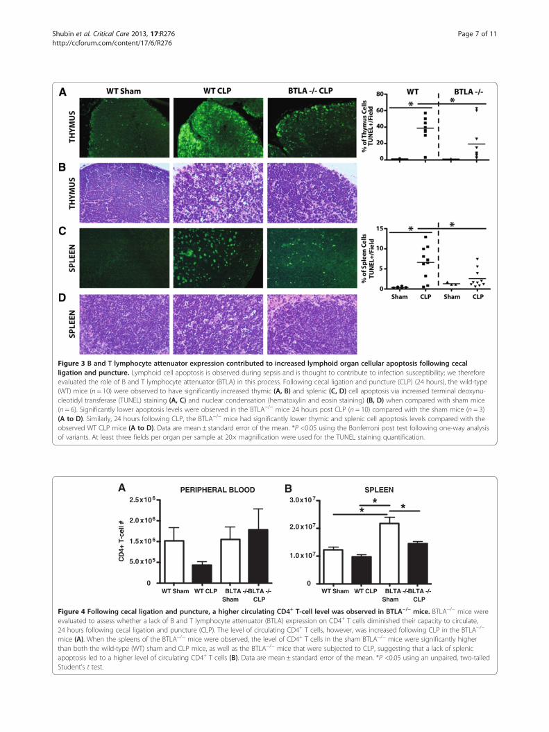
Figure 3 B and T lymphocyte attenuator expression contributed to increased lymphoid organ cellular apoptosis following cecalligation and puncture. Lymphoid cell apoptosis is observed during sepsis and is thought to contribute to infection susceptibility; we thereforeevaluated the role of B and T lymphocyte attenuator (BTLA) in this process. Following cecal ligation and puncture (CLP) (24 hours), the wild-type(WT) mice (n = 10) were observed to have significantly increased thymic (A, B) and splenic (C, D) cell apoptosis via increased terminal deoxynu-cleotidyl transferase (TUNEL) staining (A, C) and nuclear condensation (hematoxylin and eosin staining) (B, D) when compared with sham mice(n = 6). Significantly lower apoptosis levels were observed in the BTLA−/− mice 24 hours post CLP (n = 10) compared with the sham mice (n = 3)(A to D). Similarly, 24 hours following CLP, the BTLA−/− mice had significantly lower thymic and splenic cell apoptosis levels compared with theobserved WT CLP mice (A to D). Data are mean ± standard error of the mean. *P <0.05 using the Bonferroni post test following one-way analysisof variants. At least three fields per organ per sample at 20× magnification were used for the TUNEL staining quantification.
WT Sham WT CLP BLTA -/-Sham
BLTA -/-CLP
1.5x 10 6
5.0 x 10⁵
2.0 x 10⁶
2.5x 10 6
0
CD
4+ T
-cel
l #
PERIPHERAL BLOOD
1.0 x 10⁷
2.0 x 10⁷
3.0x 10 7
0
***
WT Sham WT CLP BLTA -/-Sham
BLTA -/-CLP
SPLEENBA
Figure 4 Following cecal ligation and puncture, a higher circulating CD4+ T-cell level was observed in BTLA−/− mice. BTLA−/− mice wereevaluated to assess whether a lack of B and T lymphocyte attenuator (BTLA) expression on CD4+ T cells diminished their capacity to circulate,24 hours following cecal ligation and puncture (CLP). The level of circulating CD4+ T cells, however, was increased following CLP in the BTLA−/−
mice (A). When the spleens of the BTLA−/− mice were observed, the level of CD4+ T cells in the sham BTLA−/− mice were significantly higherthan both the wild-type (WT) sham and CLP mice, as well as the BTLA−/− mice that were subjected to CLP, suggesting that a lack of splenicapoptosis led to a higher level of circulating CD4+ T cells (B). Data are mean ± standard error of the mean. *P <0.05 using an unpaired, two-tailedStudent’s t test.
Shubin et al. Critical Care 2013, 17:R276 Page 7 of 11http://ccforum.com/content/17/6/R276

Shubin et al. Critical Care 2013, 17:R276 Page 8 of 11http://ccforum.com/content/17/6/R276
demonstrated in [20], which showed that BTLA−/− Tcells have improved survival, but not improved prolifer-ative capacity) – additional studies are needed to evalu-ate the role of BTLA in CD4+ T-cell migration.
DiscussionSepsis is a deadly condition and its onset has beenthought to be the result of a hyper-inflammatory processthat often leads to an immune-suppressed state. This lat-ter state is thought to occur through a variety of intrinsicand extrinsic factors that consequently lead to a lack ofadaptive immune cell responsiveness and T-cell and B-cell loss, due in part to apoptosis [12]. Unfortunately,new therapeutics to treat sepsis and biomarkers thatmay be used to identify those patients who are at thehighest risk for developing later infections, and thusmight best benefit from such therapies, are limited orlacking. BTLA is a co-inhibitory, immune-regulatory re-ceptor, whose primary role in CD4+ T cells is thought tobe maintaining a state of immune tolerance to self-antigens [35] or resolving (suppressing) proinflammatoryresponses [31]. However, such tolerogenic activity mayinadvertently provide an immune-suppressed environ-ment that permits pathogens, such as plasmodium [36],cytomegalovirus [37], and the intracellular bacteriumListeria monogenes [38], to thrive (at least under experi-mental conditions), while also unintentionally reducingtumor (cancer) cell immune surveillance by supporting asustained state of immune suppression [39,40].Recently, we reported that BTLA expression on mye-
loid cells contributes to experimental septic mortalityin mice [28]. Here, we expand on this finding by dem-onstrating that BTLA was not only upregulated onmouse lymphocytes and contributed to cellular apop-tosis in primary and secondary lymphoid organs thatassociated with CD4+ T-cell and B-cell loss, but alsothat BTLA was seen at a higher frequency on CD4+
lymphocytes in the SIRS patients that developed subse-quent infections. These findings therefore potentially addimmune dysfunction as mediated through lymphoid cellloss during SIRS and/or sepsis to the above list of diseasestates to which augmented BTLA expression appears tocontribute.Although there appear to be promising biomarkers for
identifying which critically ill patients are septic (exam-ples being procalcitonin, C-reactive protein and solubletriggering receptor expressed on myeloid cells, amongstothers [6,7,41,42]), currently the biomarkers that delin-eate which critically ill patients are most susceptible tobecoming septic are lacking. Here we find that an in-crease in the percentage of BTLA+CD4+ T cells is muchhigher in the patients that develop subsequent nosoco-mial infections, thus implicating BTLA as a possible bio-marker for identifying which critically ill patients are
most susceptible to the development of sepsis. Only di-minished levels of monocyte human leukocyte antigen-DR (mHLA-DR) have until now been reported as beingan effective marker for identifying which critically illtrauma patients are susceptible to becoming septic [5].An approach that measures both mHLA-DR and BTLAin assessing the critically ill patient’s susceptibility tosepsis may therefore be important for a number of rea-sons. First, the nature of the regulation of these recep-tors’ expression and their roles in contributing toimmune suppression are very different. A decreasedlevel of mHLA-DR on antigen-presenting cells isthought to represent a reduced capacity by these cells toactivate the adaptive immune cells; however, increasedBTLA expression on CD4+ T cells may represent a re-duced ability for these cells to specifically function dueto BTLA’s co-inhibitory molecule effects. Additionally,the concept of combining biomarkers has shown somepotential, because a recent article demonstrating the po-tential value of combining multiple biomarkers (includ-ing procalcitonin, soluble triggering receptor expressedon myeloid cells and CD64 expression on neutrophils)for identifying and diagnosing sepsis in critically ill pa-tients was found to be much more accurate than anyone of these biomarkers alone [43]. A panel of bio-markers that includes the measurement of mHLA-DRon monocytes and BTLA on CD4+ T cells may thereforebe similarly useful in accurately predicting a patient’s in-fectious susceptibility, if not their state of immuneresponsiveness.In addition to our recent work demonstrating the im-
portance of BTLA’s contribution to septic mortality andinnate immune cell dysfunction during sepsis [28], theexpression levels of BTLA and its ligand, herpes virusentry mediator, have also been evaluated in patients whohad died from sepsis in a recent study by Boomer andcolleagues [13]. Interestingly, this group found that thepercentage of BTLA+CD4+ cells in patients who suc-cumbed to sepsis were not significantly different fromthose who died from causes other than sepsis (both wereat ~60 to 70% BTLA+); however, patients who died fromlung cancer had lung CD4+ T cells that were increasedto >80% BTLA+ [13]. The contrast in these results frompostmortem sampling to the live patient data we presenthere suggests that there may not only be differences inthe level of circulating blood CD4+BTLA+ versus theirlung CD4+ T-cell sample sites, but also in the timing ofthe sample acquisition during the sepsis process. An-other possibility is that, given very few of our patientpopulation died, BTLA expression levels may change aspatients are closer to death. Considering we found thatBTLA−/− mice were protected from lymphoid cell apop-tosis, it is also possible that many of the BTLA+CD4+ Tcells in Boomer and colleagues’ study had already

Shubin et al. Critical Care 2013, 17:R276 Page 9 of 11http://ccforum.com/content/17/6/R276
undergone apoptosis and were lost by the time thesepatients had succumbed to sepsis (compared withour data, in which we see a high level of circulatingBTLA+CD4+ lymphocytes in septic patients).Amongst BTLA, induction of other co-inhibitory re-
ceptors, including PD-1 and CTLA-4, have been demon-strated to contribute to CD4+ T-cell apoptosis followingCLP in mice, as well as in septic patients [13,24,25]. Incontrast to the mouse studies evaluating PD-1 andCTLA-4 in sepsis, we found that BTLA in mice wasconstitutively expressed on a very high percentage ofCD4+ T cells, but the intensity of this expression wasfurther increased in response to CLP, while CD4+ T cellsonly increased in the percentage of CTLA-4+ and PD-1+
expressing lymphocytes in response to CLP [24,25]. Incritically ill patients with septic shock, an increase inPD-1+CD4+ lymphocytes when compared with healthyvolunteers has also been observed [26,27]. Interestingly,a higher percentage of PD-1+CD4+ T cells correlatedwith a lower proliferation rate of CD4+ T cells [27] andblockade of the PDL-1:PD-1 pathway ex vivo, preventingCD4+ T-cell apoptosis [26]. However, neither studylooked at whether PD-1 or CTLA-4 was induced in non-septic, SIRS critically ill patients. Thus, it remains to beestablished whether PD-1 and/or CTLA-4 are also in-duced by critical injury alone prior to septic infection,like BTLA, or whether they act in a more redundantmanner during sepsis to cause further dysfunction and/or apoptotic loss of these cells.Interestingly, these data also suggest that BTLA in hu-
man CD4+ T cells has the capacity to induce themobilization of these cells into circulation, which ap-pears to contrast the findings that we and others haveobserved in mice. Alternatively, however, it has beendemonstrated that the cross-linking of BTLA on humanCD4+ T cells leads to an inability to be fully activated (viaupregulation of CD25), while also inhibiting their capacityto secrete interleukin-2, interleukin-4, interleukin-10 andinterferon-gamma [44], which appears to be consistentwith BTLA in mice [18]. Therefore, BTLA expression onCD4+ T cells may help to drive the cells into circulation inan exhausted state with a diminished capacity to be acti-vated; however, further studies are needed to provide evi-dence for this hypothesis.This study is of course not without limitations. Over-
all, prior to understanding whether using a BTLA antag-onist is a viable option for the treatment of critically illpatients, additional studies are needed to elucidatewhether the augmented BTLA expression that we ob-served on the septic and SIRS patient and mouse CD4+
T cells indeed inhibits their function (including ex vivocytokine production and upregulation of activationmarkers, such as CD69 and CD25), proliferation, or dif-ferentiation. Also, with regard to BTLA as a biomarker
for subsequent infections, significantly larger patientnumbers need to be collected to establish the specificityand sensitivity with which BTLA expression can be usedto predict the severity of disease (that is, sepsis, septicshock or severe sepsis), septic mortality, multiple organdamage, and to what particular pathogens these patientsmay be susceptible. Additionally, a previous study byZhang and colleagues found that the percentage ofBTLA+CD4+ T cells from healthy volunteers was muchhigher (~90% double positive); thus, future studies mustbe conducted with similar methods to those of Zhang andcolleagues (that is, staining technique and a similar BTLAantibody clone) to determine whether SIRS patients thatdevelop a subsequent infection have diminished BTLA ex-pression with alternative staining methods such as these[32]. This would further clarify the use of BTLA as a po-tential biomarker. Another limitation of this study wasthat, because of the relatively small sample size, patientsamples were compared at various times following ICUadmission. Although we properly compared these ICU pa-tients based on whether they were SIRS or septic, sam-pling at specific times following ICU admission and septicinsult would provide more specific information about thetiming of changes in BTLA expression on CD4+ T cellsduring SIRS and sepsis. Additionally, in our mouse datathat evaluated apoptosis against CD4+ T-cell and B-cellloss, only an indirect association of the contribution ofBTLA expression to the apoptotic cell loss of CD4+ T cellsand B cells could be drawn. Future studies, such as onesthat directly evaluate whether CD4+ T cells and B cells areprotected from the induction of apoptosis (with markerssuch as activated caspase-3 and Annexin-V) in BTLA−/−
mice compared with WT mice, should be carried out toconfirm our results. Finally, studies that adoptively trans-fer BTLA−/− CD4+ T cells into WT mice may be able toelucidate whether BTLA expression on CD4+ T cells in-appropriately drives an exhausted form of CD4+ T cellsinto circulation during experimental sepsis.
ConclusionsWe feel that the data presented here concerning BTLAexpression on CD4+ T cells in critically ill ICU patientsand experimental mice importantly add novel insight asto how ligation of BTLA may contribute to the induc-tion of the adaptive immune cell loss that is associatedwith sepsis. Further, these data illustrate the value ofBTLA as a potential therapeutic target and a possiblebiomarker for identifying the critically ill patientswhom are most susceptible to develop subsequent orsecondary infections, and thereby may allow for the de-livery of the sufficient therapies that are needed to helpprevent the morbidity and mortality associated withSIRS and sepsis.

Shubin et al. Critical Care 2013, 17:R276 Page 10 of 11http://ccforum.com/content/17/6/R276
Key messages
� The percentage of BTLA expressing circulatingperipheral blood CD4+ T cells was higher in septiccritically ill patients when compared with SIRS ICUpatients and healthy controls.
� An increased percentage of BTLA+CD4+ T cells incritically ill patients associated with poor outcomesand a longer hospital length of stay; thus, futurestudies are warranted examining BTLA expressionon CD4+ T cells as a potential biomarker forsubsequent infections and poor septic outcomes.
� BTLA expression was induced on CD4+ T cellsand B cells following experimental septic injury inmice.
� Lack of BTLA protected primary and secondarylymphoid organs from cellular apoptosis, and thiswas associated with the loss of CD4+ T cells and Bcells following CLP in mice, suggesting that BTLAmay be directly involved in CD4+ T-cell apoptosisduring experimental sepsis.
AbbreviationsBTLA: B and T lymphocyte attenuator; CD4: Cluster of differentiation 4;CLP: Cecal ligation and puncture; CTLA-4: Cytotoxic T-lymphocyte antigen-4;MFI: Mean fluorescence intensity; mHLA-DR: Monocyte human leukocyteantigen-DR; PD-1: Programmed death receptor-1; SIRS: Systemicinflammatory response syndrome; TUNEL: Terminal deoxynucleotidyltransferase; WT: Wild type.
Competing interestsThe authors declare that they have no competing interests.
Authors’ contributionsNJS, C-SC and AA conducted the general study concepts and design andwrote the manuscript. All of the experiments were performed by NJS. DSHand SFM substantially contributed to the design and conception of thehuman studies. DSH obtained the Institutional Review Board approval forthe human subjects study. All authors have given final approval for thismanuscript to be published.
AcknowledgmentsThis study was supported by the National Institutes of Health Project grantGM053209 (to AA), a fellowship from the US Department of Education,GAANN-P200A100100 funding (to NJS), and the Armand D. VersaciResearch Scholar in the Surgical Sciences Award (to SFM). The authorsthank Ms Yaping Chen for her support with the animal studies, Ms MaiTran and Ms Lydea Irwin for their assistance in the human sample proto-cols and processing of the samples, and Mr Paul Monfils (Core ResearchLaboratories, Rhode Island Hospital) for his assistance with the mouseorgan histology.
Received: 22 May 2013 Accepted: 30 October 2013Published: 29 November 2013
References1. Angus DC, Linde-Zwirble WT, Lidicker J, Clermont G, Carcillo J, Pinsky MR:
Epidemiology of severe sepsis in the United States: analysis of incidence,outcome, and associated costs of care. Crit Care Med 2001, 29:1303–1310.
2. Angus DC, Pereira CA, Silva E: Epidemiology of severe sepsis around theworld. Endocr Metab Immune Disord Drug Targets 2006, 6:207–212.
3. Levy MM, Fink MP, Marshall JC, Abraham E, Angus D, Cook D, Cohen J, OpalSM, Vincent JL, Ramsay G: SCCM/ESICM/ACCP/ATS/SIS international sepsisdefinitions conference. Crit Care Med 2001, 2003:1250–1256.
4. Angus DC: The search for effective therapy for sepsis: back to thedrawing board? JAMA 2011, 306:2614–2615.
5. Cheron A, Floccard B, Allaouchiche B, Guignant C, Poitevin F, Malcus C,Crozon J, Faure A, Guillaume C, Marcotte G, Vulliez A, Monneuse O,Monneret G: Lack of recovery in monocyte human leukocyte antigen-DRexpression is independently associated with the development ofsepsis after major trauma. Crit Care 2010, 14:R208.
6. Clec’h C, Fosse JP, Karoubi P, Vincent F, Chouahi I, Hamza L, Cupa M, CohenY: Differential diagnostic value of procalcitonin in surgical and medicalpatients with septic shock. Crit Care Med 2006, 34:102–107.
7. Harbarth S, Holeckova K, Froidevaux C, Pittet D, Ricou B, Grau GE, Vadas L,Pugin J: Diagnostic value of procalcitonin, interleukin-6, and interleukin-8in critically ill patients admitted with suspected sepsis. Am J Respir CritCare Med 2001, 164:396–402.
8. Lichtenstern C, Brenner T, Bardenheuer HJ, Weigand MA: Predictors ofsurvival in sepsis: what is the best inflammatory marker to measure? CurrOpin Infect Dis 2012, 25:328–336.
9. Hotchkiss RS, Karl IE: The pathophysiology and treatment of sepsis. N EnglJ Med 2003, 348:138–150.
10. Hotchkiss RS, Opal S: Immunotherapy for sepsis – a new approachagainst an ancient foe. N Engl J Med 2010, 363:87–89.
11. Hotchkiss RS, Swanson PE, Knudson CM, Chang KC, Cobb JP, Osborne DF,Zollner KM, Buchman TG, Korsmeyer SJ, Karl IE: Overexpression of Bcl-2 intransgenic mice decreases apoptosis and improves survival in sepsis.J Immunol 1999, 162:4148–4156.
12. Hotchkiss RS, Tinsley KW, Swanson PE, Schmieg RE Jr, Hui JJ, Chang KC,Osborne DF, Freeman BD, Cobb JP, Buchman TG, Karl IE: Sepsis-inducedapoptosis causes progressive profound depletion of B and CD4+ Tlymphocytes in humans. J Immunol 2001, 166:6952–6963.
13. Boomer JS, To K, Chang KC, Takasu O, Osborne DF, Walton AH, Bricker TL,Jarman SD 2nd, Kreisel D, Krupnick AS, Srivastava A, Swanson PE, Green JM,Hotchkiss RS: Immunosuppression in patients who die of sepsis andmultiple organ failure. JAMA 2011, 306:2594–2605.
14. Meakins JL, Pietsch JB, Bubenick O, Kelly R, Rode H, Gordon J, MacLean LD:Delayed hypersensitivity: indicator of acquired failure of host defenses insepsis and trauma. Ann Surg 1977, 186:241–250.
15. Venet F, Chung CS, Kherouf H, Geeraert A, Malcus C, Poitevin F, Bohe J,Lepape A, Ayala A, Monneret G: Increased circulating regulatory T cells(CD4+CD25+CD127−) contribute to lymphocyte anergy in septic shockpatients. Intensive Care Med 2009, 35:678–686.
16. Venet F, Chung CS, Monneret G, Huang X, Horner B, Garber M, Ayala A:Regulatory T cell populations in sepsis and trauma. J Leukoc Biol 2008,83:523–535.
17. Cavassani KA, Carson WF th, Moreira AP, Wen H, Schaller MA, Ishii M, LindellDM, Dou Y, Lukacs NW, Keshamouni VG, Hogaboam CM, Kunkel SL: The postsepsis-induced expansion and enhanced function of regulatory T cells createan environment to potentiate tumor growth. Blood 2010, 115:4403–4411.
18. Watanabe N, Gavrieli M, Sedy JR, Yang J, Fallarino F, Loftin SK, Hurchla MA,Zimmerman N, Sim J, Zang X, Murphy TL, Russell JH, Allison JP, Murphy KM:BTLA is a lymphocyte inhibitory receptor with similarities to CTLA-4 andPD-1. Nat Immunol 2003, 4:670–679.
19. Hurchla MA, Sedy JR, Gavrieli M, Drake CG, Murphy TL, Murphy KM: B and Tlymphocyte attenuator exhibits structural and expression polymorphisms andis highly induced in anergic CD4+ T cells. J Immunol 2005, 174:3377–3385.
20. Deppong C, Degnan JM, Murphy TL, Murphy KM, Green JM: B and Tlymphocyte attenuator regulates T cell survival in the lung. J Immunol2008, 181:2973–2979.
21. Mellman I, Couko G, Dranoff G: Cancer immunotherapy comes of age.Nature 2011, 480:480–489.
22. Postow MA, Callahan MK, Barker CA, Yamada Y, Yuan J, Kitano S, Mu Z,Rasalan T, Adamow M, Ritter E, Sedrak C, Jungbluth AA, Chua R, Yang AS,Roman RA, Rosner S, Benson B, Allison JP, Lesokhin AM, Gnjatic S, Wolchok JD:Immunologic correlates of the abscopal effect in a patient with melanoma.N Engl J Med 2012, 366:925–931.
23. Riley JL: PD-1 signaling in primary T cells. Immunol Rev 2009, 229:114–125.24. Brahmamdam P, Inoue S, Unsinger J, Chang KC, McDunn JE, Hotchkiss RS:
Delayed administration of anti-PD-1 antibody reverses immunedysfunction and improves survival during sepsis. J Leukoc Biol 2010,88:233–240.
25. Inoue S, Bo L, Bian J, Unsinger J, Chang K, Hotchkiss RS: Dose dependenteffect of anti-CTLA-4 on survival in sepsis. Shock 2011, 36:38–44.

Shubin et al. Critical Care 2013, 17:R276 Page 11 of 11http://ccforum.com/content/17/6/R276
26. Zhang Y, Li J, Lou J, Zhou Y, Bo L, Zhu J, Zhu K, Wan X, Cai Z, Deng X:Upregulation of programmed death-1 on T cells and programmed deathligand-1 on monocytes in septic shock patients. Crit Care 2011,15:R70.
27. Guignant C, Lepape A, Huang X, Kherouf H, Denis L, Poitevin F, Malcus C,Cheron A, Allaouchiche B, Gueyffier F, Ayala A, Monneret G, Venet F:Programmed death-1 levels correlate with increased mortality,nosocomial infection and immune dysfunctions in septic shock patients.Crit Care 2011, 15:R99.
28. Shubin NJ, Chung CS, Heffernan DS, Irwin LR, Monaghan SF, Ayala A: BTLAexpression contributes to septic morbidity and mortality by inducinginnate inflammatory cell dysfunction. J Leukoc Biol 2012,92:593–603.
29. Huang X, Venet F, Wang YL, Lepape A, Yuan Z, Chen Y, Swan R, Kherouf H,Monneret G, Chung CS, Ayala A: PD-1 expression by macrophages plays apathologic role in altering microbial clearance and the innate inflammatoryresponse to sepsis. Proc Natl Acad Sci USA 2009, 106:6303–6308.
30. Otsuki N, Kamimura Y, Hashiguchi M, Azuma M: Expression and function ofthe B and T lymphocyte attenuator (BTLA/CD272) on human T cells.Biochem Biophys Res Commun 2006, 344:1121–1127.
31. Bandyopadhyay G, De A, Laudanski K, Li F, Lentz C, Bankey P, Miller-Graziano C:Negative signaling contributes to T-cell anergy in trauma patients. Crit CareMed 2007, 35:794–801.
32. Zhang Z, Xu X, Lu J, Zhang S, Gu L, Fu J, Jin L, Li H, Zhao M, Zhang J, Wu H,Su L, Fu YX, Wang FS: B and T lymphocyte attenuator down-regulation byHIV-1 depends on type I interferon and contributes to T-cell hyperactivation.J Infect Dis 2011, 203:1668–1678.
33. Shubin NJ, Chung CS, Ayala A: B and T lymphocyte attenuator (BTLA) is acontributor to the pathological progression of sepsis [abstract].Inflamm Res 2010, 59:S22.
34. Ayala A, Perrin MM, Kisala JM, Ertel W, Chaudry IH: Polymicrobial sepsisselectively activates peritoneal but not alveolar macrophages to releaseinflammatory mediators (interleukins-1 and −6 and tumor necrosis factor).Circ Shock 1992, 36:191–199.
35. Liu X, Alexiou M, Martin-Orozco N, Chung Y, Nurieva RI, Ma L, Tian Q, Kollias G,Lu S, Graf D, Dong C: Cutting edge: a critical role of B and T lymphocyteattenuator in peripheral T cell tolerance induction. J Immunol 2009,182:4516–4520.
36. Adler G, Steeg C, Pfeffer K, Murphy TL, Murphy KM, Langhorne J, Jacobs T: Band T lymphocyte attenuator restricts the protective immune responseagainst experimental malaria. J Immunol 2011, 187:5310–5319.
37. Serriari NE, Gondois-Rey F, Guillaume Y, Remmerswaal EB, Pastor S, MessalN, Truneh A, Hirsch I, van Lier RA, Olive D: B and T lymphocyte attenuatoris highly expressed on CMV-specific T cells during infection and regu-lates their function. J Immunol 2010, 185:3140–3148.
38. Sun Y, Brown NK, Ruddy MJ, Miller ML, Lee Y, Wang Y, Murphy KM, Pfeffer K,Chen L, Kaye J, Fu YX: B and T lymphocyte attenuator tempers earlyinfection immunity. J Immunol 2009, 183:1946–1951.
39. Derre L, Rivals JP, Jandus C, Pastor S, Rimoldi D, Romero P, Michielin O,Olive D, Speiser DE: BTLA mediates inhibition of human tumor-specificCD8+ T cells that can be partially reversed by vaccination. J Clin Invest2010, 120:157–167.
40. Paulos CM, June CH: Putting the brakes on BTLA in T cell-mediatedcancer immunotherapy. J Clin Invest 2010, 120:76–80.
41. Remick DG, Bolgos GR, Siddiqui J, Shin J, Nemzek JA: Six at six: interleukin-6measured 6 h after the initiation of sepsis predicts mortality over 3 days.Shock 2002, 17:463–467.
42. Jeong SJ, Song YG, Kim CO, Kim HW, Ku NS, Han SH, Choi JY, Kim JM:Measurement of plasma sTREM-1 in patients with severe sepsis receivingearly goal-directed therapy and evaluation of its usefulness. Shock 2012,37:574–578.
43. Gibot S, Bene MC, Noel R, Massin F, Guy J, Cravoisy A, Barraud D, DeCarvalho Bittencourt M, Quenot JP, Bollaert PE, Faure G, Charles PE:Combination biomarkers to diagnose sepsis in the critically ill patient.Am J Respir Crit Care Med 2012, 186:65–71.
44. Wang XF, Chen YJ, Wang Q, Ge Y, Dai Q, Yang KF, Fang Xie, Zhou YH, HuYM, Mao YX, Zhang XG: Distinct expression and inhibitory function of Band T lymphocyte attenuator on human T cells. Tissue Antigens 2007,69:145–153.
doi:10.1186/cc13131Cite this article as: Shubin et al.: B and T lymphocyte attenuatorexpression on CD4+ T-cells associates with sepsis and subsequentinfections in ICU patients. Critical Care 2013 17:R276.
Submit your next manuscript to BioMed Centraland take full advantage of:
• Convenient online submission
• Thorough peer review
• No space constraints or color figure charges
• Immediate publication on acceptance
• Inclusion in PubMed, CAS, Scopus and Google Scholar
• Research which is freely available for redistribution
Submit your manuscript at www.biomedcentral.com/submit