RESEARCH ARTICLES Gamma Radiation Studies on … · RESEARCH ARTICLES Gamma Radiation Studies on...
14
FABAD J. Pharm. Sci., 28, 93-106, 2003 RESEARCH ARTICLES Gamma Radiation Studies on Sulfathiazole (Powder and Model-Ophthalmic Solution) Hafidha S. BENCHAABANE*, Yekta A. ÖZER*°, Meral ÖZALP**, Ekrem KILIÇ**, Mustafa POLAT***, Mustafa KORKMAZ*** 93 Gamma Radiation Studies on Sulfathiazole (Powder and Model-Ophthalmic Solution) Summary : Gamma irradiation is an excellent technique for the sterilization of pharmaceutical raw materials and products (1,2) . One of the major possible disadvantages of radiosteriliza- tion is the production of new radiolytic intermediates during the irradiation process (3) . The Radiosterilization was carried out for sulfathiazole (anti- bacterial product) powder and model-ophthalmic solution in this research. Irradiation at room temperature at radiation do- ses of 10, 25 and 40 kGy was investigated via different physi- cal, chemical, microbiological and biological techniques both in normal and accelerated stability conditions (40±2°C and 75±5 % relative humidity, 3 months). Changes in organoleptic features, pH, melting point, UV, IR, NMR, TLC, ESR, DSC characteristics, and microbiological ac- tivities of active compound at normal and accelerated stability test conditions were studied. It was observed that UV, IR, and NMR characteristics of sul- fathiazole did not change with the applied dose ranging betwe- en 10-40 kGy except λ max values of irradiated solid samples dissolved in 0.1 N HCl solution. Some radicals were detected by ESR signal intensity of solid samples. Attempt for determi- nation of radicals produced in irradiated model-ophthalmic solution by ESR technique failed due to the short life of these radicals in solution. The results obtained under accelerated stability test conditions over a period of three months were observed to be consistent with the reference values and the activity of sulfathiazole rema- ined unaffected even at the end of the test period. Microbiological and biological properties both in normal and accelerated conditions were also investigated. Based on the physical, chemical, microbological and biological results, the optimum radiation dose of 10 kGy can be applied for the sterilization of sulfathiazole powder and model-opht- halmic solution. Keywords: Sulfathiazole, Irradiation of Drugs, Radiosteriliza- tion, Gamma Irradiation, Stability. Sülfatiyazolün Gama Radyasyonu Üzerinde Çal›flmalar (Toz ve Model Oftalmik Çözelti) Özet: Gama ›fl›nlama, farmasötik ürün ve hammaddelerin ste- rilizasyonunda de¤erli bir tekniktir (1,2) . Radyosterilizasyonun muhtemel en büyük dezavantaj›, ›fl›nlama s›ras›nda yeni radyo- litik ara ürünlerin oluflmas›d›r (3) . Bu araflt›rma bir antibakteriyel olan sulfatiazol tozunun ve mo- del-oftalmik çözeltisinin radyosterilizasyonu üzerinde sürdü- rülmüfltür. Ifl›nlama oda s›cakl›¤›nda; 10, 25 ve 40 kGy dozlar- da yap›lm›fl ve normal ve h›zland›r›lm›fl stabilite flartlar›nda (40°C ve %75 ba¤›l nemde, 3 ay süreyle), farkl› fiziksel, kimya- sal, mikrobiyolojik ve biyolojik tekniklerle araflt›r›lm›flt›r. Etkin maddenin organoleptik özelliklerindeki de¤iflmeler, pH, e.d., UV, IR, NMR, ESR, DSC özellikleri, mikrobiyal aktivitesi normal ve h›zland›r›lm›fl stabilite flartlar›nda incelenmifltir. 10-40 kGy doz aral›¤›nda uygulanan ›fl›nlamayla sulfatiyazo- lün UV, IR, NMR özellikleri, 0,1 N HCI ’de çözünen örnekleri- nin λ max ’lar› d›fl›nda de¤iflme olmam›flt›r. Kat› örneklerin ESR sinyal fliddetiyle saptanan baz› radikaller oluflmufltur. Ifl›nlanan model-oftalmik çözeltilerde oluflan radikallerin ömürlerinin k›- sal›¤› nedeniyle, ESR teknolojisiyle tayin baflar›l› olmam›flt›r. Üç ay boyunca, h›zland›r›lm›fl stabilite flartlar›nda elde edilen sonuçlar referansla uyumlu bulunmufl ve test süresi sonunda bile sulfatiyazolün aktivitesi korunmufltur. Normal ve h›zland›r›lm›fl flartlarda, mikrobiyolojik ve toksiko- lojik özelliklerde incelenmifltir. Fiziksel, kimyasal, mikrobiyolojik ve biyolojik sonuçlara daya- narak Sulfatiyazol toz ve model-oftalmik çözeltisinin sterilizas- yonu için optimum 10 kGy radyasyon dozu uygulanabilece¤i sonucuna var›lm›flt›r. Anahtar kelimeler: Sulfatiyazol, ‹laçlar›n Ifl›nlanmas›, Radyo- sterilizasyon, Gama Ifl›nlama, Stabilite . * Hacettepe University, Faculty of Pharmacy, Department of Radiopharmacy, 06100 Ankara TURKEY. ** Hacettepe University, Faculty of Pharmacy, Department of Pharmaceutical Microbiology, 06100 Ankara TURKEY. *** Hacettepe University, Faculty of Science and Engineering, Department of Physics Engineering , 06100 Ankara TURKEY. ° Corresponding author • e-mail: [email protected]
Transcript of RESEARCH ARTICLES Gamma Radiation Studies on … · RESEARCH ARTICLES Gamma Radiation Studies on...

FABAD J. Pharm. Sci., 28, 93-106, 2003
RESEARCH ARTICLES
GGaammmmaa RRaaddiiaattiioonn SSttuuddiieess oonn SSuullffaatthhiiaazzoollee((PPoowwddeerr aanndd MMooddeell--OOpphhtthhaallmmiicc SSoolluuttiioonn))
Hafidha S. BENCHAABANE*, Yekta A. ÖZER*°, Meral ÖZALP**, Ekrem KILIÇ**,Mustafa POLAT***, Mustafa KORKMAZ***
93
GGaammmmaa RRaaddiiaattiioonn SSttuuddiieess oonn SSuullffaatthhiiaazzoollee ((PPoowwddeerr aanndd MMooddeell--OOpphhtthhaallmmiicc SSoolluuttiioonn))
SSuummmmaarryy :: Gamma irradiation is an excellent technique for thesterilization of pharmaceutical raw materials and products(1,2). One of the major possible disadvantages of radiosteriliza-tion is the production of new radiolytic intermediates during theirradiation process (3).The Radiosterilization was carried out for sulfathiazole (anti-bacterial product) powder and model-ophthalmic solution inthis research. Irradiation at room temperature at radiation do-ses of 10, 25 and 40 kGy was investigated via different physi-cal, chemical, microbiological and biological techniques bothin normal and accelerated stability conditions (40±2°C and75±5 % relative humidity, 3 months).Changes in organoleptic features, pH, melting point, UV, IR,NMR, TLC, ESR, DSC characteristics, and microbiological ac-tivities of active compound at normal and accelerated stabilitytest conditions were studied. It was observed that UV, IR, and NMR characteristics of sul-fathiazole did not change with the applied dose ranging betwe-en 10-40 kGy except λmax values of irradiated solid samplesdissolved in 0.1 N HCl solution. Some radicals were detectedby ESR signal intensity of solid samples. Attempt for determi-nation of radicals produced in irradiated model-ophthalmicsolution by ESR technique failed due to the short life of theseradicals in solution.The results obtained under accelerated stability test conditionsover a period of three months were observed to be consistentwith the reference values and the activity of sulfathiazole rema-ined unaffected even at the end of the test period.Microbiological and biological properties both in normal andaccelerated conditions were also investigated.Based on the physical, chemical, microbological and biologicalresults, the optimum radiation dose of 10 kGy can be appliedfor the sterilization of sulfathiazole powder and model-opht-halmic solution.KKeeyywwoorrddss:: Sulfathiazole, Irradiation of Drugs, Radiosteriliza-
tion, Gamma Irradiation, Stability.
SSüüllffaattiiyyaazzoollüünn GGaammaa RRaaddyyaassyyoonnuu ÜÜzzeerriinnddee ÇÇaall››flflmmaallaarr((TToozz vvee MMooddeell OOffttaallmmiikk ÇÇöözzeellttii))
ÖÖzzeett:: Gama ›fl›nlama, farmasötik ürün ve hammaddelerin ste-rilizasyonunda de¤erli bir tekniktir(1,2). Radyosterilizasyonunmuhtemel en büyük dezavantaj›, ›fl›nlama s›ras›nda yeni radyo-litik ara ürünlerin oluflmas›d›r (3).Bu araflt›rma bir antibakteriyel olan sulfatiazol tozunun ve mo-del-oftalmik çözeltisinin radyosterilizasyonu üzerinde sürdü-rülmüfltür. Ifl›nlama oda s›cakl›¤›nda; 10, 25 ve 40 kGy dozlar-da yap›lm›fl ve normal ve h›zland›r›lm›fl stabilite flartlar›nda(40°C ve %75 ba¤›l nemde, 3 ay süreyle), farkl› fiziksel, kimya-sal, mikrobiyolojik ve biyolojik tekniklerle araflt›r›lm›flt›r.Etkin maddenin organoleptik özelliklerindeki de¤iflmeler, pH,e.d., UV, IR, NMR, ESR, DSC özellikleri, mikrobiyal aktivitesinormal ve h›zland›r›lm›fl stabilite flartlar›nda incelenmifltir.10-40 kGy doz aral›¤›nda uygulanan ›fl›nlamayla sulfatiyazo-lün UV, IR, NMR özellikleri, 0,1 N HCI ’de çözünen örnekleri-nin λmax’lar› d›fl›nda de¤iflme olmam›flt›r. Kat› örneklerin ESRsinyal fliddetiyle saptanan baz› radikaller oluflmufltur. Ifl›nlananmodel-oftalmik çözeltilerde oluflan radikallerin ömürlerinin k›-sal›¤› nedeniyle, ESR teknolojisiyle tayin baflar›l› olmam›flt›r.Üç ay boyunca, h›zland›r›lm›fl stabilite flartlar›nda elde edilensonuçlar referansla uyumlu bulunmufl ve test süresi sonundabile sulfatiyazolün aktivitesi korunmufltur. Normal ve h›zland›r›lm›fl flartlarda, mikrobiyolojik ve toksiko-lojik özelliklerde incelenmifltir.Fiziksel, kimyasal, mikrobiyolojik ve biyolojik sonuçlara daya-narak Sulfatiyazol toz ve model-oftalmik çözeltisinin sterilizas-yonu için optimum 10 kGy radyasyon dozu uygulanabilece¤isonucuna var›lm›flt›r.AAnnaahhttaarr kkeelliimmeelleerr:: Sulfatiyazol, ‹laçlar›n Ifl›nlanmas›, Radyo-
sterilizasyon, Gama Ifl›nlama, Stabilite .
* Hacettepe University, Faculty of Pharmacy, Department of Radiopharmacy, 06100 Ankara TURKEY.** Hacettepe University, Faculty of Pharmacy, Department of Pharmaceutical Microbiology, 06100 Ankara TURKEY.*** Hacettepe University, Faculty of Science and Engineering, Department of Physics Engineering , 06100 Ankara TURKEY.° Corresponding author • e-mail: [email protected]

IINNTTRROODDUUCCTTIIOONN
Sterilization facilitates elimination or killing of themicroorganisms present in the contaminated en-vironment, and reduction of the initial contami-nation3-5.
The sterilization methods are classifed into four gro-ups:
• Heat sterilization• Filtration• Sterilization by ethylene oxide or formaldehyde
(chemical sterilization)• Radiosterilization
Radiosterilization is an interesting alternative parti-cularly where conventional methods are inadequ-ate. Certain molecules, depending on their thermo-lability, cannot be sterilized by humid heat, whileuse of ethylene oxide is less recommended for toxi-city reasons.
Some studies have demonstrated that irradiationdoes not generate the same consequences because ofthe considerable therapeutical molecules. A diffi-culty, however, is that physical and chemical chan-ges can accompany ionizing radiation3-5. It is neces-sary to describe the radicals formed by the radioste-rilization process.
The major problem of radiosterilization is the pro-duction of new radiolytic products. Therefore, ourintention was to determine and characterize thesephysical and chemical changes6,7.
Sulfathiazole is widely used in treatment as an anti-bacterial agent8,9. The aim of this study was to in-vestigate the gamma irradiation of sulfathiazole asraw material and model-ophthalmic solution as pro-ducts. The effect of gamma irradiation was determi-ned before and after the process.
A "sterile" product (like ophthalmic solution) wasprepared as a model and the gamma irradiation ef-fect was determined on this product. Additionally,
the results of raw material and product were compa-red regarding gamma irradiation effect.
Samples of powder sulfathiazole and model-opht-halmic solution irradiated at room temperature atradiation doses of 10, 25 and 40 kGy were investiga-ted by different physical, chemical and microbiolo-gical techniques under normal and accelerated sta-bility conditions6,7.
MMAATTEERRIIAALLSS AANNDD MMEETTHHOODDSS::
Solid sulfathiazole powder was provided from"YED", Yeni Ecza Deposu, Turkey.
IIrrrraaddiiaattiioonn PPrroocceessss::
All irradiations were performed at room temperatu-re (approximately 20°C) using a 60Co source (Gam-ma Cell 220) providing a dose rate of 4 kGy.h-1 in thesample located at Sarayköy Nuclear Research Cent-re of Turkish Atomic Energy Authority (TAEK) inAnkara. The dose rate was measured by Fricke dosi-meter6,7.
The samples were irradiated at doses of 10, 25 and40 kGy10.
SSttuuddiieess CCoonndduucctteedd UUnnddeerr NNoorrmmaall CCoonnddiittiioonnss
Unirradiated samples were used as controls to de-tect the changes related to the physicochemical andantimicrobial activities resulting from the action ofionizing radiation6.
AA--PPhhyyssiiccoo--CChheemmiiccaall PPrrooppeerrttiieess
pH change measurements of the reference and irra-diated solid samples were performed using 1mg.ml-1 aqueous solutions of these samples. Solubi-lity changes in boiling water, acetone, hydrochloricacid, sodium hydroxide and potassium hydroxide;and changes in melting point, color and sediment,primary aromatic amine reaction, solution appe-arance, acidity, heavy metal, loss on drying, sulfatedas and DSC (differential scanning calorimetry)
94
Benchaabane, Özer, Özalp, K›l›ç, Polat, Korkmaz

95
(General V2. 2A Du Pont 9900 Heating 10°C.min-1 atN2 atm) were also tested.
Similarly, irradiated model-ophthalmic solutionwas evaluated for appearance, pH changes, homo-geneity, foreign particles, organoleptic properties,color and odor11.
Calorimetric evaluation is one of the best methodsfor characterizing the samples; therefore, sulfathi-azole was also evaluated from this point of view.
BB-- SSppeeccttrroossccooppiicc MMeetthhooddss aanndd TTeecchhnniiqquueess
Changes in spectral properties of control and irradi-ated solid samples were studied using IR, UV,NMR12 and electron spin resonance (ESR) techniqu-es13. IR spectra were obtained for control and irradi-ated powders in KBr matrix (instrument: IR Vector22 Bruker Opus version 3)11. In UV analysis (Shi-madzu UV-160A), determination of λmax values wasperformed both in 0.1 N NaOH and 0.1 N HCl forpowder and for model-ophthalmic solution.
NMR analysis was performed using proton NMRspectrometer with the dissolving of irradiated andunirradiated powders in DMSO-d6. Tetramethylsi-lane was used as an internal standard12.
The species and amount of the molecular fragmentsor radicals processing unpaired electrons created byradiation can best be detected by ESR spectroscopy,which is frequently used to measure the absorbeddose of an irradiated product14,15.
ESR measurements were carried out using Varian E-19, X-Band ESR spectrometer USA equipped with aTE104 rectangular double cavity. All measurementswere performed using a DPPH reference sampleplaced in the front cavity. The position of the refe-rence sample in the cavity was not changed throug-hout the experiments to avoid any signal intensitymeasurements due to any possible changes in thecavity-filling factor. The spectra were double integ-rated over the magnetic field range of 3200-3320 mTto give a figure proportional to the radical numbers
in the sample. Each spectrum was corrected for va-riation in the amount of material in the "activelength" of the ESR tube, and in the spectrometer tu-ning conditions. Simulation studies based on possib-le radical species were also carried out13.
Determination of radicals produced in irradiatedmodel-ophthalmic solution samples by ESR techni-que failed due to the short life of these radicals; the-refore, UV and TLC techniques were performed todetermine degradation products.
CC-- CChhrroommaattooggrraapphhiicc MMeetthhooddss
Chromatographic (TLC) methods for identificationof sulfathiazole have been given by European Phar-macopeia (EP)16. Sulfathiazole was detected by TLCusing silica gel HR plates. Dimethylaminobenzal-dehyde solution 1g.L-1 of ethanol was used as loca-tion reagent17,18.
DD-- AAnnttiimmiiccrroobbiiaall AAccttiivviittyy SSttuuddiieess
Antibacterial activities of irradiated and unirradi-ated preparations were determined by the microdi-lution method recommended by the National Com-mittee for Clinical Laboratory Standards (NCCLS,1997).
According to this procedure, microorganism inocu-lum was first prepared, then antimicrobial activitywas determined against these reference microorga-nisms: Staphylococcus aureus (S. Aureus ATCC25923), Escherichia coli (E. Coli) (ATCC 25922), En-terococcus faecalis (E. Faecalis) (ATCC 29212), andPseudomonas Aeruginosa (Ps. Aeruginosa) (ATCC27853). The results were expressed as minimum in-hibitory concentrations (MIC).
Preparation of microorganism inoculum: Before thetest each microorganism was incubated in Mueller-Hinton broth for 2-5 hours at 35°C. Microorganismconcentration was adjusted to 0.5 McFarland stan-dard (0.5-1 x10⁸ cfu.mL-1) and final concentrationwas diluted to 5.5 x 10⁵ cfu. mL-1 in the well of mic-rotiter plates19.
FABAD J. Pharm. Sci., 28, 93-106, 2003

Microdilution broth method: 96 well u-shaped, mic-rotiter plates were used in the test. Two-fold diluti-ons of irradiated and unirradiated preparations we-re prepared in Mueller-Hinton broth in the well ofthe plates. Each samples was diluted from 1-11 wellsof the micro-titer trays (1/4 to 1/4096 dilutions). Pre-viously prepared microorganism suspensions wereadded to each well and the plates were incubated for18-24 hours at 35°C. Minimum inhibitory concentra-tions (MIC, mg. mL-1) were defined as the lowestconcentration (dilution) of the samples that inhibi-ted visible growth of the microorganism.
EE-- SStteerriilliittyy
The sterility test of irradiated samples was perfor-med according to USP XXII (United States Pharma-copoeia 1990)20. This ophthalmic solution was notsterilized as it was only a model. After irradiation ofsamples at 10, 25 and 40 kGy, sterility test was per-formed both in Soybean-casein digest medium(SCDM) and fluid thioglycollate medium (FTM) fordetermining both aerobic and anaerobic microorga-nisms. After 14 days incubation period of the dosa-ge forms, samples were evaluated for microbialgrowth6,7.
FF-- SStteerriilliittyy AAssssuurraannccee LLeevveell (( SSAALL)) DDoossee DDeetteerrmmiinnaa--ttiioonn
Since the type and the concentration of the microor-ganisms in production condition were unknown, B.Pumilus spores, which are resistant to gamma steri-lization, were used as microbial contamination mo-del.
SSttuuddiieess CCaarrrriieedd OOuutt UUnnddeerr AAcccceelleerraatteedd CCoonnddiittiioonnss
In this part of the work, studies performed under ac-celerated conditions were repeated for samples inuncapped glass tubes at high temperature (40±2)°Cand high relative humidity (75±5 %) conditions overa period of three months to investigate possible deg-radation mechanism and kinetics of irradiated pow-ders and model-ophthalmic solution during
shelf-life. Accelerated stability conditions were cho-sen according to the "guide for the stability of drugs"issued by the Ministry of Health in Turkey21. Samp-les were stored in the climate chamber continuouslyand aliquots were taken off for measurements at ro-om temperature. Unirradiated samples were used asstandard controls for comparison, and measure-ments were repeated (2nd week, 1st month, 2ndmonth and 3rd months)22.
RREESSUULLTTSS
SSttuuddiieess CCaarrrriieedd OOuutt UUnnddeerr NNoorrmmaall CCoonnddiittiioonnss
AA)) DDoossee MMaappppiinngg ooff IIrrrraaddiiaattiioonn SSoouurrccee
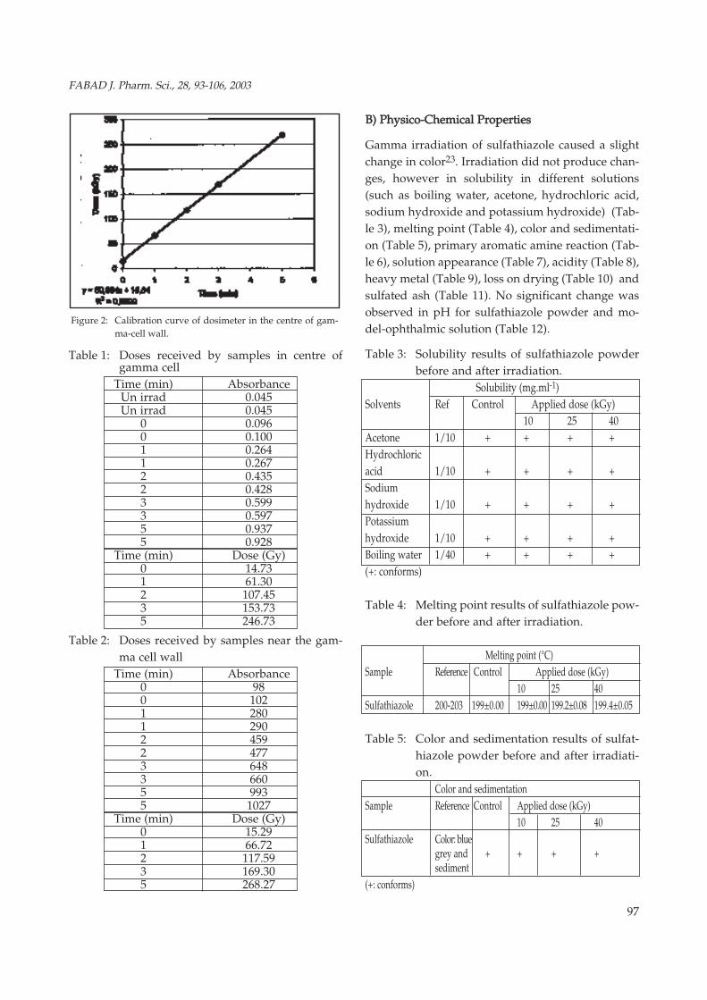
The actual doses received by samples were determi-ned by measuring the changes in absorbance. Thecorresponding doses were obtained from a calibra-ting graph (Figs 1 and 2), (Tables 1,2). Administrati-on doses were 2.8 kGy.h-1 for centre and 3.05 kGy.h-1
for the wall of gamma-cell10.
Figure 1: Calibration curve of dosimeter in the centre of gam-
ma-cell.
96
Benchaabane, Özer, Özalp, K›l›ç, Polat, Korkmaz
97
Table 1: Doses received by samples in centre ofgamma cell
Time (min) AbsorbanceUn irrad 0.045Un irrad 0.045
0 0.0960 0.1001 0.2641 0.2672 0.4352 0.4283 0.5993 0.5975 0.9375 0.928
Time (min) Dose (Gy)0 14.731 61.302 107.453 153.735 246.73
Table 2: Doses received by samples near the gam-ma cell wall
Time (min) Absorbance0 980 1021 2801 2902 4592 4773 6483 6605 9935 1027
Time (min) Dose (Gy)0 15.291 66.722 117.593 169.305 268.27
Figure 2: Calibration curve of dosimeter in the centre of gam-ma-cell wall.
BB)) PPhhyyssiiccoo--CChheemmiiccaall PPrrooppeerrttiieess
Gamma irradiation of sulfathiazole caused a slightchange in color23. Irradiation did not produce chan-ges, however in solubility in different solutions(such as boiling water, acetone, hydrochloric acid,sodium hydroxide and potassium hydroxide) (Tab-le 3), melting point (Table 4), color and sedimentati-on (Table 5), primary aromatic amine reaction (Tab-le 6), solution appearance (Table 7), acidity (Table 8),heavy metal (Table 9), loss on drying (Table 10) andsulfated ash (Table 11). No significant change wasobserved in pH for sulfathiazole powder and mo-del-ophthalmic solution (Table 12).
Table 3: Solubility results of sulfathiazole powderbefore and after irradiation.
Solubility (mg.ml-1)Solvents Ref Control Applied dose (kGy)
10 25 40Acetone 1/10 + + + +Hydrochloricacid 1/10 + + + +Sodiumhydroxide 1/10 + + + +Potassiumhydroxide 1/10 + + + +Boiling water 1/40 + + + +(+: conforms)
Table 4: Melting point results of sulfathiazole pow-der before and after irradiation.
Melting point (°C)Sample Reference Control Applied dose (kGy)
10 25 40Sulfathiazole 200-203 199±0.00 199±0.00 199.2±0.08 199.4±0.05
Table 5: Color and sedimentation results of sulfat-hiazole powder before and after irradiati-on.
Color and sedimentationSample Reference Control Applied dose (kGy)
10 25 40Sulfathiazole Color: blue
grey and + + + +sediment
(+: conforms)
FABAD J. Pharm. Sci., 28, 93-106, 2003

Table 6: Primary aromatic amine reaction results ofsulfathiazole powder before and after irra-diation
Primary aromatic amine reactionSample Reference Control Applied dose (kGy)
10 25 40Sulfathiazole Red orange
color and + + + +sediment
(+: conforms)
Table 7: Solution appearence results of sulfathiazo-le powder before and after irradiation
Solution appearanceSample Reference Control Applied dose (kGy)
10 25 40Sulfathiazole Not more
color than + + + +reference
(+: conforms)
Table 8: Acidity test results of sulfathiazole pow-der before and after irradiation
Acidity (ml)Sample Reference Control Applied dose (kGy)
10 25 40Sulfathiazole ≤0.1 0.062±0.077 0.062±0.053 0.064±0.042 0.063±0.023
Table 9: Heavy metal test results of sulfathiazolepowder before and after irradiation
Heavy metalSample Reference Control Applied dose (kGy)
10 25 40Sulfathiazole The reference
has more colorthan the blank + + + +solution of 10ppm or pbt
( +: conforms)
Table 10: Loss on drying test results of sulfathiazolepowder before and after irradiation
Loss on drying (%)Sample Reference Control Applied dose (kGy)
10 25 40Sulfathiazole ≤0.5 0.405±0.032 0.411±0.024 0.401±0.420 0.403±0.220
Table 11: Sulphated ash test results of sulfathiazolepowder before and after irradiation
Sulphated ash (%)Sample Reference Control Applied dose (kGy)
10 25 40Sulfathiazole ≤0.1 0.070±0.003 0.072±0.024 0.069±0.004 0.068±0.002
Table 12: pH values for control and irradiated pow-der and model-ophthalmic solution samp-les before and after irradiation
pHSample Reference
Values Control Applied dose (kGy)10 25 40
Powder - 8.101±0.047 8.104±0.036 8.111±0.025 8.108±0.012MOS* - 8.053±0.047 8.068±0.024 8.101±0.032 8.101±0.051* MOS: Model-ophthalmic solution
Gamma irradiation of sulfathiazole powder did notproduce changes in DSC results (Table 13) (Fig. 3).
Table 13: DSC results of sulfathiazole powder befo-re and after irradiation
Sample DSC (°C )ReferenceValues Control Applied dose (kGy)
10 25 40Powder 200-203 205.88 205.60 204.25 204.86
CC)) SSppeeccttrroossccooppiicc MMeetthhooddss aanndd TTeecchhnniiqquueess
Control and irradiated solid samples were studiedfor evaluation from the spectroscopic point of viewsuch as IR, UV, NMR and ESR24.
Figure 3: DSC thermogram of sulfathiazole powder before ir-
radiation
98
Benchaabane, Özer, Özalp, K›l›ç, Polat, Korkmaz

99
In the IR analysis at the lower radiation doses of sul-fathiazole H2N stretch bands, C=C stretch in benze-ne ring and SO2 stretch bands could be seen, for alldoses (10, 25 and 40 kGy) (Fig. 4) (Table 14).
Table 14: IR results of sulfathiazole powder beforeand after irradiation
Characteristic Peak (cm-1)
Sample Peak Reference Control Applied dose (kGy)
10 25 40
NH2 group and 3277.8- 3276.5- 3276.5- 3276.4-
N-H bonds 3100-350 3319.9 3319.2 3319.0 3318.9
NH2 vibration 1601 1595.2 1595.5 1595.8 1595.9
C-H single bonded 2853-2962 2920.9 2930.0 2931.2 2926.3
C=C aromatic 1500-1595 1495.2- 1494.8- 1494.6- 1494.6-
1573.7 1595.5 1573.6 1573.6
Asymmetric SO2 1303-1313 1281.5 1281.0 1280.6 1280.5
Symmetric SO2 1143-1155 1138.5 1135.9 1133.3 1133.0
-H 820-840 821.0 820.0 819.6 819.6
For comparison, λmax values calculated for control
and irradiated powders in both media were in good
agreement with the values given in the literature.
Although the variation with absorbed dose of the
wave number corresponding to maximum UV ab-
sorbance was not significant for samples dissolved
in 0.1N NaOH, a significant decrease was observed
for samples dissolved in 0.1N HCl (Table 15). No
significant change for model-ophthalmic solution
was observed (Table 16).
Figure 4: IR spectrum of sulfathiazole before irradiation
Table 15: λmax values calculated from UV spectra ofsulfathiazole powder before and after irra-diation
Sample λmax (nm)Medium Control Applied dose (kGy)
10 25 40NaOH 0,1N 256.00 256.00±0.01 256.20±0.04 256.74±0.03 256.65±0.03HCl 0.1 N 257.00 257.00±0.00 264.80±0.02 267.00±0.01 270.01±0.01
Table 16: λmax values calculated from UV spectra ofmodel-ophthalmic solution before and af-ter irradiation
Sample λmax (nm)Medium Control Applied dose (kGy)
10 25 40MOS* 257.00 257.00±0.00 257.10±0.01 257.6±0.05 257.8 ±0.04* MOS: Model-ophthalmic solution
Proton NMR spectra of sulfathiazole in dimethylsulfoxide DMSO-d6 containing tetramethylsilane asinternal reference consisted of different chemicalshifts which varied from two to 10 depending on thechemical environment of the related protons. Thelatter data are in good agreement with those repor-ted previously by Turczan and Medwick12 in the li-terature regarding identification of sulfonamides byNMR spectroscopy12. In the case of sulfathiazole, inthe region either containing singlets arising frommethoxyl, methyl, or methylene proton resonance,the aromatic region is distinctive and permits iden-tification (Fig. 5).
Irradiation of solid samples in the dose range of 10-40 kGy did not produce any significant effect on the
Figure 5: NMR of sulfathiazole before irradiation.
FABAD J. Pharm. Sci., 28, 93-106, 2003

100
chemical shifts of sulfathiazole protons as shown inTable 17.
Table 17: Calculated proton chemical shift valuesfor control and irradiated solid samplesbefore and after irradiation
NMR
Sample Related
Proton (s) Reference Control Applied dose (kGy)
10 25 40A 5.80 5.7 5.7 5.8 5.2B 6.64 6.5 6.5 6.5 6.5C 6.73 6.6 6.7 6.8 6.8D 7.18 7.1 7.1 7.2 7.4E 7.50 7.4 7.5 7.5 7.5
F 12.00 12.1 12.3 12.4 12.1
ESR spectra of control and irradiated solid sampleswere also investigated25. In the characterizationstudy of radicals formed by irradiation of sulfathi-azole, peak height against magnetic field value wasmeasured in the experimental ESR spectra. Accor-ding to molecular structure of sulfathiazole (Fig. 6),possible radicals and their types and structure wereestimated, and experimental ESR was plotted. Thepossible degradation pathways are given in Figure6. Depending on possible radicals, mathematicalmodels were developed and simulation studies we-re carried out. As a result of these studies, it is tho-ught that four different possible radicals (A, B, Cand D radicals) were formed by irradiation of sulfat-hiazole (Table 18).
Figure 6: Chemical structure of irradiated sulfathiazole and the
possible decomposition pathways.
Table 18: ESR results of irradiated at 40 kGy sulfat-hiazole powder
Radicals Developed formulaA (SO2)¯
B
C
D
An ionic radical has isotropic "g" value because this
radical is free and it can move fast. Movement of B
radical is limited because of the large group bonded
to sulfur atom, which is why, the B radical has noni-
sotropic "g" value. C radical is formed by break of S-
N bond. The unpaired electron is placed onto nitro-
gen N atoms. Movement of C radical is limited as
with B radicals because of the large group bonded to
nitrogen atoms. Thus, C radical has nonisotropic "g"
value. D radical is formed by the breaking of nitro-
gen and bonded hydrogen. C radical has extremely
thin structure because of hydrogen atoms bonded to
phenyl ring. In experimental ESR of bond between
phenyl and sulphur, unpaired electron placed onto
phenyl ring and this radical has isotropic "g" value.
Because of neutralization between unpaired elect-
rons, spectrum was obtained by using these four dif-
ferent radicals and simulation studies were carried
out. According to simulation studies, possible radi-
cals proposed were determined and spectroscopic
parameters of these radicals were calculated. They
are given in Table 19.
Benchaabane, Özer, Özalp, K›l›ç, Polat, Korkmaz

101
Table 19: ESR "g" value result of irradiated at 40 kGysulfathiazole powder before and after irra-diation
Radicals Intensity Half-band Extremely thin structure G value
A (1 lined, isotropic) 65.83900 1.2660 -------- G =2,0047
B (1 lined, anisotropic) 4.46130 1.3457 -------- G› =2,0098
G››=2,0098
C (6 lined, anisotropic) 0.19576 0.7925 6.5816 (for n) G› =2,0086
2.6788 (for h) G››=1,9960
D (1 lined, isotropic) 75.20100 7.5453 3.5262 (for h) G =2,0034
Theoretical ESR spectra were plotted by using thesespectroscopic parameters. Theoretical and experi-mental ESR spectra are given together in Figure 7. Itwas found that there was a good correlation betwe-en theoretical and experimental spectra. These re-sults showed that radicals formed by irradiation ofsulfathiazole samples are the same for A, B, C and Dradicals proposed in simulation studies25-28.
DD)) CChhrroommaattooggrraapphhiicc MMeetthhooddss
TLC experiments were performed using the techni-que proposed by EP 199716. For identification of sul-fathiazole, a mixture of ammoniac and butanol wasused as solvent and p-dimethylaminobenzaldehydeR solution as location reagent17, 28. This reagent pro-duces bright yellow spots in spraying with manycompounds, but heating at 100°C is necessary withsome of the compounds before the spots are visible.Rf values for unirradiated (control), irradiatedsamples and model-ophthalmic solution in the
Figure 7: ESR simulation curves of sulfothiazole powder irra-
diated at 40 kGy
applied dose range (10-40 kGy) were found signifi-cantly different. The Rf values are given in Table 20for sulfathiazole powder and model-ophthalmicsolution.
Table 20: TLC results of sulfathiazole powder beforeand after irradiation
Reference Rf
Powder Model-ophthalmic solution
Applied dose (kGy) Applied dose (kGy)
0 10 25 40 0 10 25 40
The spot obtained by
sulfathiazole was not more 0.300 0.385 0.392 0.395 0.320 0.380 0.388 0.420
intense than spot of ± ± ± ± ± ± ± ±
sulfanilamide and no 0.012 0.013 0.011 0.023 0.047 0.017 0.021 0.011
second spot appeared
EE)) AAnnttiimmiiccrroobbiiaall AAccttiivviittyy SSttuuddiieess
In the irradiated samples no activity loss was obser-ved for sulfathiazole powder and model-ophthalmicsolution .
FF)) SStteerriilliittyy
We did not observe any microbial growth in sterilitytest for all radiation doses (10, 25 and 40 kGy). Ana-erobic and aerobic microorganisms did not generatein the studied media.
SStteerriilliittyy AAssssuurraannccee LLeevveell (( SSAALL)) DDoossee DDeetteerrmmiinnaattii--oonn
SAL test performed on B. Pumilus spores (106 cfu. mL -1)infected samples did not work.
SSttuuddiieess CCaarrrriieedd OOuutt UUnnddeerr AAcccceelleerraatteedd SSttaabbiilliittyyCCoonnddiittiioonnss
Experimental results showed that physico-chemicalproperties such as color, odor, solubility in differentsolvents (boiling water, acetone, hydrochloric acid,sodium hydroxide and potassium hydroxide) (Tab-le 21), melting point (Table 22), color and sedimen-tation, primary aromatic amine reaction, solutionappearance, acidity (Table 23), heavy metal, loss on
FABAD J. Pharm. Sci., 28, 93-106, 2003
Samp
leSu
lfathi
azole

drying (Table 24), sulphated ash (Table 25) and DSC(Table 26), of control and irradiated solid and mo-del-ophthalmic solutions did not change under acce-lerated stability test conditions, however ph valuesdid, as shown in Table 27.
Table 21: Solubity results of sulfathiazole powderunder accelerated conditions
Time Solvent Solubility (ml)
(day) Ref Applied dose (kGy)10 25 40
0 + + +14 + + +28 1/10 + + +60 + + +90 + + +0 + + +14 + + +28 1/10 + + +60 + + +90 + + +0 + + +14 + + +28 1/10 + + +60 + + +90 + + +0 + + +14 + + +28 1/10 + + +60 + + +90 + + +0 + + +14 + + +28 1/40 + + +60 + + +90 + + +(+: conforms)
Table 22: Melting point results of sulfathiazole pow-der under accelerated conditions
Time Melting point (°C)(day) Control Applied dose (kGy)
10 25 400 199 199.00±0.00 199.20±0.08 199.40±0.0514 199 198.08±0.18 198.25±0.38 198.16±0.2328 199 198.41±0.18 198.25±0.38 198.16±0.2360 199 200.60±0.48 199.20±0.74 199.00±0.6390 199 199.81±0.40 199.20±0.74 199.80±0.74Table 23: Acidity results of sulfathiazole powder
under accelerated conditions
Time Acidity (ml)
(day) Control Applied dose (kGy)
10 25 40
0 0.062±0.077 0.062±0.053 0.064±0.042 0.063±0.023
14 0.062±0.077 0.063±0.031 0.067±0.001 0.067±0.001
28 0.062±0.077 0.066±0.007 0.062±0.005 0.065±0.005
60 0.062±0.077 0.064±0.001 0.069±0.009 0.069±0.001
90 0.062±0.077 0.063±0.001 0.068±0.001 0.070±0.001
Table 24: Loss on drying results of sulfathiazolepowder under accelerated conditions
Time Loss on drying (%)
(day) Ref Control Applied dose (kGy)
10 25 40
0 0.405±0.032 0.411±0.024 0.401±0.042 0.403±0.22
14 0.410±0.022 0.385±0.029 0.401±0.024 0.420±0.036
28 ≤0.5 0.409±0.011 0.401±0.012 0.399±0.004 0.399±0.005
60 0.415±0.002 0.401±0.019 0.423±0.036 0.411±0.024
90 0.411±0.021 0.400±0.003 0.400±0.001 0.417±0.036
Table 25: Sulphated ash results of sulfathiazolepowder under accelerated conditions
Time Sulphated ash (%)
(day) Ref Control Applied dose (kGy)
10 25 40
0 0.070±0.003 0.072±0.024 0.069±0.004 0.068±0.002
14 0.073±0.001 0.069±0.002 0.069±0.007 0.069±0.001
28 ≤ 0.1 0.077±0.005 0.069±0.005 0.059±0.004 0.069±0.004
60 0.077±0.010 0.069±0.004 0.069±0.006 0.060±0.001
90 0.072±0.001 0.068±0.004 0.069±0.004 0.068±0.004
Table 26: DSC results of sulfathiazole powder underaccelerated conditions
Time DSC (°C)
(day) Ref Control Applied dose (kGy)
10 25 40
0 205.88 205.60 204.25 204.86
28 200-203 205.88 205.60 203.25 203.66
60 205.88 202.96 203.56 203.62
90 205.88 204.41 204.37 203.37
Table 27: pH values for control and irradiated solids
102
Benchaabane, Özer, Özalp, K›l›ç, Polat, Korkmaz
Ace
tone
Hyd
ro ch
loric
acid
Sodi
umhy
drox
ide
Pota
ssiu
mhy
drox
ide
Boili
ngw
ater

103
stored at accelerated conditionsTime PH
(day) Powder Model-ophthalmic solution
Applied dose (kGy) Applied dose (kGy)
0 10 25 40 0 10 25 40
0 8.101 8.104 8.111 8.108 8.053 8.068 8.101 8.101
±0.047 ±0.036 ±0.025 ±0.012 ±0.047 ±0.024 ±0.032 ±0.051
14 8.1 7.678 7.728 7.801 8.051 8.223 8.19 8.183
±0.02 ±0.029 ±0.051 ±0.01 ±0.02 ±0.012 ±0.005 ±0.004
28 8.00 8.006 8.013 8.676 8.052 8.216 8.183 8.178
±0.02 ±0.016 ±0.007 ±0.03 ±0.04 ±0.013 ±0.012 ±0.014
60 8.011 8.273 8.265 8.263 8.052 8.316 8.166 8.033
±0.03 ±0.007 ±0.007 ±0.004 ±0.03 ±0.068 ±0.047 ±0.074
90 8.001 8.028 8.05 8.035 8.053 8.25 8.283 8.3
±0.02 ±0.014 ±0.016 ±0.012 ±0.04 ±0.095 ±0.089 ±0.115
Although λmax values of control and irradiated solidpowders dissolved in 0.1 N NAOH were found toexhibit no changes overall the stability studies, thatof control samples dissolved in 0.1 N HCI experien-ced a meaningful increase (Table 28) in the first we-ek storage period, then stayed approximately cons-tant6. However, the changes in lmax values of irra-diated samples were less prounouced. λmax valuesof control and irradiated model-ophthalmic solutionwere found to exhibit no changes throughout thestability studies. The amounts of sulfathiazole areshown in Table 29.
Table 28: λmax values of sulfathiazole powder underaccelerated conditions
Solvent Time (day) λmax (nm)
Control Applied dose (kGy)
10 25 40
NaOH 0.1N 0 256.00±0.01 256.20±0.04 256.74±0.03 256.65±0.0314 256.01±0.01 256.00±0.04 240.00±0.03 256.20±0.0428 256.01±0.05 257.00±0.02 256.00±0.03 240.00±0.0860 256.04±0.03 258.40±0.01 256.50±0.01 256.10±0.0290 256.01±0.01 239.90±0.07 257.60±0.07 265.50±0.05
HCl 0.1N 0 257.00±0.00 264.80±0.02 267.00±0.01 270.01±0.0114 257.01±0.01 279.10±0.01 264.80±0.08 277.40±0.0228 257.00±0.05 280.00±0.03 279.00±0.06 265.80±0.0160 257.02±0.04 278.60±0.02 281.00±0.01 279.00±0.0290 257.00±0.01 267.20±0.04 277.80±0.03 280.50±0.07
Table 29: Sulfathiazole determination of model-oph-
thalmic solution under accelerated conditions.Time (Day) Amount of sulfathiazole (mg. mL-1)
Control Applied dose (kGy)
10 25 40
0 0.042±0.013 0.046±0.022 0.047±0.019 0.045±0.019
14 0.044±0.020 0.042±0.018 0.045±0.013 0.043±0.024
28 0.042±0.017 0.048±0.014 0.044±0.010 0.040±0.018
60 0.041±0.019 0.044±0.011 0.042±0.017 0.041±0.014
90 0.044±0.022 0.044±0.024 0.046±0.020 0.046±0.019
FT-IR (Table 30) and NMR (Table 31) spectra of cont-rol and irradiated samples stored for three monthsunder stability test conditions were found to exhibitcharacteristic features of the spectra obtained forsamples stored at normal environmental conditions.
Table 30: Amount of sulfathiazole powder under ac-celerated conditions.
Sample Characteristic Peak (cm-1)
Peak Reference Applied dose (kGy)
10 25 40
0 14 28 60 90 0 14 28 60 90 0 14 28 60 90
NH2 group and
N-H bonds 3100-3500 + + + + + + + + + + + + + + +
NH2 vibration 1601 + + + + + + + + + + + + + + +
C-H single bonded 2853-2962 + + + + + + + + + + + + + + +
C=C aromatic 1500-1595 + + + + + + + + + + + + + + +
Asymmetric SO2 1303-1313 + + + + + + + + + + + + + + +
Symmetric SO2 1143-1155 + + + + + + + + + + + + + + +
-H 820-840 + + + + + + + + + + + + + + +
(+: conforms )
Table 31: NMR results of sulfathiazole powder un-der accelerated conditions
Sample Related NMR
Proton Reference Applied dose (kGy)
(s) 10 25 40
0 14 28 60 90 0 14 28 60 90 0 14 28 60 90
A ~5.80 + + + + + + + + + + + + + + +
B ~6.64 + + + + + + + + + + + + + + +
C ~6.73 + + + + + + + + + + + + + + +
D ~7.18 + + + + + + + + + + + + + + +
E ~7.50 + + + + + + + + + + + + + + +
F ~12.00 + + + + + + + + + + + + + + +
(+: conforms)
As emphasized in the previous section of the pre-
FABAD J. Pharm. Sci., 28, 93-106, 2003

sent work, unirradiated (control) solid samples donot exhibit any ESR signal. Storing of these samplesat stability test conditions, i.e., at high temperatureand high relative humidity, does not create anychanges in this feature. However, storing irradiatedsamples in the same conditions have been observedto cause a decrease in the ESR signal intensities ofthe samples due to the decay of radiolytic interme-diates created during the irradiation. The possibledegradation pathways are given in Figure 6. The re-sults obtained for solid sulfathiazole at the dose of40 kGy are given in Figure 7. As can be seen, ESRsignal intensity decay curve exhibits biphasic cha-racter just at the beginning, unstable radiolytic pro-duct decay completely, than the more stable onesdominate on the decay curve. However, at the endof the storing period (90th day) all the radiolytic in-termediates decay almost completely.
Rf values determined by TLC method of control andirradiated solid and model-ophthalmic solutionsamples stored at stability test conditions are foundto be independent of storage time, have a meaning-ful increase in the second week of storage period,and then stay approximately constant within the ex-perimental error limits (Table 32).
Table 32: TLC results of sulfathiazole powder andmodel-ophthalmic solution under accele-rated conditions.
Time Rf
(Day) Powder Model-ophthalmic solution
Applied dose (kGy) Applied dose (kGy)
0 10 25 40 0 10 25 40
0 0.320 0.380 0.388 0.420 0.300 0.385 0.392 0.395
±0.047 ±0.017 ±0.021 ±0.011 ±0.012 ±0.013 ±0.011 ±0.023
14 0.420 0.479 0.444 0.444 0.322 0.516 0.515 0.493
±0.003 ±0.005 ±0.004 ±0.004 ±0.004 ±0.007 ±0.003 ±0.005
28 0.426 0.507 0.515 0.515 0.334 0.528 0.536 0.537
±0.008 ±0.002 ±0.003 ±0.003 ±0.010 ±0.003 ±0.005 ±0.012
60 0.435 0.523 0.526 0.526 0.327 0.578 0.562 0.544
±0.002 ±0.003 ±0.003 ±0.003 ±0.014 ±0.002 ±0.003 ±0.003
90 0.421 0.534 0.533 0.533 0.338 0.509 0.513 0.527
±0.005 ±0.06 ±0.003 ±0.003 ±0.012 ±0.09 ±0.009 ±0.009
During the stability period, the sterility test was fo-
und to be the same for all applied doses at the begin-ning and after the storage period.
DDIISSCCUUSSSSIIOONN
SSttuuddiieess CCaarrrriieedd OOuutt UUnnddeerr NNoorrmmaall CCoonnddiittiioonnss
Color change in the irradiated substances is thesimplest and most helpful observation to obtein in-formation about possible radiolytical intermediatesproduced in these substances upon irradiation29,30.Based on the fact that color change was observed inirradiated samples in the applied dose region of 10-40 kGy, it can be concluded that radiolytical inter-mediates are produced by irradiation of powders(Fig. 7)4. Gamma radiation transfers its energy indi-rectly to the target in the solution. Radicals produ-ced by the direct action of radiation on water mole-cules are the principal elements in the degradationof aqueous solutions31. In its direct action, gammaradiation ejects electrons from water molecules. Po-sitively charged water molecules react in their turn,react with unchanged water molecules and radicals;mainly OH ¯ is produced32.
The latter is very strong oxidants and they play prin-cipal role in the degradation of aqueous systems.This feature of water molecules makes the aqueoussystems more sensitive to radiolysis7. When evalu-ating of experimental results concerning pH, it wasfound that no change was observed in the irradiatedsolid and model-ophthalmic solution samples8. Ra-diation did not cause any change in solubility of sul-fathiazole powder in boiling water, acetone,hydrochloric acid, sodium hydroxide and potassi-um hydroxide nor in its melting point.
UV spectra of control sulfathiazole in acidic and ba-sic media exhibit two λmax values at about 240-256nm and 256-281 nm, respectively (Table 28). Obser-vation of λmax appearing at nearly the same wave-lentgth even after irradiation indicates that sulfathi-azole is conserved in the irradiated samples; howe-ver, the same is not true for substitution rings. Na-mely, this ring is affected, to a large extent, by gam-ma radiation. Comparison of the proton chemical
104
Benchaabane, Özer, Özalp, K›l›ç, Polat, Korkmaz

105
shifts of control and irradiated samples given inTable 10 shows that gamma radiation cannot produ-ce significant changes in the electronic environmentof the protons of sulfathiazole molecules.
The presence of ESR signal in the irradiated but notin the control sample definitively points out the pro-duction of radiolytic intermediates in solid samplesupon irradiation. The ESR spectra of irradiatedsamples consist of a signal resonance line with ashoulder at low magnetic field and it is distinguis-hable from noise even at the lowest applied dose (10kGy). However, the short life radicals decay imme-diately after the second of irradiation and thereforethe recorded experimental spectra are due to thelong-life radicals. G value which represents radiati-on yield of solid sulfathiazole.
SSttuuddiieess CCaarrrriieedd OOuutt UUnnddeerr AAcccceelleerraatteedd CCoonnddiittiioonnss
Physico-chemical properties of solid sulfathiazoleand model-ophthalmic solution were observed asnot changing in the stability test experiments. Thefact that solubility and melting point of unirradiated(control) and irradiated solid samples did not chan-ge. The fact that pH values did not significantlychange for sulfathiazole powder and model-opht-halmic solution, demonstrates that accelerated stabi-lity test conditions have similar effects on unirradi-ated and irradiated samples.
UV spectra of control (unirradiated) sulfathiazole inacidic and basic media exhibited two λmax values atabout 257 nm and 256 nm, respectively, under acce-lerated conditions (Table 28). λmax of irradiated so-lid samples dissolved in basic medium did not chan-ge, but increased in acidic medium in applied doseand in storage time. Observation of λmax appearingat nearly the same wavelength even after irradiationindicates that sulfathiazole is conserved in the irra-diated samples; however, the same is not true forsubstitution rings. Namely, this ring is affected to alarge extent from gamma radiation. Comparison ofthe proton chemical shifts of control and irradiatedsamples given in Table 17 shows that gamma radi-ation cannot produce significant changes in theelectronic environment of the protons of sulfonami-
de molecules, λmax of irradiated model-ophthalmicsolution staying constant.
The biphasic character of ESR signal intensity decaycurve of irradiated solid samples under acceleratedstability test conditions reflects the existance of fourradicals of different decay characteristics. Althoughthe g factors and corresponding line shapes of theseradicals are similar, they have different features.
It is concluded that irradiation of sulfathiazole pow-der and model-ophthalmic solution did not produceany changes in the antimicrobial activities. Becauseof the solubility problems of sulfathiazole formulati-ons, it was not possible to count the number of themicro-organisms (bioburden). Therefore, sal couldnot be determined preciously. The dose of 10 kGycould be applied to our powder and model-ophthal-mic solution of sulfathiazole, and this is a lower do-se level than the one mentioned (25 kGy) in EP11.
CCOONNCCLLUUSSIIOONN
When the physico-chemical properties of the irradi-ated substances are analyzed, it is observed that sul-fathiazole powder and model-ophthalmic solutionare not affected by the irradiation. While evaluatingthe effect of irradiation on the antimicrobial activiti-es of irradiated samples, no activity loss was obser-ved with the increase in radiation dose. Negative re-sult of the tests concerning of B. Pumilus in uninfec-ted and infected dosage forms by the spores of B.Pumilus indicates that radiation dose of 10 kGy canbe applied to our model-ophthalmic solution of sul-fathiazole without any changes.
The three month stability test showing that free radi-cals formed in irradiated samples during the stabilityperiod supports that in the accelerated conditions irra-diated samples are not very affected by the irradiationwhen compared to the unirradiated samples.
AACCKKNNOOWWLLEEDDGGEEMMEENNTT
This study was carried out as a project from IAEA-Alg/8/010.The authors wish to thank IAEA/Vienna for valuab-
FABAD J. Pharm. Sci., 28, 93-106, 2003

le support, and Assoc. Prof. Dr. Nesrin Gökhan, Dr.Cengiz Uzun, Pharm (M.Sci) Melike Ekizo¤lu,Pharm. Ekrem K›l›ç, and Pharm.Yasemin Dündarfor their excellent help.
RReeffeerreenncceess 1. IAEA Report of the Consultant’s Meeting on Radiation
Sterilization in the Pharmaceutical Industry, Vienna,9-11,1996.
2. IAEA Manual on Radiation Sterilization of Medicaland Biological Materials. Technical Report, Vienna,149, 1973.
3. IAEA Conferences Proceedings. Industrial Applicati-on of Radioisotopes and Radiation Technology. Gre-noble, Vienna, 10-8. 1981.
4. Piccerelle P, Reynier JP, Joachim J, Tilquin B, Raffi J.Radio-sterilisation de médicaments Intérét, législationet travaux a entreprendre, J. Pharm. Belg., 55, 131-136,2000.
5. Burger F, Pouliquen-Sonaglia I, D’avino S, Raffi J. Etu-de de l’irradiation gamma du collagéne en phase soli-de et en phase liquise, J. Pharm. Belg., 55 ,145-154,2000.
6. Olguner G (Mercano¤lu), Özer AY, Özalp M, Ekizog-lu M, Çolak fi, Korkmaz M. Radiosterilization of sulfo-namides-II: Determination of the effects of gamma ir-radiation on commercial sulfonamide preparations (InPress).
7. Olguner G (Mercano¤lu), Özer AY, Çolak fi, KorkmazM, Barbarin N, Tilquin B, Özalp M, Ekizoglu M. Radi-osterilization of sulfonamides-I: Determination of theeffects of gamma irradiation on solid sulfonamides (InPress).
8. Martindale, Extra Pharmacopoeia (29th ed) The Phar-maceutical press. London pp:301-6;4953, 1989.
9. The Merck Index (11th ed), Merck & Co Inc, USA,1414, 1989.
10. Final Report of the Co-ordinated Research Project onCharacterization and Evaluation of High Dose Dosi-metry Techniques for Quality Assurance in RadiationProcessing. Dosimetry for Radiation Processing. IA-EA-Tec Doc-1156, 2000.
11. European Pharmacopoeia ( 3rd ed), Suisse Edition, Jo-uve, France, 1593, 1997.
12. Turczan J, Medwick T. Identification of sulfonamidesby NMR spectroscopy, J. Pharm. Sci., 61, 434-440, 1972.
13. Polat M, Korkmaz M. Kinetics of the radicals inducedin gamma-irradiated naproxen sodium and apranax.Applicability of ESR technique to monitor radiosterili-zation of naproxen sodium-containing drugs, Int. J.Pharm. 244, 169-179. 2002.
14. Phillips GO, Power DM, Stewart M. Effect of δ irradi-ation on sodium sulphacetamide, Radiat. Res., 46, 236-
250, 1971.15. Phillips GO, Power DM, Stewart M. Effect of δ irradi-
ation on sodium sulphacetamide. Radiat. Res., 53, 204-213, 1973.
16. Clarke EGC, Humphreys DJ. A note on the identifica-tion of sulphonamides by thin-layer chromatography,J. Pharm. Pharmacol., 22, 845-847, 1970.
17. Klein HR, Marder WJ. TLC identification of sulphona-mides, J. Pharm. Sci., 60, 448-451,1971.
18. Gazso IG. Microbiological Parameters and RadiationMicrobiology in Process and Quality Control. NationalTraining Course on Industrial Radiation Sterilization.Ankara, 1992.
19. United States Pharmacopeia (USP XVIII). The Pharma-ceutical Press, London, 1237-1238, 1985.
20. Jacobs GP, Leupin K. Irradiation of drugs, Pharm. Ac-ta. Helv., 49, 12-20,1974.
21. Gazso LG. Guidelines and Criteria Standard in Radi-ation Processing, Process and Quality Control-SterilityAssurance, "National Training Course on IndustrialSterilization" Ankara, 1-13, 1992.
22. Varshney I, Patel KM. Effects of ionizing radiations ona pharmaceutical compound, chloramphenicol. Radi-al. Phys. Chem., 43, 471-480, 1994.
23. Sunshine I (ed.). Handbook of Spectrophotometric Da-ta of Drugs, CRC Press, London, 139, 1981.
24. Köseo¤lu R, Köseo¤lu E, Köksal F. Electron paramag-netic resonance of some δ-irradiated drugs, Appl. Ra-diat. and Isot., 58, 63-68, 2003.
25. Gopal NGS, Pate KM, Sharma G, Bhalla HL, PamelaAW, Hilmy N. Radiation guide for radiation steriliza-tion of pharmaceuticals and decontamination of rawmaterials, Phys.Chem., 32, 619-622, 1988.
26. Gibella M, Grucq AS, Tilquin B, Stocker P, Lesgards G.Electron spin resonance studies of some irradiatedpharmaceuticals, Radiat. Phys. Chem., 58, 69-76, 2000.
27. Lawrence JR, Donald MS. Effect of δ-radiation on se-lected pharmaceuticals, J. Pharm. Sci., 56, 1967.
28. Jacobs GP. A review of the effect of gamma irradiationon pharmaceutical materials, J. Biomater. Appl.,10, 59-96, 1995.
29. Lazier J, Darbord JC. Theoretical basis of choice of thedose in radiation sterilization of supplies, Phys.Chem., 31, 727-739, 1988.
30. Laizier J, Steinmetz AC, Vincent F, Darbord, JC. Pro-position d’une méthode de determination experimen-tale de la dose en radiostérilisation, S.t.p. Pharma. 4,778-784,1988.
31. Berk F, Özer, AY. Radiation sterilization I: Radiosteri-lization of medical devices, J. Pharm. Sci., 24, 223-231,1999.
32. Clark’s Isolation and Identification of Drug Substan-ces, Pharmaceutical Press, London, 981, 1986.
106
Benchaabane, Özer, Özalp, K›l›ç, Polat, Korkmaz