RESEARCH ARTICLES (1) THE TRIANGLE PROJECT · PDF fileslide on which was deposited a small ......
10
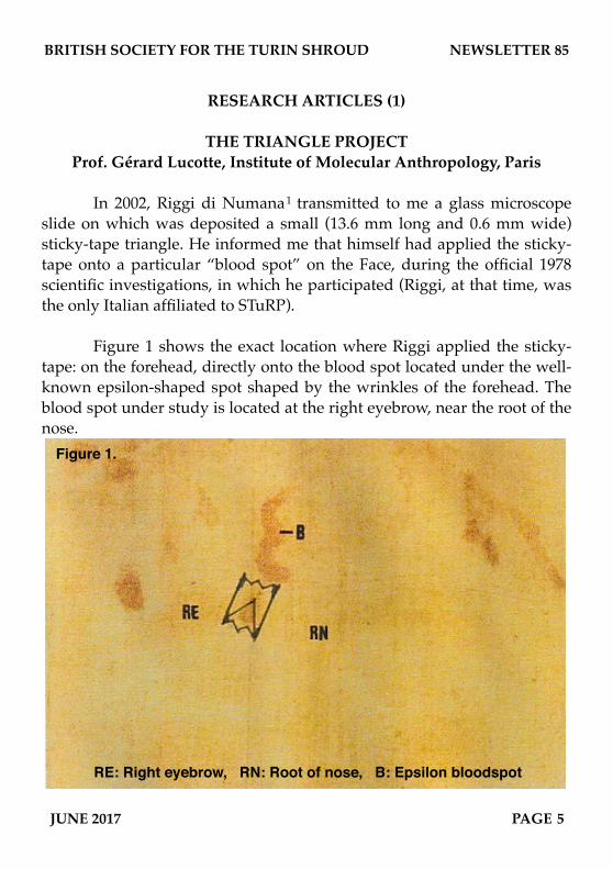
RESEARCH ARTICLES (1) THE TRIANGLE PROJECT Prof. Gérard Lucotte, Institute of Molecular Anthropology, Paris In 2002, Riggi di Numana 1 transmitted to me a glass microscope slide on which was deposited a small (13.6 mm long and 0.6 mm wide) sticky-tape triangle. He informed me that himself had applied the sticky- tape onto a particular “blood spot” on the Face, during the official 1978 scientific investigations, in which he participated (Riggi, at that time, was the only Italian affiliated to STuRP). Figure 1 shows the exact location where Riggi applied the sticky- tape: on the forehead, directly onto the blood spot located under the well- known epsilon-shaped spot shaped by the wrinkles of the forehead. The blood spot under study is located at the right eyebrow, near the root of the nose. RE: Right eyebrow, RN: Root of nose, B: Epsilon bloodspot Figure 1. BRITISH SOCIETY FOR THE TURIN SHROUD NEWSLETTER 85 JUNE 2017 PAGE 5
Transcript of RESEARCH ARTICLES (1) THE TRIANGLE PROJECT · PDF fileslide on which was deposited a small ......
RESEARCH ARTICLES (1)
THE TRIANGLE PROJECTProf. Gérard Lucotte, Institute of Molecular Anthropology, Paris
! In 2002, Riggi di Numana1 transmitted to me a glass microscope slide on which was deposited a small (13.6 mm long and 0.6 mm wide) sticky-tape triangle. He informed me that himself had applied the sticky-tape onto a particular “blood spot” on the Face, during the official 1978 scientific investigations, in which he participated (Riggi, at that time, was the only Italian affiliated to STuRP).
! Figure 1 shows the exact location where Riggi applied the sticky-tape: on the forehead, directly onto the blood spot located under the well-known epsilon-shaped spot shaped by the wrinkles of the forehead. The blood spot under study is located at the right eyebrow, near the root of the nose.
RE: Right eyebrow, RN: Root of nose, B: Epsilon bloodspot
Figure 1.
BRITISH SOCIETY FOR THE TURIN SHROUD NEWSLETTER 85
JUNE 2017 PAGE 5

! After taking his sample, Riggi cut a small triangle from the sticky-tape. The reason why is that he thought that a small particle that he observed with a magnifying glass, of rounded form and red-brown colour, clearly visible at the triangle top was a red blood cell; Riggi charged me to the analyse this “hematy.”
! During the year 2002, I collaborated with Thierry Derouin (Phanerology Department of the Museum of Natural History, Paris) to obtain some in colour photographs (in optical microscopy) of the various parts of the triangle. This first work was achieved while the sticky-tape triangle was deposited on the glass slide, using a Zeiss photomicroscope (model III, 1972). Some views, in cross-polarized light, were obtained with the petrographic version of this microscope.
! Since 2003, we have realized further electronic microscopy studies (in collaboration with Thierry Thomasset, Laboratory of Physico-chemical analysis, UST University of Compiègne) on the numerous particles of the triangle. For these observations and analyses, the triangle was mounted upside down on a substratum of a sterile dedicated paper backing, held in place by Scotch tape. Two SEM (Scanning Electron Microscopy) apparatus were used:! 1) A Philips XL. 30 instrument, Environmental version. GSE (Gaseous Secondary Electrons) and BSE (Back Scattered Electrons) procedures were used, the last one to detect heavy elements. Elemental analysis of the particles and its sub-parts were achieved by using EDX (Energy Dispersive X-ray spectroscopy), this SEM being equipped with a Bruker AXS-EDX (PGT system analysis: Spirit model, of Princeton Gamma Technology). ! 2) Since 2016 we have used another SEM apparatus (FEI model Quanta 25 of FEG, probe model X-flash 6/30), both in LFD (Large Field Detector) and in CBS (Circular Back Scattering).
! Figure 2 shows, at low magnifications, two photographs of the triangle (the upper photograph, in optical microscopy; the lower photograph, in electronic microscopy). On both these photographs we can
BRITISH SOCIETY FOR THE TURIN SHROUD NEWSLETTER 85
PAGE 6 JUNE 2017
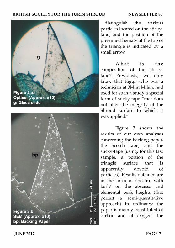
distinguish the various particles located on the sticky-tape; and the position of the presumed hematy at the top of the triangle is indicated by a small arrow.
! W h a t i s t h e composition of the sticky-tape? Previously, we only knew that Riggi, who was a technician at 3M in Milan, had used for such a study a special form of sticky-tape “that does not alter the integrity of the Shroud surface to which it was applied.”
! Figure 3 shows the results of our own analyses concerning the backing paper, the Scotch tape, and the sticky-tape (using, for this last sample, a portion of the triangle surface that is apparently devoid of particles). Results obtained are in the form of spectra, with ke/V on the abscissa and elemental peak heights (that permit a semi-quantitative approach) in ordinates: the paper is mainly constituted of carbon and of oxygen (the
BRITISH SOCIETY FOR THE TURIN SHROUD NEWSLETTER 85
JUNE 2017 PAGE 7
Figure 2.a.Optical (Approx. x10)g: Glass slide
Figure 2.b.SEM (Approx. x10)bp: Backing Paper
bp

organic component); the Scotch tape is also mainly constituted of carbon and oxygen, but with a small peak of aluminium (which is a usual minor component of many industrial Scotch tapes); the sticky-tape is mainly constituted of carbon, with a little peak of oxygen. Our conclusion about the constitution of the sticky-tape of the triangle is that it is practically a matter of pure (organic) hydrocarbon.
Figure 3. Elemental compositions of constituents.
C: Carbon, O: Oxygen, Al: Aluminium
2 - Scotch tape
3 - sticky-tape sample
1 - backing paper
BRITISH SOCIETY FOR THE TURIN SHROUD NEWSLETTER 85
PAGE 8 JUNE 2017

! Figure 4 represents an enlarged SEM photograph of the triangle. Because of the large number of particles it contains, it was subdivided into 19 areas (from A to S). Table 1 (next page) summarizes the numbers of particles (1µm across or larger) deposited on each division.
! For each of these particles, we now hold: ! a) An optical photograph, that informs us about the aspect, the size and colour of the particle; polarized light studies show whether it birefracts or not.! b) An SEM photograph, that informs us about the aspect, the superficial topology and the size of the particle.! c) An EDX analysis, that informs us about the elemental composition of the particle.
! Concerning now the presumed hematy located at the top of
the triangle that Riggi asked me to study, we have now precise information; this particle (named a5 because it is located in the A area and is the fifth particle under study) is of rounded form, a little more than 10µm in diameter) and of a red-brown colour. However it is not in fact a red blood cell: it is a mineral/metallic particle, an aluminosilicate iron-rich formation with a some tormented surface and little lead inclusions. It is assimilable to what was previously documented as a “McCrone red microbille” (McCrone, 2000)2, rich in iron oxide. This particle represents a unique type on the triangle surface.
Figure 4.Area Index
BRITISH SOCIETY FOR THE TURIN SHROUD NEWSLETTER 85
JUNE 2017 PAGE 9

Table 1: Numbers of particles found on each division of the sample.
Area No. Area No. Area No. Area No. Area No.
A 54 E 182 I 86 M 87 Q 35
B 101 F 75 J 93 N 55 R 44
C 53 G 108 K 108 O 51 S 33
D 42 H 80 L 98 P 97 Total 1482
! There is a great diversity (in aspect, colour and elemental composition) in the various particles loaded on the triangle. Table 2 summarizes (somewhat artificially) twelve different categories to which they have been assigned.
Table 2: Particles categorised into twelve broad categories. Categories Characterization References
1. Minerals mainly clays Lucotte, 2012
2. Metals mainly iron, but also gold, silver, copper, brass, etc.
3. Textile fibers five linen fibers Lucotte, 2015a
4. Bacterias recent
5. Cyanophycaes both recent and ancient
6. Marine micro-organisms Diatoms, Dinophycaes, Coccoliths
7. Pollens and spores at least six pollen species; twenty-one modern spores
Lucotte, 2015 b
8. Eggs of insects and Acaris
9. Human remains - red blood cells - skin cells - one hair portion
Lucotte, 2015c Lucotte, 2016 Lucotte and Thomasset, 2017
10. Organic mineral and metallic pigments11. Manufactured materials plastic, glass, plaster, brick,
cement, etc.12. Household chemicals
BRITISH SOCIETY FOR THE TURIN SHROUD NEWSLETTER 85
PAGE 10 JUNE 2017

! Minerals constitute, in a first estimation (Lucotte, 2012)3, about a quarter of the set. Textile fibers consist of five linen fibers, one long silk fiber, and one (tripartite) synthetic fiber (Lucotte, 2012).4 There are at least six pollen species, and twenty-one modern spores (Lucotte, 2015b).5 Particle a5 located at the top of the triangle is the single example of a mineral particle that is specially iron-rich. Numerous biological samples were observed and characterized: modern Bacterias, recent and ancient Cyanophycaes, Diatoms, Dinophycaes, Coccoliths and other marine micro-organisms, eggs of Insects and Acaris, etc.
! Because they could constitute remains of the Man who bled on the Shroud, a special attention was reserved to particles of human origin: red blood cells (Lucotte, 2015c) 6, at least three pieces of skin debris (Lucotte, 2016) 7 and one section of hair (Lucotte and Thomasset, 2017).8 One possible fragment of human bone and/or cartilage is currently under study (Lucotte and Thomasset, in preparation).
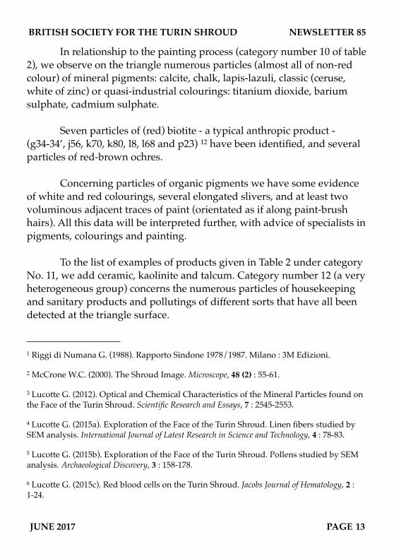
! A very important question to resolve concerns the nature of the red-brown colour. Figure 5 shows a colour photograph of the triangle (deposited on the backing-paper), and an enlargement of it. On these photographs, taken in optical microscopy, the triangle appears as red-
bp
Figure 5. Photograph of the triangle showing red-brown colour and enlargement. Circled: sample, s: Scotch tape, bp: backing paper.
BRITISH SOCIETY FOR THE TURIN SHROUD NEWSLETTER 85
JUNE 2017 PAGE 11
brown coloured. The enlarged photograph shows that this colour seems mainly concentrated on the particle groups located in the middle part of the triangle.
! This colour of the sticky-tape triangle is the same as that of the corresponding blood spot (and that of the other blood spots of the Face, and even of the rest of the body). Two alternative explanations have been given in the past to account for the red colour of the “blood stains”: the first explanation was given by Heller and Adler 9,10, who made desperate efforts to explain them by suggesting a novel result of the trauma of crucifixion, proposing that it was a mixture of bilirubin and an exotic complex of oxidised met-hemoglobin, that they called “parahemic”. The opposite position is held by McCrone and Skirius, mainly on the basis of polarized light microscopy studies of the red particles of the Shroud. They proposed that this red-brown colour was mainly due to aggregates of crystalline particles of hematite (an iron-oxide mineral) and vermillion (a pigment constituted of cinnabar mineral).
! The most recent results we have obtained (Lucotte, Derouin and Thomasset, 2016) 11 establish that neither red blood cells, nor hematite and cinnabar particles, can explain the whole observed red-brown colour of the triangle:! Of the twenty-five red blood cells (or groups of red blood cells) we have observed, only two of them (e22 and g25) are of red colour in optical microscopy.! We have observed only two well-characterized particles of hematite (l39 and o7) and only one particle (k56) of cinnabar.
! To explain the whole red-brown colour of the triangle, we are currently studying (Lucotte, Derouin, Thomasset, in preparation) the elemental compositions and distributions of some yellow-red clays: goethite, montmorillonite, illite, and above all a special sort of phosphorite. They are very numerous on the surface of the triangle and - because of the iron oxide - of yellow, red and red-brown colours.
BRITISH SOCIETY FOR THE TURIN SHROUD NEWSLETTER 85
PAGE 12 JUNE 2017
! In relationship to the painting process (category number 10 of table 2), we observe on the triangle numerous particles (almost all of non-red colour) of mineral pigments: calcite, chalk, lapis-lazuli, classic (ceruse, white of zinc) or quasi-industrial colourings: titanium dioxide, barium sulphate, cadmium sulphate.
! Seven particles of (red) biotite - a typical anthropic product - (g34-34’, j56, k70, k80, l8, l68 and p23) 12 have been identified, and several particles of red-brown ochres.
! Concerning particles of organic pigments we have some evidence of white and red colourings, several elongated slivers, and at least two voluminous adjacent traces of paint (orientated as if along paint-brush hairs). All this data will be interpreted further, with advice of specialists in pigments, colourings and painting.
! To the list of examples of products given in Table 2 under category No. 11, we add ceramic, kaolinite and talcum. Category number 12 (a very heterogeneous group) concerns the numerous particles of housekeeping and sanitary products and pollutings of different sorts that have all been detected at the triangle surface.
BRITISH SOCIETY FOR THE TURIN SHROUD NEWSLETTER 85
JUNE 2017 PAGE 13
1 Riggi di Numana G. (1988). Rapporto Sindone 1978/1987. Milano : 3M Edizioni.
2 McCrone W.C. (2000). The Shroud Image. Microscope, 48 (2) : 55-61.
3 Lucotte G. (2012). Optical and Chemical Characteristics of the Mineral Particles found on the Face of the Turin Shroud. Scientific Research and Essays, 7 : 2545-2553.
4 Lucotte G. (2015a). Exploration of the Face of the Turin Shroud. Linen fibers studied by SEM analysis. International Journal of Latest Research in Science and Technology, 4 : 78-83.
5 Lucotte G. (2015b). Exploration of the Face of the Turin Shroud. Pollens studied by SEM analysis. Archaeological Discovery, 3 : 158-178.
6 Lucotte G. (2015c). Red blood cells on the Turin Shroud. Jacobs Journal of Hematology, 2 : 1-24.
BRITISH SOCIETY FOR THE TURIN SHROUD NEWSLETTER 85
PAGE 14 JUNE 2017
7 Lucotte G. (2016). Skin debris on the Face of the Turin Shroud : a SEM-EDX analysis. Archaeological Discovery, 4 : 103-117.
8 Lucotte G., Thomasset T. (2017). Scanning electron microscopic characterization and elemental analysis of one hair located on the Face of the Turin Shroud. Archaeological Discovery, 5 : 1-21.
9 Heller J.H., Adler A.D. (1980). Blood of the Shroud of Turin. Applied Optics, 19 : 2742-2744.
10 Heller J.H., Adler A.D. (1981). A chemical investigation of the Shroud of Turin. Canadian Society of Forensic Science Journal, 14 : 81-103.
11 McCrone W.C., Skirius C. (1980). Light microscopical studies of the Turin Shroud. Microscope, 28 : 105-112.
12 Lucotte G., Derouin T., Thomasset T. (2016). Hematite, biotite and cinnabar on the Face of the Turin Shroud : microscopy and SEM-EDX analysis. Open Journal of Applied Sciences, 6 : 601-625.
dadadadadcbcbcbcbc
Editor’s Note on Prof. Lucotte’s article
! In the last issue I speculated that the sliver of tape being examined by Prof. Lucotte may have come from the ‘hooverings’ that Giovanni Riggi di Numana made with a specially adapted micro-vacuum-cleaner in 1978. However Prof. Lucotte makes it clear that it actually came from a sticky tape being laid directly on the face of the Shroud. The 1978 scientific investigation is often generally assumed to have been almost exclusively American, whose respect for the sanctity of the Shroud forbad direct contact with the face, even to the extent of a heated row between John Jackson and Max Frei, as the latter attempted to take pollen samples from it. Even contact with the rest of the Shroud was carefully moderated by the pressure-controllable apparatus Ray Rogers used to apply his own sticky tape (although he had originally intended to take samples from the face himself - perhaps the argument between Jackson and Frei made him change his mind). However, before the Americans were allowed access to ‘their’ relic, it is clear that some quite punishing investigations were carried out, by Riggi di Numana and Pier Luigi Baima Bollone among others, including invasive tape samples and the extraction of actual threads from significant places around the image.
dadadadadcbcbcbcbc