Research Article Significance of Not Detected but...
8
Research Article Significance of (Not Detected but Amplified) Results by Real-Time PCR Method for HPV DNA Detection Taek Soo Kim, 1 Mi Suk Lim, 2 Yun Ji Hong, 2 Sang Mee Hwang, 2 Kyoung Un Park, 2,3 Junghan Song, 2,3 and Eui-Chong Kim 1,3 1 Department of Laboratory Medicine, Seoul National University Hospital, 101 Daehak-ro, Jongno-gu, Seoul 03080, Republic of Korea 2 Department of Laboratory Medicine, Seoul National University Bundang Hospital, 82 Gumi-ro, 173 Beon-gil, Bundang-gu, Seongnam-si, Gyeonggi-do 13620, Republic of Korea 3 Department of Laboratory Medicine, Seoul National University College of Medicine, 101 Daehak-ro, Jongno-gu, Seoul 03080, Republic of Korea Correspondence should be addressed to Kyoung Un Park; [email protected] Received 24 March 2016; Accepted 21 November 2016 Academic Editor: Patrizia Cardelli Copyright © 2016 Taek Soo Kim et al. is is an open access article distributed under the Creative Commons Attribution License, which permits unrestricted use, distribution, and reproduction in any medium, provided the original work is properly cited. Human papillomavirus (HPV) infection is an important etiologic factor in cervical carcinogenesis. Various HPV DNA detection methods have been evaluated for clinicopathological level. For the specimens with normal cytological finding, discrepancies among the detection methods were frequently found and adequate interpretation can be difficult. 6,322 clinical specimens were submitted and evaluated for real-time PCR and Hybrid Capture 2 (HC2). 573 positive or “Not Detected but Amplified” (NDBA) specimens by real-time PCR were additionally tested using genetic analyzer. For the reliability of real-time PCR, 325 retests were performed. Optimal cut-off cycle threshold ( ) value was evaluated also. 78.7% of submitted specimens showed normal or nonspecific cytological finding. e distributions of HPV types by real-time PCR were not different between positive and NDBA cases. For positive cases by fragment analysis, concordance rates with real-time PCR and HC2 were 94.2% and 84.2%. In NDBA cases, fragment analysis and real-time PCR showed identical results in 77.0% and HC2 revealed 27.6% of concordance with fragment analysis. Optimal cut-off value was different for HPV types. NDBA results in real-time PCR should be regarded as equivocal, not negative. e adjustment of cut-off value for HPV types will be helpful for the appropriate result interpretation. 1. Introduction Persistent infection with one or more carcinogenic types of human papillomavirus (HPV) is an important etiologic factor in the development of cervical intraepithelial neoplasia and the progression to cervical cancer [1–3], the third common cause of cancer mortality in women worldwide [4]. HPV infection causes virtually all cases of cervical cancer and a less-defined, smaller fraction of vaginal, vulvar, penile, and anal cancers. Moreover, cervical infection with high risk HPV is associated with preterm birth and placental abnormalities in pregnant women [5]. Cytopathology has provoked the marked reduction of cervical cancer mortality, but its sensitivity is actually lower than that of HPV DNA assays [6]. Based upon this agreement, some researchers insisted that the screening interval could be extended to 6 years (10 years for women aged 50 and over) in HPV testing replaced cytology as the primary screening test [7]. Until now, more than 100 HPV types have been identified and fully sequenced [8]. Approximately 40 HPV types infect the anogenital tract and fiſteen HPV types, 16, 18, 31, 33, 35, 39, 45, 51, 52, 56, 58, 59, 68, 73, and 82, are considered as highly oncogenic (high risk HPV) and HPV types 26, 53, and 66 as probably oncogenic, while HPV types 6, 11, 40, 42, 43, 44, 54, 61, 70, 72, 81, and CP6108 are classified as viruses with low oncogenic potential (low risk HPV) [9]. As well as in nearly all abnormal cytology samples, high risk HPV DNA has been detected in a high percent of cytological “negative for intraepithelial lesion or malignancy (NILM)” samples [10, 11]. In other words, HPV is known to be detectable in virtually all abnormal cervical samples; how about in NILM samples? So we evaluated “Not Detected but Amplified (NDBA)” results Hindawi Publishing Corporation BioMed Research International Volume 2016, Article ID 5170419, 7 pages http://dx.doi.org/10.1155/2016/5170419
Transcript of Research Article Significance of Not Detected but...
Research ArticleSignificance of (Not Detected but Amplified) Results byReal-Time PCR Method for HPV DNA Detection
Taek Soo Kim,1 Mi Suk Lim,2 Yun Ji Hong,2 Sang Mee Hwang,2 Kyoung Un Park,2,3
Junghan Song,2,3 and Eui-Chong Kim1,3
1Department of Laboratory Medicine, Seoul National University Hospital, 101 Daehak-ro, Jongno-gu, Seoul 03080, Republic of Korea2Department of Laboratory Medicine, Seoul National University Bundang Hospital, 82 Gumi-ro, 173 Beon-gil, Bundang-gu,Seongnam-si, Gyeonggi-do 13620, Republic of Korea3Department of Laboratory Medicine, Seoul National University College of Medicine, 101 Daehak-ro, Jongno-gu,Seoul 03080, Republic of Korea
Correspondence should be addressed to Kyoung Un Park; [email protected]
Received 24 March 2016; Accepted 21 November 2016
Academic Editor: Patrizia Cardelli
Copyright © 2016 Taek Soo Kim et al. This is an open access article distributed under the Creative Commons Attribution License,which permits unrestricted use, distribution, and reproduction in any medium, provided the original work is properly cited.
Human papillomavirus (HPV) infection is an important etiologic factor in cervical carcinogenesis. Various HPV DNA detectionmethods have been evaluated for clinicopathological level. For the specimens with normal cytological finding, discrepancies amongthe detection methods were frequently found and adequate interpretation can be difficult. 6,322 clinical specimens were submittedand evaluated for real-time PCR andHybrid Capture 2 (HC2). 573 positive or “Not Detected but Amplified” (NDBA) specimens byreal-time PCR were additionally tested using genetic analyzer. For the reliability of real-time PCR, 325 retests were performed.Optimal cut-off cycle threshold (𝐶
𝑇) value was evaluated also. 78.7% of submitted specimens showed normal or nonspecific
cytological finding. The distributions of HPV types by real-time PCR were not different between positive and NDBA cases. Forpositive cases by fragment analysis, concordance rates with real-time PCR and HC2 were 94.2% and 84.2%. In NDBA cases,fragment analysis and real-time PCR showed identical results in 77.0% and HC2 revealed 27.6% of concordance with fragmentanalysis. Optimal cut-off 𝐶
𝑇value was different for HPV types. NDBA results in real-time PCR should be regarded as equivocal,
not negative. The adjustment of cut-off 𝐶𝑇value for HPV types will be helpful for the appropriate result interpretation.
1. Introduction
Persistent infection with one or more carcinogenic types ofhumanpapillomavirus (HPV) is an important etiologic factorin the development of cervical intraepithelial neoplasia andthe progression to cervical cancer [1–3], the third commoncause of cancer mortality in women worldwide [4]. HPVinfection causes virtually all cases of cervical cancer anda less-defined, smaller fraction of vaginal, vulvar, penile,and anal cancers. Moreover, cervical infection with highrisk HPV is associated with preterm birth and placentalabnormalities in pregnant women [5]. Cytopathology hasprovoked the marked reduction of cervical cancer mortality,but its sensitivity is actually lower than that of HPV DNAassays [6]. Based upon this agreement, some researchersinsisted that the screening interval could be extended to 6
years (10 years for women aged 50 and over) in HPV testingreplaced cytology as the primary screening test [7].
Until now, more than 100 HPV types have been identifiedand fully sequenced [8]. Approximately 40 HPV types infectthe anogenital tract and fifteenHPV types, 16, 18, 31, 33, 35, 39,45, 51, 52, 56, 58, 59, 68, 73, and 82, are considered as highlyoncogenic (high risk HPV) and HPV types 26, 53, and 66 asprobably oncogenic, while HPV types 6, 11, 40, 42, 43, 44, 54,61, 70, 72, 81, and CP6108 are classified as viruses with lowoncogenic potential (low risk HPV) [9]. As well as in nearlyall abnormal cytology samples, high risk HPV DNA hasbeen detected in a high percent of cytological “negative forintraepithelial lesion ormalignancy (NILM)” samples [10, 11].In other words, HPV is known to be detectable in virtually allabnormal cervical samples; how about in NILM samples? Sowe evaluated “Not Detected but Amplified (NDBA)” results
Hindawi Publishing CorporationBioMed Research InternationalVolume 2016, Article ID 5170419, 7 pageshttp://dx.doi.org/10.1155/2016/5170419
2 BioMed Research International
that could be low-copy of high risk HPV DNA and/or cross-reaction with nonhigh risk HPV types, when using real-timePCR method compared with the results for other assays.
2. Materials and Methods
2.1. Study Population. From April 2010 to July 2012, 6,322clinical specimens were submitted for HPV DNA test atSeoul National University Bundang Hospital. 814 specimensshowing positive andNDBAresults by real-timePCRmethodwere evaluated in this study.
2.2. Papanicolaou (Pap) Tests. All womenwere first subjectedto a conventional cervicovaginal Pap smear. Pap smearabnormalities were interpreted and classified by using the2001 Bethesda System [12]. An additional sample for thedetection of HPV DNA was taken from the cervix by usingthe sampling kit for the Hybrid Capture 2 (HC2; Qiagen,Hilden, Germany).
2.3. HPV Detection by Real-Time PCR. TheAbbott RealTimeHigh Risk HPV test (Abbott, Wiesbaden, Germany) wasperformed with the fully automated nucleic acid preparationinstrumentm2000sp (Abbott) and the real-time PCR instru-ment m2000rt (Abbott) by following the manufacturer’sinstructions as previously described [13]. The assay uses fourchannels for the detection of fluorescent signals: one for thedetection of an internal control (136-bp region of human 𝛽-globin), a second one for the detection of HPV 16, a thirdone for the detection of HPV 18, and a fourth one for thedetection of the high risk common 12 HPV types, that is, 31,33, 35, 39, 45, 51, 52, 56, 58, 59, 66, and 68. PCR amplificationof HPV targets was achieved using a modified GP5+/6+primer mixture consisting of three forward and two reverseprimers.The assay cut-off is set at a fixed cycle threshold (𝐶
𝑇)
value of 32, which is already established by the manufacturer.On the interpretation of amplification curve, amplificationabove the target 𝐶
𝑇value refers to new term “Not Detected
but Amplified (NDBA).” For 325 of 814 specimens showingNDBA or positive results, the specimens were refrigerated at4∘C. After 2 or 4 days, DNA extraction and real-time PCRwere repeated by the same technologist.
2.4. HR HPV Detection by HC2 Assay. HC2 test was alsoperformed on the Digene Specimen Transport Medium(STM; Qiagen, Hilden, Germany) specimen throughout thestudy in accordance with the manufacturer’s instructions andas previously described [14]. Specimens are stored in the STMtubes at 4∘C until use. The hybridization RNA probes usedwere directed against high risk HPV types 16, 18, 31, 33, 35, 39,45, 51, 52, 56, 58, 59, and 68, as described by themanufacturer.Samples were classified as high risk HPV DNA positive if therelative light unit/cut-off (RLU/CO) ratio reading obtainedfrom the luminometer was 1.0 or greater.
2.5. HPV Detection Using Genetic Analyzer. Using 3130xlgenetic analyzer (Applied Biosystems, Foster, USA), fragmentanalysis were performed. To detect the HPV type(s) presentin a sample, the samples showing positive and NDBA results
by real-time PCR were tested additionally and this methodis capable of recognizing 18 different HPV types including 13high risk (16, 18, 31, 33, 35, 39, 45, 51, 52, 56, 58, 59, and 68) and5 low risk types. DNAwas extracted usingQIAamp viral RNAkit (Qiagen, Hilden, Germany) and amplified through 40cycles consisting of 30 sec at 95∘C, 90 sec at 60∘C, and 90 secat 72∘C. PCR products were purified with 1𝜇L of shrimpalkaline phosphatase (SAP) under the condition of 35min at35∘C followed by 15min at 65∘C. Purified PCR products wereanalyzed with GeneMapper software version 4.0 (AppliedBiosystems, Foster, USA).
2.6. Statistical Analysis. Statistical analysis of the data wasperformed and included the 2-tailed chi-square test forcomparison of NDBA results and positive results in real-timePCR. The statistics were calculated using Analyse-it (version2.30, Analyse-it Software, Ltd., Leeds, UK). Statistical signif-icance was set at a level of < 0.05.
3. Results
3.1. SpecimenDemographics. Totally, 6,322 clinical specimensfrom health promotion center (6,036, 95.5%), the depart-ment of obstetrics and gynecology (272, 4.3%), and otherdepartments (14, 0.2%) were submitted at the Departmentof Laboratory Medicine, Seoul National University BundangHospital. Through the chart reviews, Pap smear results werereported as “negative for intraepithelial lesion or malig-nancy (NILM)” in 4516 (71.4%) specimens, “reactive cellularchange” in 117 (1.9%) specimens, “reactive cellular changesassociated with inflammation (includes typical repair)” in347 (5.5%) specimens, “high grade squamous intraepitheliallesion (HSIL)” in 7 (0.1%) specimens, “low grade squamousintraepithelial lesion (LSIL)” in 26 (0.4%) specimens, “atypi-cal squamous cells of undetermined significance (ASC-US)”in 115 (1.8%) specimens, “atrophy” in 251 (3.0%) specimens,“chronic cervicitis” in 3 (0.1%) specimens, and the descriptivereports not mentioned above in 749 (11.9%) specimens. In191 (3.0%) specimens, Pap smear results were not reported ortests were not performed.
3.2. Concordance among Multiple Methods. Positive orNDBA results in real-time PCR were shown in 816 (12.9%)specimens. For 763 (12.1%) specimens, fragment analysiswas performed and high risk HPV types were identified in582 (9.2%) specimens. Positivity for HC2 was shown in 544(8.6%) specimens and high risk HPV types for real-timePCR were identified in 593 (9.4%) specimens. In 479 (7.6%)specimens, HC2 and real-time PCR revealed concordantresults (positive in HC2 and high risk HPV types in real-timePCR).
In 593 (9.4%) and 221 (3.5%) out of 6,322 specimens,positive and NDBA results for high risk HPV types by real-time PCR were obtained and described with mean 𝐶
𝑇value
(Table 1). The distribution of high risk HPV type for positiveand NDBA results are not significantly different (𝑝 > 0.05).
In NDBA results by real-time PCR, HC2 shows corre-spondence rate of 20.4% in HRC, 21.1% in type 16, and 38.1%in type 18, respectively. For 14mixedHPV type, HC2 revealed
BioMed Research International 3
Table 1: Distribution of high risk HPV types for positive and NDBA results by real-time PCR.
Type 16 Type 18 HRC Type 16 & HRC Type 18 & HRC Types 16 & 18 TotalPositive 55 33 482 12 10 1 593𝐶𝑇 Mean 25.1 26.5 25.6 25.0/26.9 25.2/25.4 24.1/23.2
NDBA 19 21 167 9 5 0 221𝐶𝑇 Mean 34.6 34.9 34.0 34.6/34.3 35.5/34.0 —
negativity. In positive results by real-time PCR, HC2 revealedthe positivity in 82.0% (HRC), 72.7% (type 16), 66.7% (type18), and 95.7% (mixed type), respectively (Table 2).
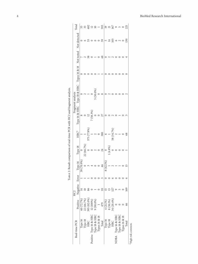
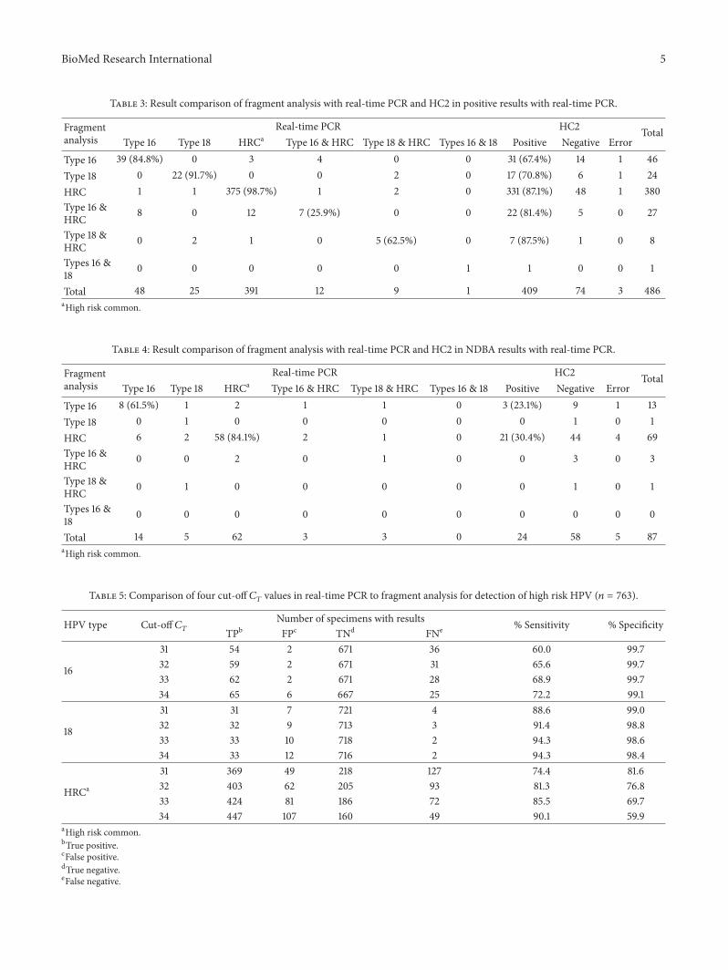
For the results by fragment analysis, real-time PCR showsthe detection rates of 79.7% (type 16), 92% (type 18), 96.4%(HRC), and 32.5% (mixed type) including NDBA. HC2detected 57.6% (type 16), 68.0% (type 18), 78.4% (HRC), and75.0% (mixed type) for types identified by fragmented anal-ysis (Table 3). Overall, real-time PCR detects correctly 516(90.1%) of 573 fragments analysis results. On the other hand,HC2 detects 433 (75.6%) of fragment analysis. For NDBAresults identified as high risk HPV by fragment analysis,real-time PCR revealed 77.0% (67/87) of concordance rate,whereas HC2 showed 27.6% (24/87) (Table 4).
3.3. Result Interpretation by Cycle Threshold (𝐶𝑇) Changes.
Up to the change of cut-off 𝐶𝑇from 31 to 34, sensitivity
and specificity of real-time PCR were described in Table 5.When drawing ROC decision plot, areas under the ROCcurve (AUCs) were 0.86 for HPV type 16, 0.98 for HPV type18, and 0.76 for HRC. Optimal cut-off 𝐶
𝑇values were 35.58
in type 16, 34.01 in type 18, and 31.99 in high risk commontypes.
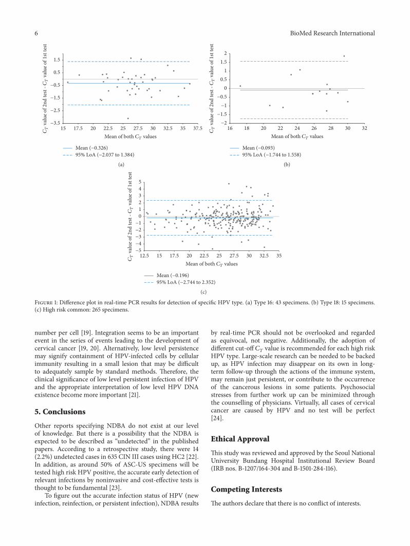
3.4. Repeatability. For evaluation of repeatability, retests wereperformed in 325 specimens showing the presence of type 16HPVDNA, type 18 HPVDNA, or HRCHPVDNA includingNDBA results. For type 16 HPVDNA, retests were done in 53specimens and amplification curvewas observed in 43 (81.1%)specimens. As shown in Figure 1, SD difference was 0.873and upper and lower margins of 95% limits of agreementwere 1.384 and −2.037, respectively. In 43 specimens showingamplification, 5 retest results showed 𝐶
𝑇value larger than
32. For 10 specimens, no amplification curve was observedat repetition, and they showed initial 𝐶
𝑇value larger than 32.
In case of type 18, retests were performed in 22 specimensand 15 specimens showed amplification at repetition.No casesrevealed 𝐶
𝑇value larger than 32 and 7 cases did not show
any amplification at repetition.The initial𝐶𝑇values in 7 cases
were larger than 32. In 15 specimens, SD difference was 0.842and upper and lowermargins of 95% limits of agreementwere1.558 and −1.744, respectively.
In case of HRCHPVDNA, 265 specimens showed ampli-fication at repetition and SD of difference was 1.3. The upperand lower margins of 95% limits of agreement were 2.352 and−2.744, respectively. In 17 specimens, no amplification wasobserved at repetition. For 10 specimens showing initial 𝐶
𝑇
value larger than 32, 3 specimens showed 𝐶𝑇value under
32 at repetition. For 18 specimens larger than 32 𝐶𝑇value at
repetition, 11 specimens showed initial𝐶𝑇value under 32. Ten
of 17 specimens showing no amplification showed 𝐶𝑇values
larger than 32 at initial test.
4. Discussion
Differently from other previous evaluation studies, our studypopulation was mainly limited to the specimens showing thenormal or nonspecific cytological findings (NILM, reactivecellular change, atrophy, etc.). So, the cytological or patho-logic finding was not helpful for the prediction of HPVexistence in this study. HPV load and cumulative incidenceof cervical lesion are known to be significantly correlated[15, 16]. At the view of guideline change, the position ofHPV DNA test moves from the adjunctive test method tocotest method. Although PCR has been the “gold standard”technique in HPV diagnostics, the disadvantages of PCRare its extremely high analytical sensitivity and potential forcontamination, leading to false-positive results [14]. But, asrevealed in NDBA results, real-time PCR results showingamplification curve above𝐶
𝑇were needed to be reconsidered
carefully. The accuracy of detection of high risk HPV isknown to be significantly higher with Abbott RealTimeHigh Risk HPV than HC2 [17]. HC2 technology measuressensitivity versus defined clinical endpoints (CIN 3+/SCC)and ensures reporting of positive results only when risk ofdisease progression exists. The limit of detection of HC2 is5,000 copies/mL; it ismuch lowerwhen compared to less than10 copies of PCR [18].
Also, in results showing specific HPV types by fragmentanalysis, real-time PCR shows higher concordance rate thanHC2. Particularly, in specimens showing NDBA, HC2 tendsto reveal negative results much more frequently. Consideringthe distributions of HPV types in NDBA and positive results,the concordance rates between fragment analysis and real-time PCR, and the results of repeatability tests, NDBA resultsshould be regarded as equivocal or positive, not as negative.
According to AUC value for the 𝐶𝑇change, appropriate
𝐶𝑇was different for HPV types. In HRC, 𝐶
𝑇of 32 is appro-
priate as described by manufacturer, but in types 16 and 18,𝐶𝑇of 33 or 34 will be more suitable.In 167 results with negative result by HC2 and NDBA by
real-timePCR, 58 (34.7%) resultswere assigned to the specifichigh risk HPV types by fragment analysis. Out of 58 results,52 (89.6%) high risk HPV types were detected by real-timePCR and fragment analysis. Using HC2 only, false-negativeresults can be reported in specimens with low level persistentinfection.The clinical relevance and implications of low levelpersistence of HPV are not clearly known, nor is the causeof low level persistence. In a previous study by Collins et al.,integration of HPV 16 resulted in a markedly lower viral copy
4 BioMed Research International
Table2:Re
sultcomparis
onof
real-timeP
CRwith
HC2
andfragmentanalysis
.
Real-timeP
CRHC2
Fragmentanalysis
Total
Positive
Negative
Error
Type
16Ty
pe18
HRC
aTy
pe16
&HRC
Type
18&HRC
Types16&18
Not
teste
dNot
detected
Positive
Type
1640
(72.7%
)14
139
(70.9%
)0
18
00
70
55Ty
pe18
22(66.7%
)10
10
22(66.7%
)1
02
02
633
HRC
395(82.0%
)86
13
0375(77.8
%)
121
038
53482
Type
16&HRC
12(100%)
00
40
17(58.3%
)0
00
012
Type
18&HRC
9(90.0%
)1
00
22
05(50.0%
)0
10
10Ty
pes16&18
10
00
00
00
10
01
Total
479
111
346
24380
278
148
59593
NDBA
Type
164(21.1%)
150
8(42.1%
)0
60
00
05
19Ty
pe18
8(38.1%
)13
01
1(4.8%
)1
02
02
1421
HRC
34(20.4%
)127
62
058
(34.7%
)2
00
2103
167
Type
16&HRC
09
01
02
00
00
69
Type
18&HRC
05
01
01
10
00
25
Types16&18
00
00
00
00
00
00
Total
46169
613
168
32
04
130
221
a Highris
kcommon
.
BioMed Research International 5
Table 3: Result comparison of fragment analysis with real-time PCR and HC2 in positive results with real-time PCR.
Fragmentanalysis
Real-time PCR HC2 TotalType 16 Type 18 HRCa Type 16 & HRC Type 18 & HRC Types 16 & 18 Positive Negative Error
Type 16 39 (84.8%) 0 3 4 0 0 31 (67.4%) 14 1 46Type 18 0 22 (91.7%) 0 0 2 0 17 (70.8%) 6 1 24HRC 1 1 375 (98.7%) 1 2 0 331 (87.1%) 48 1 380Type 16 &HRC 8 0 12 7 (25.9%) 0 0 22 (81.4%) 5 0 27
Type 18 &HRC 0 2 1 0 5 (62.5%) 0 7 (87.5%) 1 0 8
Types 16 &18 0 0 0 0 0 1 1 0 0 1
Total 48 25 391 12 9 1 409 74 3 486aHigh risk common.
Table 4: Result comparison of fragment analysis with real-time PCR and HC2 in NDBA results with real-time PCR.
Fragmentanalysis
Real-time PCR HC2 TotalType 16 Type 18 HRCa Type 16 & HRC Type 18 & HRC Types 16 & 18 Positive Negative Error
Type 16 8 (61.5%) 1 2 1 1 0 3 (23.1%) 9 1 13Type 18 0 1 0 0 0 0 0 1 0 1HRC 6 2 58 (84.1%) 2 1 0 21 (30.4%) 44 4 69Type 16 &HRC 0 0 2 0 1 0 0 3 0 3
Type 18 &HRC 0 1 0 0 0 0 0 1 0 1
Types 16 &18 0 0 0 0 0 0 0 0 0 0
Total 14 5 62 3 3 0 24 58 5 87aHigh risk common.
Table 5: Comparison of four cut-off 𝐶𝑇values in real-time PCR to fragment analysis for detection of high risk HPV (𝑛 = 763).
HPV type Cut-off 𝐶𝑇
Number of specimens with results % Sensitivity % SpecificityTPb FPc TNd FNe
16
31 54 2 671 36 60.0 99.732 59 2 671 31 65.6 99.733 62 2 671 28 68.9 99.734 65 6 667 25 72.2 99.1
18
31 31 7 721 4 88.6 99.032 32 9 713 3 91.4 98.833 33 10 718 2 94.3 98.634 33 12 716 2 94.3 98.4
HRCa
31 369 49 218 127 74.4 81.632 403 62 205 93 81.3 76.833 424 81 186 72 85.5 69.734 447 107 160 49 90.1 59.9
aHigh risk common.bTrue positive.cFalse positive.dTrue negative.eFalse negative.
6 BioMed Research International
Mean (−0.326)95% LoA (−2.037 to 1.384)
17.5 20 22.5 25 27.5 30 32.5 35 37.515−3.5
−2.5
−1.5
−0.5
0.5
1.5
Mean of both CT values
CT
valu
e of 2
nd te
st -C
Tva
lue o
f 1st
test
(a)
Mean (−0.093)95% LoA (−1.744 to 1.558)
18 20 22 24 26 28 30 3216−2
−1.5−1
−0.50
0.51
1.52
CT
valu
e of 2
nd te
st -C
Tva
lue o
f 1st
test
Mean of both CT values
(b)
15 17.5 20 22.5 25 27.5 30 32.5 3512.5−5−4−3−2−1
012345
Mean (−0.196)95% LoA (−2.744 to 2.352)
CT
valu
e of 2
nd te
st -C
Tva
lue o
f 1st
test
Mean of both CT values
(c)
Figure 1: Difference plot in real-time PCR results for detection of specific HPV type. (a) Type 16: 43 specimens. (b) Type 18: 15 specimens.(c) High risk common: 265 specimens.
number per cell [19]. Integration seems to be an importantevent in the series of events leading to the development ofcervical cancer [19, 20]. Alternatively, low level persistencemay signify containment of HPV-infected cells by cellularimmunity resulting in a small lesion that may be difficultto adequately sample by standard methods. Therefore, theclinical significance of low level persistent infection of HPVand the appropriate interpretation of low level HPV DNAexistence become more important [21].
5. Conclusions
Other reports specifying NDBA do not exist at our levelof knowledge. But there is a possibility that the NDBA isexpected to be described as “undetected” in the publishedpapers. According to a retrospective study, there were 14(2.2%) undetected cases in 635 CIN III cases using HC2 [22].In addition, as around 50% of ASC-US specimens will betested high risk HPV positive, the accurate early detection ofrelevant infections by noninvasive and cost-effective tests isthought to be fundamental [23].
To figure out the accurate infection status of HPV (newinfection, reinfection, or persistent infection), NDBA results
by real-time PCR should not be overlooked and regardedas equivocal, not negative. Additionally, the adoption ofdifferent cut-off 𝐶
𝑇value is recommended for each high risk
HPV type. Large-scale research can be needed to be backedup, as HPV infection may disappear on its own in long-term follow-up through the actions of the immune system,may remain just persistent, or contribute to the occurrenceof the cancerous lesions in some patients. Psychosocialstresses from further work up can be minimized throughthe counselling of physicians. Virtually, all cases of cervicalcancer are caused by HPV and no test will be perfect[24].
Ethical Approval
This study was reviewed and approved by the Seoul NationalUniversity Bundang Hospital Institutional Review Board(IRB nos. B-1207/164-304 and B-1501-284-116).
Competing Interests
The authors declare that there is no conflict of interests.
BioMed Research International 7
Acknowledgments
This study is funded by Seoul National University BundangHospital Research Fund (Grants nos. 11-2012-011 and 02-2013-068).
References
[1] G. Y. F. Ho, R. Bierman, L. Beardsley, C. J. Chang, and R. D.Burk, “Natural history of cervicovaginal papillomavirus infec-tion in young women,” The New England Journal of Medicine,vol. 338, no. 7, pp. 423–428, 1998.
[2] N. F. Schlecht, S. Kulaga, J. Robitaille et al., “Persistent humanpapillomavirus infection as a predictor of cervical intraepithe-lial neoplasia,” Journal of the AmericanMedical Association, vol.286, no. 24, pp. 3106–3114, 2001.
[3] L. A. Koutsky, K. K. Holmes, C. W. Critchlow et al., “A cohortstudy of the risk of cervical intraepithelial neoplasia grade 2or 3 in relation to papillomavirus infection,” The New EnglandJournal of Medicine, vol. 327, no. 18, pp. 1272–1278, 1992.
[4] M. Arbyn, X. Castellsague, S. de sanjose et al., “Worldwideburden of cervical cancer in 2008,” Annals of Oncology, vol. 22,no. 12, pp. 2675–2686, 2011.
[5] Z. Zuo, S. Goel, and J. E. Carter, “Association of cervicalcytology andHPVDNA status during pregnancy with placentalabnormalities and preterm birth,” American Journal of ClinicalPathology, vol. 136, no. 2, pp. 260–265, 2011.
[6] M. F. Evans, C. S.-C. Adamson, L. M. Schned et al., “HPV isdetectable in virtually all abnormal cervical cytology samplesafter reinvestigation of HPV negatives with multiple alternativePCR tests,” Diagnostic Molecular Pathology, vol. 19, no. 3, pp.144–150, 2010.
[7] H. C. Kitchener, C. Gilham, A. Sargent et al., “A comparisonof HPV DNA testing and liquid based cytology over threerounds of primary cervical screening: extended follow up in theARTISTIC trial,” European Journal of Cancer, vol. 47, no. 6, pp.864–871, 2011.
[8] E.-M. De Villiers, C. Fauquet, T. R. Broker, H.-U. Bernard, andH. Zur Hausen, “Classification of papillomaviruses,” Virology,vol. 324, no. 1, pp. 17–27, 2004.
[9] N. Munoz, F. X. Bosch, S. de Sanjose et al., “Epidemiologicclassification of human papillomavirus types associated withcervical cancer,”TheNew England Journal of Medicine, vol. 348,no. 6, pp. 518–527, 2003.
[10] A.-B.Moscicki, L.Widdice, Y.Ma et al., “Comparison of naturalhistories of human papillomavirus detected by clinician-andself-sampling,” International Journal of Cancer, vol. 127, no. 8,pp. 1882–1892, 2010.
[11] S. de Sanjose, M. Diaz, X. Castellsague et al., “Worldwideprevalence and genotype distribution of cervical human papil-lomavirus DNA in women with normal cytology: a meta-analysis,” Lancet Infectious Diseases, vol. 7, no. 7, pp. 453–459,2007.
[12] D. Solomon, D. Davey, R. Kurman et al., “The 2001 bethesdasystem: terminology for reporting results of cervical cytology,”Journal of the AmericanMedical Association, vol. 287, no. 16, pp.2114–2119, 2002.
[13] M. Poljak, A. Ostrbenk, K. Seme et al., “Comparison of clinicaland analytical performance of the Abbott RealTime HighRisk HPV test to the performance of hybrid capture 2 inpopulation-based cervical cancer screening,” Journal of ClinicalMicrobiology, vol. 49, no. 5, pp. 1721–1729, 2011.
[14] S.-M. Kulmala, S. Syrjanen, I. Shabalova et al., “Human papil-lomavirus testing with the hybrid capture 2 assay and PCR asscreening tools,” Journal of Clinical Microbiology, vol. 42, no. 6,pp. 2470–2475, 2004.
[15] S. Monnier-Benoit, V. Dalstein, D. Riethmuller, N. Lalaoui, C.Mougin, and J. L. Pretet, “Dynamics of HPV16DNA load reflectthe natural history of cervical HPV-associated lesions,” Journalof Clinical Virology, vol. 35, no. 3, pp. 270–277, 2006.
[16] M. Schmitt, C. Depuydt, I. Benoy et al., “Multiple humanpapillomavirus infections with high viral loads are associatedwith cervical lesions but do not differentiate grades of cervicalabnormalities,” Journal of ClinicalMicrobiology, vol. 51, no. 5, pp.1458–1464, 2013.
[17] S. Huang, B. Erickson, N. Tang et al., “Clinical performance ofAbbott RealTime High Risk HPV test for detection of high-grade cervical intraepithelial neoplasia in women with abnor-mal cytology,” Journal of Clinical Virology, vol. 45, supplement1, pp. S19–S23, 2009.
[18] A. Molijn, B. Kleter, W. Quint, and L.-J. Van Doorn, “Moleculardiagnosis of human papillomavirus (HPV) infections,” Journalof Clinical Virology, vol. 32, supplement 1, pp. S43–S51, 2005.
[19] S. I. Collins, C. Constandinou-Williams, K. Wen et al., “Dis-ruption of the E2 gene is a common and early event In thenatural history of cervical human papillomavirus infection: alongitudinal cohort study,” Cancer Research, vol. 69, no. 9, pp.3828–3832, 2009.
[20] W. Li, W. Wang, M. Si et al., “The physical state of HPV16infection and its clinical significance in cancer precursor lesionand cervical carcinoma,” Journal of Cancer Research andClinicalOncology, vol. 134, no. 12, pp. 1355–1361, 2008.
[21] B. Weaver, M. Shew, B. Qadadri et al., “Low-level persistence ofhuman papillomavirus 16 DNA in a cohort of closely followedadolescent women,” Journal of Medical Virology, vol. 83, no. 8,pp. 1362–1369, 2011.
[22] M. Motamedi, G. Bohmer, H. H. Neumann, and R. von Wasi-elewski, “CIN III lesions and regression: retrospective analysisof 635 cases,” BMC Infectious Diseases, vol. 15, no. 1, article 541,2015.
[23] M. F. Evans, C. S.-C. Adamson, J. L. Papilio, T. L. S. John, G.Leiman, andK. Cooper, “Distribution of human papillomavirustypes inThinPrep papanicolaou tests classified according to theBethesda 2001 terminology and correlations with patient ageand biopsy outcomes,” Cancer, vol. 106, no. 5, pp. 1054–1064,2006.
[24] W. Kinney, M. H. Stoler, and P. E. Castle, “Patient safety andthe next generation of HPV DNA tests,” American Journal ofClinical Pathology, vol. 134, no. 2, pp. 193–199, 2010.

Submit your manuscripts athttp://www.hindawi.com
Hindawi Publishing Corporationhttp://www.hindawi.com Volume 2014
Anatomy Research International
PeptidesInternational Journal of
Hindawi Publishing Corporationhttp://www.hindawi.com Volume 2014
Hindawi Publishing Corporation http://www.hindawi.com
International Journal of
Volume 2014
Zoology
Hindawi Publishing Corporationhttp://www.hindawi.com Volume 2014
Molecular Biology International
GenomicsInternational Journal of
Hindawi Publishing Corporationhttp://www.hindawi.com Volume 2014
The Scientific World JournalHindawi Publishing Corporation http://www.hindawi.com Volume 2014
Hindawi Publishing Corporationhttp://www.hindawi.com Volume 2014
BioinformaticsAdvances in
Marine BiologyJournal of
Hindawi Publishing Corporationhttp://www.hindawi.com Volume 2014
Hindawi Publishing Corporationhttp://www.hindawi.com Volume 2014
Signal TransductionJournal of
Hindawi Publishing Corporationhttp://www.hindawi.com Volume 2014
BioMed Research International
Evolutionary BiologyInternational Journal of
Hindawi Publishing Corporationhttp://www.hindawi.com Volume 2014
Hindawi Publishing Corporationhttp://www.hindawi.com Volume 2014
Biochemistry Research International
ArchaeaHindawi Publishing Corporationhttp://www.hindawi.com Volume 2014
Hindawi Publishing Corporationhttp://www.hindawi.com Volume 2014
Genetics Research International
Hindawi Publishing Corporationhttp://www.hindawi.com Volume 2014
Advances in
Virolog y
Hindawi Publishing Corporationhttp://www.hindawi.com
Nucleic AcidsJournal of
Volume 2014
Stem CellsInternational
Hindawi Publishing Corporationhttp://www.hindawi.com Volume 2014
Hindawi Publishing Corporationhttp://www.hindawi.com Volume 2014
Enzyme Research
Hindawi Publishing Corporationhttp://www.hindawi.com Volume 2014
International Journal of
Microbiology