Research Article Segmentation of Bone with Region...
14
Research Article Segmentation of Bone with Region Based Active Contour Model in PD Weighted MR Images of Shoulder Aysun Sezer, 1 Hasan Basri Sezer, 2 and Songul Albayrak 1 1 Computer Engineering Department, Yildiz Technical University, 34220 Istanbul, Turkey 2 Orthopaedic and Traumatology Clinic, Sisli Hamidiye Etfal Training and Research Hospital, 34360 Istanbul, Turkey Correspondence should be addressed to Aysun Sezer; [email protected] Received 14 February 2015; Revised 6 April 2015; Accepted 13 April 2015 Academic Editor: Chuangyin Dang Copyright © 2015 Aysun Sezer et al. is is an open access article distributed under the Creative Commons Attribution License, which permits unrestricted use, distribution, and reproduction in any medium, provided the original work is properly cited. Proton density (PD) weighted MR images present inhomogeneity problem, low signal to noise ratio (SNR) and cannot define bone borders clearly. Segmentation of PD weighted images is hampered with these properties of PD weighted images which even limit the visual inspection. e purpose of this study is to determine the effectiveness of segmentation of humeral head from axial PD MR images with active contour without edge (ACWE) model. We included 219 images from our original data set. We extended the use of speckle reducing anisotropic diffusion (SRAD) in PD MR images by estimation of standard deviation of noise (SDN) from ROI. To overcome the problem of initialization of the initial contour of these region based methods, the location of the initial contour was automatically determined with use of circular Hough transform. For comparison, signed pressure force (SPF), fuzzy C-means, and Gaussian mixture models were applied and segmentation results of all four methods were also compared with the manual segmentation results of an expert. Experimental results on our own database show promising results. is is the first study in the literature to segment normal and pathological humeral heads from PD weighted MR images. 1. Introduction Shoulder instability constitutes an important mass of the shoulder surgery in orthopedics. Shoulder joint has a very wide range of motion and it is susceptible to instability because of less congruent bony relations. Shoulder instability is a condition in which relation between glenoid and humeral head is lost in a position or under physiological joint reaction forces. Instability may cause joint dysfunction and may result in dislocation of the joint which may cause further soſt tissue and bone damage making disruption of anatomical structures worse. Instability of shoulder joint is a result of disruption in bony structures or soſt tissues. MRI is widely used to detect shoulder pathologies because it is able to demonstrate both bony and soſt tissue pathologies which is essential to make accurate diagnosis. e humeral head being part of the bony structure of the shoulder joint plays an important role in shoulder instability. Segmentation of bone has primary importance in order to define the anatomical borders and location of the lesions. Different imaging modalities are able to detect bony struc- tures. Computerized Tomography (CT) and T1 weighted images were studied to be successful in bone segmentation in the literature. P´ erez et al. used a hybrid, statistical, and geometrical model based on Chan-Vese to segment bone and soſt tissues in T1 coronal shoulder MR images. ey reported that their model provided correct classification of bone but poor classification of soſt tissues [1]. Nguyen et al. studied sagittal T1 weighted shoulder images. ey used an integrated region based and gradient based supervised method to segment humeral head. eir average success rate measured over entire database was 96.32% [2]. Chaoui et al. segmented bony structures by using a recognition based segmentation method from CT images with 96% success rate [3]. ere is no single modality which can demonstrate bony edges and bone edema with high performance at the same time. CT and T1 weighted MR images are more successful in demonstrating bone edges. Transition between soſt tissues and bone is sharper in CT and T1 weighted MRI. However Hindawi Publishing Corporation Computational and Mathematical Methods in Medicine Volume 2015, Article ID 754894, 13 pages http://dx.doi.org/10.1155/2015/754894
Transcript of Research Article Segmentation of Bone with Region...

Research ArticleSegmentation of Bone with Region Based Active Contour Modelin PD Weighted MR Images of Shoulder
Aysun Sezer1 Hasan Basri Sezer2 and Songul Albayrak1
1Computer Engineering Department Yildiz Technical University 34220 Istanbul Turkey2Orthopaedic and Traumatology Clinic Sisli Hamidiye Etfal Training and Research Hospital 34360 Istanbul Turkey
Correspondence should be addressed to Aysun Sezer sezeraysungmailcom
Received 14 February 2015 Revised 6 April 2015 Accepted 13 April 2015
Academic Editor Chuangyin Dang
Copyright copy 2015 Aysun Sezer et al This is an open access article distributed under the Creative Commons Attribution Licensewhich permits unrestricted use distribution and reproduction in any medium provided the original work is properly cited
Proton density (PD) weighted MR images present inhomogeneity problem low signal to noise ratio (SNR) and cannot define boneborders clearly Segmentation of PD weighted images is hampered with these properties of PD weighted images which even limitthe visual inspection The purpose of this study is to determine the effectiveness of segmentation of humeral head from axial PDMR images with active contour without edge (ACWE) model We included 219 images from our original data set We extendedthe use of speckle reducing anisotropic diffusion (SRAD) in PD MR images by estimation of standard deviation of noise (SDN)from ROI To overcome the problem of initialization of the initial contour of these region based methods the location of the initialcontour was automatically determined with use of circular Hough transform For comparison signed pressure force (SPF) fuzzyC-means and Gaussian mixture models were applied and segmentation results of all four methods were also compared with themanual segmentation results of an expert Experimental results on our own database show promising results This is the first studyin the literature to segment normal and pathological humeral heads from PD weighted MR images
1 Introduction
Shoulder instability constitutes an important mass of theshoulder surgery in orthopedics Shoulder joint has a verywide range of motion and it is susceptible to instabilitybecause of less congruent bony relations Shoulder instabilityis a condition in which relation between glenoid and humeralhead is lost in a position or under physiological joint reactionforces Instability may cause joint dysfunction andmay resultin dislocation of the joint which may cause further soft tissueand bone damagemaking disruption of anatomical structuresworse Instability of shoulder joint is a result of disruption inbony structures or soft tissues
MRI is widely used to detect shoulder pathologiesbecause it is able to demonstrate both bony and soft tissuepathologieswhich is essential tomake accurate diagnosisThehumeral head being part of the bony structure of the shoulderjoint plays an important role in shoulder instability
Segmentation of bone has primary importance in orderto define the anatomical borders and location of the lesions
Different imaging modalities are able to detect bony struc-tures Computerized Tomography (CT) and T1 weightedimages were studied to be successful in bone segmentationin the literature Perez et al used a hybrid statistical andgeometrical model based on Chan-Vese to segment boneand soft tissues in T1 coronal shoulder MR images Theyreported that their model provided correct classification ofbone but poor classification of soft tissues [1] Nguyen etal studied sagittal T1 weighted shoulder images They usedan integrated region based and gradient based supervisedmethod to segment humeral head Their average success ratemeasured over entire database was 9632 [2] Chaoui etal segmented bony structures by using a recognition basedsegmentation method from CT images with 96 success rate[3]
There is no single modality which can demonstrate bonyedges and bone edema with high performance at the sametime CT and T1 weighted MR images are more successfulin demonstrating bone edges Transition between soft tissuesand bone is sharper in CT and T1 weighted MRI However
Hindawi Publishing CorporationComputational and Mathematical Methods in MedicineVolume 2015 Article ID 754894 13 pageshttpdxdoiorg1011552015754894
2 Computational and Mathematical Methods in Medicine
these modalities cannot supply sufficient information aboutbone edema Axial PD weighted MRI of shoulder is themostly used sequence being able to present both bonyand soft tissue components of instability synchronously PDweighted MRI is also able to demonstrate bony edema whichis a sign of the magnitude and location of bone trauma anda common source of pain However there is a very smoothtransition zone between bone and soft tissues making seg-mentation of bone edges more difficult
Another parameter that aggravates the segmentationprocess is noise PD weighted MR images have poorer SNRthan T1 weighted MR and CT images Many researchers andnumerous applications in medical images have been devotedto use nonlinear diffusion filters such as anisotropic diffusion(AD) and speckle reducing anisotropic diffusion (SRAD) toreduce noisewhile preserving image edges [4ndash6] ADmethodhas been applied to reduce noise in CT [7] MR [8] andultrasound images with good results ADmethod encouragesdiffusion in the homogeneous region while inhibiting diffu-sion at edges However it is not directional and leaves somenoise in the neighborhood of the edges
SRADmethod is directional that it inhibits smoothing indirections perpendicular to edges and encourages smoothingparallel to the edges not leaving noise in the vicinity of edgesSRADwas introduced as a useful technique to reduce specklenoise in ultrasound and satellite images successfully [4 6]Not only speckle noise but also other types of noise can bereduced by SRAD in case the standard deviation of noise(SDN) is estimated [9]
There is a wide variety of methods proposed to segmentbackground and the object pixels among which the activecontour model (ACM) was one of the most successfulmethods The basic idea of the ACM is to evolve a curveunder some constraints to extract the desired object based onenergy minimizing method Existing AC methods for imagesegmentation can be categorized into two classes edge basedmodels [10 11] and region based models [12ndash15]
Edge basedmodels utilize image gradient as an additionalconstraint to construct force to direct the motion of thecontour These models usually have edge based stoppingconstraint to intercept the contour evolution on the objectboundaries [10] High noise and weak or deficient borders ofhumeral head in PD images make edge based methods lesssuitable
Region based active contour methods have many advan-tages over the edge based methods First the region basedmethods do not depend on the image gradient and utilizeimage statistical information inside and outside the contourto control evolution They can satisfactorily segment imageswith weak edges and without edges Second the segmenta-tion result is less dependent on the location of the initialcontour and they can effectively detect the exterior andinterior boundaries simultaneously Chan-Vese model is oneof the most popular region based models which is based ona simplified Mumford-Shah segmentation techniques [12]Chan-Vese model has been successfully applied to binaryphase segmentation with assumption that each image regionis statistically homogeneous This assumption is a limitationof its application in inhomogeneous regions In order to solve
this limitation of Chan-Vese model Vese and Chan [14] andTsai et al [15] proposed piecewise smooth (PS) models whichcan deal with part of the problem [12] However PS modelshave high computational costsMoreover segmentation resultis still dependent on the initial contour placement
Li at al proposed a local binary fitting (LBF) model tosolve the problem caused by intensity inhomogeneity [16]LBF model uses local statistical information especially thelocal intensity mean in a region based active contour modelHowever the intensity inhomogeneity problem cannot besolved with using particular uniform distribution becauseintensity nonuniformity in a desired object can show vari-ations in different positions Use of more than one kind ofspecific distribution is needed to describe the variation ofintensity nonuniformity in each region To solve this problemNi et al proposed control of evolution of contour by usingthe histogram of the intensity however they experiencedlimitations in segmentation of natural images [17 18] Ge etal defined a new regularization term with the anisotropicdiffusion process based on the structure tensor and theduality theory They computed the statistical information ofmagnitude of gradient to approximate the variation of theintensity by the anisotropic diffusion process [13] Hybridregion based active contour (HRBAC)models were proposedrecently to segment images with intensity inhomogeneity[19 20] HRBAC models like SPF model [21] and geodesicintensity fitting model [22] combine merits of the traditionalgeodesic active contour (GAC)model which is an edge basedactive contour model and region based Chan-Vese modelDespite all of these models which were intended to solve theintensity inhomogeneity problem it is yet to be solved
The common problem of all these methods is sensitivityto initialization The problem of initial contour was studiedbyWang et alThey proposed to combine the local and globalintensity information [23]The limitation of the studywas thedeviation of the real object boundarywhen the initial contourwas close to the object boundaries Liu and Peng proposedto use degraded Chan-Vese model the segmentation resultof which is taken as initial contour of proposed local regionbased Chan-Vese model Their model can segment imageswith intensity inhomogeneity although this method is com-putationally efficient the success rates are still sensitive to theinitial contour [24]
In this paper we studied humeral head (bone) segmen-tation in axial PD MRI of shoulder In the first step weapplied SRADmethod and homomorphic filter SRAD is verysensitive to the estimation of SDN and normally calculatedin a homogenous area [4 9] We estimated this parameterfrom area of interest where there is more tissue intensitythan background to reduce Rician noise By this way weextended use of SRAD in PD MR images Noise reductionwith SRAD makes a good contribution to the correctionof inhomogeneity because the image noise is a factor thataffects quantity of inhomogeneity But noise reduction withSRAD alone is not enough for satisfactory correction of inho-mogeneity in the image Thus we supported SRAD methodwith homomorphic filter This filter separates low and highfrequency components in an image and enhances changes inhigh frequency component and suppresses changes in low
Computational and Mathematical Methods in Medicine 3
(a) (b) (c)
Figure 1 PD weighted axial MR images of left shoulder with (a) normal humeral head (b) edematous humeral head and (c) humeral headwith Hill-Sachs lesion
frequency component to obtain more homogenous imagesMoreover the homomorphic filter is in its essences a linearfilter and is quintessentially used for noise reduction or signalfeature extraction if the signal (PDMR image) is distorted byadditive noise
Region based active contour models have ability tosegment very weak edges of humeral head but they aresensitive to the location of initial circle In the second stepthis problem was handled with the automatic detection ofthe initial contour in each shoulder image We used circularHough transform to automatically detect the location ofthe humeral head from PD weighted MR images Resultparameters obtained by circular Hough transform were usedto identify the ROI and the place of initial contour
In the last step we applied ACWEmethod [12] to segmenthumeral head in the ROI We compared results of ACWEwith SPF and clustering methods of fuzzy C-means (FCM)and Gaussian mixture model (GMM) ACWE has a globalsegmentation property and provides to segment all objectsin the ROI SPF has a local property which provides toonly extract a desired object by setting the initial contoursurrounding or intersecting the humeral head boundaries[21] All segmentation results were comparedwith themanualsegmentation results of an orthopedic specialist
Note that some part of results of the segmentation ofnormal humeral heads in this paper was reported in ourrecent conference paper [25] This paper is organized as fol-lows In Section 2 description ofmaterials andmethods stepsof preprocessing segmentation and postprocessingmethodsare described Experimental results and evaluation are givenin Section 3 followed by some discussion in Section 4 Thispaper is summarized in Section 5
2 Material and Methods
MRI is a standard of protocol in evaluation of the shoulderinstability We used 2D PD weighted MR images of shoulderwhich can successfully demonstrate the pathologies thatcause shoulder instability MRI has the capacity to present 3Dimages however pathological changes in shoulder instability
may be hidden in 3D images 3D MRI has no advantage over2D MRI in demonstrating the whole pathology
The humeral head is the part of the bony structure of theshoulder joint The pathologies of humeral head in shoulderinstability can be categorized as bone edema and disruptionof bone borders (eg Hill-Sachs lesion) In this study wesegmented normal humeral heads humeral heads with boneedema and Hill-Sachs lesion from 2D axial PD MR images
A normal humeral head image of left shoulder is shownin Figure 1(a) Humeral head with edema is demonstratedin Figure 1(b) The amount and the location of bone edemachange according to patient Hill-Sachs lesion occurs whenthe humeral head has a compression fracture where the dis-tribution of intensity values increases and shape of humeralhead changes (Figure 1(c))
We included shoulder MR images of 219 patients whohave been admitted to Sisli Hamidiye Etfal Training andResearch Hospital We used 15 Tesla PD weighted MRimages The size of the DICOM image was 256 times 256 pixelsThe slice thickness was 4 millimeters We grouped patientsaccording to appearance of humeral head as normal edema-tous and Hill-Sachs deformity The numbers of the patientswere 81 in normal group 100 in edematous group and 38in Hill-Sachs group Segmentation of humeral head from PDweighted MR images was not studied in literature and therewas no available database The original data set was collectedby authors Segmentation process in this study may becategorized into three steps as preprocessing segmentationand postprocessing (Figure 2)
21 Steps of Image Preprocessing
211 Noise Reduction Using Speckle Reducing AnisotropicDiffusion (SRAD) MR images can be categorized into twoclasses high resolution with high SNR and high resolutionwith low SNR High resolution with high SNR is a conse-quence of increased acquisition time of MRI which is notfeasible due to the patient comfort Noise reduction in PDweightedMR images has an important effect on segmentationsuccess as much as correction of intensity inhomogeneity
4 Computational and Mathematical Methods in Medicine
SRAD
Homomorphic filter
Determination of ROI
ACWE method
SPF method
FCM method
GMM method
Preprocessing step Segmentation step Postprocessing step
Morphological operations
Connected component
labelling
Anatomical knowledge
Database Result
Figure 2 The flow chart of the segmentation steps by ACWE and the comparison methods of SPF FCM and GMM
(a) (b) (c) (d)
Figure 3 (a) Orginal PDweightedMRI (b) image obtained after SRADmethod (c) result of the homomorphic filter and (d) result obtainedafter using SRAD method and homomorphic filter
Owing to the fact that the distribution of the noise in PDimages is not Gaussian noise problem cannot be solvedsufficiently by traditional noise reduction methods such asGaussian filter and median filter [8]
PD weighted MR images have a very smooth transitionzone between soft tissues and bone (Figure 3(a)) There isneed of a method which decreases noise without deterio-rating the important anatomical and pathological details ofhumeral head ADmethod was proved to produce successfulresults in medical imaging area however AD method leavessome remaining noise in the vicinity of edges after filtering[4 7 8] We used SRAD which is a directional method thatovercomes this problem by inhibiting smoothing parallel toedge and enhancing smoothing in perpendicular direction(Figure 3(b))
SRADmethod demonstrates good speckle noise suppres-sion in ultrasound satellite images and MRI The weak sideof SRAD method is its sensitivity to the SDN Through theaccurate estimation of SDN SRADmethod can be applied forall kinds of noise not only for speckle noise [9] SRADmethodis a combination of AD and speckle reducing Lee filter
The partial differential equation (PDE) of AD is given asfollows in continuous domain where 119868
0is the initial image
119888( ) is the diffusion coefficient nabla is gradient operator and divis divergence operator
120597119868
120597119905
= div [119888 (|nabla119868|) nabla119868] (1)
119868 (119905 = 0) = 1198680 (2)
We used SRAD to decrease noise in the PD weighted MRimages Given an intensity image 119868
0(119909 119910) having finite power
and no zero values over the image support domain Ω theoutput image 119868(119909 119910 119905) is evolved according to the followingformat of the PDE of SRAD [4] where 119868(119909 119910 119905) is theintensity image estimated at position119909 119910 at the diffusion time119905 120597Ω denotes the border of Ω 119899 is the outher normal to the120597Ω
120597119868 (119909 119910 119905)
120597119905
= div [119888 (119902) nabla119868 (119909 119910 119905)] (3)
119868 (119909 119910 0) = 1198680(119909 119910)
(
120597119868 (119909 119910 119905)
120597 119899
)
100381610038161003816100381610038161003816100381610038161003816120597Ω
= 0
(4)
SRAD uses the format of the PDE of AD Selection ofdiffusion coefficient is the main difference between AD andSRAD methods ((1) and (3))
119888 (119902)
= [
1
1 + [1199022(119909 119910 119905) minus 119902
2
0(119905)] [119902
2
0(119905) (1 + 119902
2
0(119905))]
]
(5)
SRAD inherits instantaneous coefficient of variation (ICOV)and speckle scale function prototype of Lee filter ICOVserves as the edge detector ICOV is a function of the image
Computational and Mathematical Methods in Medicine 5
normalized gradient magnitude nabla119868119868 and normalized Lapla-cian nabla2119868119868 defined in relation with the adaptive coefficient ofthe Lee filter [4] expressed as follows
119902 (119909 119910 119905) = radic
(12) (|nabla119868| 119868)2minus (116) (nabla
2119868119868)
2
[1 + (14) (nabla2119868119868)2]
(6)
Speckle scale function 1199020(119905) controls amount of smoothing
applied to the image by SRAD and computed from a homo-geneous region of fully developed speckle by
1199020(119905) =
radicvar [119911 (119905)]119911 (119905)
(7)
But we have calculated speckle scale function on the fore-ground where the humeral head is dominant than othertissues This way we assure that the background has noeffect over the estimation of Rician noise present in MRIThe var[119911(119905)] and 119911(119905) are intensity variance and mean overhumeral head region at time 119905
212 Homomorphic Filter Despite progress in the scannertechnology MR images still have imperfections like lowSNR intensity inhomogeneity due to the bias field andother artifacts Intensity inhomogeneity adversely affectsvisual inspection and segmentationNumerousmethods havebeen introduced to eliminate intensity inhomogeneity in MRimages [26] Homomorphic filtering assumes that intensityinhomogeneity is a low frequency artifact that can be sepa-rated fromhigh frequency signal by lowpass filtering Perez etal used homomorphic filter in the image preprocessing stepto segment bone and soft tissues on T1 weighted MR imageswith good results [1]
According to homomorphic filtering model each pixelvalue 119891(119909 119910) can be expressed as the product of a low fre-quency illumination component 119894(119909 119910) and a high frequencyreflectance component 119903(119909 119910) as follows
119891 (119909 119910) = 119894 (119909 119910) 119903 (119909 119910) (8)
The homomorphic system for image enhancement is basedon the separation of independent low and high frequencycomponents To facilitate their separate processing by takinglogarithmic transform of (8) thus
119892 (119909 119910) = ln (119891 (119909 119910)) = ln [119894 (119909 119910) 119903 (119909 119910)]
= ln (119894 (119909 119910)) + ln (119903 (119909 119910)) (9)
Fourier transform is applied to (9) where119866(119906 V) 119868(119906 V) and119877(119906 V) represent the Fourier transform of 119892(119909 119910) ln(119894(119909 119910))and ln(119903(119909 119910)) respectively Consider
119866 (119906 V) = 119868 (119906 V) + 119877 (119906 V) (10)
Applying a high pass filter 119867(119906 V) in the frequency domainto enhance changes in reflectance and suppressing changes inillumination are as follows
119866 (119906 V) = 119868 (119906 V)119867 (119906 V) + 119877 (119906 V)119867 (119906 V) (11)
We used a Gaussian high pass filter instead of Butterworthhigh pass filter normally used in homomorphic filtering toremove the illumination component Inverse Fourier trans-form is applied to (11) to obtain 119892(119906 V) which represent theinverse Fourier transform of 119866(119906 V)
119892 (119906 V) = (119906 V) + 119903 (119906 V) (12)
Exponential form exp(119892(119906 V)) is used to reverse logarithmiceffect to obtain the image after enhancement
Our strategy for correction of inhomogeneity is to use thehomomorphic filter to eliminate intensity inhomogeneity inthe preprocessing step just after noise reduction with SRADmethod (Figures 3(c) and 3(d)) We combined advantages ofSRAD and homomorphic filter before application of regionbased active contour models
213 Determination of Region of Interest (ROI) The Houghtransform is widely used in pattern recognition and computervision for the detection of regular curves Humeral head has around cross-sectional shape in axial PDMR images CircularHough transform has an ability to detect circular shapesbased on parametric equation of circle [27] This methodprovides detection of humeral head for each PDweightedMRimage without affecting image noise (Figure 4) This methodis also very tolerant to gaps in feature boundary descriptionsthus it can also find defective shape of humeral head resultingfrom the edema and deformity
Figure 4(b) shows detection of humeral head from a PDweighted left shoulder MR image The shape of the humeralhead is not exactly a circle thus some parts of the area ofinterest may remain out of the circle defined by circularHough transform To involve whole humeral head in ROI weenlarged radius of circle found by circular Hough transform
Common problem of region based active contour modelsis dependence of segmentation success on the initial contourplacement [23 24] We achieved this problem by automati-cally determining location of initial contour in the ROI
22 Segmentation Methods
221 Active Contours without Edges (ACWE)Model ACWEknown also as Chan-Vese model is a region based activecontour model and has successful applications in manypapers and fields [1 7 12] ACWE model utilizes statisticalinformation inside and outside the contour instead of imagegradient
An evolving curve 119862 = 120597120596 with 120596 sub Ω an opensubset inside (119862) denotes the region 120596 which represents theforeground pixels and outside (119862) denotes the region Ω 120596
which represents the background pixels Chan-Vese model isformulated byminimizing the following energy functionwiththe level set formulation [12] (14)
119865 (1198881 1198882 119862)
= 1205821int
Ω
10038161003816100381610038161199060(119909 119910) minus 119888
1
1003816100381610038161003816
2119867(120601 (119909 119910)) 119889119909 119889119910
+ 1205822int
Ω
10038161003816100381610038161199060(119909 119910) minus 119888
2
1003816100381610038161003816
2(1 minus 119867 (120601 (119909 119910))) 119889119909 119889119910
(13)
6 Computational and Mathematical Methods in Medicine
(a) (b)
Figure 4 (a) Orginal MR image of left shoulder and (b) detection of humeral head by using the circular Hough transform
With the level set method we assume
119862 = 120597120596 = (119909 119910) isin Ω 120601 (119909 119910) = 0
120596 = (119909 119910) isin Ω 120601 (119909 119910) gt 0
Ω 120596 = (119909 119910) isin Ω 120601 (119909 119910) lt 0
(14)
The image 1199060is assumed to be composed of two regions
of constant intensities 1198881(the average intensities inside the
contour) and 1198882(the average intensities outside the contour)
The constants 1198881and 1198882can be found by minimizing (13)
1198881(120601) =
intΩ1199060119867(120601 (119909 119910)) 119889119909 119889119910
intΩ119867(120601 (119909 119910)) 119889119909 119889119910
(15)
1198882(120601) =
intΩ1199060(1 minus 119867 (120601 (119909 119910))) 119889119909 119889119910
intΩ(1 minus 119867 (120601 (119909 119910))) 119889119909 119889119910
(16)
Using the Heaviside function 119867(120601) and the Dirac function120575(120601) defined by Chan and Vese the energy function can beexpressed as follows
120597120593
120597119905
= 120575 (120601)
sdot [120583nabla(
nabla120601
1003816100381610038161003816nabla120601
1003816100381610038161003816
) minus V minus 1205821(1 minus 1198881)2+ 1205822(1 minus 1198882)2]
(17)
where V ge 0 1205821ge 0 120582
2ge 0 and 120583 ge 0 are fixed parameters
V parameter is used to increase propagation speed 1205821and 120582
2
parameters control the force inside and outside the contour 120583manipulates the smoothness of zero level set [12] If 120583 value islarge it detects only large objects while when 120583 value is smallsmall sized objects can also be detected
ACWE model can satisfactorily segment a desired objecthaving weak and deficient edges This property of ACWEmodel matches up with our needs because PD images have avery poor transition between soft tissues and bones and some
part of the border of bone is destroyed especially in imageswhich has bone edema and cortical deformity On the otherhand PD images have a low SNR than other MR modalitieswhich is also suitable for ACWE model which has ability tosegment noisy images
The weak points of this model are the assumption of eachimage region as homogeneous and sensitivity to initializationSome related methods were proposed to solve intensityinhomogeneity problem [13 16] Some of them deal withpart of the problem and to some extent they could performsegmentation of inhomogeneous objects Our data set wascomposed of humeral heads presenting awidemorphologicalvariance due to traumatic changes in borders and edema inthe bone Therefore the variance of the location and magni-tude of bone and edema would render previously proposedmethods less efficient in the humeral head segmentationfrom PD weighted MR images Instead of correction ofinhomogeneity during application of region basedmodels weapplied segmentationmethods after intensity inhomogeneitywas decreased with preprocessing Besides these methodscannot provide a reasonable solution to the initialization ofthe initial contour [24]
When ACWE method was applied to the original PDweighted axial MR images segmentation of humeral headwas notably unsatisfactory as shown in Figure 5(b) SRADmethod was applied to decrease noise and homomorphicfilter was used to obtain more homogeneous images In thenext step initial contour was automatically initialized in thehumeral head A better segmentation result was reached asshown in Figure 5(c)The humeral head was segmented fromdetermined ROI with the same preprocessing step As shownin Figure 5(d) segmentation success of humeral head wasincreased Figure 5(d) also demonstrates some white pixelsin the humeral head representing the edematous area of thehumeral head
222 Signed Pressure Force (SPF) Model SPF model isa combination of GAC [11] and region based Chan-Vese
Computational and Mathematical Methods in Medicine 7
(a) (b) (c) (d)
Figure 5 (a) Orginal PDweightedMR image of shoulder (b) binary segmentation result of ACWEmethod without preprocessing operation(c) segmentation result with ACWE after application of the SRAD and homomorphic filter and (d) segmentation result of ACWE in ROIwith the same preprocessing steps in (c)
model [12] and possesses a local segmentation propertySPF model is able to segment selectively the desired objectby setting initial contour intersecting or surrounding thedesired boundaries This model has advantages over GACand Chan-Vese model GAC model utilizes image gradientto construct an edge stopping function whereas SPF modeluses statistical information to intercept contour evolution onthe object boundaries Zhang et al stated that SPF methodwas capable of handling objects with weak or damagedboundaries Furthermore they suggested that SPF modelwas able to segment objects with inhomogeneous interiorintensity compared to Chan-Vese model [21] In our data setsome of the humeral heads had edema inside and weak ordamaged surrounding edges We applied SPF model to ourdata set and compared advantages of global segmentationproperty of ACWEmethod with local segmentation propertyof SPF
SPF function was defined as follows
spf (119868 (119909)) =119868 (119909) minus (1198881
+ 1198882) 2
max (1003816100381610038161003816119868 (119909) minus (119888
1+ 1198882) 2
1003816100381610038161003816)
(18)
where 1198881and 1198882are defined in (15) and (16) respectively The
level set formulation of SPF model is as follows
120597120593
120597119905
= spf (119868 (119909)) sdot (nabla120601
1003816100381610038161003816nabla120601
1003816100381610038161003816
+ 120572)1003816100381610038161003816nabla120601
1003816100381610038161003816+ nablaspf (119868 (119909))
sdot nabla120601
(19)
where 120572 is the balloon force term to shrink and expand thecontour
223 Gaussian Mixture Model (GMM) GMM is widelyapplied for segmentation inmany fieldsWe used GMM clus-tering model to segment humeral head and the parameters ofthemodel were estimated by using expectationmaximization(EM) algorithm [28] We applied this model in ROI definedby circular Hough transform (Figure 6(a))
Let us observe image in a vector 119909119895 119895 = 1 2 119899 and119894 isin 1 2 119896 and 119896 is the number of regions The mixtureof Gaussian distribution is assumed as in the following form
119891 (119909) =
119896
sum
119894=11015840
119901119894119873(119909 | 120583
119894 1205902
119894) (20)
119901119894gt 0 are weights such thatsum119896
119894=1119901119894= 1
119873(120583119894 1205902
119894) =
1
120590radic2120587
exp(minus (119909 minus 120583
119894)2
21205902
119894
) (21)
Steps of EM-MAP Algorithm (1) Any classification methodcould be used to initialize parameter of 120579(0) In our case weused 119870-means clustering method to define initial parameterof means (120583(0)
119896) variances 120590(0)
119896 and weights 119901(0)
119896
120579(0)
= (119901(0)
1 119901
(0)
119896 120583(0)
1 120583
(0)
119896 120590(0)
1 120590
(0)
119896) (22)
(2) E-step 119901(119903)119894
is the discrete prior probability in stage 119903and119901(119903+1)119894119895
is the discrete posterior probability in the next stagewhich is calculated by the Bayes rule as follows
119901(119903+1)
119894119895= 119901(119903+1)
(119894 | 119909119895) =
119901(119903)
119894119873(119909119895| 120583(119903)
119894 1205902(119903)
119894)
119891 (119909119895)
(23)
(3) M-step
119901(119903+1)
119894=
1
119899
119899
sum
119895=1
119901(119903)
119894119895
120583(119903+1)
119894=
sum119899
119895=1119901(119903+1)
119894119895119909119895
119899119901(119903+1)
119894
2(119903+1)
119894=
sum119899
119895=1119901(119903+1)
119894119895(119909119895minus 120583(119903+1)
119894)
2
119899119901(119903+1)
119894
(24)
8 Computational and Mathematical Methods in Medicine
(a) (b) (c)
Figure 6 (a) Demonstration of orginal MR images of shoulder in determined ROI by circular Hough transform (b) Segmentation result byusing FCMmethod (c) Segmenation result of GMM
(4) E-step and M-step iterated until convergence to thearbitrary error
sum
119894
1198902
119894lt 120576 (25)
GMMprovides to segment normal humeral head imageswithtwo labels (Figure 6(c))
224 Fuzzy C-Means (FCM)Method FCMmethod providesto assign pixels to each cluster based on minimizing thesum of distances from each pixel to every cluster centroidweighted by its corresponding membership The objectivecost function was minimized by assigning high member-ship values to pixels whose intensities are close to thecentroid of its particular class and low membership valuesare assigned when the pixels are far from the centroid Let119883 = (119909
1 1199092 119909
119873) represent an image with 119873 pixels to be
partitioned into 119888 cluster [29] The cost function is defined asfollows
119869 =
119873
sum
119895=1
119888
sum
119894=1
119906119898
119894119895
10038171003817100381710038171003817119909119895minus V119894
10038171003817100381710038171003817
2
(26)
where 119906119894119895defines themembership of pixel 119909
119895in the 119894th cluster
and V119894is the cluster center Fuzzy coefficient represented by119898
and 119898 = 2 is used in this study The membership function isupdated iteratively by
119906119894119895=
1
sum119888
119896=1(
10038171003817100381710038171003817119909119895minus V119894
10038171003817100381710038171003817
10038171003817100381710038171003817119909119895minus V119896
10038171003817100381710038171003817)
2(119898minus1) (27)
Each cluster center is updated as follows
V119894=
sum119873
119895=1119906119898
119894119895119909119895
sum119873
119895=1119906119898
119894119895
(28)
FCM model provides to segment normal humeral headimages with two labels (Figure 6(b))
23 Postprocessing The segmented object after use of ACWEor SPF contains soft tissue components especially tendonswhich have a similar intensity value with humeral head(Figures 7(a) and 7(b)) After enlargement of the circle foundwith the circular Hough transform the scapular edge was alsolabeled as humeral head (Figures 8(a) and 8(b)) Scapula andsome parts of tendons are located at the borders of the ROIcircle To eliminate scapula and tendon regions we appliedspecificmorphological operations and connected componentlabeling method in the light of anatomical localization oftendons
We determined the side of the humeral head by usinghistogram of an image The scapula is located at the samedirection with the shoulder side and in the periphery of thecircle According to the left and right shoulder images wemaydefine the location of scapula area by using radius and centerof circle which was obtained from circular Hough transformIn the scapula area we have done some erosion operationswith circular structuring element Connected componentlabeling method was used to detect connected regions in thecomplement of the binary image Next the boundary of thehumerus was labeled in red color (Figures 8(c) 8(d) and8(e)) In the last step the white pixels resulting from the boneedema were filled to get the final result
3 Experimental Results and Evaluation
Experiments were carried out withMATLAB 711 inWindowsXP platform on a 25GHz Intel (R) core (TM) personalcomputer with 4GB of RAM The manual segmentationtool was prepared in MATLAB environment and performedby using a mouse To evaluate the success of segmentationwe compared the segmentation results with the manuallysegmented areas determined by the orthopedic specialistSimilarity of segmented images is compared by Sorensen-Dice metric to find the success rates
We segmented normal and edematous humeral headsand humeral heads with Hill-Sachs deformity with ACWEand SPF methods (Figures 9(a) 9(b) 9(c) and 9(d))
Computational and Mathematical Methods in Medicine 9
(a) (b)
Figure 7 (a) Axial PD weighted MRI of shoulder (b) biceps and subscapularis tendons are demonstrated in red area The boundary ofscapula and humeral head is colored in green and blue respectively
(a) (b) (c)
(d) (e)
Figure 8 Steps of postprocessing (a) ROI as a result of circular Hough transform (b) The segmentation result in the ROI area (c) Theresult of specific morphological operation and connected component labelling (d) The filling of white pixels resulting from bone edema (e)Segmentation result
The parameters used for ACWE are 120583 = 02 1205821= 07 120582
2= 1
V = 1 and 120576 = 15 1205821and 120582
2parameters affect the uniformity
and the resulting force inside and outside the contourrespectively In our study we set 120582
1lt 1205822because we had
uniform backgrounds and foreground containing varyinggrayscale objects In order to detect smaller sized objects weassigned a small value to 120583 parameter The parameters usedfor SPF method are 119901 = 1 120590 = 2 119870 = 5 and 120576 = 15
10 Computational and Mathematical Methods in Medicine
(a) (b) (c) (d)
Figure 9 Segmentation results of normal edematous humeral heads and humeral head with Hill-Sachs deformity were demonstrated infirst second and third rows respectively Column (a) original images (b) manual segmentation results by an expert (c) segmentation resultsof ACWE method and (d) segmentation results of SPF method
Table 1 Segmentation results of three groups of humeral heads before and after postprocessing operation
Methods anddata
Average success rate before postprocessing [] Average success rate after postprocessing []
Normalhumeral head
Humeralhead withedema
Humeralhead withHill-Sachslesion
Averagesuccess rate
Normalhumeral head
Humeralhead withedema
Humeralhead withHill-Sachslesion
Averagesuccess rate
Number ofdata 81 100 38 219 81 100 38 219
ACWEmethod 9079 8840 8653 8895 9217 9020 8890 9070
SPF method 8842 8718 8325 8695 9030 8932 8471 8888
We operated ACWE and SPF methods on determinedROI in 81 normal axial PD MR images The average successrates of the ACWE and SPF before postprocessing accordingto Sorensen-Dice metric were 9079 and 8842 respec-tively After postprocessing operation segmentation results
were increased to 9217 in ACWE and 9030 in SPFmethod as shown in Table 1
We applied ACWE and SPF methods also in 100 ede-matous humeral heads and 38 humeral heads having Hill-Sachs deformity The success rate of ACWE and SPF were
Computational and Mathematical Methods in Medicine 11
Table 2 Segmentation results obtained by using ACWE SPF GMM and FCMmethods
Methods Success rate for 81 normal MR images []ACWEmethod SPF method GMMmethod FCMmethod
Average success rate 9217 9030 8325 7683Maximum segmentation success 9727 9612 8754 8359Minimum segmentation success 8576 8291 6439 5918
8840ndash8653 and 8718ndash8325 in edematous and defectivehumeral heads respectively Success rates of both of themethods increased approximately by 1-2 in edematous anddefective humeral heads after postprocessing operation asshown in Table 1
FCM and GMM were applied to whole data set how-ever they provided unsatisfactory results even with normalimages (Table 2) As predicted unsuccessful results werealso obtained in edematous and defective humeral headsFCM and GMM are vulnerable to pathologic changes inthe humeral head like edema and Hill-Sachs deformitybecause of the changes in intensity of bone and blurredborders and deficient edges in these conditions
4 Discussion
The ideal MRI study to evaluate shoulder instability is 2Daxial PD slices in clinical studies PD weighted images aregratifying in demonstration of bony edema that may helpclinicians in the explanation of shoulder pain or the amountand place of trauma
Segmentation of the humeral head from axial PDweighted shoulder MRI slices is complicated by four mainreasons The first and global reason is the innate feature PDweighted MRI which has a low SNR The other importantlimitation is the soft transition between bony humeral headand surrounding soft tissuesThe third difficulty results fromthe additional trauma to the humeral head that may causebone edema in the ROI in variable amount and distributionChanging amount and distribution of edema aggravatesintensity inhomogeneity problem The fourth issue that mayhassle bony segmentation is the change of the shape of thehumeral head after dislocation (Hill-Sachs lesion) Thesedifficulties may also have adverse effects on visual inspection
We recognized that without preprocessing step ACWEand SPF were unsuccessful in the entire PD MR imagesIn the image processing the image noise is a factor thataffects inhomogeneity quantity SRAD method is preferablebecause it decreases noise while protecting weak edges ofhumeral head Application of SRAD to the Rician noise ina PD weighted MR image was carried out by estimatingSDN from foreground The use of homomorphic filter afterSRAD further decreases inhomogeneity Even though thedecrease in the intensity inhomogeneity and the reduction ofnoise cannot be detected easily with pure inspection (Figures3(b) and 3(c)) there is a remarkable and visually inspectableprogress in the results of segmentation of preprocessedimages when compared to the unprocessed images as shownin Figures 5(b) and 5(c) After noise reduction and decreased
inhomogeneity in PD images ACWE and SPF would beestimated to work on PD weighted images
The problem of the initialization of the initial contour isanother issue to be solved to reach the desired success ratesACWE and SPF can be operated with high success rates forsegmentation of round cross-sectional objects with predeter-mination of the location of the initial contour automaticallywith circular Hough transform
The most important obstacle affecting the success of thesegmentation was the soft tissues (biceps and subscapularistendons) and the remaining of scapula in the neighborhoodof humeral head It is very hard to eliminate the tendon tissuesfrom the humeral head because of the similarity of theirintensities in PD weighted images In the postprocessing stepwe eliminated surrounding tissues with a specific morpho-logical operation and using the knowledge of the anatomicalstructures
In case of edema the intensity of the bone area increasesand the borders of bone area blur Deficient edges haveadditive effect on hampering the segmentation process Theoverall results of segmentation in edematous and defectiveheads were lower than normal group by 197ndash327 forACWEmethod and by 098ndash561 for SPFmethod respec-tively Moreover edema of the humeral head was labeled inwhite causing white pixels in the segmented humeral headresembling oversegmentation Even in the humeral headsselected as visually normal there may be some minor edemawhich is hard to detect by visual inspection causing smallareas of white pixels (Figure 5(d))
Although in some cases like in Figures 9(c) and 9(d) thereis not a visually inspectable difference between segmentationsuccess of ACWE and SPF the average success rate of ACWEwas approximately 2ndash4 superior to SPF method in allcases Although this difference seems small it is importantin medical decision making (Table 2) because deformity ofthe humeral head in Hill-Sachs lesion and location of edemain posttraumatic cases have a peripheric placement in thesegmented humeral head which renders slight improvementscritical in reaching the correct diagnosis
The number of iterations and computational time ofthe ACWE and SPF methods when operated in ROI were650-2392 sec and 120-1572 sec respectively Although thecomputational cost of ACWE method was higher its successrate was also higher than SPFmethod Determination of ROIincreased the success of the segmentation by decreasing thestudied area and time to the desired limits A number ofiterations decrease by approximately tenfolds by determiningROI area
The clustering methods of FCM and GMM were able tosegment humeral head in normal group with lower success
12 Computational and Mathematical Methods in Medicine
rates than ACWE and SPFThese methods could not providereasonable results in edematous humeral heads or humeralheads with Hill-Sachs deformity
5 Conclusions
In this study humeral head segmentation was performed onPD weighted axial slices of the shoulder MRI There is noperfect method to visualize shoulder instability yet there isa perfect method to segment the humeral head To overcomethe problems of imaging and to increase the success rates ofthe ACWE and SPFmethods preprocessing and postprocess-ing steps have crucial importanceOur original data set whichis composed of patients is therefore a limitation of this studyManual segmentation with a mouse is the other limitationThere remains some impurities after manual segmentationwhich may affect the control group data negatively alsoaffecting the final result However the overall results of ourstudy are still promising Correct segmentation of humeralhead may help to diagnose the humeral head defects whichmay be a subject of a further study
Conflict of Interests
The authors declare that there is no conflict of interestsregarding the publication of this paper
References
[1] G Perez J F Garamendi R M Diez and E Schiavi ldquoAmultiphase approach to MRI shoulder images classificationrdquoin Biomedical Engineering Systems and Technologies vol 25 ofCommunications in Computer and Information Science pp 321ndash329 Springer Berlin Germany 2009
[2] N T Nguyen D Laurendeau and A Branzan-Albu ldquoA newsegmentation method for MRI images of the shoulder jointrdquo inProceedings of the 4th Canadian Conference on Computer andRobot Vision (CRV rsquo07) pp 329ndash338 May 2007
[3] J Chaoui C Hamitouche E Stindel and C RouxldquoRecognition-based segmentation and registration methodfor image guided shoulder surgeryrdquo in Proceedings of the33rd Annual International Conference of the IEEE Engineeringin Medicine and Biology Society (EMBS rsquo11) pp 6212ndash6215September 2011
[4] Y Yu and S T Acton ldquoSpeckle reducing anisotropic diffusionrdquoIEEE Transactions on Image Processing vol 11 no 11 pp 1260ndash1270 2002
[5] Q Sun J AHossack J Tang and S T Acton ldquoSpeckle reducinganisotropic diffusion for 3D ultrasound imagesrdquo ComputerizedMedical Imaging and Graphics vol 28 no 8 pp 461ndash470 2004
[6] B C Yoo J G Ryu H K Park and T H Nishimura ldquoMulti-scale based adaptive SRAD for ultrasound images enhance-mentrdquo in Proceedings of the World Congress on Engineering andComputer Science pp 258ndash266 2008
[7] A Morar F Moldoveanu and E Groller ldquoImage segmentationbased on active contours without edgesrdquo in Proceedings ofthe IEEE 8th International Conference on Intelligent ComputerCommunication and Processing (ICCP rsquo12) pp 213ndash220 Cluj-Napoca Romania September 2012
[8] T Jinshan S Qingling L Jun and C Yongyan ldquoAn adaptiveanisotropic diffusion filter for noise reduction inMR imagesrdquo inProceedings of the IEEE International Conference on Mechatron-ics and Automation (ICMA 07) pp 1299ndash1304 Harbin ChinaAugust 2007
[9] K Krissian and S Aja-Fernandez ldquoNoise-driven anisotropicdiffusion filtering of MRIrdquo IEEE Transactions on Image Process-ing vol 18 no 10 pp 2265ndash2274 2009
[10] C Li C Xu C Gui andM D Fox ldquoLevel set evolution withoutre-initialization a new variational formulationrdquo in Proceedingsof the IEEE Computer Society Conference on Computer Visionand Pattern Recognition (CVPR rsquo05) pp 430ndash436 San DiegoCalif USA June 2005
[11] N Paragios and R Deriche ldquoGeodesic active contours and levelsets for the detection and tracking of moving objectsrdquo IEEETransactions on Pattern Analysis and Machine Intelligence vol22 no 3 pp 266ndash280 2000
[12] T F Chan and L A Vese ldquoActive contours without edgesrdquo IEEETransactions on Image Processing vol 10 no 2 pp 266ndash2772001
[13] Q Ge L Xiao J Zhang and Z H Wei ldquoAn improvedregion-based model with local statistical features for imagesegmentationrdquoPattern Recognition vol 45 no 4 pp 1578ndash15902012
[14] L A Vese and T F Chan ldquoA multiphase level set frameworkfor image segmentation using the Mumford and Shah modelrdquoInternational Journal of Computer Vision vol 50 no 3 pp 271ndash293 2002
[15] A Tsai A Yezzi Jr and A S Willsky ldquoCurve evolutionimplementation of the Mumford-Shah functional for imagesegmentation denoising interpolation and magnificationrdquoIEEE Transactions on Image Processing vol 10 no 8 pp 1169ndash1186 2001
[16] C Li C-Y Kao J C Gore and Z Ding ldquoMinimization ofregion-scalable fitting energy for image segmentationrdquo IEEETransactions on Image Processing vol 17 no 10 pp 1940ndash19492008
[17] K Ni X Bresson T Chan and S Esedoglu ldquoLocal histogrambased segmentation using the wasserstein distancerdquo Interna-tional Journal of Computer Vision vol 84 no 1 pp 97ndash111 2009
[18] J Kim I Fisher A Yezzi M Cetin and A S Willsky ldquoAnonparametric statistical method for image segmentation usinginformation theory and curve evolutionrdquo IEEE Transactions onImage Processing vol 14 no 10 pp 1486ndash1502 2005
[19] T Liu H Xu W Jin Z Liu Y Zhao and W Tian ldquoMedicalimage segmentation based on a hybrid region-based activecontour modelrdquo Computational and Mathematical Methods inMedicine vol 2014 Article ID 890725 10 pages 2014
[20] F Akram J H Kim H U Lim and K N Choi ldquoSegmentationof intensity inhomogeneous brain MR images using active con-toursrdquo Computational and Mathematical Methods in Medicinevol 2014 Article ID 194614 14 pages 2014
[21] K Zhang L Zhang H Song and W Zhou ldquoActive contourswith selective local or global segmentation a new formulationand level set methodrdquo Image and Vision Computing vol 28 no4 pp 668ndash676 2010
[22] H Xu T Liu and G Wang ldquoHybrid geodesic region-basedactive contours for image segmentationrdquo Computers and Elec-trical Engineering vol 40 no 3 pp 858ndash869 2014
Computational and Mathematical Methods in Medicine 13
[23] L Wang C Li Q Sun D Xia and C-Y Kao ldquoActive contoursdriven by local and global intensity fitting energy with applica-tion to brain MR image segmentationrdquo Computerized MedicalImaging and Graphics vol 33 no 7 pp 520ndash531 2009
[24] S Liu and Y Peng ldquoA local region-based ChanndashVese modelfor image segmentationrdquo Pattern Recognition vol 45 no 7 pp2769ndash2779 2012
[25] A SezerH B Sezer and S Albayrak ldquoSegmentation of humeralhead from axial proton density weighted shoulderMRIrdquo in 10thInternational Symposium onMedical Information Processing andAnalysis vol 9287 of Proceedings of SPIE January 2015
[26] U Vovk F Pernus and B Likar ldquoA review of methods for cor-rection of intensity inhomogeneity in MRIrdquo IEEE Transactionson Medical Imaging vol 26 no 3 pp 405ndash421 2007
[27] R C Gonzalez and R E Woods Digital Image ProcessingPrentice Hall 2002
[28] R Farnoosh and B Zarpak ldquoImage segmentation using Gaus-sian mixture modelrdquo International Journal of Engineering Sci-ence vol 19 no 1-2 pp 29ndash32 2008
[29] K-S Chuang H-L Tzeng S Chen J Wu and T-J ChenldquoFuzzy c-means clustering with spatial information for imagesegmentationrdquo Computerized Medical Imaging and Graphicsvol 30 no 1 pp 9ndash15 2006
Submit your manuscripts athttpwwwhindawicom
Stem CellsInternational
Hindawi Publishing Corporationhttpwwwhindawicom Volume 2014
Hindawi Publishing Corporationhttpwwwhindawicom Volume 2014
MEDIATORSINFLAMMATION
of
Hindawi Publishing Corporationhttpwwwhindawicom Volume 2014
Behavioural Neurology
EndocrinologyInternational Journal of
Hindawi Publishing Corporationhttpwwwhindawicom Volume 2014
Hindawi Publishing Corporationhttpwwwhindawicom Volume 2014
Disease Markers
Hindawi Publishing Corporationhttpwwwhindawicom Volume 2014
BioMed Research International
OncologyJournal of
Hindawi Publishing Corporationhttpwwwhindawicom Volume 2014
Hindawi Publishing Corporationhttpwwwhindawicom Volume 2014
Oxidative Medicine and Cellular Longevity
Hindawi Publishing Corporationhttpwwwhindawicom Volume 2014
PPAR Research
The Scientific World JournalHindawi Publishing Corporation httpwwwhindawicom Volume 2014
Immunology ResearchHindawi Publishing Corporationhttpwwwhindawicom Volume 2014
Journal of
ObesityJournal of
Hindawi Publishing Corporationhttpwwwhindawicom Volume 2014
Hindawi Publishing Corporationhttpwwwhindawicom Volume 2014
Computational and Mathematical Methods in Medicine
OphthalmologyJournal of
Hindawi Publishing Corporationhttpwwwhindawicom Volume 2014
Diabetes ResearchJournal of
Hindawi Publishing Corporationhttpwwwhindawicom Volume 2014
Hindawi Publishing Corporationhttpwwwhindawicom Volume 2014
Research and TreatmentAIDS
Hindawi Publishing Corporationhttpwwwhindawicom Volume 2014
Gastroenterology Research and Practice
Hindawi Publishing Corporationhttpwwwhindawicom Volume 2014
Parkinsonrsquos Disease
Evidence-Based Complementary and Alternative Medicine
Volume 2014Hindawi Publishing Corporationhttpwwwhindawicom
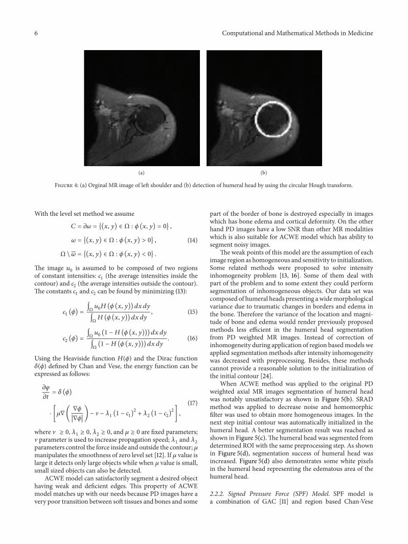


![Detecting and Determining the Types of Hand Bone Fracture ... · techniques has been introduced in [6]. The techniques are thresholding, region-based methods, edge-based segmentation](https://static.fdocuments.in/doc/165x107/5e77ea47af7fe033065d5d24/detecting-and-determining-the-types-of-hand-bone-fracture-techniques-has-been.jpg)