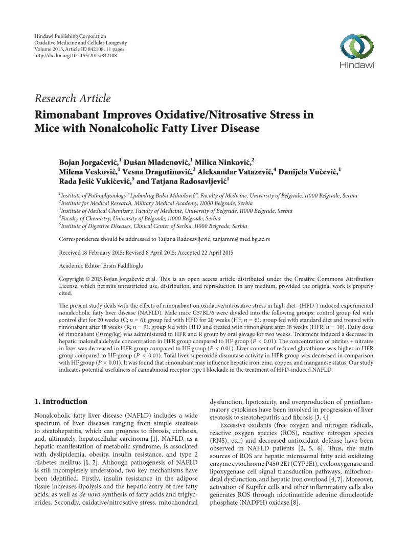
Research Article Rimonabant Improves Oxidative/Nitrosative...
12
Research Article Rimonabant Improves Oxidative/Nitrosative Stress in Mice with Nonalcoholic Fatty Liver Disease Bojan JorgaIeviT, 1 Dušan MladenoviT, 1 Milica NinkoviT, 2 Milena VeskoviT, 1 Vesna DragutinoviT, 3 Aleksandar VatazeviT, 4 Danijela VuIeviT, 1 Rada JešiT VukiTeviT, 5 and Tatjana RadosavljeviT 1 1 Institute of Pathophysiology “Ljubodrag Buba Mihailovi´ c”, Faculty of Medicine, University of Belgrade, 11000 Belgrade, Serbia 2 Institute for Medical Research, Military Medical Academy, 11000 Belgrade, Serbia 3 Institute of Medical Chemistry, Faculty of Medicine, University of Belgrade, 11000 Belgrade, Serbia 4 Faculty of Chemistry, University of Belgrade, 11000 Belgrade, Serbia 5 Institute of Digestive Diseases, Clinical Center of Serbia, 11000 Belgrade, Serbia Correspondence should be addressed to Tatjana Radosavljevi´ c; [email protected] Received 18 February 2015; Revised 8 April 2015; Accepted 22 April 2015 Academic Editor: Ersin Fadillioglu Copyright © 2015 Bojan Jorgaˇ cevi´ c et al. is is an open access article distributed under the Creative Commons Attribution License, which permits unrestricted use, distribution, and reproduction in any medium, provided the original work is properly cited. e present study deals with the effects of rimonabant on oxidative/nitrosative stress in high diet- (HFD-) induced experimental nonalcoholic fatty liver disease (NAFLD). Male mice C57BL/6 were divided into the following groups: control group fed with control diet for 20 weeks (C; =6); group fed with HFD for 20 weeks (HF; =6); group fed with standard diet and treated with rimonabant aſter 18 weeks (R; =9); group fed with HFD and treated with rimonabant aſter 18 weeks (HFR; = 10). Daily dose of rimonabant (10mg/kg) was administered to HFR and R group by oral gavage for two weeks. Treatment induced a decrease in hepatic malondialdehyde concentration in HFR group compared to HF group ( < 0.01). e concentration of nitrites + nitrates in liver was decreased in HFR group compared to HF group ( < 0.01). Liver content of reduced glutathione was higher in HFR group compared to HF group ( < 0.01). Total liver superoxide dismutase activity in HFR group was decreased in comparison with HF group ( < 0.01). It was found that rimonabant may influence hepatic iron, zinc, copper, and manganese status. Our study indicates potential usefulness of cannabinoid receptor type 1 blockade in the treatment of HFD-induced NAFLD. 1. Introduction Nonalcoholic fatty liver disease (NAFLD) includes a wide spectrum of liver diseases ranging from simple steatosis to steatohepatitis, which can progress to fibrosis, cirrhosis, and, ultimately, hepatocellular carcinoma [1]. NAFLD, as a hepatic manifestation of metabolic syndrome, is associated with dyslipidemia, obesity, insulin resistance, and type 2 diabetes mellitus [1, 2]. Although pathogenesis of NAFLD is still incompletely understood, two key mechanisms have been identified. Firstly, insulin resistance in the adipose tissue increases lipolysis and the hepatic entry of free fatty acids, as well as de novo synthesis of fatty acids and triglyc- erides. Secondly, oxidative/nitrosative stress, mitochondrial dysfunction, lipotoxicity, and overproduction of proinflam- matory cytokines have been involved in progression of liver steatosis to steatohepatitis and fibrosis [3, 4]. Excessive oxidants (free oxygen and nitrogen radicals, reactive oxygen species (ROS), reactive nitrogen species (RNS), etc.) and decreased antioxidant defense have been observed in NAFLD patients [2, 5, 6]. us, the main sources of ROS are hepatic microsomal fatty acid oxidizing enzyme cytochrome P450 2E1 (CYP2E1), cyclooxygenase and lipoxygenase cell signal transduction pathways, mitochon- drial dysfunction, and hepatic iron overload [4, 7]. Moreover, activation of Kupffer cells and other inflammatory cells also generates ROS through nicotinamide adenine dinucleotide phosphate (NADPH) oxidase [8]. Hindawi Publishing Corporation Oxidative Medicine and Cellular Longevity Volume 2015, Article ID 842108, 11 pages http://dx.doi.org/10.1155/2015/842108
Transcript of Research Article Rimonabant Improves Oxidative/Nitrosative...
Research ArticleRimonabant Improves Oxidative/Nitrosative Stress inMice with Nonalcoholic Fatty Liver Disease
Bojan JorgaIeviT,1 Dušan MladenoviT,1 Milica NinkoviT,2
Milena VeskoviT,1 Vesna DragutinoviT,3 Aleksandar VatazeviT,4 Danijela VuIeviT,1
Rada JešiT VukiTeviT,5 and Tatjana RadosavljeviT1
1 Institute of Pathophysiology “Ljubodrag Buba Mihailovic”, Faculty of Medicine, University of Belgrade, 11000 Belgrade, Serbia2Institute for Medical Research, Military Medical Academy, 11000 Belgrade, Serbia3Institute of Medical Chemistry, Faculty of Medicine, University of Belgrade, 11000 Belgrade, Serbia4Faculty of Chemistry, University of Belgrade, 11000 Belgrade, Serbia5Institute of Digestive Diseases, Clinical Center of Serbia, 11000 Belgrade, Serbia
Correspondence should be addressed to Tatjana Radosavljevic; [email protected]
Received 18 February 2015; Revised 8 April 2015; Accepted 22 April 2015
Academic Editor: Ersin Fadillioglu
Copyright © 2015 Bojan Jorgacevic et al. This is an open access article distributed under the Creative Commons AttributionLicense, which permits unrestricted use, distribution, and reproduction in any medium, provided the original work is properlycited.
The present study deals with the effects of rimonabant on oxidative/nitrosative stress in high diet- (HFD-) induced experimentalnonalcoholic fatty liver disease (NAFLD). Male mice C57BL/6 were divided into the following groups: control group fed withcontrol diet for 20 weeks (C; 𝑛 = 6); group fed with HFD for 20 weeks (HF; 𝑛 = 6); group fed with standard diet and treated withrimonabant after 18 weeks (R; 𝑛 = 9); group fed with HFD and treated with rimonabant after 18 weeks (HFR; 𝑛 = 10). Daily doseof rimonabant (10mg/kg) was administered to HFR and R group by oral gavage for two weeks. Treatment induced a decrease inhepatic malondialdehyde concentration in HFR group compared to HF group (𝑃 < 0.01). The concentration of nitrites + nitratesin liver was decreased in HFR group compared to HF group (𝑃 < 0.01). Liver content of reduced glutathione was higher in HFRgroup compared to HF group (𝑃 < 0.01). Total liver superoxide dismutase activity in HFR group was decreased in comparisonwith HF group (𝑃 < 0.01). It was found that rimonabant may influence hepatic iron, zinc, copper, andmanganese status. Our studyindicates potential usefulness of cannabinoid receptor type 1 blockade in the treatment of HFD-induced NAFLD.
1. Introduction
Nonalcoholic fatty liver disease (NAFLD) includes a widespectrum of liver diseases ranging from simple steatosisto steatohepatitis, which can progress to fibrosis, cirrhosis,and, ultimately, hepatocellular carcinoma [1]. NAFLD, as ahepatic manifestation of metabolic syndrome, is associatedwith dyslipidemia, obesity, insulin resistance, and type 2diabetes mellitus [1, 2]. Although pathogenesis of NAFLDis still incompletely understood, two key mechanisms havebeen identified. Firstly, insulin resistance in the adiposetissue increases lipolysis and the hepatic entry of free fattyacids, as well as de novo synthesis of fatty acids and triglyc-erides. Secondly, oxidative/nitrosative stress, mitochondrial
dysfunction, lipotoxicity, and overproduction of proinflam-matory cytokines have been involved in progression of liversteatosis to steatohepatitis and fibrosis [3, 4].
Excessive oxidants (free oxygen and nitrogen radicals,reactive oxygen species (ROS), reactive nitrogen species(RNS), etc.) and decreased antioxidant defense have beenobserved in NAFLD patients [2, 5, 6]. Thus, the mainsources of ROS are hepatic microsomal fatty acid oxidizingenzyme cytochrome P450 2E1 (CYP2E1), cyclooxygenase andlipoxygenase cell signal transduction pathways, mitochon-drial dysfunction, and hepatic iron overload [4, 7]. Moreover,activation of Kupffer cells and other inflammatory cells alsogenerates ROS through nicotinamide adenine dinucleotidephosphate (NADPH) oxidase [8].
Hindawi Publishing CorporationOxidative Medicine and Cellular LongevityVolume 2015, Article ID 842108, 11 pageshttp://dx.doi.org/10.1155/2015/842108
2 Oxidative Medicine and Cellular Longevity
Endocannabinoid system (ECS) is a signaling cascadeconsisting of cannabinoid receptors (G-protein-coupled can-nabinoid receptor type 1 /CB1/ and type 2 /CB2/), endocanna-binoids (derivatives of arachidonic acid, N-arachidonoylethanolamine, also known as anandamide /ANA/, and 2-arachidonoyl glycerol /2-AG/), and several enzymes involvedin the synthesis and degradation of endocannabinoids [9–13].
The presence of CB1 receptors in the liver has beenconfirmed, and both ANA and 2-AG have been detectedin different types of liver cells, including hepatocytes andstellate cells [14–17]. A number of studies show that ECSis important in NAFLD pathogenesis and other obesity-related metabolic disorders, due to its crucial role in theregulation of metabolism and energy homeostasis [9, 11, 16,18–20].Thus, dysregulation of ECS in human obesity leads toincreasing hedonic and homeostatic food intake, weight gain,and triglyceride accumulation in the liver and adipose tissue,with consequent hepatic steatosis and insulin resistance [9,15, 18, 20, 21]. Apart from modulating lipid metabolism, theECSmodulates glucose metabolism by promoting pancreatichormonal secretion that in turn favours glucose uptake bytissues such as the liver and white adipose tissue, leadingto fatty acid synthesis and energy storage [13]. Additionally,ECS is involved inmodulating the immune and inflammatoryresponses [11, 22–25].
The liver is identified as a primary site for endocannabi-noid-mediated modulation of lipogenesis [26]. In fact, theactivation of the CB1 receptor increases the expression oflipogenic genes in the liver, which is the main source of denovo fatty acid synthesis in the body [15, 17, 26]. It is suggestedthat hepatic CB1 receptors are implicated in the developmentof fatty liver, hypertriglyceridaemia, and other metabolicabnormalities in diet-induced obesity [12, 13, 17, 20, 26–30].It is reported that CB1 receptor antagonism may protectthe liver from high fat diet- (HFD-) induced phenomenasuch as hepatic steatosis [31–33], insulin resistance [26, 33],and glucose intolerance [13]. Having in mind that deletionof CB1 receptors in the liver decreases hepatic lipogenesisand ameliorates hypercholesterolemia, hypertriglyceridemia,and hepatocellular damage [12, 15], it has been hypothesizedrecently that the hepatic ECS may represent a target for thetreatment of NAFLD [11, 34].
The ability of ECS to promote oxidative/nitrosative stressproduction and modulate antioxidant capacity in kidneyand cardiac tissue has received great attention [25, 35, 36].It is also suggested that inhibition of CB1 receptors mayexert beneficial effects in renal diseases associated withoxidative/nitrosative stress [25]. However, at present nothingis known about the precise role of ECS in initiation and prop-agation of oxidative/nitrosative hepatic injury. Based on thisbackground, the aim of the present study was to investigatethe effect of CB1 receptor blockade on oxidative/nitrosativestress in mice with NAFLD.
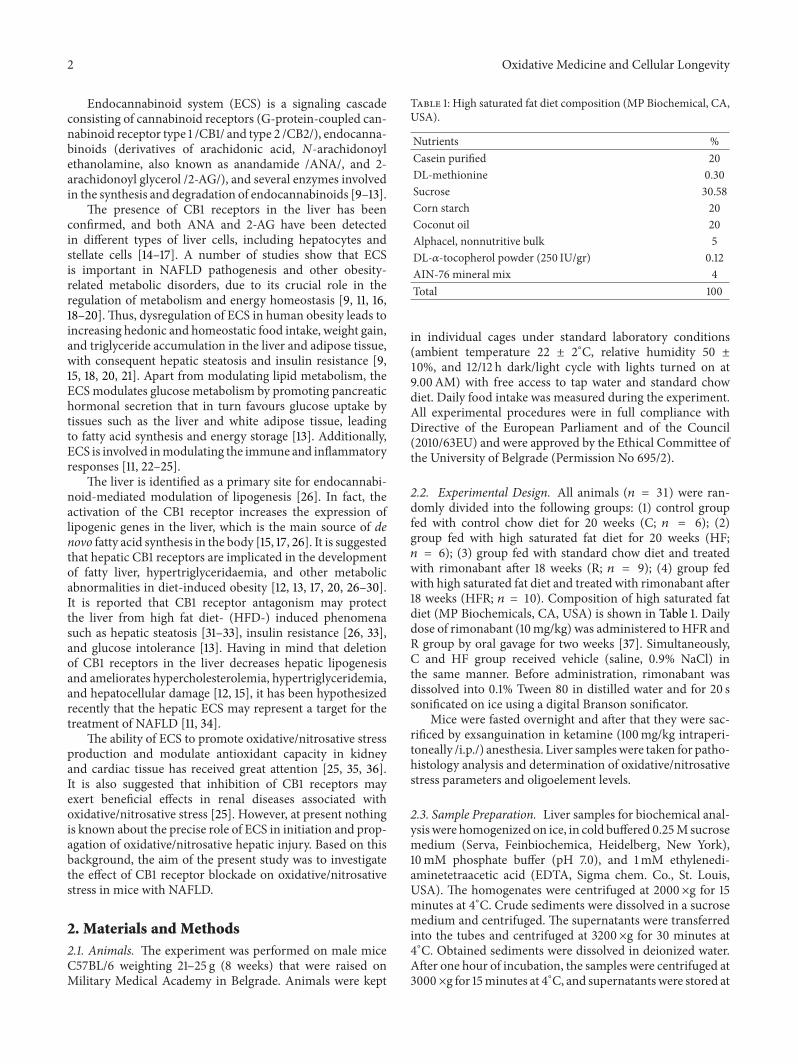
2. Materials and Methods2.1. Animals. The experiment was performed on male miceC57BL/6 weighting 21–25 g (8 weeks) that were raised onMilitary Medical Academy in Belgrade. Animals were kept
Table 1: High saturated fat diet composition (MP Biochemical, CA,USA).
Nutrients %Casein purified 20DL-methionine 0.30Sucrose 30.58Corn starch 20Coconut oil 20Alphacel, nonnutritive bulk 5DL-𝛼-tocopherol powder (250 IU/gr) 0.12AIN-76 mineral mix 4Total 100
in individual cages under standard laboratory conditions(ambient temperature 22 ± 2∘C, relative humidity 50 ±10%, and 12/12 h dark/light cycle with lights turned on at9.00AM) with free access to tap water and standard chowdiet. Daily food intake was measured during the experiment.All experimental procedures were in full compliance withDirective of the European Parliament and of the Council(2010/63EU) and were approved by the Ethical Committee ofthe University of Belgrade (Permission No 695/2).
2.2. Experimental Design. All animals (𝑛 = 31) were ran-domly divided into the following groups: (1) control groupfed with control chow diet for 20 weeks (C; 𝑛 = 6); (2)group fed with high saturated fat diet for 20 weeks (HF;𝑛 = 6); (3) group fed with standard chow diet and treatedwith rimonabant after 18 weeks (R; 𝑛 = 9); (4) group fedwith high saturated fat diet and treated with rimonabant after18 weeks (HFR; 𝑛 = 10). Composition of high saturated fatdiet (MP Biochemicals, CA, USA) is shown in Table 1. Dailydose of rimonabant (10mg/kg) was administered to HFR andR group by oral gavage for two weeks [37]. Simultaneously,C and HF group received vehicle (saline, 0.9% NaCl) inthe same manner. Before administration, rimonabant wasdissolved into 0.1% Tween 80 in distilled water and for 20 ssonificated on ice using a digital Branson sonificator.
Mice were fasted overnight and after that they were sac-rificed by exsanguination in ketamine (100mg/kg intraperi-toneally /i.p./) anesthesia. Liver sampleswere taken for patho-histology analysis and determination of oxidative/nitrosativestress parameters and oligoelement levels.
2.3. Sample Preparation. Liver samples for biochemical anal-ysis were homogenized on ice, in cold buffered 0.25M sucrosemedium (Serva, Feinbiochemica, Heidelberg, New York),10mM phosphate buffer (pH 7.0), and 1mM ethylenedi-aminetetraacetic acid (EDTA, Sigma chem. Co., St. Louis,USA). The homogenates were centrifuged at 2000×g for 15minutes at 4∘C. Crude sediments were dissolved in a sucrosemedium and centrifuged. The supernatants were transferredinto the tubes and centrifuged at 3200×g for 30 minutes at4∘C. Obtained sediments were dissolved in deionized water.After one hour of incubation, the samples were centrifuged at3000×g for 15minutes at 4∘C, and supernatantswere stored at
Oxidative Medicine and Cellular Longevity 3
−70∘C. Proteins were determined by the Lowrymethod usingbovine serum albumin as the standard [38].
2.4. Biochemical Parameters2.4.1. Determination of Hepatic Oxidative/Nitrosative StressParameters. Lipid peroxidation, measured as malondialde-hyde (MDA) level, was determined spectrophotometricallyin a reaction with thiobarbituric acid as described by Girottiet al. [39]. The results are expressed as nmol of MDA permilligram of proteins (nmol/mg protein).
The concentration of nitrites + nitrates (NO𝑥) as a
measure of NO production was determined by using Griessreagent. After reduction of the nitrates, the total nitrites werereacted with sulfanilamide and N-(1-naphthyl) ethylenedi-amine to produce an azo dye, which was measured spec-trophotometrically at 492 nm [40].
Activity of total superoxide dismutase (EC1.15.1.1.; SOD)in the liver was measured spectrophotometrically, as an inhi-bition of epinephrine autooxidation at 480 nm.After additionof 10mM epinephrine (Sigma, St. Louis, USA), analysis wasperformed in the sodium carbonate buffer (50mM, pH 10.2;Serva, Feinbiochemica, Heidelberg, New York) containing0.1mM EDTA (Sigma, St. Louis, USA). Samples for man-ganese SOD (MnSOD) were previously treated with 8mMKCN (Sigma, St. Louis, USA) and then analysed as previouslydescribed [41]. Activity of copper/zincSOD (Cu/ZnSOD)wasdetermined as difference between the activities of total SODand MnSOD. All enzyme activities are expressed as units permilligram of protein (U/mg prot.).
The content of reduced glutathione (GSH) was deter-mined spectrophotometrically using 5,5-dithio-bis-2-nitrob-enzoic acid (DTNB). DTNB reacts with aliphatic thiol com-pounds at pH 8.0 forming yellow p-nitrophenol anion whoseabsorption is measured spectrophotometrically at 412 nm[42]. Results are expressed as nmol per milligram of proteins(nmol/mg protein).
2.4.2. Determination of Oligoelement Contents in the Liver.A MDS-2000 system microwave digestion lab station wasused for digesting the samples of tissue with 10mL ofdiluted nitric acid (1 : 1), followed with addition of 10mLconcentrated nitric acid. Samples were treated accordingto the program of 3 steps. This was done over the periodof 20min with the power of 630 ± 30W and within thetemperature range of 140–160∘C. After complete digestionsamples were transferred in 50mL volumetric flask [43].
Zn and Cu liver content was determined by meansof flame atomic absorption spectrometry (FAAS), whilegraphite furnace atomic absorption spectrometry (GF AAS)was applied for determining ofMn and iron (Fe) liver content[43].
Oligoelement liver content is expressed as milligram perkilogram (mg/kg).
All chemicals for the digestion of the samples were ofthe analytical reagent grade and were supplied by J.T. BakerChemical Co. (Phillipsburg, NJ, USA).
2.5. Pathohistological Analysis. Liver tissue was incubatedin 10% formalin solution at room temperature. After the
fixation, the liver samples were processed by the standardmethod. Tissues were incorporated in paraffin sectionedat 5 𝜇m and then stained with Hematoxylin-Eosin (HE)and prepared for light microscopy analysis. All sampleswere evaluated by an experienced pathohistologist who wasblinded to the experiment. Preparations were analyzed andphotographed using a combined photobinocular light micro-scope Olympus BX51 equipped with Artcore 500MI artery,Co. Ltd. Japan Camera.
2.6. Statistical Analysis. All results are expressed as means ±SD. As the normal distribution of parameters was confirmedby Kolmogorov-Smirnov test, one-way analysis of variance(ANOVA) with Tukey’s post hoc test was used for testingthe difference among groups. The difference was consideredsignificant if 𝑃 < 0.05. For statistical analysis computersoftware SPSS 15.0 was used.
3. Results
3.1. Effect of Rimonabant on Liver/Body Weight Ratio. Therewas no statistically significant difference in liver/body weightratio between C group (4.65 ± 0.33%) and R group (4.54 ±0.41%) (𝑃 = 0.955), as well as in HF group (4.86±0.44%) andHFR group (4.67 ± 0.38%) (𝑃 = 0.907).
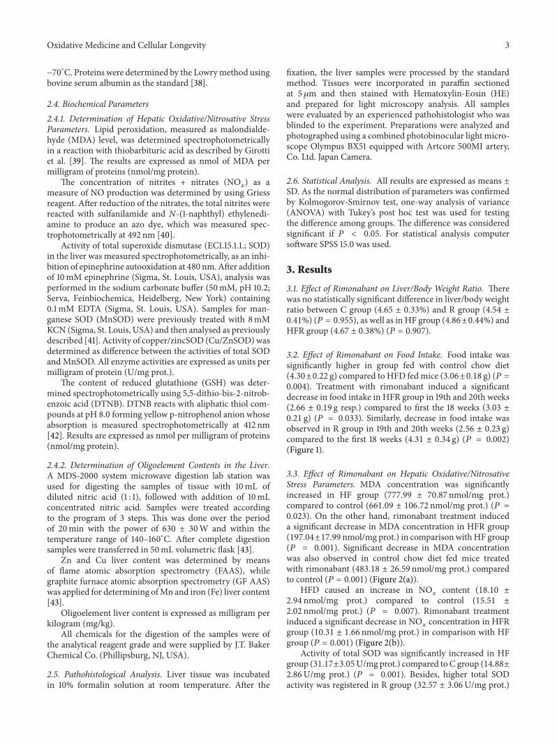
3.2. Effect of Rimonabant on Food Intake. Food intake wassignificantly higher in group fed with control chow diet(4.30±0.22 g) compared to HFD fedmice (3.06±0.18 g) (𝑃 =0.004). Treatment with rimonabant induced a significantdecrease in food intake in HFR group in 19th and 20th weeks(2.66 ± 0.19 g resp.) compared to first the 18 weeks (3.03 ±0.21 g) (𝑃 = 0.033). Similarly, decrease in food intake wasobserved in R group in 19th and 20th weeks (2.56 ± 0.23 g)compared to the first 18 weeks (4.31 ± 0.34 g) (𝑃 = 0.002)(Figure 1).
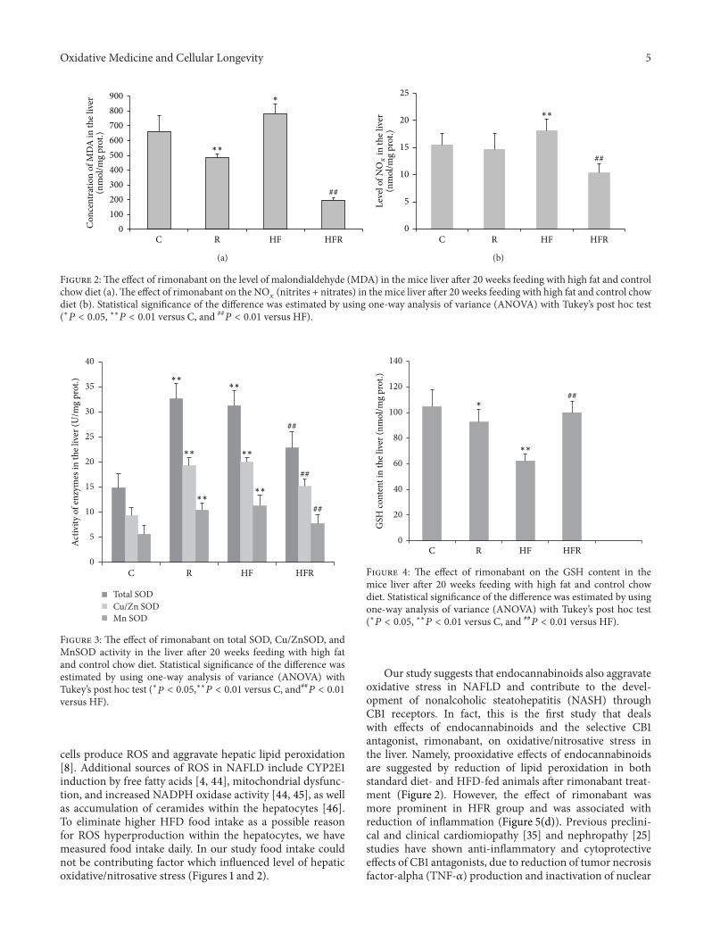
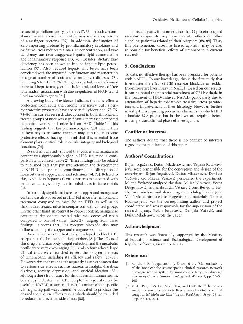
3.3. Effect of Rimonabant on Hepatic Oxidative/NitrosativeStress Parameters. MDA concentration was significantlyincreased in HF group (777.99 ± 70.87 nmol/mg prot.)compared to control (661.09 ± 106.72 nmol/mg prot.) (𝑃 =0.023). On the other hand, rimonabant treatment induceda significant decrease in MDA concentration in HFR group(197.04±17.99 nmol/mg prot.) in comparison withHF group(𝑃 = 0.001). Significant decrease in MDA concentrationwas also observed in control chow diet fed mice treatedwith rimonabant (483.18 ± 26.59 nmol/mg prot.) comparedto control (𝑃 = 0.001) (Figure 2(a)).
HFD caused an increase in NO𝑥
content (18.10 ±2.94 nmol/mg prot.) compared to control (15.51 ±2.02 nmol/mg prot.) (𝑃 = 0.007). Rimonabant treatmentinduced a significant decrease in NO
𝑥concentration in HFR
group (10.31 ± 1.66 nmol/mg prot.) in comparison with HFgroup (𝑃 = 0.001) (Figure 2(b)).
Activity of total SOD was significantly increased in HFgroup (31.17±3.05U/mg prot.) compared toC group (14.88±2.86U/mg prot.) (𝑃 = 0.001). Besides, higher total SODactivity was registered in R group (32.57 ± 3.06U/mg prot.)
4 Oxidative Medicine and Cellular Longevity
Table 2: The effect of rimonabant on concentration of oligoelements in the liver.
Groups Concentration of oligoelements (mg/kg)Fe Zn Mg Cu
C 82.90 ± 4.83 36.69 ± 5.02 1.27 ± 0.01 2.66 ± 0.15R 88.09 ± 6.22 45.79 ± 6.16∗∗ 0.06 ± 0.01∗∗ 4.04 ± 0.48∗∗
HF 110.59 ± 10.07∗∗ 29.10 ± 2.38∗ 2.41 ± 0.23∗∗ 4.33 ± 0.24∗∗
HFR 70.46 ± 10.07## 50.10 ± 8.65## 10.01 ± 1.99## 5.53 ± 1.38##
Fe—iron; Zn—zinc; Mg—manganese; Cu—copper.C-control group fed with control chow diet for 20 weeks; R-group fed with standard chow diet and treated with rimonabant after 18 weeks; HF-group fed withhigh saturated fat diet for 20 weeks; HFR- group fed with high saturated fat diet and treated with rimonabant after 18 weeks.Significance of the difference was estimated by using one-way analysis of variance (ANOVA) with Tukey’s post hoc test (∗𝑃 < 0.05, ∗∗𝑃 < 0.01 vs. C, and##𝑃 < 0.01 vs. HF).
5
4.5
4
3.5
3
2.5
2
1.5
1
0.5
0
Aver
age f
ood
inta
ke (g
/day
)
HFC R HFR
1–18 weeks19-20 weeks
∗∗ ∗
##
##
Figure 1: The effects of control chow and high fat diet on foodintake during 20 weeks of treatment (C, HF, R, and HFR). Statisticalsignificance of the difference was estimated by using one-wayanalysis of variance (ANOVA) with Tukey’s post hoc test (∗𝑃 < 0.05versus first 18 weeks; ∗∗𝑃 < 0.01 versus first 18 weeks; ##
𝑃 < 0.01
versus control group).
compared to control (𝑃 = 0.001). On the other hand, totalSOD activity in HFR group (22.84 ± 3.27U/mg prot.) wassignificantly decreased in comparison with HF group (𝑃 =0.004). Analysis of SOD isoenzymes revealed that expressionofMnSODandCu/ZnSOD followed the changes of total SODactivity in all groups (Figure 3).
GSH content was significantly decreased in HF group(62.43 ± 8.86 nmol/mg prot.) in comparison with control(104.59 ± 13.16 nmol/mg prot.) (𝑃 = 0.003). However,GSH level was significantly higher in HFR group (100.09 ±5.39 nmol/mg prot.) compared to HF group (𝑃 = 0.009).Besides, decrease in GSH concentration was observed inR group (92.46 ± 10.19 nmol/mg prot.) when compared tocontrol (𝑃 = 0.041) (Figure 4).
3.4. Effect of Rimonabant on Liver Oligoelement Contents. Itwas found a significant decline in Zn content in HF group
(29.10 ± 2.38mg/kg) in comparison with C group (36.69 ±5.02mg/kg) (𝑃 = 0.033). However, Zn content in HFR group(50.1 ± 8.65mg/kg) was higher compared to HF group (𝑃 =0.001) and also in R group (45.79 ± 6.16mg/kg) compared toC group (𝑃 = 0.009) (Table 2).
It was shown a significant HFD-induced increase in Fecontent (110.59 ± 10.07mg/kg) when compared to control(82.90 ± 4.83mg/kg) (𝑃 = 0.004). In contrast, treatment withrimonabant reduced Fe content inHFR (70.46±10.07mg/kg)in comparison with HF group (𝑃 = 0.003) (Table 2).
Cu content in HF group (4.33 ± 0.24mg/kg) was sig-nificantly increased in comparison with control (2.66 ±0.15mg/kg) (𝑃 = 0.001). Significant increase in Cu con-tent was also observed in HFR group (5.53 ± 1.38mg/kg)compared to HF group (𝑃 = 0.005), as well as in R group(4.04 ± 0.48mg/kg) in comparison with C group (𝑃 = 0.001)(Table 2).
Similarly to Cu content, HFD induced an increase in Mncontent (2.41±0.23mg/kg) compared to control value (1.27±0.01mg/kg) (𝑃 < 0.003). Administration of rimonabantincreased Mn content in HFR group (10.01 ± 1.99mg/kg) incomparison with HF group, too (𝑃 < 0.001). In contrast toCu content, Mn content in R group (0.06 ± 0.01mg/kg) wasdecreased when compared to C group (𝑃 < 0.001) (Table 2).
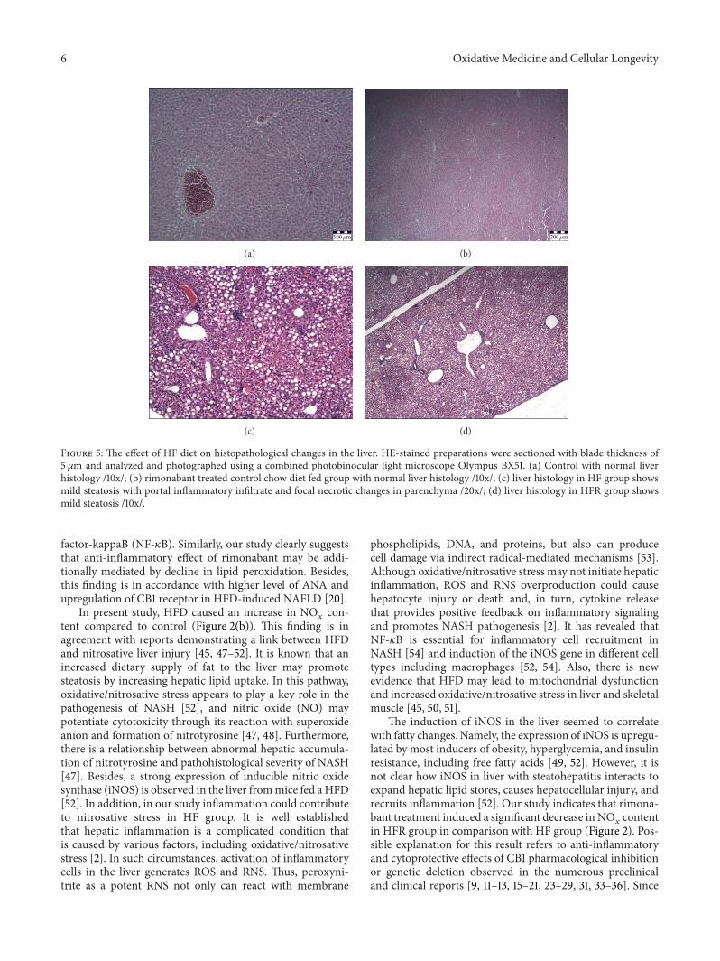
3.5. Pathohistological Findings. There were no pathohistolog-ical changes in the liver in C and R groups (Figure 5(a) and5(b)). HFD caused mild hepatic steatosis with portal inflam-matory infiltrate and focal necrotic changes in parenchyme(Figure 5(c)). In contrast to HF group, mild steatosis andno inflammatory infiltrate in portal space and focal necroticchanges in the liver parenchyma were found in HFR group(Figure 5(d)).
4. Discussion
Results of our study clearly showed that HFD induced lipidperoxidation and nitrosative stress in the liver that are asso-ciated with steatosis and portal inflammation (Figure 5(c)).The role of oxidative stress in the pathogenesis of NAFLD iswell known and is bidirectionally linked with inflammation[5–8]. Lipid peroxidation alongwith cytokine production andFas ligand induction causes hepatocyte injury and apoptosis[7]. Besides, activated Kupffer cells and other inflammatory
Oxidative Medicine and Cellular Longevity 5
HF HFR
Con
cent
ratio
n of
MD
A in
the l
iver
(nm
ol/m
g pr
ot.)
900
800
700
600
500
400
300
200
100
0
∗∗
∗
##
C R
(a)
HF HFR
∗∗
##
25
20
15
10
5
0
(nm
ol/m
g pr
ot.)
C R
Leve
l ofN
Ox
in th
e liv
er
(b)
Figure 2:The effect of rimonabant on the level of malondialdehyde (MDA) in the mice liver after 20 weeks feeding with high fat and controlchow diet (a).The effect of rimonabant on the NO
𝑥
(nitrites + nitrates) in the mice liver after 20 weeks feeding with high fat and control chowdiet (b). Statistical significance of the difference was estimated by using one-way analysis of variance (ANOVA) with Tukey’s post hoc test(∗𝑃 < 0.05, ∗∗𝑃 < 0.01 versus C, and ##
𝑃 < 0.01 versus HF).
0
5
10
15
20
25
30
35
40
Total SODCu/Zn SODMn SOD
HF HFR
Activ
ity o
f enz
ymes
in th
e liv
er (U
/mg
prot
.) ∗∗
∗∗
∗∗
∗∗
∗∗
∗∗
##
##
##
C R
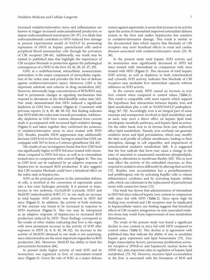
Figure 3: The effect of rimonabant on total SOD, Cu/ZnSOD, andMnSOD activity in the liver after 20 weeks feeding with high fatand control chow diet. Statistical significance of the difference wasestimated by using one-way analysis of variance (ANOVA) withTukey’s post hoc test (∗𝑃 < 0.05,∗∗𝑃 < 0.01 versus C, and##𝑃 < 0.01versus HF).
cells produce ROS and aggravate hepatic lipid peroxidation[8]. Additional sources of ROS in NAFLD include CYP2E1induction by free fatty acids [4, 44], mitochondrial dysfunc-tion, and increased NADPH oxidase activity [44, 45], as wellas accumulation of ceramides within the hepatocytes [46].To eliminate higher HFD food intake as a possible reasonfor ROS hyperproduction within the hepatocytes, we havemeasured food intake daily. In our study food intake couldnot be contributing factor which influenced level of hepaticoxidative/nitrosative stress (Figures 1 and 2).
0
20
40
60
80
100
120
140
HF HFR
GSH
cont
ent i
n th
e liv
er (n
mol
/mg
prot
.)
∗∗
∗##
C R
Figure 4: The effect of rimonabant on the GSH content in themice liver after 20 weeks feeding with high fat and control chowdiet. Statistical significance of the difference was estimated by usingone-way analysis of variance (ANOVA) with Tukey’s post hoc test(∗𝑃 < 0.05, ∗∗𝑃 < 0.01 versus C, and ##
𝑃 < 0.01 versus HF).
Our study suggests that endocannabinoids also aggravateoxidative stress in NAFLD and contribute to the devel-opment of nonalcoholic steatohepatitis (NASH) throughCB1 receptors. In fact, this is the first study that dealswith effects of endocannabinoids and the selective CB1antagonist, rimonabant, on oxidative/nitrosative stress inthe liver. Namely, prooxidative effects of endocannabinoidsare suggested by reduction of lipid peroxidation in bothstandard diet- and HFD-fed animals after rimonabant treat-ment (Figure 2). However, the effect of rimonabant wasmore prominent in HFR group and was associated withreduction of inflammation (Figure 5(d)). Previous preclini-cal and clinical cardiomiopathy [35] and nephropathy [25]studies have shown anti-inflammatory and cytoprotectiveeffects of CB1 antagonists, due to reduction of tumor necrosisfactor-alpha (TNF-𝛼) production and inactivation of nuclear
6 Oxidative Medicine and Cellular Longevity
100𝜇m
(a)
200𝜇m
(b)
(c) (d)
Figure 5: The effect of HF diet on histopathological changes in the liver. HE-stained preparations were sectioned with blade thickness of5 𝜇m and analyzed and photographed using a combined photobinocular light microscope Olympus BX51. (a) Control with normal liverhistology /10x/; (b) rimonabant treated control chow diet fed group with normal liver histology /10x/; (c) liver histology in HF group showsmild steatosis with portal inflammatory infiltrate and focal necrotic changes in parenchyma /20x/; (d) liver histology in HFR group showsmild steatosis /10x/.
factor-kappaB (NF-𝜅B). Similarly, our study clearly suggeststhat anti-inflammatory effect of rimonabant may be addi-tionally mediated by decline in lipid peroxidation. Besides,this finding is in accordance with higher level of ANA andupregulation of CB1 receptor in HFD-induced NAFLD [20].
In present study, HFD caused an increase in NO𝑥con-
tent compared to control (Figure 2(b)). This finding is inagreement with reports demonstrating a link between HFDand nitrosative liver injury [45, 47–52]. It is known that anincreased dietary supply of fat to the liver may promotesteatosis by increasing hepatic lipid uptake. In this pathway,oxidative/nitrosative stress appears to play a key role in thepathogenesis of NASH [52], and nitric oxide (NO) maypotentiate cytotoxicity through its reaction with superoxideanion and formation of nitrotyrosine [47, 48]. Furthermore,there is a relationship between abnormal hepatic accumula-tion of nitrotyrosine and pathohistological severity of NASH[47]. Besides, a strong expression of inducible nitric oxidesynthase (iNOS) is observed in the liver frommice fed a HFD[52]. In addition, in our study inflammation could contributeto nitrosative stress in HF group. It is well establishedthat hepatic inflammation is a complicated condition thatis caused by various factors, including oxidative/nitrosativestress [2]. In such circumstances, activation of inflammatorycells in the liver generates ROS and RNS. Thus, peroxyni-trite as a potent RNS not only can react with membrane
phospholipids, DNA, and proteins, but also can producecell damage via indirect radical-mediated mechanisms [53].Although oxidative/nitrosative stress may not initiate hepaticinflammation, ROS and RNS overproduction could causehepatocyte injury or death and, in turn, cytokine releasethat provides positive feedback on inflammatory signalingand promotes NASH pathogenesis [2]. It has revealed thatNF-𝜅B is essential for inflammatory cell recruitment inNASH [54] and induction of the iNOS gene in different celltypes including macrophages [52, 54]. Also, there is newevidence that HFD may lead to mitochondrial dysfunctionand increased oxidative/nitrosative stress in liver and skeletalmuscle [45, 50, 51].
The induction of iNOS in the liver seemed to correlatewith fatty changes. Namely, the expression of iNOS is upregu-lated by most inducers of obesity, hyperglycemia, and insulinresistance, including free fatty acids [49, 52]. However, it isnot clear how iNOS in liver with steatohepatitis interacts toexpand hepatic lipid stores, causes hepatocellular injury, andrecruits inflammation [52]. Our study indicates that rimona-bant treatment induced a significant decrease in NO
𝑥content
in HFR group in comparison with HF group (Figure 2). Pos-sible explanation for this result refers to anti-inflammatoryand cytoprotective effects of CB1 pharmacological inhibitionor genetic deletion observed in the numerous preclinicaland clinical reports [9, 11–13, 15–21, 23–29, 31, 33–36]. Since
Oxidative Medicine and Cellular Longevity 7
increased oxidative/nitrosative stress and inflammation areknown to trigger increased endocannabinoid production orimpair endocannabinoid inactivation [55–57], it is likely thatendocannabinoids contribute to HFD-induced liver damagein present experimental model of NAFLD by promotingexpression of iNOS in hepatic parenchymal cells and/orperipheral blood mononuclear cells through the activationof CB1 receptors [58–61]. Additionally, our result may berelated to published data that highlight the importance ofCB1 receptor blockade in protection against the pathologicalconsequences of a HFD in the liver [11, 16, 20, 25–27, 29].
GSH, as a multifunctional, intracellular, nonenzymaticantioxidant, is the major component of intracellular regula-tion of the redox state and provides the first line of defenseagainst oxidative/nitrosative injury. Moreover, GSH is theimportant substrate and cofactor in drug metabolism [62].However, abnormally large concentrations of ROS/RNS maylead to permanent changes in signaling mechanisms thatrespond to alterations in the thiol/disulfide redox state [63].Our study demonstrated that HFD induced a significantdepletion in GSH liver content (Figure 4). Consistent withprevious reports [2, 6, 45, 48, 50–52], this finding indicatesthatHFD shifts the redox state towards prooxidant. Addition-ally, depletion in GSH liver content obtained from currentstudy is accompanied with increased lipid oxidative damage(Figure 2) and taken together they confirmed the existenceof oxidative/nitrosative stress in mice treated with HFD[52]. Besides, possible iNOS suppression may additionallydecrease GSH level in the liver, having in mind that GSH canconjugate with NO to form a S-nitroso-glutathione [64, 65].
The results of our investigation found that liver GSH levelwas significantly higher in HFD fedmice treated with rimon-abant compared to HFD fed mice, as well as in rimonabanttreated mice in comparison with control (Figure 4). This risein GSH level can be explained by an adaptive response ofhepatocytes to increased ROS production. It also suggeststhat CB1 receptor blockade could have a beneficial effect onthe redox state in hepatocytes.
SOD, as the principal enzyme in the antioxidant defenseof cells, is involved in the conversion of superoxide anioninto a less toxic hydrogen peroxide. It is present in hepa-tocytes in two isoforms, Cu/ZnSOD (cytosolic SOD) andMnSOD (mitochondrial SOD) [7]. In our study an increasein total hepatic SOD activity was observed in HFD fedmice (Figure 3). In addition, the activity of both isoformsof this enzyme was found to be increased in response toHFD treatment (Figure 3). This rise may be interpretedas an adaptive response of hepatocytes to increased ROSproduction induced by HFD. These findings correspond tothe results of other studies indicating that liver is the organwith most prominent increase in the activity of SOD afterexposure to HFD [4, 6, 8, 45, 50–52]. An increase in theactivity of MnSOD obtained in our study is not surprising,sincemitochondria are themost important organelles in ROSproduction [45]. Moreover, MnSOD has ability to limit theperoxynitrite formation [66].
In present study higher activity of total SOD and itsisoenzymes was registered in liver of rimonabant-treatedmice (Figure 3). Given the role of SOD, as a major defense
system against superoxide, it seems that increase in its activityupon the action of rimonabant improved antioxidant defensesystem in the liver and makes hepatocytes less sensitiveto oxidative/nitrosative damage. This result is similar tothe documented data which reports that blockade of CB1receptors may exert beneficial effects in renal and cardiacdiseases associated with oxidative/nitrosative stress [25, 35,36].
In the present study total hepatic SOD activity andits isoenzymes were significantly decreased in HFD fedmice treated with rimonabant in comparison with micetreated with HFD (Figure 3). This decrease of total hepaticSOD activity, as well as depletion in both mitochondrialand cytosolic SOD activity, indicates that blockade of CB1receptors may modulate liver antioxidant capacity withoutinfluence on SOD activity.
In the current study, HFD caused an increase in ironliver content when compared to control values (Table 2).This result is compatible with most reports which supportthe hypothesis that interactions between hepatic iron andlipid metabolism play a role in NASH/NAFLD pathophysi-ology [67–70]. Accordingly, iron is an integral part of someenzymes and transporters involved in lipid metabolism and,as such, may exert a direct effect on hepatic lipid load,intrahepatic metabolic pathways, and hepatic lipid secretion.On the other hand, iron in its ferrous form may indirectlyaffect lipid metabolism. Namely, iron overload can generateoxidative stress and lipid peroxidation, which may modifythe fatty acid profile of cellular membranes, leading to theirdisruption, damage to cell organelles, and impairment ofmitochondrial oxidative metabolism [68]. It is suggestedthat the free radicals that form may cause a change in theratio of saturated to unsaturated membrane phospholipids,leading to alterations in membrane fluidity [63]. This in turnmay affect the activity of the embedded enzymes, so theyrespond to oxidative stress differently by altering their activity[71]. Besides, iron accumulation has a proinflammatoryand profibrogenic role by activating Kupffer cells to releaseinflammatory cytokines and by activating hepatic stellatecells, which can culminate in the replacement of parenchymaltissue with connective tissue [72].
Our study has shown that administration of rimonabantto HFD fedmice reduced hepatic iron content in comparisonwith mice fed with HFD (Table 2). Since upon high fatfeeding iron overload and CB1 receptors may be implicatedin hepatocellular injury, our finding suggests that beneficialeffects of CB1 receptor antagonism on iron-associated oxida-tive stress may result from improvement of iron metabolismdisturbances.
The result of the present study was found a significantdecline in zinc content in mice fed with HFD compared tocontrol values (Table 2). This decline is in agreement withpublished data that indicate the ability of zinc to modulatehepatic gene expression and lipid homeostasis [73]. Zincfinger transcription factors, peroxisome proliferation activa-tor receptor-𝛼 (PPAR-𝛼) and hepatocyte nuclear factor-4𝛼(HNF-4𝛼), play important roles in regulation of hepatic lipidmetabolism [73, 74]. However, excessive lipid accumulationin the liver is associated with the formation of ROS and
8 Oxidative Medicine and Cellular Longevity
release of proinflammatory cytokines [7, 75]. In such circum-stance, hepatic accumulation of fat may impaire expressionof zinc-finger proteins [75]. In addition, dysfunction ofzinc-importing proteins by proinflammatory cytokines andoxidative stress reduces plasma zinc concentration, and zincdeficiency can thus exaggerate hepatic lipid accumulationand inflammatory response [73, 76]. Besides, dietary zincdeficiency has been shown to induce hepatic lipid perox-idation [77]. Also, reduced hepatic zinc levels have beencorrelated with the impaired liver function and regenerationin a great number of acute and chronic liver diseases [76],including NAFLD [74, 76]. Thus, as expected, zinc deficiencyincreased hepatic triglyceride, cholesterol, and levels of freefatty acids in associationwith downregulation of PPAR-𝛼 andlipid metabolism genes [73].
A growing body of evidence indicates that zinc offers aprotection from acute and chronic liver injury, but its hep-atoprotective properties have not been fully identified [73, 76,78–80]. In current research zinc content in both rimonabanttreated groups of mice was significantly increased comparedto control values and mice fed on HFD (Table 2). Thisfinding suggests that the pharmacological CB1 inactivationin hepatocytes in some manner may contribute to zincprotective effects, having in mind that this essential traceelement plays a critical role in cellular integrity and biologicalfunctions [76].
Results in our study showed that copper and manganesecontent was significantly higher in HFD fed mice in com-parison with control (Table 2). These findings may be relatedto published data that put into attention the developmentof NAFLD as a potential contributor to the disruption ofhomeostasis of copper, zinc, and selenium [74, 78]. Related tothis, NAFLD in hepatitis C virus patients is associated withoxidative damage, likely due to imbalances in trace metals[74].
In our study significant increase in copper andmanganesecontent was also observed in HFD fed mice after rimonabanttreatment compared to mice fed on HFD, as well as inrimonabant treated mice in comparison with control group.On the other hand, in contrast to copper content, manganesecontent in rimonabant treated mice was decreased whencompared to control values (Table 2). Judging from thesefindings, it seems that CB1 receptor blockade also mayinfluence on hepatic copper and manganese status.
Rimonabant was the first drug developed to block CB1receptors in the brain and in the periphery [81].The effects ofthis drug on human bodyweight reduction and themetabolicprofile were very encouraging [82] and so four related largeclinical trials were launched to test the long-term effectsof rimonabant, including its efficacy and safety [83–86].However, rimonabant has subsequently been withdrawn dueto serious side effects, such as nausea, arthralgia, diarrhea,dizziness, anxiety, depression, and suicidal ideation [87].Although there is no future for rimonabant in human health,our study indicates that CB1 receptor antagonists may beuseful in NAFLD treatment. It is still unclear which specificCB1-signaling pathways should be activated to produce thedesired therapeutic effects versus which should be excludedto reduce the unwanted side effects [88].
In recent years, it becomes clear that G-protein-coupledreceptor antagonists may have agonistic effects on othersignaling pathways related to their receptors [88, 89]. Thus,this phenomenon, known as biased agonism, may be alsoresponsible for beneficial effects of rimonabant in currentresearch.
5. Conclusions
To date, no effective therapy has been proposed for patientswith NAFLD. To our knowledge, this is the first study thatinvestigates the effect of CB1 receptor blockade on oxida-tive/nitrosative liver injury in NAFLD. Based on our results,it can be noted the potential usefulness of CB1 blockade inthe treatment of HFD-induced NAFLD, particularly due toattenuation of hepatic oxidative/nitrosative stress parame-ters and improvement of liver histology. However, furtherinvestigations regarding precise mechanisms by which HFDstimulate ECS production in the liver are required beforemoving toward clinical phase of investigation.
Conflict of Interests
The authors declare that there is no conflict of interestsregarding the publication of this paper.
Authors’ Contributions
Bojan Jorgacevic, Dusan Mladenovic, and Tatjana Radosavl-jevic were responsible for the conception and design of theexperiment. Bojan Jorgacevic, Dusan Mladenovic, DanijelaVucevic, and Milena Veskovic performed the experiment.Milena Veskovic analyzed the data. Milica Ninkovic, VesnaDragutinovic, and Aleksandar Vatazevic contributed to bio-chemical analysis and describing methodology. Rada JesicVukicevic contributed to reagents and materials. TatjanaRadosavljevic was the corresponding author and projectcoordinator and was responsible for the supervision of theresearch group. Bojan Jorgacevic, Danijela Vucevic, andDusan Mladenovic wrote the paper.
Acknowledgment
This research was financially supported by the Ministryof Education, Science and Technological Development ofRepublic of Serbia, Grant no. 175015.
References
[1] R. Juluri, R. Vuppalanchi, J. Olson et al., “Generalizabilityof the nonalcoholic steatohepatitis clinical research networkhistologic scoring system for nonalcoholic fatty liver disease,”Journal of Clinical Gastroenterology, vol. 45, no. 1, pp. 55–58,2011.
[2] M.-H. Pan, C.-S. Lai, M.-L. Tsai, and C.-T. Ho, “Chemopre-vention of nonalcoholic fatty liver disease by dietary naturalcompounds,”MolecularNutrition andFoodResearch, vol. 58, no.1, pp. 147–171, 2014.
Oxidative Medicine and Cellular Longevity 9
[3] J. C. Fraulob, R. Ogg-Diamantino, C. Fernandes-Santos, M. B.Aguila, and C. A. Mandarim-de-Lacerda, “A mouse model ofmetabolic syndrome: insulin resistance, fatty liver and Non-Alcoholic Fatty Pancreas Disease (NAFPD) in C57BL/6 micefed a high fat diet,” Journal of Clinical Biochemistry and Nutri-tion, vol. 46, no. 3, pp. 212–223, 2010.
[4] J. Aubert, K. Begriche, L. Knockaert, M. A. Robin, and B. Fro-menty, “Increased expression of cytochrome P450 2E1 in nonal-coholic fatty liver disease: mechanisms and pathophysiologicalrole,” Clinics and Research in Hepatology and Gastroenterology,vol. 35, no. 10, pp. 630–637, 2011.
[5] S. Seki, T. Kitada, and H. Sakaguchi, “Clinicopathologicalsignificance of oxidative cellular damage in non-alcoholic fattyliver diseases,” Hepatology Research, vol. 33, no. 2, pp. 132–134,2005.
[6] K. Madan, P. Bhardwaj, S. Thareja, S. D. Gupta, and A. Saraya,“Oxidant stress and antioxidant status among patients withnonalcoholic fatty liver disease (NAFLD),” Journal of ClinicalGastroenterology, vol. 40, no. 10, pp. 930–935, 2006.
[7] A. P. Rolo, J. S. Teodoro, and C. M. Palmeira, “Role of oxidativestress in the pathogenesis of nonalcoholic steatohepatitis,” FreeRadical Biology and Medicine, vol. 52, no. 1, pp. 59–69, 2012.
[8] A. Gornicka, G. Morris-Stiff, S. Thapaliya, B. G. Papouchado,M. Berk, and A. E. Feldstein, “Transcriptional profile of genesinvolved in oxidative stress and antioxidant defense in a dietarymurine model of steatohepatitis,” Antioxidants and Redox Sig-naling, vol. 15, no. 2, pp. 437–445, 2011.
[9] F. J. Bermudez-Silva, P. Cardinal, and D. Cota, “The role of theendocannabinoid system in the neuroendocrine regulation ofenergy balance,” Journal of Psychopharmacology, vol. 26, no. 1,pp. 114–124, 2012.
[10] A. C. Howlett, “The cannabinoid receptors,” Prostaglandins andOther Lipid Mediators, vol. 68-69, pp. 619–631, 2002.
[11] U. Pagotto, G.Marsicano,D. Cota, B. Lutz, andR. Pasquali, “Theemerging role of the endocannabinoid system in endocrineregulation and energy balance,” Endocrine Reviews, vol. 27, no.1, pp. 73–100, 2006.
[12] C. Quarta, R. Mazza, S. Obici, R. Pasquali, and U. Pagotto,“Energy balance regulation by endocannabinoids at central andperipheral levels,” Trends in Molecular Medicine, vol. 17, no. 9,pp. 518–526, 2011.
[13] S. Y. Romero-Zerbo and F. J. Bermudez-Silva, “Cannabinoids,eating behaviour, and energy homeostasis,” Drug Testing andAnalysis, vol. 6, no. 1-2, pp. 52–58, 2014.
[14] L. Hanus, Y. Avraham, D. Ben-Shushan, O. Zolotarev, E. M.Berry, and R. Mechoulam, “Short-term fasting and prolongedsemistarvation have opposite effects on 2-AG levels in mousebrain,” Brain Research, vol. 983, no. 1-2, pp. 144–151, 2003.
[15] D. Osei-Hyiaman, J. Liu, L. Zhou et al., “Hepatic CB1 receptoris required for development of diet-induced steatosis, dyslipi-demia, and insulin and leptin resistance in mice,”The Journal ofClinical Investigation, vol. 118, no. 9, pp. 3160–3169, 2008.
[16] E. Tibirica, “The multiple functions of the endocannabinoidsystem: a focus on the regulation of food intake,” Diabetology& Metabolic Syndrome, vol. 2, article 5, 2010.
[17] C. Li, P. M. Jones, and S. J. Persaud, “Role of the endocannabi-noid system in food intake, energy homeostasis and regulationof the endocrine pancreas,” Pharmacology & Therapeutics, vol.129, no. 3, pp. 307–320, 2011.
[18] A. Mallat, F. Teixeira-Clerc, V. Deveaux, S. Manin, and S.Lotersztajn, “The endocannabinoid system as a key mediator
during liver diseases: new insights and therapeutic openings,”British Journal of Pharmacology, vol. 163, no. 7, pp. 1432–1440,2011.
[19] T. C. Kirkham and C. M. Williams, “Endogenous cannabinoidsand appetite,” Nutrition Research Reviews, vol. 14, no. 1, pp. 65–86, 2001.
[20] R. Vettor and C. Pagano, “The role of the endocannabinoidsystem in lipogenesis and fatty acid metabolism,” Best Practice& Research: Clinical Endocrinology &Metabolism, vol. 23, no. 1,pp. 51–63, 2009.
[21] Q.Wang, X.D. Perrard, J. L. Perrard et al., “Effect of the cannabi-noid receptor-1 antagonist rimonabant on inflammation inmicewith diet-induced obesity,” Obesity, vol. 19, no. 3, pp. 505–513,2011.
[22] L. Walter, A. Franklin, A. Witting et al., “Nonpsychotropiccannabinoid receptors regulate microglial cell migration,” Jour-nal of Neuroscience, vol. 23, no. 4, pp. 1398–1405, 2003.
[23] T. W. Klein, C. Newton, K. Larsen et al., “The cannabinoidsystem and immune modulation,” Journal of Leukocyte Biology,vol. 74, no. 4, pp. 486–496, 2003.
[24] F. Massa, G. Marsicano, H. Hermana et al., “The endogenouscannabinoid systemprotects against colonic inflammation,”TheJournal of Clinical Investigation, vol. 113, no. 8, pp. 1202–1209,2004.
[25] P. Mukhopadhyay, H. Pan, M. Rajesh et al., “CB 1 cannabinoidreceptors promote oxidative/nitrosative stress, inflammationand cell death in a murine nephropathy model,” British Journalof Pharmacology, vol. 160, no. 3, pp. 657–668, 2010.
[26] D.Osei-Hyiaman,M.DePetrillo, P. Pacher et al., “Endocannabi-noid activation at hepatic CB1 receptors stimulates fatty acidsynthesis and contributes to diet-induced obesity,” Journal ofClinical Investigation, vol. 115, no. 5, pp. 1298–1305, 2005.
[27] D. Cota, “CB1 receptors: emerging evidence for central andperipheral mechanisms that regulate energy balance, metab-olisms, and cardiovascular health,” Diabetes/MetabolismResearch and Reviews, vol. 23, no. 7, pp. 507–517, 2007.
[28] A. D. de Kloet and S. C. Woods, “Minireview: endocannabi-noids and their receptors as targets for obesity therapy,”Endocrinology, vol. 150, no. 6, pp. 2531–2536, 2009.
[29] V. di Marzo and J.-P. Despres, “CB1 antagonists for obesity—what lessons have we learned from rimonabant?” NatureReviews Endocrinology, vol. 5, no. 11, pp. 633–638, 2009.
[30] J. A. Harrold, T. M. Dovey, J. E. Blundell, and J. C. G. Halford,“CNS regulation of appetite,”Neuropharmacology, vol. 63, no. 1,pp. 3–17, 2012.
[31] A. A. Izzo and M. Camilleri, “Emerging role of cannabinoidsin gastrointestinal and liver diseases: basic and clinical aspects,”Gut, vol. 57, no. 8, pp. 1140–1155, 2008.
[32] J. P. Despres, R. Ross, G. Boka, N. Almeras, and I. Lemieux,“ADAGIO-Lipids Investigators. Effects of rimonabant onthe high-triacylglycerol/low-HDL-cholesterol dyslipidemia,intraabdominal adiposity, and liver fat: the ADAGIO-lipidsTrial,” Arteriosclerosis, Thrombosis, and Vascular Biology, vol.29, no. 3, pp. 416–423, 2009.
[33] A. Bartelt, P. Orlando, C. Mele et al., “Altered endocannabinoidsignalling after a high-fat diet in 𝐴𝑝𝑜𝑒−/− mice: relevanceto adipose tissue inflammation, hepatic steatosis and insulinresistance,” Diabetologia, vol. 54, no. 11, pp. 2900–2910, 2011.
[34] R. F. Schwabe, “Endocannabinoids promote hepatic lipogenesisand steatosis through CB1 receptors,”Hepatology, vol. 42, no. 4,pp. 959–961, 2005.
10 Oxidative Medicine and Cellular Longevity
[35] P. Mukhopadhyay, M. Rajesh, S. Batkai et al., “CB1 cannabinoidreceptors promote oxidative stress and cell death in murinemodels of doxorubicin-induced cardiomyopathy and in humancardiomyocytes,” Cardiovascular Research, vol. 85, no. 4, pp.773–784, 2010.
[36] D. H. Nam,M. H. Lee, J. E. Kim et al., “Blockade of cannabinoidreceptor 1 improves insulin resistance, lipid metabolism, anddiabetic nephropathy in db/db mice,” Endocrinology, vol. 153,no. 3, pp. 1387–1396, 2012.
[37] Z. Pang, N. N.Wu,W. Zhao et al., “The central cannabinoid CB1receptor is required for diet-induced obesity and rimonabant’santiobesity effects inmice,”Obesity, vol. 19, no. 10, pp. 1923–1934,2011.
[38] O. H. Lowry, N. J. Rosenbrough, A. L. Farr, and R. J. Randall,“Protein measurement with the folin phenol reagent,” TheJournal of Biological Chemistry, vol. 193, no. 1, pp. 265–275, 1951.
[39] M. J. Girotti, N. Khan, and B. A. McLellan, “Early measurementof systemic lipid peroxidation products in the plasma of majorblunt trauma patients,” Journal of Trauma, vol. 31, no. 1, pp. 32–35, 1991.
[40] J. B. Hibbs Jr., R. R. Taintor, Z. Vavrin, and E.M. Rachlin, “Nitricoxide: a cytotoxic activated macrophage effector molecule,”Biochemical and Biophysical Research Communications, vol. 157,no. 1, pp. 87–94, 1988.
[41] M. Sun and S. Zigman, “An improved spectrophotometric assayfor superoxide dismutase based on epinephrine autoxidation,”Analytical Biochemistry, vol. 90, no. 1, pp. 81–89, 1978.
[42] M. E. Anderson, “Tissue glutathione,” in The DTNB-GSSG Re-ductase Recycling Assay for Total Glutathione (GSH+1/2GSSG),R. A. Greenwald, Ed., pp. 317–323, CRC Press, Boca Raton, Fla,USA, 1986.
[43] S. M. Enamorado-Baez, J. M. Abril, and J. M. Gomez-Guzman,“Determination of 25 trace element concentrations in biologicalreference materials by ICP-MS following different microwave-assisted acid digestion methods based on scaling masses ofdigested samples,” ISRN Analytical Chemistry, vol. 2013, ArticleID 851713, 14 pages, 2013.
[44] B. Jorgacevic, D. Mladenovic, M. Ninkovic et al., “Dynamics ofoxidative/nitrosative stress in mice with methionine-choline-deficient diet-induced nonalcoholic fatty liver disease,” Human& Experimental Toxicology, vol. 33, no. 7, pp. 701–709, 2014.
[45] L.V. Yuzefovych, S. I.Musiyenko,G. L.Wilson, and L. I. Rachek,“Mitochondrial DNA damage and dysfunction, and oxidativestress are associated with endoplasmic reticulum stress, proteindegradation and apoptosis in high fat diet-induced insulinresistancemice,”PLoSONE, vol. 8, no. 1, Article ID e54059, 2013.
[46] M. Pagadala, T. Kasumov, A. J.McCullough, N. N. Zein, and J. P.Kirwan, “Role of ceramides in nonalcoholic fatty liver disease,”Trends in Endocrinology andMetabolism, vol. 23, no. 8, pp. 365–371, 2012.
[47] C. Garcia-Monzon, E. Martın-Perez, O. L. Iacono et al., “Char-acterization of pathogenic and prognostic factors of nonalco-holic steatohepatitis associated obesity,” Journal of Hepatology,vol. 33, no. 5, pp. 716–724, 2000.
[48] C.Garcıa-Monzon, P. L.Majano, I. Zubia, P. Sanz,A.Apolinario,and R. Moreno-Otero, “Intrahepatic accumulation of nitroty-rosine in chronic viral hepatitis is associated with histologicalseverity of liver disease,” Journal of Hepatology, vol. 32, no. 2,pp. 331–338, 2000.
[49] M. Fujimoto, N. Shimizu, K. Kunii, J. A. J. Martyn, K. Ueki,and M. Kaneki, “A role for iNOS in fasting hyperglycemia and
impaired insulin signaling in the liver of obese diabetic mice,”Diabetes, vol. 54, no. 5, pp. 1340–1348, 2005.
[50] E. J. Anderson, M. E. Lustig, K. E. Boyle et al., “MitochondrialH2
O2
emission and cellular redox state link excess fat intake toinsulin resistance in both rodents and humans,” The Journal ofClinical Investigation, vol. 119, no. 3, pp. 573–581, 2009.
[51] R. S. Rector, J. P. Thyfault, G. M. Uptergrove et al., “Mitochon-drial dysfunction precedes insulin resistance and hepatic steato-sis and contributes to the natural history of non-alcoholic fattyliver disease in an obese rodent model,” Journal of Hepatology,vol. 52, no. 5, pp. 727–736, 2010.
[52] S.-K. Ha and C. Chae, “Inducible nitric oxide distribution inthe fatty liver of a mouse with high fat diet-induced obesity,”Experimental Animals, vol. 59, no. 5, pp. 595–604, 2010.
[53] P. Pacher, J. S. Beckman, and L. Liaudet, “Nitric oxide andperoxynitrite in health and disease,” Physiological Reviews, vol.87, no. 1, pp. 315–424, 2007.
[54] D. Cai, M. Yuan, D. F. Frantz et al., “Local and systemic insulinresistance resulting from hepatic activation of IKK-beta andNF-kappaB,” Nature Medicine, vol. 11, no. 2, pp. 183–190, 2005.
[55] V. Di Marzo, “Targeting the endocannabinoid system: toenhance or reduce?” Nature Reviews Drug Discovery, vol. 7, no.5, pp. 438–455, 2008.
[56] J. Liu, L. Wang, J. Harvey-White et al., “Multiple pathwaysinvolved in the biosynthesis of anandamide,” Neuropharmacol-ogy, vol. 54, no. 1, pp. 1–7, 2008.
[57] P. Pacher and G. Hasko, “Endocannabinoids and cannabinoidreceptors in ischaemia-reperfusion injury and precondition-ing,” British Journal of Pharmacology, vol. 153, no. 2, pp. 252–262, 2008.
[58] S. Batkai, Z. Jarai, J. A. Wagner et al., “Endocannabinoidsacting at vascular CB1 receptors mediate the vasodilated statein advanced liver cirrhosis,” Nature Medicine, vol. 7, no. 7, pp.827–832, 2001.
[59] A. De Laurentiis, J. Fernandez Solari, C. Mohn, M. ZorrillaZubilete, and V. Rettori, “Endocannabinoid system participatesin neuroendocrine control of homeostasis,”NeuroImmunoMod-ulation, vol. 17, no. 3, pp. 153–156, 2010.
[60] R. Jalan, S. W. M. Olde Damink, J. C. Ter Steege et al., “Acuteendotoxemia following transjugular intrahepatic stent-shuntinsertion is associatedwith systemic and cerebral vasodilatationwith increased whole body nitric oxide production in criticallyill cirrhotic patients,” Journal of Hepatology, vol. 54, no. 2, pp.265–271, 2011.
[61] P. Pacher and G. Kunos, “Modulating the endocannabinoidsystem in human health and disease—successes and failures,”The FEBS Journal, vol. 280, no. 9, pp. 1918–1943, 2013.
[62] O. W. Griffith, “Biologic and pharmacologic regulation ofmammalian glutathione synthesis,” Free Radical Biology andMedicine, vol. 27, no. 9-10, pp. 922–935, 1999.
[63] M. Valko, D. Leibfritz, J. Moncol, M. T. D. Cronin, M. Mazur,and J. Telser, “Free radicals and antioxidants in normal physio-logical functions and human disease,”The International Journalof Biochemistry & Cell Biology, vol. 39, no. 1, pp. 44–84, 2007.
[64] L. Castro, V.Demicheli, V. Tortora, andR. Radi, “Mitochondrialprotein tyrosine nitration,” Free Radical Research, vol. 45, no. 1,pp. 37–52, 2011.
[65] G.Wu, Y.-Z. Fang, S. Yang, J. R. Lupton, andN. D. Turner, “Glu-tathione metabolism and its implications for health,” Journal ofNutrition, vol. 134, no. 3, pp. 489–492, 2004.
Oxidative Medicine and Cellular Longevity 11
[66] J. Nesovic-Ostojic, D. Mladenovic, M. Ninkovic et al., “Theeffects of cold-induced stress on liver oxidative injury duringbinge drinking,”Human & Experimental Toxicology, vol. 31, no.4, pp. 387–396, 2012.
[67] Y. Sumida, T. Yoshikawa, and T. Okanoue, “Role of hepatic ironin non-alcoholic steatohepatitis,” Hepatology Research, vol. 39,no. 3, pp. 213–222, 2009.
[68] U. Ahmed, P. S. Latham, and P. S. Oates, “Interactions betweenhepatic iron and lipid metabolism with possible relevance tosteatohepatitis,” World Journal of Gastroenterology, vol. 18, no.34, pp. 4651–4658, 2012.
[69] C. Datz, T. K. Felder, D. Niederseer, and E. Aigner, “Ironhomeostasis in the metabolic syndrome,” European Journal ofClinical Investigation, vol. 43, no. 2, pp. 215–224, 2013.
[70] H. Tsuchiya, Y. Ebata, T. Sakabe, S. Hama, K. Kogure, and G.Shiota, “High-fat, high-fructose diet induces hepatic iron over-load via a hepcidin-independent mechanism prior to the onsetof liver steatosis and insulin resistance inmice,”Metabolism, vol.62, no. 1, pp. 62–69, 2013.
[71] S. Brunet, L. Thibault, E. Delvin, W. Yotov, M. Bendayan, andE. Levy, “Dietary iron overload and induced lipid peroxidationare associated with impaired plasma lipid transport and hepaticsterol metabolism in rats,” Hepatology, vol. 29, no. 6, pp. 1809–1817, 1999.
[72] K. Otogawa, K. Kinoshita, H. Fujii et al., “Erythrophagocytosisby liver macrophages (Kupffer cells) promotes oxidative stress,inflammation, and fibrosis in a rabbit model of steatohepati-tis: implications for the pathogenesis of human nonalcoholicsteatohepatitis,”TheAmerican Journal of Pathology, vol. 170, no.3, pp. 967–980, 2007.
[73] W. Zhong, Y. Zhao, X. Sun, Z. Song, C. J. McClain, and Z.Zhou, “Dietary zinc deficiency exaggerates ethanol-inducedliver injury in mice: involvement of intrahepatic and extrahep-atic factors,” PLoS ONE, vol. 8, no. 10, Article ID e76522, 2013.
[74] C.-H. Guo, P.-C. Chen, and W.-S. Ko, “Status of essential traceminerals and oxidative stress in viral hepatitis C patients withnonalcoholic fatty liver disease,” International Journal ofMedicalSciences, vol. 10, no. 6, pp. 730–737, 2013.
[75] B. Schnabl, B. Czech, D. Valletta, T. S. Weiss, G. Kirovski, andC. Hellerbrand, “Increased expression of Zinc finger protein267 in non-alcoholic fatty liver disease,” International Journal ofClinical and Experimental Pathology, vol. 4, no. 7, pp. 661–666,2011.
[76] C. Zhang, X. Lu, Y. Tan et al., “Diabetes-induced hepaticpathogenic damage, inflammation, oxidative stress, and insulinresistancewas exacerbated in zinc deficientmousemodel,”PLoSONE, vol. 7, no. 12, Article ID e49257, 2012.
[77] Z. Zhou, “Zinc and alcoholic liver disease,” Digestive Diseases,vol. 28, no. 6, pp. 745–750, 2010.
[78] C.-C. Lin, J.-F. Huang, L.-Y. Tsai, and Y.-L. Huang, “Selenium,iron, copper, and zinc levels and copper-to-zinc ratios in serumof patients at different stages of viral hepatic diseases,” BiologicalTrace Element Research, vol. 109, no. 1, pp. 15–23, 2006.
[79] X. Miao, W. Sun, Y. Fu, L. Miao, and L. Cai, “Zinc homeostasisin the metabolic syndrome and diabetes,” Frontiers of Medicinein China, vol. 7, no. 1, pp. 31–52, 2013.
[80] S. Abediankenari, M. Ghasemi, M. M. Nasehi, S. Abedi, andV. Hosseini, “Determination of trace elements in patients withchronic Hepatitis B,” Acta Medica Iranica, vol. 49, no. 10, pp.667–669, 2011.
[81] U. Pagotto and R. Pasquali, “Fighting obesity and associatedrisk factors by antagonising cannabinoid type 1 receptors,” TheLancet, vol. 365, no. 9468, pp. 1363–1364, 2005.
[82] V. Di Marzo and I. Matias, “Endocannabinoid control of foodintake and energy balance,” Nature Neuroscience, vol. 8, no. 5,pp. 585–589, 2005.
[83] L. Van Gaal, A. Rissanen, A. Scheen, O. Ziegler, and S. Rossner,“Effects of the cannabinoid-1 receptor blocker rimonabant onweight reduction and cardiovascular risk factors in overweightpatients: 1-year experience from the RIO-Europe study,” TheLancet, vol. 365, no. 9468, pp. 1389–1397, 2005.
[84] J.-P. Despres, A. Golay, and L. Sjostrom, “Effects of rimonabanton metabolic risk factors in overweight patients with dyslipi-demia,” The New England Journal of Medicine, vol. 353, no. 20,pp. 2121–2134, 2005.
[85] F. X. Pi-Sunyer, L. J. Aronne, H. M. Heshmati, J. Devin, andJ. Rosenstock, “Effect of rimonabant, a cannabinoid-1 receptorblocker, on weight and cardiometabolic risk factors in over-weight or obese patients RIO-North America: a randomizedcontrolled trial,” Journal of the American Medical Association,vol. 295, no. 7, pp. 761–775, 2006.
[86] A. J. Scheen, N. Finer, P. Hollander, M. D. Jensen, and L. F. VanGaal, “Efficacy and tolerability of rimonabant in overweight orobese patients with type 2 diabetes: a randomised controlledstudy,”The Lancet, vol. 368, no. 9548, pp. 1660–1672, 2006.
[87] B.M. Y. Cheung, T. T. Cheung, andN. R. Samaranayake, “Safetyof antiobesity drugs,”Therapeutic Advances in Drug Safety, vol.4, no. 4, pp. 171–181, 2013.
[88] B. D. Hudson, T. E. Hebert, and M. E. M. Kelly, “Ligand-and heterodimer-directed signaling of the CB1 cannabinoidreceptor,”Molecular Pharmacology, vol. 77, no. 1, pp. 1–9, 2010.
[89] B. Bosier, G. G. Muccioli, E. Hermans, and D. M. Lambert,“Functionally selective cannabinoid receptor signalling: thera-peutic implications and opportunities,” Biochemical Pharmacol-ogy, vol. 80, no. 1, pp. 1–12, 2010.

Submit your manuscripts athttp://www.hindawi.com
Stem CellsInternational
Hindawi Publishing Corporationhttp://www.hindawi.com Volume 2014
Hindawi Publishing Corporationhttp://www.hindawi.com Volume 2014
MEDIATORSINFLAMMATION
of
Hindawi Publishing Corporationhttp://www.hindawi.com Volume 2014
Behavioural Neurology
EndocrinologyInternational Journal of
Hindawi Publishing Corporationhttp://www.hindawi.com Volume 2014
Hindawi Publishing Corporationhttp://www.hindawi.com Volume 2014
Disease Markers
Hindawi Publishing Corporationhttp://www.hindawi.com Volume 2014
BioMed Research International
OncologyJournal of
Hindawi Publishing Corporationhttp://www.hindawi.com Volume 2014
Hindawi Publishing Corporationhttp://www.hindawi.com Volume 2014
Oxidative Medicine and Cellular Longevity
Hindawi Publishing Corporationhttp://www.hindawi.com Volume 2014
PPAR Research
The Scientific World JournalHindawi Publishing Corporation http://www.hindawi.com Volume 2014
Immunology ResearchHindawi Publishing Corporationhttp://www.hindawi.com Volume 2014
Journal of
ObesityJournal of
Hindawi Publishing Corporationhttp://www.hindawi.com Volume 2014
Hindawi Publishing Corporationhttp://www.hindawi.com Volume 2014
Computational and Mathematical Methods in Medicine
OphthalmologyJournal of
Hindawi Publishing Corporationhttp://www.hindawi.com Volume 2014
Diabetes ResearchJournal of
Hindawi Publishing Corporationhttp://www.hindawi.com Volume 2014
Hindawi Publishing Corporationhttp://www.hindawi.com Volume 2014
Research and TreatmentAIDS
Hindawi Publishing Corporationhttp://www.hindawi.com Volume 2014
Gastroenterology Research and Practice
Hindawi Publishing Corporationhttp://www.hindawi.com Volume 2014
Parkinson’s Disease
Evidence-Based Complementary and Alternative Medicine
Volume 2014Hindawi Publishing Corporationhttp://www.hindawi.com