Research Article Polysaccharides from Enteromorpha...
13
Research Article Polysaccharides from Enteromorpha prolifera Improve Glucose Metabolism in Diabetic Rats Wenting Lin, 1 Wenxiang Wang, 2,3 Dongdong Liao, 1 Damiao Chen, 1 Pingping Zhu, 1 Guoxi Cai, 4,5 and Aoyagi Kiyoshi 6 1 Department of Nutrition and Health Care, School of Public Health, Fujian Medical University, Fuzhou, Fujian 350108, China 2 Department of Health Inspection and Quarantine, School of Public Health, Fujian Medical University, Fuzhou, Fujian 350108, China 3 Fujian Province Key Laboratory of Environment and Health, School of Public Health, Fujian Medical University, Fuzhou, Fujian 350108, China 4 Institute of Tropical Medicine, Nagasaki University, Nagasaki 852-8523, Japan 5 Nagasaki Prefectural Institute of Environmental Research and Public Health, Nagasaki 2-1306-11, Japan 6 Department of Public Health, Nagasaki University Graduate School of Biomedical Sciences, Nagasaki 852-8523, Japan Correspondence should be addressed to Wenxiang Wang; [email protected] Received 22 June 2015; Revised 22 July 2015; Accepted 26 July 2015 Academic Editor: Mark A. Yorek Copyright © 2015 Wenting Lin et al. is is an open access article distributed under the Creative Commons Attribution License, which permits unrestricted use, distribution, and reproduction in any medium, provided the original work is properly cited. is study investigated the effects of polysaccharides from Enteromorpha prolifera (PEP) on glucose metabolism in a rat model of diabetes mellitus (DM). PEP (0, 150, 300, and 600 mg/kg) was administered intragastrically to rats for four weeks. Aſter treatment, fasting blood glucose (FBG) and insulin (INS) levels were measured, and the insulin sensitivity index (ISI) was calculated. e morphopathological changes in the pancreas were observed. Serum samples were collected to measure the oxidant-antioxidant status. e mRNA expression levels of glucokinase (GCK) and insulin receptor (InsR) in liver tissue and glucose transporter type 4 (GLUT-4) and adiponectin (APN) in adipose tissue were determined. Compared with the model group, the FBG and INS levels were lower, the ISI was higher, and the number of islet -cells was significantly increased in all the PEP groups. In the medium- and high-dose PEP groups, MDA levels decreased, and the enzymatic activities of SOD and GSH-Px increased. e mRNA expression of InsR and GCK increased in all the PEP groups; APN mRNA expression increased in the high-dose PEP group, and GLUT-4 mRNA expression increased in adipose tissue. ese findings suggest that PEP is a potential therapeutic agent that can be utilized to treat DM. 1. Introduction Diabetes mellitus (DM) is characterized by endocrine- induced metabolic disorders with complex etiology [1]. e major pathophysiological issue for DM patients is insufficient insulin (INS) secretion associated with the destruction of pancreatic islet -cells and insulin resistance (IR), ultimately resulting in complications in numerous systems, such as the cardiovascular and renal systems [2, 3]. DM-associated cardiovascular complications have the highest disability and mortality rates and have caused the greatest damage of all the chronic complications in China [4]. Until now, DM patients had to consistently receive injections of exogenous INS or take oral hypoglycemic drugs, and this long-term treatment induces drug side effects and creates a significant economic burden. erefore, it is worth exploring diet composition and nutritional status to prevent and/or identify therapies to treat this chronic disease, as it is seldom studied. Polysaccharides, which belong to a class of essential organic compounds that form the base of all living creatures, are generated by combining multiple monosaccharides of the same type or of different types [5]. With advancements in related research, the activity of marine algal polysaccharides has become a hot research topic worldwide [6, 7]. Marine algae are rich in polysaccharides, which account for more than 50% of their dry weight. Studies have demonstrated that marine algal polysaccharides are predominantly sulfated Hindawi Publishing Corporation Journal of Diabetes Research Volume 2015, Article ID 675201, 12 pages http://dx.doi.org/10.1155/2015/675201
Transcript of Research Article Polysaccharides from Enteromorpha...
Research ArticlePolysaccharides from Enteromorpha prolifera Improve GlucoseMetabolism in Diabetic Rats
Wenting Lin1 Wenxiang Wang23 Dongdong Liao1 Damiao Chen1 Pingping Zhu1
Guoxi Cai45 and Aoyagi Kiyoshi6
1Department of Nutrition and Health Care School of Public Health Fujian Medical University Fuzhou Fujian 350108 China2Department of Health Inspection and Quarantine School of Public Health FujianMedical University Fuzhou Fujian 350108 China3Fujian Province Key Laboratory of Environment and Health School of Public Health Fujian Medical University FuzhouFujian 350108 China4Institute of Tropical Medicine Nagasaki University Nagasaki 852-8523 Japan5Nagasaki Prefectural Institute of Environmental Research and Public Health Nagasaki 2-1306-11 Japan6Department of Public Health Nagasaki University Graduate School of Biomedical Sciences Nagasaki 852-8523 Japan
Correspondence should be addressed to Wenxiang Wang wangwenxiangmailfjmueducn
Received 22 June 2015 Revised 22 July 2015 Accepted 26 July 2015
Academic Editor Mark A Yorek
Copyright copy 2015 Wenting Lin et al This is an open access article distributed under the Creative Commons Attribution Licensewhich permits unrestricted use distribution and reproduction in any medium provided the original work is properly cited
This study investigated the effects of polysaccharides from Enteromorpha prolifera (PEP) on glucose metabolism in a rat model ofdiabetes mellitus (DM) PEP (0 150 300 and 600mgkg) was administered intragastrically to rats for four weeks After treatmentfasting blood glucose (FBG) and insulin (INS) levels were measured and the insulin sensitivity index (ISI) was calculated Themorphopathological changes in the pancreas were observed Serum samples were collected to measure the oxidant-antioxidantstatus The mRNA expression levels of glucokinase (GCK) and insulin receptor (InsR) in liver tissue and glucose transporter type4 (GLUT-4) and adiponectin (APN) in adipose tissue were determined Compared with the model group the FBG and INS levelswere lower the ISI was higher and the number of islet 120573-cells was significantly increased in all the PEP groups In the medium- andhigh-dose PEP groups MDA levels decreased and the enzymatic activities of SOD and GSH-Px increased The mRNA expressionof InsR and GCK increased in all the PEP groups APN mRNA expression increased in the high-dose PEP group and GLUT-4mRNA expression increased in adipose tissue These findings suggest that PEP is a potential therapeutic agent that can be utilizedto treat DM
1 Introduction
Diabetes mellitus (DM) is characterized by endocrine-induced metabolic disorders with complex etiology [1] Themajor pathophysiological issue for DMpatients is insufficientinsulin (INS) secretion associated with the destruction ofpancreatic islet 120573-cells and insulin resistance (IR) ultimatelyresulting in complications in numerous systems such asthe cardiovascular and renal systems [2 3] DM-associatedcardiovascular complications have the highest disability andmortality rates and have caused the greatest damage of all thechronic complications in China [4] Until now DM patientshad to consistently receive injections of exogenous INS ortake oral hypoglycemic drugs and this long-term treatment
induces drug side effects and creates a significant economicburdenTherefore it is worth exploring diet composition andnutritional status to prevent andor identify therapies to treatthis chronic disease as it is seldom studied
Polysaccharides which belong to a class of essentialorganic compounds that form the base of all living creaturesare generated by combining multiple monosaccharides of thesame type or of different types [5] With advancements inrelated research the activity of marine algal polysaccharideshas become a hot research topic worldwide [6 7] Marinealgae are rich in polysaccharides which account for morethan 50 of their dry weight Studies have demonstratedthat marine algal polysaccharides are predominantly sulfated
Hindawi Publishing CorporationJournal of Diabetes ResearchVolume 2015 Article ID 675201 12 pageshttpdxdoiorg1011552015675201
2 Journal of Diabetes Research
polysaccharides [8] the antioxidant activity of sulfated glu-cans is greater than that of regular glucans [9] Previousstudies have shown that many marine algal polysaccharidesincluding laminarin 13 exopolysaccharide of Porphyrid-ium cruentum 18 Spirulina polysaccharides 22 and alginicsodium diester 25 play a role in lowering blood sugar andtreating DM complications such as hyperlipidemia [10ndash13]However different marine algal polysaccharides contain dif-ferent functional agents therefore they may work to preventor treat DM by different mechanisms and further studies inthis area are warranted
Enteromorpha prolifera belongs to the phylum Chloro-phyta class Chlorophyceae order Ulvales and genus Entero-morpha It is a large natural wild green alga that grows inoffshore shoals and is widely distributed in rock pools inriver mouths and tidal zones Enteromorpha prolifera hasbeen recognized as an edible andmedicinal alga since ancienttimes It is highly abundant and currently themajority growsand dies without humanmanagement or control Enteromor-pha prolifera naturally has a strong reproductive capacitywhich can have a significant negative impact on the envi-ronment and aquaculture [14] The large-scale outbreak oflarge green algae such as Enteromorpha prolifera is nowcalled ldquogreen tiderdquo which is considered to be a type ofmarine disaster similar to red tide [15] However if these algaecould be utilized then waste could be turned into a valuableresource Enteromorpha prolifera polysaccharides (PEP) aresoluble sulfated heteropolysaccharides Previous studies havedemonstrated that PEP have numerous functional bioactiv-ities such as strong antibacterial anti-inflammatory antitu-mor and antioxidant functions as well as the abilities to rein-force immunity and regulate blood lipid levels [16ndash19] How-ever whether PEP has an anti-DMpharmacological effect hasbeen seldom studied
Based on these observations the purpose of this studywas to determine the anti-DM effects of PEP in a rat modelof DM and investigate the possible underlying mechanismsOur study provides experimental evidence for the clinicaltreatment of DM and the theoretical basis for the develop-ment and application of PEP
2 Material and Methods
21 Animals Ninety six-week-old male Sprague-Dawley(SD) rats weighting 90plusmn10 gwere obtained from the ShanghaiSLAC Laboratory Animal Co LTD The experimental proto-col was approved by the Animal Care and Use Committee ofFujianMedical UniversityThe experimental procedureswerecarried out in accordance with international guidelines forthe care and use of laboratory animals
22 High-Sugar High-Fat Diet A high-sugar high-fat dietwas prepared based on the following formula that wasslightly modified from a previous study [20] 65 basal feed20 sucrose 10 lard 25 egg yolk powder and 25cholesterol The basal feed was prepared and provided by theLaboratory Animal Center of Fujian Medical University themain components were 30 corn 21 soybean cake 10
bran 28 wheat middlings 9 fish powder 17 CaHPO4
03 salt 15 yeast and 1 fish liver oil
23 Chemicals PEP was extracted and prepared in ourlaboratory from Enteromorpha prolifera that was collectedfrom the coastal region of Fujian China using the waterextraction-alcohol precipitation method The obtained PEPsample was a soluble polysaccharide Streptozotocin (STZ)was obtained fromSigmaChemicalCo (St LouisMOUSA)All other reagents were of analytical grade
24 Experimental Design After one week of acclimatizationthe rats were randomly divided into the control group often rats and model groups of eighty rats Then the controlgroup was fed a normal diet and the model group wasfed the above high-sugar high-fat diet for four weeks Afterfour weeks the model group was injected intraperitoneallywith 30mgkg streptozotocin (STZ) [21] after 72 h plasmaglucose levels were determined and a glucose concentrationge200mgdL indicated that the hyperglycemic rat (DM)model is established [22] Subsequently 50 successful DMmodel rats were divided into five groups of ten rats eachthe model metformin hydrochloride (metformin HC) andlow- medium- and high-dose PEP groups The metforminHC group received 500mgkg metformin hydrochlorideintragastrically and the low- medium- and high-dose PEPgroups were dosed intragastrically with PEP at 150mgkg300mgkg and 600mgkg (distilled water as the solvent)respectively once per day The model and control groupsreceived equivalent distilled water intragastrically The dosesof PEP used in this study were selected based on our previousstudies and in accordance with other previous reports [2324] The intervention period was four weeks At the end ofthe intervention the rats were fasted for 12 hours and thensacrificed by decapitation and the blood samples perirenalwhite adipose tissue liver and pancreatic tissue samples wereharvested and stored at minus80∘C until use
25 Characterization of Polysaccharides from Enteromorphaprolifera DEAE-cellulose and Sephadex-100 were used topurify and separate the PEP The structural characteristicsof PEP were observed using infrared spectrum analysis Thecompositions of the PEP monosaccharides were determinedby gas chromatographic analysis
26 Fasting Blood Glucose (FBG) Measurements and the OralGlucose Tolerance Test (OGTT) After the rats were fastedfor 12 hours the blood sample was collected and FBG wasdetermined on days 0 7 14 21 and 28 after the interventiontreatment using a blood glucose meter on the caudal vein[25] After measuring the FBG on day 28 the rats in eachgroupwere dosed intragastrically with a 50 glucose solution(2 gkg) the blood glucose levels after 05 1 and 2 h weremeasured for the OGTT [26]
27 Fasting Insulin (FINS) Measurements and the InsulinSensitivity Index (ISI) After the rats were fasted for 12 hoursthe blood sample was collected and the serum FINS levelsfor all rats in each group were measured using the ELISA
Journal of Diabetes Research 3
method The ISI is the reciprocal of the product derivedfrom the multiplication of the FBG and INS levels Becausethis index demonstrated a skewed distribution the naturallogarithm was used ISI = Ln 1(FBG times FINS) [27]
28 Determination of Serum Malondialdehyde (MDA) SuperOxide Dismutase (SOD) and Glutathione Peroxidase (GSH-Px) Malondialdehyde (MDA) content was assayed usingthe TBA method of Buege and Aust [28] SOD activity wasmeasured using the method of Misra and Fridovich [29]Glutathione peroxidase (GPH-PX) activity was assayed usingthe method of Rotruck et al [30] These biomolecules werequantified using test kits that were obtained from theNanjingJiancheng Bioengineering Institute (Nanjing China)
29 Histopathological Examination of the Pancreas Pancre-atic tissues were fixed in a 10 neutral formalin solutionfor 24 h After being washed overnight with running waterthe samples were embedded in paraffin sectioned bakedand stained with hematoxylin and eosin (HampE) A specificantibody against insulinproinsulin was utilized to observethe residual 120573-cells in islets by using immunohistochemicalmethod (IHC) The 120573-cells in islets were calculated asintegrated optical density per area (mean integrated opticaldensity) using the Image-ProPlus 45 image analysis software
210 Detecting the mRNA Expression of Related Genes inAdipose and Liver Tissue The mRNA levels of GCK InsRGLUT-4 and APN were determined using real-time PCRTotal RNA was extracted from each sample (ten samples pergroup) using the RNAiso Plus reagent (TaKaRa Biotechnol-ogy Co Ltd Dalian China) according to the manufacturerrsquosinstructions RNA was reverse-transcribed into cDNA usingthe PrimeScript Reverse Transcriptase Reagent (TaKaRaBiotechnology Co Ltd Dalian China) according to themanufacturerrsquos instructions Briefly the following substrateswere placed into a tube 20120583g of RNA in 2120583L 10 120583L of 50120583Moligo-dT primer 10 120583L of PrimeScript RT Enzyme Mix10 120583L of 100120583M random hexamers 50120583L of 5 x PrimeScriptBuffer and 100 120583L of RNase-free dH
2O for a total volume of
20120583L The mixture was incubated at 37∘C for 15min and at85∘C for 5 sec After reverse transcription the samples werestored at minus20∘C until polymerase chain reaction (PCR) wasperformed
Real-time fluorescent quantitative PCR was carried outwith the SYBRGreen I fluorescent dye reagent (SYBR PremixEx Taq II TaKaRa Biotechnology Co Ltd) and an ABISystem Sequence Detector 7500 (Applied Biosystems IncUSA) 120573-actin was used as an internal standard to normalizeall samples for potential variations in mRNA content Ampli-fications were performed under the following conditionsStage 1 95∘C for 30 sec 1 cycle Stage 2 95∘C for 10 secand 60∘C for 34 sec 40 cycles The relative expression levelsof GCK InsR GLUT-4 and APN were normalized by theinternal standard120573-actin and expressed as a percentage of the120573-actin value The results are given as the mean plusmn standarddeviation The oligonucleotide sequences of primers usedfor detecting the mRNA expression of these markers arepresented below
120573-actin
Forward Primer51015840-GGAGATTACTGCCCTGGCTCCTA-31015840
Reverse Primer51015840-GACTCATCGTACTCCTGCTTGCTG-31015840
GCK
Forward Primer51015840-AGTATGACCGGATGGTGGATGAA-31015840
Reverse Primer51015840-CCAGCTTAAGCAGCACAAGTCGTA-31015840
InsR
Forward Primer51015840-TCATGGATGGAGGCTATCTGGA-31015840
Reverse Primer51015840-TCCTTGAGCAGGTTGACGATTTC-31015840
GLUT-4
Forward Primer51015840-CCGGGACACTATACCCTATTCA-31015840
Reverse Primer51015840-AGGACCAGTGTCCCAGTCACTC-31015840
APN
Forward Primer51015840-CTGTCTGTACGAGTGCCAGTGGA-31015840
Reverse Primer51015840-CTTCATGACTGGGCAGGATTAAGAG-31015840
211 Statistical Analysis All data are expressed as the mean plusmnstandard deviation The IBM SPSS 190 software was usedto analyze the data Differences among the groups werecompared by a one-way ANOVA An LSD test was used tofurther analyze the differences between the two groups ifequal variances were assumed If equal variances were notassumed Dunnettrsquos C test was used instead A 119875 value of lessthan 005 was considered significant
3 Results
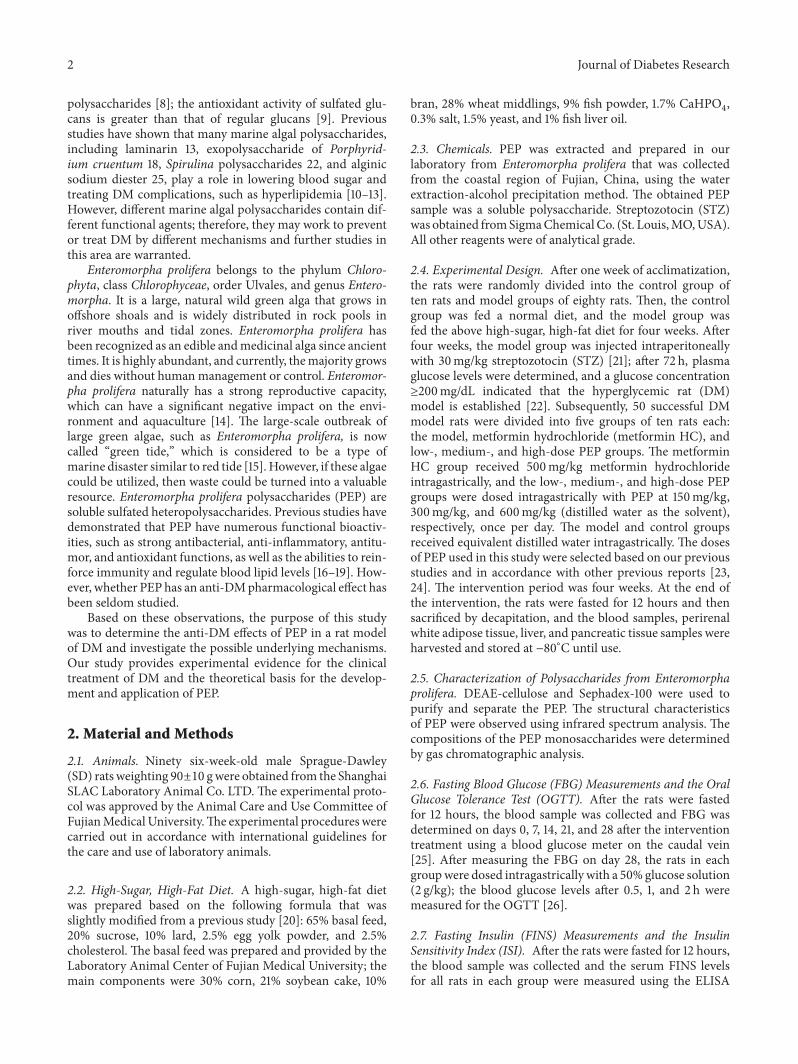
31 Characterization of Polysaccharides from Enteromorphaprolifera PEP was separated to four components PEP-1 PEP-2 PEP-3 and PEP-4 by DEAE-cellulose methods(Figure 1(a)) PEP2 is the major component at up to81 PEP-2 shows a single symmetric peak when it flowsthrough Sephadex-100 (Figure 1(b)) Infrared spectrum anal-ysis shows that PEP-2 is a sulfate ester-containing polysaccha-ride (Figure 1(c)) Gas chromatographic analysis shows thatthe monosaccharides of PEP-2 were rhamnose glucuronicacid arabinose fucose xylose and glucose (Figures 1(d1)and 1(d2)) The proportions of the monosaccharides are512 132 338 162 1 103 respectively
4 Journal of Diabetes Research
0
1
2
3
4
5
6
7
0
02
04
06
08
1
12
14
1 5 9 13 17 21 25 29 33 37 41 45 49
(mol
L)
A v
alue
Time (min)
PEP (A value) NaCl (molL)
PEP-1
PEP-2
PEP-3 PEP-4
(a) (b)
(c)
(d1) (d2)
0
01
02
03
04
05
06
07
08
1 5 9 13 17 21 25 29 33 37
A v
alue
Time (min)
PEP
PEP-2
98
96
94
92
90
88
86
84
82
80
78
76
Tran
smitt
ance
()
3000 2000 1500 1000 500
Wavenumber (cmminus1)
PEP-2
25 75 125 175 225 275 325 375minus050
000050100150200250300350400450500
005001000150020002500300035004000
Time (min)25 75 125 175 225 275 325 375
Time (min)
Rhamnose179Glucuronic acid183
Arabinose187Fucose214
Galacturonic acid229
Mannose285
Xylose290
Glucose293Galactose304
times104
(120583V
)
minus050
000050100150200250300350400450500times104
(120583V
)
(∘C)
005001000150020002500300035004000
(∘C)
Rhamnose179Glucuronic acid183
Arabinose187Fucose214 Xylose290
Glucose293
ChromatogramChromatogram
Figure 1 Characterization of polysaccharides from Enteromorpha prolifera (a) shows four peaks in the eluting curve PEP-1 PEP-2 PEP-3and PEP-4 The peak area of PEP-2 is the largest and contains 161mg of polysaccharides (b) shows the basic graph of PEP-2 which has asingle symmetrical elution peak (c) shows the wavenumbers of 338602 cmminus1 as the stretching vibration of OndashH 292384 cmminus1 285328 cmminus1and 145436 cmminus1 as the vibration of ndashCH2ndash The 139054 cmminus1 and 123213 cmminus1 symmetric stretching vibrations corresponded to two S=Ogroups of sulfate groups Furthermore 84809 cmminus1 and 76989 cmminus1 are the antisymmetric and symmetric stretching vibrations of the sulfategroups CndashO and SndashO (d1) shows that the chromatogram of different standard monosaccharides (d2) shows that PEP-2 is mainly composedof rhamnose glucuronic acid arabinose fucose xylose and glucose
Journal of Diabetes Research 5
Table 1 The effect of PEP on body weight food consumption and drinking water intake in DM rats
Group Body weight (g) Food consumption (gdaydam) Drinking water intake (mLdaydam)Control 51775 plusmn 2957lowast 1126 plusmn 205lowast 1521 plusmn 171lowast
Model 35912 plusmn 5769 985 plusmn 138 3418 plusmn 503
Metformin HC 35473 plusmn 5861 929 plusmn 171 3079 plusmn 583
150mgkg PEP 38220 plusmn 4036 937 plusmn 226 2896 plusmn 839
300mgkg PEP 37593 plusmn 4358 898 plusmn 163 2468 plusmn 529lowast
600mgkg PEP 35860 plusmn 3508 883 plusmn 169 1661 plusmn 282lowast
A DM model was established by feeding rats a high-fat diet and injecting them with a low dose of STZ After the model was established PEP (150 300 and600mgkg) was administered intragastrically for 28 d The body weight food consumption and drinking water intake were measured The data are presentedas the mean plusmn standard deviation (119899 = 10 rats) Compared with the model group lowast119875 lt 005 compared with the control group 119875 lt 005
Table 2 The effect of PEP on FBG levels in DM rats
Group FBG (mgdL)Day 0 Day 7 Day 14 Day 21 Day 28
Control 15534 plusmn 351lowast 17676 plusmn 3852lowast 16164 plusmn 2718lowast 16110 plusmn 2988lowast 13878 plusmn 2862lowast
Model 32148 plusmn 461 25848 plusmn 3582 24876 plusmn 3960 28368 plusmn 3798 25812 plusmn 5868
Metformin HC 32886 plusmn 1458lowast 14832 plusmn 1566lowast 15138 plusmn 1638lowast 15318 plusmn 1638lowast 15048 plusmn 1908lowastamp
150mgkg PEP 29736 plusmn 4086 18072 plusmn 1962lowast 16776 plusmn 4140lowast 19476 plusmn 3402lowast 16200 plusmn 3330lowastamp
300mgkg PEP 29142 plusmn 3654 15948 plusmn 2826lowast 15228 plusmn 3690lowast 17208 plusmn 3888lowast 14904 plusmn 3456lowastamp
600mgkg PEP 32508 plusmn 4464 17514 plusmn 2610lowast 16074 plusmn 2916lowast 17334 plusmn 3132lowast 15876 plusmn 3780lowastamp
A DM model was established by feeding rats a high-fat diet and injecting them with a low dose of STZ After the model was established PEP (150 300 and600mgkg) was administered intragastrically for 28 d Blood samples from the caudal vein were collected to determine FBG levels using a blood glucose meterThe data are presented as the mean plusmn standard deviation (119899 = 10 rats) Compared with the model group lowast119875 lt 005 compared with the control group 119875 lt005 compared with day 0 amp119875 lt 005
32The Effect of PEP on BodyWeight Food Consumption andDrinking Water Intake in DM Rats During the experimentgut dysfunction and death were not observed in DM ratsHowever body weight and daily food consumption weresignificantly lower in the model group compared with thecontrol group (119875 lt 005) On the contrary the daily drinkingwater intake was significantly higher than that in the controlgroup (119875 lt 005) Compared with the model group drinkingwater intakes were lower in the medium- and high-dose PEPgroups (119875 lt 005) Body weight and food consumption werenot significantly different between the low- medium- andhigh-dose PEP groups the metformin HC group and themodel group (119875 gt 005) (Table 1)
33 The Effects of PEP on FBG and OGTT in DM RatsBefore the intragastric treatment (on day 0) the FBG wassignificantly higher in the model metformin HC and PEPgroups than in the control group (119875 lt 005) One weekafter the intragastric intervention the FBG levels decreasedin all groups however from weeks 1 through 4 rats inthe model group had a higher FBG than rats in the othergroups After four weeks of intragastric treatment the FBGwas lower in the PEP and metformin HC groups than inthe model group (119875 lt 005) Compared with day 0 theFBG levels in the low- medium- and high-dose PEP groupsand in the metformin HC group decreased (119875 lt 005)(Table 2) The OGTT indicated that the blood glucose levelsin all the rats peaked 30min after glucose injection andessentially recovered within 2 h Compared with the model
0
50
100
150
200
250
300
350
400
0 05 1 2
Bloo
d gl
ucos
e (m
gdL
)
Time (h)
ModelHPEPMPEP
LPEPMel HCControl
Figure 2The effect of PEP on OGTT in DM rats A DMmodel wasestablished by feeding rats a high-fat diet and injecting them with alow dose of STZ After the model was established PEP (0 150 300and 600mgkg) was administered intragastrically as an interventionfor 28 d (119899 = 10 rats) Subsequently the OGTT was performedControl control group Model model group Mel HC metforminHC group LPEP low-dose PEP group MPEP medium-dose PEPgroup HPEP high-dose PEP group
group the blood glucose levels recovered faster in the PEPand metformin HC groups but the differences between thePEP groups were not significant (Figure 2)
6 Journal of Diabetes Research
(a) Control (b) Model (c) Metformin HC
(d) 150mgkg (e) 300mgkg (f) 600mgkg
Figure 3 The effect of PEP on the histopathological structure of the pancreas in DM rats (HampE staining) A DM model was established byfeeding rats a high-fat diet and injecting them with a low dose of STZ After the model was established PEP (0 150 300 and 600mgkg)was administered intragastrically as an intervention for 28 d (119899 = 10 rats) The pancreatic tissues were collected embedded sectioned andstained with HampE (a) control control group (b) model model group (c) metformin HC metformin HC group (d) 150mgkg low-dosePEP group (e) 300mgkg medium-dose PEP group (f) 600mgkg high-dose PEP group
Table 3 The effect of PEP on FINS levels and the ISI in DM rats
Group FINS (mUL) ISIControl 327 plusmn 020lowast minus305 plusmn 030lowast
Model 363 plusmn 025 minus387 plusmn 032
Metformin HC 339 plusmn 027lowast minus292 plusmn 013lowast
150mgkg PEP 355 plusmn 029 minus333 plusmn 032lowast
300mgkg PEP 359 plusmn 015 minus318 plusmn 031lowast
600mgkg PEP 339 plusmn 023lowast minus321 plusmn 044lowast
A DM model was established by feeding rats a high-fat diet and injectingthem with a low dose of STZ After the model was established PEP (150300 and 600mgkg) was administered intragastrically as an intervention for28 d Blood samples from angular vein were collected to measure the FBGand FINS levels with the ELISA kit and the ISI was calculated The data arepresented as the mean plusmn standard deviation (119899 = 10 rats) Compared withthe model group lowast119875 lt 005 compared with the control group 119875 lt 005
34 The Effect of PEP on Serum FINS Levels and the ISI inDM Rats At the end of the experiment FINS levels werehigher and the ISI was significantly lower in the model groupcompared with the control group (119875 lt 005) Compared withthemodel group FINS levels were lower in the high-dose PEPand the metformin HC groups (119875 lt 005) The ISI was lowerin the low- medium- and high-dose PEP andmetforminHCgroups than in the model group (119875 lt 005) (Table 3)
35TheEffect of PEP on SerumMDALevels and the EnzymaticActivity of SOD andGSH-Px in DMRats Comparedwith the
control group the MDA levels were higher and the GSH-Pxactivity was lower in the model group (119875 lt 005) althoughthere were no significant differences in SOD enzymaticactivity (119875 gt 005) it exhibited a decreasing trend Comparedwith the model group the MDA levels in the low- medium-and high-dose PEP and metformin HC groups were lower(119875 lt 005)TheSODenzymatic activity in the low-medium-and high-dose PEP groups increased The GSH-Px activityincreased in themedium- and high-dose PEP andmetforminHC groups (Table 4)
36 The Effect of PEP on the Histopathological Changes inthe Pancreas in DM Rats Light microscopy following HampEstaining indicated that the pancreatic acini and islets wereintact in the control group In themodel group the islets werescattered the islet volume was significantly decreased mostislet cells were atrophic and more lightly stained there werefewer cells the cytoplasm was significantly decreased thenuclei exhibited large-scale condensation and lymphocyticfiltration was extensive around and invading into the isletsIn themetforminHC group themorphological changes werenot as severe as those in the model group In the medium-and high-dose PEP groups the number of islets and 120573-cellswas significantly increased the morphology and structurewere essentially normal the cytoplasm was homogenous andin greater quantity the nucleoli were clear and occasionallya small amount of lymphocytic infiltration was observed(Figure 3)
Journal of Diabetes Research 7
Table 4 The effect of PEP on MDA SOD and GSH-Px in DM rats
Group MDA (nmolmL) SOD (UmL) GSH-Px (120583molL)Control 509 plusmn 063lowast 25268 plusmn 2492lowast 480000 plusmn 44946lowast
Model 594 plusmn 089 24345 plusmn 1932 368000 plusmn 38865
Metformin HC 505 plusmn 051lowast 23990 plusmn 2809 416457 plusmn 58916lowast
150mgkg PEP 503 plusmn 045lowast 27131 plusmn 2337lowast 397943 plusmn 39713
300mgkg PEP 425 plusmn 067lowast 27263 plusmn 2225lowast 413257 plusmn 48374lowast
600mgkg PEP 456 plusmn 096lowast 26909 plusmn 2605lowast 430400 plusmn 34172lowast
A DM model was established by feeding rats a high-fat diet and injecting them with a low dose of STZ After the model was established PEP (150 300 and600mgkg) was administered intragastrically as an intervention for 28 d Blood samples from angular vein were collected to quantitate the enzymatic activityof MDA (nmolmL) SOD (UmL) and GSH-Px (120583molL) in serum The data are presented as the mean plusmn standard deviation (119899 = 10 rats) Compared withthe model group lowast119875 lt 005 compared with the control group 119875 lt 005
The immunohistochemistry results indicated that in thepancreatic islets the positively stained cells containing cyto-plasmic bright red granules were120573-cells In the control groupthe islets were predominantly round or oval with differentsizes defined edges and intact morphology the islets werefull of copious bright red granules and the staining was darkIn the model group the islet structure was damaged theedges were irregular the islet area was decreased there weresignificantly fewer bright red granules and the staining waslight Compared with the model group islets with darkerstaining were present in the metformin HC group and therewere significantly more bright red granules similar resultswere observed for the low- medium- and high-dose PEPgroups (Figure 4(a)) The mean integrated optical density of120573-cells in islets was significantly lower in the model groupcompared with the control group (119875 lt 005) Comparedwith the model group the mean integrated optical densitiesof 120573-cells were significant increased in the low- medium-and high-dose groups (119875 lt 005) However the differencesbetween the model group andmetforminHC group were notsignificant (Figure 4(b))
37 The Effect of PEP on the mRNA Expression of GCK andInsR in Liver Tissue and of APN and GLUT-4 in AdiposeTissue in DM Rats Compared with the model group InsRmRNA expression in the liver in the low- medium- andhigh-dose PEP groups significantly increased (119875 lt 005) andGCK mRNA expression in the liver in the low- medium-and high-dose PEP and the metformin HC groups increased(119875 lt 005) APN mRNA expression in adipose tissue washigher in the high-dose PEP group than in the model group(119875 lt 005) GLUT-4 mRNA expression in adipose tissue inthe low- medium- and high-dose PEP and the metforminHC groups was not significantly different from that in themodel group (119875 gt 005) but GLUT-4 expression exhibited anincreasing trend wherein the expression level increased withincreasing PEP concentration (Figure 5)
4 Discussion
The etiology of DM is complex It is primarily consideredto result from a combination of genetic factors and variousenvironmental factors [31] There is no drug or method forthe prevention or treatment of DM that has a significant
therapeutic effect few toxic side effects and an acceptablesafety profile [32ndash34] Therefore studying nutritious com-pounds that have minimal toxic side effects and can havelong-term use is important for the prevention and treatmentof DM
Due to the different adsorption capacities of polysac-charides by the DEAE cellulose method and the differentretention capabilities of polysaccharides by the Sephadex-100gelmethod we separated and highly purified polysaccharidesfrom Enteromorpha prolifera By infrared spectrum analysisthe main components of the polysaccharides were carbohy-drates which not only are rich in sulfate esters but also con-tain ether compounds and unsaturated carbon chains whichis in accord with previous studies [35] The sulfate contentof polysaccharides from Enteromorpha prolifera was 192 inour study whichwas higher than that of previous studies [35]Gas chromatographic analysis showed that the monosac-charides of PEP were rhamnose glucuronic acid arabinosefucose xylose and glucose which is inconsistent with a pre-vious study in which galactose and mannose were observed[35] The discrepancies in these results may be due to differ-ences in the species origin and season of Enteromorpha
High blood glucose levels hyperinsulinemia (in the earlystages) and low insulin sensitivity are the salient features ofDM Impaired glucose tolerance (IGT) occurs in the middleof the transition from normal to DM and is a primary featureof glucose metabolic disorders The mechanism by whichIGT occurs may be associated with IR and impaired islet120573-cell function [36] The rapid changes in glucose toleranceandor blood sugar levels are prone to induce DM thereforeOGTTs and blood glucose levels can be utilized to monitorand control the occurrence anddevelopment ofDM[37]Thisstudy confirmed that PEP reduced blood glucose levels inDM rats and significantly improved IGT with similar effectsas the hypoglycemic agent metformin HC in particularprominent effects were observed in the medium-dose PEPgroup Serum insulin levels were significantly reduced in thehigh-dose PEP group and insulin sensitivity was significantlyhigher in all the PEP intervention groups Furthermore PEPintervention alleviated the diabetes symptom of drinkingmore water without changing caloric intake and inducinggut dysfunction for food consumption and body weighthad no changed These results indicated that PEP affectedthe regulation of glucose metabolism by improving IGT
8 Journal of Diabetes Research
(A) Control (B) Model (C) Metformin HC
(D) 150mgkg (E) 300mgkg (F) 600mgkg
(a)
0
005
01
015
02
025
03
Control Model Metformin
Inte
grat
ed o
ptic
al d
ensit
yar
ea
PEP
lowastlowast
lowast lowastlowast
150mgkg
300mgkg
600mgkg
(b)
Figure 4 The effect of PEP on the histopathological structure of the pancreas in DM rats (IHC) A DM model was established by feedingrats a high-fat diet and injecting them with a low dose of STZ After the model was established PEP (0 150 300 and 600mgkg) wasadministered intragastrically as an intervention for 28 d (119899 = 10 rats) The pancreatic tissues were collected embedded and sectioned anda specific antibody against insulinproinsulin was utilized to observe the residual 120573-cells in islets by using immunohistochemical method(IHC) (A) control control group (B) model model group (C) metformin HC metformin HC group (D) 150mgkg low-dose PEP group(E) 300mgkgmedium-dose PEP group (F) 600mgkg high-dose PEP groupThe arrow indicates islet tissue (a) shows the histopathologicalstructure of the pancreas in DM rats by IHC (b) shows the mean integrated optical density of 120573-cells in islets in DM rats using the Image-ProPlus 45 image analysis software
promoting insulin secretion by islet 120573-cells and enhancinginsulin sensitivity
High glucose can cause the nonenzymatic oxidation ofproteins and induce oxidative stress [38] Chronic exposureto high glucose levels increases the oxidation of glucoseitself which produces enediol and dihydroxy compoundsas well as a significant amount of ROS [39] Therefore inthe model used in the study the high-glucose high-fat dietthat was fed to the rats stimulated oxidative stress moreover
in combination with the STZ injection that damaged thepancreas the oxidative damage in the pancreas as well as liverwas exacerbated resulting in DM SOD is a key intracellularantioxidant that removes superoxide anion radicals in thebody [40] GSH-Px is a scavenger that protects the structureand functional integrity of the cell membrane by reducinghydrogen peroxide and lipid hydroperoxides [41] MDAlevels reflect the degree of lipid peroxidation in the bodyand indirectly reflect the degree of cellular damage [42]
Journal of Diabetes Research 9
0030609121518
InsR GCK
Relat
ive m
RNA
leve
l
ControlModelMetformin HC
lowast lowast lowast
lowast
lowastlowast
0030609121518
GLUT-4 APN
Rela
tive m
RNA
leve
l
lowast
lowast
lowast
150mgkg PEP300mgkg PEP600mgkg PEP
ControlModelMetformin HC
150mgkg PEP300mgkg PEP600mgkg PEP
Figure 5 The effect of PEP on the mRNA expression of GCK and InsR in the liver and APN and GLUT-4 in adipose tissue in DM rats ADMmodel was established by feeding rats a high-fat diet and injecting them with a low dose of STZ After the model was established PEP (0150 300 and 600mgkg) was administered intragastrically as an intervention for 28 d Subsequently liver and adipose tissue was collectedmRNAwas extracted reverse transcribed into cDNA and analyzed by real-time PCR to determine the mRNA expression levels in the rats ineach groupThemRNA expression levels were determined using the relative quantificationmethod (120573-actin was used as the internal control)The data are presented as the mean plusmn standard deviation (119899 = 10 rats) Compared with the control group lowast119875 lt 005 compared with themodel group 119875 lt 005
In this study compared with the control group MDA levelsincreased GSH-Px activity decreased and SOD activityexhibited a decreasing trend in the model group indicatingthat there was more severe oxidative damage in the DMrats potentially attributable to the oxidative stress inducedby the high-sugar high-fat diet and STZ After four weeksof PEP intervention MDA levels significantly decreased andSOD and GSH-Px activities increased in a dose-dependentmanner in all the PEP treatment groups compared with themodel groupThese results indicated that PEP inhibited lipidperoxidation in the cell membrane relieved cellular oxidativedamage and promoted the functional recovery of tissues byenhancing antioxidant activity and promoting the clearanceof free radicals and peroxidation products such as MDA
The pathological examination of the pancreas revealedthat the number of pancreatic islet cells in the rats in thehigh- and medium-dose PEP groups significantly increasedcompared with the model group whereas low-dose PEPhad a slightly weaker effect The cellular morphology of thepancreatic islet cells was nearly normal in the rats in thehigh- and medium-dose PEP groups This result confirmedthat PEP facilitated the repair of tissue damage and exerted aprotective function in the pancreas
It is currently understood that insulin acts by interactingwith the InsR on the cell membrane (predominantly in hepa-tocytes adipocytes and muscle cells) and that its functionsrely on receptor density and affinity in the cell membraneTherefore InsR is the first key point of action for insulinand the InsR gene is critical for studies of IR [43] Our studydemonstrated that mRNA level of InsR was significantlyhigher in the low- medium- and high-dose PEP groups thanthat in the model group this result was consistent with themajority of previous studies that have reported a reduction inboth the number and affinity for insulin of membrane InsRin liver and muscle tissue of DM rats with IR [44] The bloodsugar and insulin levels in the model group remained highindicating that PEP reversed IR by upregulating the number
of InsR Further studies are needed to elucidate the precisemechanisms
GCK is a key enzyme in the regulation of glucosemetabolism and insulin secretion and is the first rate-limitingenzyme in glycolysis It catalyzes the phosphorylation ofglucose to glucose-6-phosphate and this process is a pre-requisite for liver glycogen synthesis The functional loss ordecreased expression of GCK suppresses glucose utilizationin the liver and decreases insulin secretion from islet 120573-cells Studies have reported that GCK is critical for beta cellhyperplasia under the condition of high-fat diet-induced IR[45] Therefore mutations in the GCK gene may be a causeof DM Conversely enhancing GCK gene expression mayhave a therapeutic effect on DM Our study demonstratedthat significantly higher GCKmRNA expression levels in thelivers of rats in the low-medium- and high-dose PEP groupscompared with those in the model group the difference ofGCK mRNA expression levels between the high-dose groupand the metformin HC group was most dramatic Previousstudy showed that dietary phenolic compounds altered glu-cose metabolism and decreased the risk of type 2 diabetes byincreasing the level of glucokinase mRNA [46] Therefore itis possible that PEP improved glucosemetabolism inDM ratsby upregulating GCK mRNA expression
GLUT-4 is a glycoprotein of 509 amino acids thatis highly expressed in insulin-sensitive tissues includingmuscle adipose and liver These tissues also have highlevels of glucose uptake and utilization these processes areregulated by blood glucose and insulin [47] Transmembraneglucose transport is mediated by GLUT-4 Under restingconditions 90 of GLUT-4 molecules are distributed on theinner cell membrane and only a small portion is on theouter cell membrane When the insulin stimulation signal istransmitted into cells GLUT-4 translocates from the inner tothe outer membrane therefore increasing glucose transport[48] When GLUT-4 function is impaired glucose uptake aswell as utilization in peripheral tissues decreases particularly
10 Journal of Diabetes Research
in skeletal muscle and adipose tissue leading to postreceptorIR Therefore GLUT-4 dysfunction is a major contributor tothe onset of DM [49] This study determined that GLUT-4 mRNA expression in adipose tissue decreased in the ratmodels despite the fact that the differences between theintervention groups and the model group were not signifi-cant PEP intervention induced GLUT-4 mRNA expressionto various extents Previous study showed that the alterationsin GLUT-4 mRNA level in adipose tissue contributed to thephysiological activities of n-3 PUFA in preventing body fataccumulation and in regulating glucose metabolism in rats[50] Therefore it is possible that PEP improved glucosemetabolism by upregulating the GLUT-4 mRNA expression
APN also termed Acrip30AMP1GBP28 is a cytokinethat is secreted by adipocytes APN is composed of 247amino acids the APN gene is located on 3q27 and encodessusceptibility genes for DM and metabolic syndrome [51]APN is a protective factor for IR and therefore it is highlycorrelated with insulin sensitivity and plays an importantrole in IR Similar to insulin APN promotes glucose and fatutilization and energy consumption in muscle and inhibitsgluconeogenesis and the synthesis of fatty acids and choles-terol in the liver [52] APN is predominantly expressed inadipocytes and the APN gene is only expressed in whiteadipose tissue therefore changes in adipocyte number canaffect APN expression It has been demonstrated in animalmodels that injecting APN into lipoatrophic mice partiallyreversed IR [53] and that the ability to control blood sugar lev-els with insulin in transgenic rats with high APN expressionsignificantly increased [54] In humans the direct injectionof APN reduces blood sugar levels [55] and increased APNincreases insulin sensitivity in obese DM and IR patientsTherefore adipocytokines such as APN reach target organsvia various pathways to induce IR-related diseases andthe downregulation of related factors might prevent IR byimproving insulin sensitivity In this study we determinedthat the difference in APN gene expression between the high-dose PEP group and themodel groupwas significant whereasAPN gene expression was higher in the low- and medium-dose PEP groups than in the model group but did not reachsignificance These results indicated that PEP improved IRand enhanced insulin sensitivity by upregulating APN geneexpression in rat white adipose tissue and that it therefore haspotential for the prevention of DM
Previous study had showed that Enteromorpha compressaand polysaccharide fraction from Enteromorpha proliferaexhibit potent antioxidant activity [17 18] These studiesindicate that PEP might directly exhibit antioxidant activityto alleviate oxidative damage in the pancreas and finallyimprove glucose metabolism However PEP might improveglucose metabolism by upregulating the InsR GCK APNand GLUT-4 mRNA expression in our present study whichmay alleviate oxidative damage for high glucose ultimatelyresult in nonenzymatic oxidation of proteins and induceoxidative stress [38] Therefore in our present study it isunclear whether the improvements in glucose drive theimprovements in oxidative measures or vice versa Furtherwork especially in vitro study in this area is warranted
5 Conclusion
Our results indicate that PEP can improve glucose meta-bolism in DM ratsThemechanismmay relate to the antioxi-dant activity of PEP and its ability to regulate the mRNA levelof InsR GCK APN and GLUT-4 gene in liver and adiposetissue These findings may suggest that PEP could be usefulfor the therapy of diabetes mellitus
Abbreviations
PEP Polysaccharides from Enteromorpha proliferaDM Diabetes mellitusSTZ StreptozotocinFBG Fasting blood glucoseINS InsulinISI Insulin sensitivity indexGCK GlucokinaseInsR Insulin receptorGLUT-4 Glucose transporter type 4APN AdiponectinIR Insulin resistanceSOD Superoxide dismutaseGSH-Px Glutathione peroxidaseMDA MalondialdehydeRT-PCR Reverse transcription polymerase chain reactionOGTT Oral glucose tolerance testROS Reactive oxygen species
Conflict of Interests
All authors declared no conflict of interests
Acknowledgments
This project was supported by the Natural Science Founda-tion of Fujian Province (2015J01306) and the National SparkProgram (2011GA720014) China
References
[1] S Di Yacovo C Garcia-Vidal D Viasus et al ldquoClinical featuresetiology and outcomes of community-acquired pneumonia inpatients with diabetes mellitusrdquoMedicine vol 92 no 1 pp 42ndash50 2013
[2] V N Titov ldquoPhylogenesis etiology and pathogenesis of insulinresistance Differences from type II diabetes mellitusrdquo VestnikRossiiskoi Akademii Meditsinskikh Nauk no 4 pp 65ndash73 2012
[3] K Stankov D Benc and D Draskovic ldquoGenetic and epigeneticfactors in etiology of diabetes mellitus type 1rdquo Pediatrics vol132 no 6 pp 1112ndash1122 2013
[4] L Guo Y Cheng X Wang et al ldquoAssociation betweenmicroalbuminuria and cardiovascular disease in type 2 diabetesmellitus of the Beijing Han nationalityrdquo Acta Diabetologica vol49 supplement 1 pp S65ndashS71 2012
[5] V A Sharpatyi ldquoRadiochemistry of polysaccharides (review)rdquoRadiatsionnaia Biologiia Radioecologiia vol 39 no 1 pp 156ndash161 1999
[6] H Itoh H Noda H Amano C Zhuaug T Mizuno and H ItoldquoAntitumor activity and immunological properties of marine
Journal of Diabetes Research 11
algal polysaccharides especially fucoidan prepared from Sar-gassum thunbergii of phaeophyceaerdquo Anticancer Research vol13 no 6 pp 2045ndash2052 1993
[7] A J Mann R L Hahnke S Huang et al ldquoThe genome ofthe alga-associated marine flavobacterium Formosa agariphilaKMM 3901T reveals a broad potential for degradation of algalpolysaccharidesrdquo Applied and Environmental Microbiology vol79 no 21 pp 6813ndash6822 2013
[8] D-HNgo and S-K Kim ldquoSulfated polysaccharides as bioactiveagents from marine algaerdquo International Journal of BiologicalMacromolecules vol 62 pp 70ndash75 2013
[9] E Tsiapali SWhaley J Kalbfleisch H E Ensley IW Browderand D L Williams ldquoGlucans exhibit weak antioxidant activitybut stimulate macrophage free radical activityrdquo Free RadicalBiology and Medicine vol 30 no 4 pp 393ndash402 2001
[10] D-Q Jin G Li J-S Kim C-S Yong J-A Kim and K HuhldquoPreventive effects ofLaminaria japonica aqueous extract on theoxidative stress and xanthine oxidase activity in streptozotocin-induced diabetic rat liverrdquo Biological and Pharmaceutical Bul-letin vol 27 no 7 pp 1037ndash1040 2004
[11] B D Dumelod R P B Ramirez C L P Tiangson E B Barriosand L N Panlasigui ldquoCarbohydrate availability of arroz caldowith 120582-carrageenanrdquo International Journal of Food Sciences andNutrition vol 50 no 4 pp 283ndash289 1999
[12] P Parikh U Mani and U Iyer ldquoRole of Spirulina in the controlof glycemia and lipidemia in type 2 diabetes mellitusrdquo Journalof Medicinal Food vol 4 no 4 pp 193ndash199 2001
[13] D Zhang I Fujii C Lin et al ldquoThe stimulatory activitiesof polysaccharide compounds derived from algae extracts oninsulin secretion in vitrordquo Biological and Pharmaceutical Bul-letin vol 31 no 5 pp 921ndash924 2008
[14] A Lin S Shen J Wang and B Yan ldquoReproduction diversityof Enteromorpha proliferardquo Journal of Integrative Plant Biologyvol 50 no 5 pp 622ndash629 2008
[15] J Blomster S Back D P Fewer et al ldquoNovel morphologyin Enteromorpha (Ulvophyceae) forming green tidesrdquo TheAmerican Journal of Botany vol 89 no 11 pp 1756ndash1763 2002
[16] S Celikler S Tas O Vatan S Ziyanok-Ayvalik G Yildiz andR Bilaloglu ldquoAnti-hyperglycemic and antigenotoxic potentialof Ulva rigida ethanolic extract in the experimental diabetesmellitusrdquo Food and Chemical Toxicology vol 47 no 8 pp 1837ndash1840 2009
[17] S M M Shanab E A Shalaby and E A El-FayoumyldquoEnteromorpha compressa exhibits potent antioxidant activityrdquoJournal of Biomedicine and Biotechnology vol 2011 Article ID726405 11 pages 2011
[18] Z Tang H Gao S Wang S Wen and S Qin ldquoHypolipi-demic and antioxidant properties of a polysaccharide fractionfromEnteromorpha proliferardquo International Journal of BiologicalMacromolecules vol 58 pp 186ndash189 2013
[19] H-B Cho H-H Lee O-H Lee H-S Choi J-S Choi andB-Y Lee ldquoClinical and microbial evaluation of the effects ongingivitis of a mouth rinse containing an enteromorpha linzaextractrdquo Journal ofMedicinal Food vol 14 no 12 pp 1670ndash16762011
[20] L E Kuo M Czarnecka J B Kitlinska J U Tilan RKvetnansky and Z Zukowska ldquoChronic stress combinedwith a high-fathigh-sugar diet shifts sympathetic signalingtoward neuropeptide Y and leads to obesity and the metabolicsyndromerdquo Annals of the New York Academy of Sciences vol1148 pp 232ndash237 2008
[21] X Xiang Z Wang Y Zhu L Bian and Y Yang ldquoDosage ofstreptozotocin in inducing rat model of type 2 diabetes mel-litusrdquo Wei Sheng Yan Jiu vol 39 no 2 pp 138ndash142 2010(Chinese)
[22] R Li T Liang L Xu Y Li S Zhang and X Duan ldquoProtectiveeffect of cinnamon polyphenols against STZ-diabetic mice fedhigh-sugar high-fat diet and its underlying mechanismrdquo Foodand Chemical Toxicology vol 51 no 1 pp 419ndash425 2013
[23] J Wei S Wang G Liu et al ldquoPolysaccharides from Entero-morpha prolifera enhance the immunity of normal micerdquoInternational Journal of Biological Macromolecules vol 64 pp1ndash5 2014
[24] W T Lin and D D Liao ldquoEffects of enteromorpharsquos polysac-charide on lipid metabolism and adiponectin gene expressionin type 2 diabetic ratsrdquo Acta Nutrimenta Sinica vol 35 pp 181ndash185 2013 (Chinese)
[25] G S Dhatt M M Agarwal Y Othman and S C NairldquoPerformance of the roche accu-chek active glucose meter toscreen for gestational diabetes mellitus using fasting capillarybloodrdquoDiabetes Technology andTherapeutics vol 13 no 12 pp1229ndash1233 2011
[26] J Codina M Vall and E Herrera ldquoComparative changes withage of the fasting response in circulating ketone bodies glucoseand insulin and oral glucose tolerance test in the ratrdquo Com-parative Biochemistry and Physiology Part A Physiology vol71 no 2 pp 231ndash236 1982
[27] MGutt C L Davis S B Spitzer et al ldquoValidation of the insulinsensitivity index (ISI0120) comparison with other measuresrdquoDiabetes Research and Clinical Practice vol 47 no 3 pp 177ndash184 2000
[28] J A Buege and S D Aust ldquoMicrosomal lipid peroxidationrdquoMethods in Enzymology vol 52 pp 302ndash310 1978
[29] H PMisra and I Fridovich ldquoThe role of superoxide anion in theautoxidation of epinephrine and a simple assay for superoxidedismutaserdquoThe Journal of Biological Chemistry vol 247 no 10pp 3170ndash3175 1972
[30] J T Rotruck A L Pope H E Ganther A B Swanson D GHafeman and W G Hoekstra ldquoSelenium biochemical role asa component of glutathione peroxidaserdquo Science vol 179 no4073 pp 588ndash590 1973
[31] D Asbury and A E Kalderon ldquoEtiology of insulin-dependentdiabetes mellitus a review of the current literaturerdquoThe Journalof the Arkansas Medical Society vol 79 no 8 pp 281ndash285 1983
[32] C Zou and H Hu ldquoUse of pioglitazone in the treatment ofdiabetes effect on cardiovascular riskrdquoVascular Health and RiskManagement vol 9 no 1 pp 429ndash433 2013
[33] S A Stein E M Lamos and S N Davis ldquoA review of theefficacy and safety of oral antidiabetic drugsrdquo Expert Opinion onDrug Safety vol 12 no 2 pp 153ndash175 2013
[34] M S Islam and R D Wilson ldquoExperimentally induced rodentmodels of type 2 diabetesrdquo Methods in Molecular Biology vol933 pp 161ndash174 2012
[35] B Li S Liu R Xing et al ldquoDegradation of sulfated polysac-charides from Enteromorpha prolifera and their antioxidantactivitiesrdquo Carbohydrate Polymers vol 92 no 2 pp 1991ndash19962013
[36] A Kazemnejad Z Batvandi and J Faradmal ldquoComparisonof artificial neural network and binary logistic regression fordetermination of impaired glucose tolerancediabetesrdquo EasternMediterranean Health Journal vol 16 no 6 pp 615ndash620 2010
12 Journal of Diabetes Research
[37] E Bartoli G P Fra and G P C Schianca ldquoThe oral glucosetolerance test (OGTT) revisitedrdquo European Journal of InternalMedicine vol 22 no 1 pp 8ndash12 2011
[38] T Nishikawa D Edelstein X L Du et al ldquoNormalizingmitochondrial superoxide production blocks three pathways ofhyperglycaemic damagerdquo Nature vol 404 no 6779 pp 787ndash790 2000
[39] T J Guzik S Mussa D Gastaldi et al ldquoMechanisms ofincreased vascular superoxide production in human diabetesmellitus role of NAD(P)H oxidase and endothelial nitric oxidesynthaserdquo Circulation vol 105 no 14 pp 1656ndash1662 2002
[40] H Yamamoto P Zhao and K Inoue ldquoOrigin of two isoprenoidunits in a lavandulyl moiety of sophoraflavanone G fromSophora flavescens cultured cellsrdquo Phytochemistry vol 60 no3 pp 263ndash267 2002
[41] J Q Wu T R Kosten and X Y Zhang ldquoFree radicals antiox-idant defense systems and schizophreniardquo Progress in Neuro-Psychopharmacology and Biological Psychiatry vol 46 pp200ndash206 2013
[42] D C Wallace ldquoAnimal models for mitochondrial diseaserdquoMethods in Molecular Biology vol 197 pp 3ndash54 2002
[43] R Mioni B Mozzanega M Granzotto et al ldquoInsulin receptorand glucose transporters mRNA expression throughout themenstrual cycle in human endometrium a physiological andcyclical condition of tissue insulin resistancerdquo GynecologicalEndocrinology vol 28 no 12 pp 1014ndash1018 2012
[44] G Sesti M Federici D Lauro P Sbraccia and R LauroldquoMolecular mechanism of insulin resistance in type 2 diabetesmellitus role of the insulin receptor variant formsrdquo DiabetesMetabolism Research and Reviews vol 17 no 5 pp 363ndash3732001
[45] Y Terauchi I Takamoto N Kubota et al ldquoGlucokinase andIRS-2 are required for compensatory 120573 cell hyperplasia inresponse to high-fat diet-induced insulin resistancerdquo Journal ofClinical Investigation vol 117 no 1 pp 246ndash257 2007
[46] K Valentova N T Truong A Moncion I de Waziers andJ Ulrichova ldquoInduction of glucokinase mRNA by dietaryphenolic compounds in rat liver cells in vitrordquo Journal of Agri-cultural and Food Chemistry vol 55 no 19 pp 7726ndash7731 2007
[47] B J Morgan S Y Chai and A L Albiston ldquoGLUT4 associatedproteins as therapeutic targets for diabetesrdquo Recent Patents onEndocrine Metabolic amp Immune Drug Discovery vol 5 no 1pp 25ndash32 2011
[48] J Stockli D J Fazakerley and D E James ldquoGLUT4 exocytosisrdquoJournal of Cell Science vol 124 no 24 pp 4147ndash4159 2011
[49] K Kurokawa and Y Oka ldquoInsulin resistance and glucosetransporterrdquo Nihon Rinsho vol 58 pp 310ndash314 2000
[50] Y Takahashi and T Ide ldquoDietary n-3 fatty acids affect mRNAlevel of brown adipose tissue uncoupling protein 1 and whiteadipose tissue leptin and glucose transporter 4 in the ratrdquoBritishJournal of Nutrition vol 84 no 2 pp 175ndash184 2000
[51] A B Goldfine and C R Kahn ldquoAdiponectin linking the fat cellto insulin sensitivityrdquo The Lancet vol 362 no 9394 pp 1431ndash1432 2003
[52] A Haynes A Frederick and A Chirkoff ldquoAPN plan improvesoutcome for pregnant patient with congenital heart diseaserdquoAACN Advanced Critical Care vol 23 no 2 pp 142ndash154 2012
[53] H Su W B Lau and X-L Ma ldquoHypoadiponectinaemia indiabetes mellitus type 2 molecular mechanisms and clinicalsignificancerdquo Clinical and Experimental Pharmacology andPhysiology vol 38 no 12 pp 897ndash904 2011
[54] M C Jong P J Voshol M Muurling et al ldquoProtection fromobesity and insulin resistance in mice overexpressing humanapolipoproteinC1rdquoDiabetes vol 50 no 12 pp 2779ndash2785 2001
[55] T P Combs A H Berg S Obici P E Scherer and L RossettildquoEndogenous glucose production is inhibited by the adipose-derived protein Acrp30rdquo The Journal of Clinical Investigationvol 108 no 12 pp 1875ndash1881 2001
Submit your manuscripts athttpwwwhindawicom
Stem CellsInternational
Hindawi Publishing Corporationhttpwwwhindawicom Volume 2014
Hindawi Publishing Corporationhttpwwwhindawicom Volume 2014
MEDIATORSINFLAMMATION
of
Hindawi Publishing Corporationhttpwwwhindawicom Volume 2014
Behavioural Neurology
EndocrinologyInternational Journal of
Hindawi Publishing Corporationhttpwwwhindawicom Volume 2014
Hindawi Publishing Corporationhttpwwwhindawicom Volume 2014
Disease Markers
Hindawi Publishing Corporationhttpwwwhindawicom Volume 2014
BioMed Research International
OncologyJournal of
Hindawi Publishing Corporationhttpwwwhindawicom Volume 2014
Hindawi Publishing Corporationhttpwwwhindawicom Volume 2014
Oxidative Medicine and Cellular Longevity
Hindawi Publishing Corporationhttpwwwhindawicom Volume 2014
PPAR Research
The Scientific World JournalHindawi Publishing Corporation httpwwwhindawicom Volume 2014
Immunology ResearchHindawi Publishing Corporationhttpwwwhindawicom Volume 2014
Journal of
ObesityJournal of
Hindawi Publishing Corporationhttpwwwhindawicom Volume 2014
Hindawi Publishing Corporationhttpwwwhindawicom Volume 2014
Computational and Mathematical Methods in Medicine
OphthalmologyJournal of
Hindawi Publishing Corporationhttpwwwhindawicom Volume 2014
Diabetes ResearchJournal of
Hindawi Publishing Corporationhttpwwwhindawicom Volume 2014
Hindawi Publishing Corporationhttpwwwhindawicom Volume 2014
Research and TreatmentAIDS
Hindawi Publishing Corporationhttpwwwhindawicom Volume 2014
Gastroenterology Research and Practice
Hindawi Publishing Corporationhttpwwwhindawicom Volume 2014
Parkinsonrsquos Disease
Evidence-Based Complementary and Alternative Medicine
Volume 2014Hindawi Publishing Corporationhttpwwwhindawicom
2 Journal of Diabetes Research
polysaccharides [8] the antioxidant activity of sulfated glu-cans is greater than that of regular glucans [9] Previousstudies have shown that many marine algal polysaccharidesincluding laminarin 13 exopolysaccharide of Porphyrid-ium cruentum 18 Spirulina polysaccharides 22 and alginicsodium diester 25 play a role in lowering blood sugar andtreating DM complications such as hyperlipidemia [10ndash13]However different marine algal polysaccharides contain dif-ferent functional agents therefore they may work to preventor treat DM by different mechanisms and further studies inthis area are warranted
Enteromorpha prolifera belongs to the phylum Chloro-phyta class Chlorophyceae order Ulvales and genus Entero-morpha It is a large natural wild green alga that grows inoffshore shoals and is widely distributed in rock pools inriver mouths and tidal zones Enteromorpha prolifera hasbeen recognized as an edible andmedicinal alga since ancienttimes It is highly abundant and currently themajority growsand dies without humanmanagement or control Enteromor-pha prolifera naturally has a strong reproductive capacitywhich can have a significant negative impact on the envi-ronment and aquaculture [14] The large-scale outbreak oflarge green algae such as Enteromorpha prolifera is nowcalled ldquogreen tiderdquo which is considered to be a type ofmarine disaster similar to red tide [15] However if these algaecould be utilized then waste could be turned into a valuableresource Enteromorpha prolifera polysaccharides (PEP) aresoluble sulfated heteropolysaccharides Previous studies havedemonstrated that PEP have numerous functional bioactiv-ities such as strong antibacterial anti-inflammatory antitu-mor and antioxidant functions as well as the abilities to rein-force immunity and regulate blood lipid levels [16ndash19] How-ever whether PEP has an anti-DMpharmacological effect hasbeen seldom studied
Based on these observations the purpose of this studywas to determine the anti-DM effects of PEP in a rat modelof DM and investigate the possible underlying mechanismsOur study provides experimental evidence for the clinicaltreatment of DM and the theoretical basis for the develop-ment and application of PEP
2 Material and Methods
21 Animals Ninety six-week-old male Sprague-Dawley(SD) rats weighting 90plusmn10 gwere obtained from the ShanghaiSLAC Laboratory Animal Co LTD The experimental proto-col was approved by the Animal Care and Use Committee ofFujianMedical UniversityThe experimental procedureswerecarried out in accordance with international guidelines forthe care and use of laboratory animals
22 High-Sugar High-Fat Diet A high-sugar high-fat dietwas prepared based on the following formula that wasslightly modified from a previous study [20] 65 basal feed20 sucrose 10 lard 25 egg yolk powder and 25cholesterol The basal feed was prepared and provided by theLaboratory Animal Center of Fujian Medical University themain components were 30 corn 21 soybean cake 10
bran 28 wheat middlings 9 fish powder 17 CaHPO4
03 salt 15 yeast and 1 fish liver oil
23 Chemicals PEP was extracted and prepared in ourlaboratory from Enteromorpha prolifera that was collectedfrom the coastal region of Fujian China using the waterextraction-alcohol precipitation method The obtained PEPsample was a soluble polysaccharide Streptozotocin (STZ)was obtained fromSigmaChemicalCo (St LouisMOUSA)All other reagents were of analytical grade
24 Experimental Design After one week of acclimatizationthe rats were randomly divided into the control group often rats and model groups of eighty rats Then the controlgroup was fed a normal diet and the model group wasfed the above high-sugar high-fat diet for four weeks Afterfour weeks the model group was injected intraperitoneallywith 30mgkg streptozotocin (STZ) [21] after 72 h plasmaglucose levels were determined and a glucose concentrationge200mgdL indicated that the hyperglycemic rat (DM)model is established [22] Subsequently 50 successful DMmodel rats were divided into five groups of ten rats eachthe model metformin hydrochloride (metformin HC) andlow- medium- and high-dose PEP groups The metforminHC group received 500mgkg metformin hydrochlorideintragastrically and the low- medium- and high-dose PEPgroups were dosed intragastrically with PEP at 150mgkg300mgkg and 600mgkg (distilled water as the solvent)respectively once per day The model and control groupsreceived equivalent distilled water intragastrically The dosesof PEP used in this study were selected based on our previousstudies and in accordance with other previous reports [2324] The intervention period was four weeks At the end ofthe intervention the rats were fasted for 12 hours and thensacrificed by decapitation and the blood samples perirenalwhite adipose tissue liver and pancreatic tissue samples wereharvested and stored at minus80∘C until use
25 Characterization of Polysaccharides from Enteromorphaprolifera DEAE-cellulose and Sephadex-100 were used topurify and separate the PEP The structural characteristicsof PEP were observed using infrared spectrum analysis Thecompositions of the PEP monosaccharides were determinedby gas chromatographic analysis
26 Fasting Blood Glucose (FBG) Measurements and the OralGlucose Tolerance Test (OGTT) After the rats were fastedfor 12 hours the blood sample was collected and FBG wasdetermined on days 0 7 14 21 and 28 after the interventiontreatment using a blood glucose meter on the caudal vein[25] After measuring the FBG on day 28 the rats in eachgroupwere dosed intragastrically with a 50 glucose solution(2 gkg) the blood glucose levels after 05 1 and 2 h weremeasured for the OGTT [26]
27 Fasting Insulin (FINS) Measurements and the InsulinSensitivity Index (ISI) After the rats were fasted for 12 hoursthe blood sample was collected and the serum FINS levelsfor all rats in each group were measured using the ELISA
Journal of Diabetes Research 3
method The ISI is the reciprocal of the product derivedfrom the multiplication of the FBG and INS levels Becausethis index demonstrated a skewed distribution the naturallogarithm was used ISI = Ln 1(FBG times FINS) [27]
28 Determination of Serum Malondialdehyde (MDA) SuperOxide Dismutase (SOD) and Glutathione Peroxidase (GSH-Px) Malondialdehyde (MDA) content was assayed usingthe TBA method of Buege and Aust [28] SOD activity wasmeasured using the method of Misra and Fridovich [29]Glutathione peroxidase (GPH-PX) activity was assayed usingthe method of Rotruck et al [30] These biomolecules werequantified using test kits that were obtained from theNanjingJiancheng Bioengineering Institute (Nanjing China)
29 Histopathological Examination of the Pancreas Pancre-atic tissues were fixed in a 10 neutral formalin solutionfor 24 h After being washed overnight with running waterthe samples were embedded in paraffin sectioned bakedand stained with hematoxylin and eosin (HampE) A specificantibody against insulinproinsulin was utilized to observethe residual 120573-cells in islets by using immunohistochemicalmethod (IHC) The 120573-cells in islets were calculated asintegrated optical density per area (mean integrated opticaldensity) using the Image-ProPlus 45 image analysis software
210 Detecting the mRNA Expression of Related Genes inAdipose and Liver Tissue The mRNA levels of GCK InsRGLUT-4 and APN were determined using real-time PCRTotal RNA was extracted from each sample (ten samples pergroup) using the RNAiso Plus reagent (TaKaRa Biotechnol-ogy Co Ltd Dalian China) according to the manufacturerrsquosinstructions RNA was reverse-transcribed into cDNA usingthe PrimeScript Reverse Transcriptase Reagent (TaKaRaBiotechnology Co Ltd Dalian China) according to themanufacturerrsquos instructions Briefly the following substrateswere placed into a tube 20120583g of RNA in 2120583L 10 120583L of 50120583Moligo-dT primer 10 120583L of PrimeScript RT Enzyme Mix10 120583L of 100120583M random hexamers 50120583L of 5 x PrimeScriptBuffer and 100 120583L of RNase-free dH
2O for a total volume of
20120583L The mixture was incubated at 37∘C for 15min and at85∘C for 5 sec After reverse transcription the samples werestored at minus20∘C until polymerase chain reaction (PCR) wasperformed
Real-time fluorescent quantitative PCR was carried outwith the SYBRGreen I fluorescent dye reagent (SYBR PremixEx Taq II TaKaRa Biotechnology Co Ltd) and an ABISystem Sequence Detector 7500 (Applied Biosystems IncUSA) 120573-actin was used as an internal standard to normalizeall samples for potential variations in mRNA content Ampli-fications were performed under the following conditionsStage 1 95∘C for 30 sec 1 cycle Stage 2 95∘C for 10 secand 60∘C for 34 sec 40 cycles The relative expression levelsof GCK InsR GLUT-4 and APN were normalized by theinternal standard120573-actin and expressed as a percentage of the120573-actin value The results are given as the mean plusmn standarddeviation The oligonucleotide sequences of primers usedfor detecting the mRNA expression of these markers arepresented below
120573-actin
Forward Primer51015840-GGAGATTACTGCCCTGGCTCCTA-31015840
Reverse Primer51015840-GACTCATCGTACTCCTGCTTGCTG-31015840
GCK
Forward Primer51015840-AGTATGACCGGATGGTGGATGAA-31015840
Reverse Primer51015840-CCAGCTTAAGCAGCACAAGTCGTA-31015840
InsR
Forward Primer51015840-TCATGGATGGAGGCTATCTGGA-31015840
Reverse Primer51015840-TCCTTGAGCAGGTTGACGATTTC-31015840
GLUT-4
Forward Primer51015840-CCGGGACACTATACCCTATTCA-31015840
Reverse Primer51015840-AGGACCAGTGTCCCAGTCACTC-31015840
APN
Forward Primer51015840-CTGTCTGTACGAGTGCCAGTGGA-31015840
Reverse Primer51015840-CTTCATGACTGGGCAGGATTAAGAG-31015840
211 Statistical Analysis All data are expressed as the mean plusmnstandard deviation The IBM SPSS 190 software was usedto analyze the data Differences among the groups werecompared by a one-way ANOVA An LSD test was used tofurther analyze the differences between the two groups ifequal variances were assumed If equal variances were notassumed Dunnettrsquos C test was used instead A 119875 value of lessthan 005 was considered significant
3 Results
31 Characterization of Polysaccharides from Enteromorphaprolifera PEP was separated to four components PEP-1 PEP-2 PEP-3 and PEP-4 by DEAE-cellulose methods(Figure 1(a)) PEP2 is the major component at up to81 PEP-2 shows a single symmetric peak when it flowsthrough Sephadex-100 (Figure 1(b)) Infrared spectrum anal-ysis shows that PEP-2 is a sulfate ester-containing polysaccha-ride (Figure 1(c)) Gas chromatographic analysis shows thatthe monosaccharides of PEP-2 were rhamnose glucuronicacid arabinose fucose xylose and glucose (Figures 1(d1)and 1(d2)) The proportions of the monosaccharides are512 132 338 162 1 103 respectively
4 Journal of Diabetes Research
0
1
2
3
4
5
6
7
0
02
04
06
08
1
12
14
1 5 9 13 17 21 25 29 33 37 41 45 49
(mol
L)
A v
alue
Time (min)
PEP (A value) NaCl (molL)
PEP-1
PEP-2
PEP-3 PEP-4
(a) (b)
(c)
(d1) (d2)
0
01
02
03
04
05
06
07
08
1 5 9 13 17 21 25 29 33 37
A v
alue
Time (min)
PEP
PEP-2
98
96
94
92
90
88
86
84
82
80
78
76
Tran
smitt
ance
()
3000 2000 1500 1000 500
Wavenumber (cmminus1)
PEP-2
25 75 125 175 225 275 325 375minus050
000050100150200250300350400450500
005001000150020002500300035004000
Time (min)25 75 125 175 225 275 325 375
Time (min)
Rhamnose179Glucuronic acid183
Arabinose187Fucose214
Galacturonic acid229
Mannose285
Xylose290
Glucose293Galactose304
times104
(120583V
)
minus050
000050100150200250300350400450500times104
(120583V
)
(∘C)
005001000150020002500300035004000
(∘C)
Rhamnose179Glucuronic acid183
Arabinose187Fucose214 Xylose290
Glucose293
ChromatogramChromatogram
Figure 1 Characterization of polysaccharides from Enteromorpha prolifera (a) shows four peaks in the eluting curve PEP-1 PEP-2 PEP-3and PEP-4 The peak area of PEP-2 is the largest and contains 161mg of polysaccharides (b) shows the basic graph of PEP-2 which has asingle symmetrical elution peak (c) shows the wavenumbers of 338602 cmminus1 as the stretching vibration of OndashH 292384 cmminus1 285328 cmminus1and 145436 cmminus1 as the vibration of ndashCH2ndash The 139054 cmminus1 and 123213 cmminus1 symmetric stretching vibrations corresponded to two S=Ogroups of sulfate groups Furthermore 84809 cmminus1 and 76989 cmminus1 are the antisymmetric and symmetric stretching vibrations of the sulfategroups CndashO and SndashO (d1) shows that the chromatogram of different standard monosaccharides (d2) shows that PEP-2 is mainly composedof rhamnose glucuronic acid arabinose fucose xylose and glucose
Journal of Diabetes Research 5
Table 1 The effect of PEP on body weight food consumption and drinking water intake in DM rats
Group Body weight (g) Food consumption (gdaydam) Drinking water intake (mLdaydam)Control 51775 plusmn 2957lowast 1126 plusmn 205lowast 1521 plusmn 171lowast
Model 35912 plusmn 5769 985 plusmn 138 3418 plusmn 503
Metformin HC 35473 plusmn 5861 929 plusmn 171 3079 plusmn 583
150mgkg PEP 38220 plusmn 4036 937 plusmn 226 2896 plusmn 839
300mgkg PEP 37593 plusmn 4358 898 plusmn 163 2468 plusmn 529lowast
600mgkg PEP 35860 plusmn 3508 883 plusmn 169 1661 plusmn 282lowast
A DM model was established by feeding rats a high-fat diet and injecting them with a low dose of STZ After the model was established PEP (150 300 and600mgkg) was administered intragastrically for 28 d The body weight food consumption and drinking water intake were measured The data are presentedas the mean plusmn standard deviation (119899 = 10 rats) Compared with the model group lowast119875 lt 005 compared with the control group 119875 lt 005
Table 2 The effect of PEP on FBG levels in DM rats
Group FBG (mgdL)Day 0 Day 7 Day 14 Day 21 Day 28
Control 15534 plusmn 351lowast 17676 plusmn 3852lowast 16164 plusmn 2718lowast 16110 plusmn 2988lowast 13878 plusmn 2862lowast
Model 32148 plusmn 461 25848 plusmn 3582 24876 plusmn 3960 28368 plusmn 3798 25812 plusmn 5868
Metformin HC 32886 plusmn 1458lowast 14832 plusmn 1566lowast 15138 plusmn 1638lowast 15318 plusmn 1638lowast 15048 plusmn 1908lowastamp
150mgkg PEP 29736 plusmn 4086 18072 plusmn 1962lowast 16776 plusmn 4140lowast 19476 plusmn 3402lowast 16200 plusmn 3330lowastamp
300mgkg PEP 29142 plusmn 3654 15948 plusmn 2826lowast 15228 plusmn 3690lowast 17208 plusmn 3888lowast 14904 plusmn 3456lowastamp
600mgkg PEP 32508 plusmn 4464 17514 plusmn 2610lowast 16074 plusmn 2916lowast 17334 plusmn 3132lowast 15876 plusmn 3780lowastamp
A DM model was established by feeding rats a high-fat diet and injecting them with a low dose of STZ After the model was established PEP (150 300 and600mgkg) was administered intragastrically for 28 d Blood samples from the caudal vein were collected to determine FBG levels using a blood glucose meterThe data are presented as the mean plusmn standard deviation (119899 = 10 rats) Compared with the model group lowast119875 lt 005 compared with the control group 119875 lt005 compared with day 0 amp119875 lt 005
32The Effect of PEP on BodyWeight Food Consumption andDrinking Water Intake in DM Rats During the experimentgut dysfunction and death were not observed in DM ratsHowever body weight and daily food consumption weresignificantly lower in the model group compared with thecontrol group (119875 lt 005) On the contrary the daily drinkingwater intake was significantly higher than that in the controlgroup (119875 lt 005) Compared with the model group drinkingwater intakes were lower in the medium- and high-dose PEPgroups (119875 lt 005) Body weight and food consumption werenot significantly different between the low- medium- andhigh-dose PEP groups the metformin HC group and themodel group (119875 gt 005) (Table 1)
33 The Effects of PEP on FBG and OGTT in DM RatsBefore the intragastric treatment (on day 0) the FBG wassignificantly higher in the model metformin HC and PEPgroups than in the control group (119875 lt 005) One weekafter the intragastric intervention the FBG levels decreasedin all groups however from weeks 1 through 4 rats inthe model group had a higher FBG than rats in the othergroups After four weeks of intragastric treatment the FBGwas lower in the PEP and metformin HC groups than inthe model group (119875 lt 005) Compared with day 0 theFBG levels in the low- medium- and high-dose PEP groupsand in the metformin HC group decreased (119875 lt 005)(Table 2) The OGTT indicated that the blood glucose levelsin all the rats peaked 30min after glucose injection andessentially recovered within 2 h Compared with the model
0
50
100
150
200
250
300
350
400
0 05 1 2
Bloo
d gl
ucos
e (m
gdL
)
Time (h)
ModelHPEPMPEP
LPEPMel HCControl
Figure 2The effect of PEP on OGTT in DM rats A DMmodel wasestablished by feeding rats a high-fat diet and injecting them with alow dose of STZ After the model was established PEP (0 150 300and 600mgkg) was administered intragastrically as an interventionfor 28 d (119899 = 10 rats) Subsequently the OGTT was performedControl control group Model model group Mel HC metforminHC group LPEP low-dose PEP group MPEP medium-dose PEPgroup HPEP high-dose PEP group
group the blood glucose levels recovered faster in the PEPand metformin HC groups but the differences between thePEP groups were not significant (Figure 2)
6 Journal of Diabetes Research
(a) Control (b) Model (c) Metformin HC
(d) 150mgkg (e) 300mgkg (f) 600mgkg
Figure 3 The effect of PEP on the histopathological structure of the pancreas in DM rats (HampE staining) A DM model was established byfeeding rats a high-fat diet and injecting them with a low dose of STZ After the model was established PEP (0 150 300 and 600mgkg)was administered intragastrically as an intervention for 28 d (119899 = 10 rats) The pancreatic tissues were collected embedded sectioned andstained with HampE (a) control control group (b) model model group (c) metformin HC metformin HC group (d) 150mgkg low-dosePEP group (e) 300mgkg medium-dose PEP group (f) 600mgkg high-dose PEP group
Table 3 The effect of PEP on FINS levels and the ISI in DM rats
Group FINS (mUL) ISIControl 327 plusmn 020lowast minus305 plusmn 030lowast
Model 363 plusmn 025 minus387 plusmn 032
Metformin HC 339 plusmn 027lowast minus292 plusmn 013lowast
150mgkg PEP 355 plusmn 029 minus333 plusmn 032lowast
300mgkg PEP 359 plusmn 015 minus318 plusmn 031lowast
600mgkg PEP 339 plusmn 023lowast minus321 plusmn 044lowast
A DM model was established by feeding rats a high-fat diet and injectingthem with a low dose of STZ After the model was established PEP (150300 and 600mgkg) was administered intragastrically as an intervention for28 d Blood samples from angular vein were collected to measure the FBGand FINS levels with the ELISA kit and the ISI was calculated The data arepresented as the mean plusmn standard deviation (119899 = 10 rats) Compared withthe model group lowast119875 lt 005 compared with the control group 119875 lt 005
34 The Effect of PEP on Serum FINS Levels and the ISI inDM Rats At the end of the experiment FINS levels werehigher and the ISI was significantly lower in the model groupcompared with the control group (119875 lt 005) Compared withthemodel group FINS levels were lower in the high-dose PEPand the metformin HC groups (119875 lt 005) The ISI was lowerin the low- medium- and high-dose PEP andmetforminHCgroups than in the model group (119875 lt 005) (Table 3)
35TheEffect of PEP on SerumMDALevels and the EnzymaticActivity of SOD andGSH-Px in DMRats Comparedwith the
control group the MDA levels were higher and the GSH-Pxactivity was lower in the model group (119875 lt 005) althoughthere were no significant differences in SOD enzymaticactivity (119875 gt 005) it exhibited a decreasing trend Comparedwith the model group the MDA levels in the low- medium-and high-dose PEP and metformin HC groups were lower(119875 lt 005)TheSODenzymatic activity in the low-medium-and high-dose PEP groups increased The GSH-Px activityincreased in themedium- and high-dose PEP andmetforminHC groups (Table 4)
36 The Effect of PEP on the Histopathological Changes inthe Pancreas in DM Rats Light microscopy following HampEstaining indicated that the pancreatic acini and islets wereintact in the control group In themodel group the islets werescattered the islet volume was significantly decreased mostislet cells were atrophic and more lightly stained there werefewer cells the cytoplasm was significantly decreased thenuclei exhibited large-scale condensation and lymphocyticfiltration was extensive around and invading into the isletsIn themetforminHC group themorphological changes werenot as severe as those in the model group In the medium-and high-dose PEP groups the number of islets and 120573-cellswas significantly increased the morphology and structurewere essentially normal the cytoplasm was homogenous andin greater quantity the nucleoli were clear and occasionallya small amount of lymphocytic infiltration was observed(Figure 3)
Journal of Diabetes Research 7
Table 4 The effect of PEP on MDA SOD and GSH-Px in DM rats
Group MDA (nmolmL) SOD (UmL) GSH-Px (120583molL)Control 509 plusmn 063lowast 25268 plusmn 2492lowast 480000 plusmn 44946lowast
Model 594 plusmn 089 24345 plusmn 1932 368000 plusmn 38865
Metformin HC 505 plusmn 051lowast 23990 plusmn 2809 416457 plusmn 58916lowast
150mgkg PEP 503 plusmn 045lowast 27131 plusmn 2337lowast 397943 plusmn 39713
300mgkg PEP 425 plusmn 067lowast 27263 plusmn 2225lowast 413257 plusmn 48374lowast
600mgkg PEP 456 plusmn 096lowast 26909 plusmn 2605lowast 430400 plusmn 34172lowast
A DM model was established by feeding rats a high-fat diet and injecting them with a low dose of STZ After the model was established PEP (150 300 and600mgkg) was administered intragastrically as an intervention for 28 d Blood samples from angular vein were collected to quantitate the enzymatic activityof MDA (nmolmL) SOD (UmL) and GSH-Px (120583molL) in serum The data are presented as the mean plusmn standard deviation (119899 = 10 rats) Compared withthe model group lowast119875 lt 005 compared with the control group 119875 lt 005
The immunohistochemistry results indicated that in thepancreatic islets the positively stained cells containing cyto-plasmic bright red granules were120573-cells In the control groupthe islets were predominantly round or oval with differentsizes defined edges and intact morphology the islets werefull of copious bright red granules and the staining was darkIn the model group the islet structure was damaged theedges were irregular the islet area was decreased there weresignificantly fewer bright red granules and the staining waslight Compared with the model group islets with darkerstaining were present in the metformin HC group and therewere significantly more bright red granules similar resultswere observed for the low- medium- and high-dose PEPgroups (Figure 4(a)) The mean integrated optical density of120573-cells in islets was significantly lower in the model groupcompared with the control group (119875 lt 005) Comparedwith the model group the mean integrated optical densitiesof 120573-cells were significant increased in the low- medium-and high-dose groups (119875 lt 005) However the differencesbetween the model group andmetforminHC group were notsignificant (Figure 4(b))
37 The Effect of PEP on the mRNA Expression of GCK andInsR in Liver Tissue and of APN and GLUT-4 in AdiposeTissue in DM Rats Compared with the model group InsRmRNA expression in the liver in the low- medium- andhigh-dose PEP groups significantly increased (119875 lt 005) andGCK mRNA expression in the liver in the low- medium-and high-dose PEP and the metformin HC groups increased(119875 lt 005) APN mRNA expression in adipose tissue washigher in the high-dose PEP group than in the model group(119875 lt 005) GLUT-4 mRNA expression in adipose tissue inthe low- medium- and high-dose PEP and the metforminHC groups was not significantly different from that in themodel group (119875 gt 005) but GLUT-4 expression exhibited anincreasing trend wherein the expression level increased withincreasing PEP concentration (Figure 5)
4 Discussion
The etiology of DM is complex It is primarily consideredto result from a combination of genetic factors and variousenvironmental factors [31] There is no drug or method forthe prevention or treatment of DM that has a significant
therapeutic effect few toxic side effects and an acceptablesafety profile [32ndash34] Therefore studying nutritious com-pounds that have minimal toxic side effects and can havelong-term use is important for the prevention and treatmentof DM
Due to the different adsorption capacities of polysac-charides by the DEAE cellulose method and the differentretention capabilities of polysaccharides by the Sephadex-100gelmethod we separated and highly purified polysaccharidesfrom Enteromorpha prolifera By infrared spectrum analysisthe main components of the polysaccharides were carbohy-drates which not only are rich in sulfate esters but also con-tain ether compounds and unsaturated carbon chains whichis in accord with previous studies [35] The sulfate contentof polysaccharides from Enteromorpha prolifera was 192 inour study whichwas higher than that of previous studies [35]Gas chromatographic analysis showed that the monosac-charides of PEP were rhamnose glucuronic acid arabinosefucose xylose and glucose which is inconsistent with a pre-vious study in which galactose and mannose were observed[35] The discrepancies in these results may be due to differ-ences in the species origin and season of Enteromorpha
High blood glucose levels hyperinsulinemia (in the earlystages) and low insulin sensitivity are the salient features ofDM Impaired glucose tolerance (IGT) occurs in the middleof the transition from normal to DM and is a primary featureof glucose metabolic disorders The mechanism by whichIGT occurs may be associated with IR and impaired islet120573-cell function [36] The rapid changes in glucose toleranceandor blood sugar levels are prone to induce DM thereforeOGTTs and blood glucose levels can be utilized to monitorand control the occurrence anddevelopment ofDM[37]Thisstudy confirmed that PEP reduced blood glucose levels inDM rats and significantly improved IGT with similar effectsas the hypoglycemic agent metformin HC in particularprominent effects were observed in the medium-dose PEPgroup Serum insulin levels were significantly reduced in thehigh-dose PEP group and insulin sensitivity was significantlyhigher in all the PEP intervention groups Furthermore PEPintervention alleviated the diabetes symptom of drinkingmore water without changing caloric intake and inducinggut dysfunction for food consumption and body weighthad no changed These results indicated that PEP affectedthe regulation of glucose metabolism by improving IGT
8 Journal of Diabetes Research
(A) Control (B) Model (C) Metformin HC
(D) 150mgkg (E) 300mgkg (F) 600mgkg
(a)
0
005
01
015
02
025
03
Control Model Metformin
Inte
grat
ed o
ptic
al d
ensit
yar
ea
PEP
lowastlowast
lowast lowastlowast
150mgkg
300mgkg
600mgkg
(b)
Figure 4 The effect of PEP on the histopathological structure of the pancreas in DM rats (IHC) A DM model was established by feedingrats a high-fat diet and injecting them with a low dose of STZ After the model was established PEP (0 150 300 and 600mgkg) wasadministered intragastrically as an intervention for 28 d (119899 = 10 rats) The pancreatic tissues were collected embedded and sectioned anda specific antibody against insulinproinsulin was utilized to observe the residual 120573-cells in islets by using immunohistochemical method(IHC) (A) control control group (B) model model group (C) metformin HC metformin HC group (D) 150mgkg low-dose PEP group(E) 300mgkgmedium-dose PEP group (F) 600mgkg high-dose PEP groupThe arrow indicates islet tissue (a) shows the histopathologicalstructure of the pancreas in DM rats by IHC (b) shows the mean integrated optical density of 120573-cells in islets in DM rats using the Image-ProPlus 45 image analysis software
promoting insulin secretion by islet 120573-cells and enhancinginsulin sensitivity
High glucose can cause the nonenzymatic oxidation ofproteins and induce oxidative stress [38] Chronic exposureto high glucose levels increases the oxidation of glucoseitself which produces enediol and dihydroxy compoundsas well as a significant amount of ROS [39] Therefore inthe model used in the study the high-glucose high-fat dietthat was fed to the rats stimulated oxidative stress moreover
in combination with the STZ injection that damaged thepancreas the oxidative damage in the pancreas as well as liverwas exacerbated resulting in DM SOD is a key intracellularantioxidant that removes superoxide anion radicals in thebody [40] GSH-Px is a scavenger that protects the structureand functional integrity of the cell membrane by reducinghydrogen peroxide and lipid hydroperoxides [41] MDAlevels reflect the degree of lipid peroxidation in the bodyand indirectly reflect the degree of cellular damage [42]
Journal of Diabetes Research 9
0030609121518
InsR GCK
Relat
ive m
RNA
leve
l
ControlModelMetformin HC
lowast lowast lowast
lowast
lowastlowast
0030609121518
GLUT-4 APN
Rela
tive m
RNA
leve
l
lowast
lowast
lowast
150mgkg PEP300mgkg PEP600mgkg PEP
ControlModelMetformin HC
150mgkg PEP300mgkg PEP600mgkg PEP
Figure 5 The effect of PEP on the mRNA expression of GCK and InsR in the liver and APN and GLUT-4 in adipose tissue in DM rats ADMmodel was established by feeding rats a high-fat diet and injecting them with a low dose of STZ After the model was established PEP (0150 300 and 600mgkg) was administered intragastrically as an intervention for 28 d Subsequently liver and adipose tissue was collectedmRNAwas extracted reverse transcribed into cDNA and analyzed by real-time PCR to determine the mRNA expression levels in the rats ineach groupThemRNA expression levels were determined using the relative quantificationmethod (120573-actin was used as the internal control)The data are presented as the mean plusmn standard deviation (119899 = 10 rats) Compared with the control group lowast119875 lt 005 compared with themodel group 119875 lt 005
In this study compared with the control group MDA levelsincreased GSH-Px activity decreased and SOD activityexhibited a decreasing trend in the model group indicatingthat there was more severe oxidative damage in the DMrats potentially attributable to the oxidative stress inducedby the high-sugar high-fat diet and STZ After four weeksof PEP intervention MDA levels significantly decreased andSOD and GSH-Px activities increased in a dose-dependentmanner in all the PEP treatment groups compared with themodel groupThese results indicated that PEP inhibited lipidperoxidation in the cell membrane relieved cellular oxidativedamage and promoted the functional recovery of tissues byenhancing antioxidant activity and promoting the clearanceof free radicals and peroxidation products such as MDA
The pathological examination of the pancreas revealedthat the number of pancreatic islet cells in the rats in thehigh- and medium-dose PEP groups significantly increasedcompared with the model group whereas low-dose PEPhad a slightly weaker effect The cellular morphology of thepancreatic islet cells was nearly normal in the rats in thehigh- and medium-dose PEP groups This result confirmedthat PEP facilitated the repair of tissue damage and exerted aprotective function in the pancreas
It is currently understood that insulin acts by interactingwith the InsR on the cell membrane (predominantly in hepa-tocytes adipocytes and muscle cells) and that its functionsrely on receptor density and affinity in the cell membraneTherefore InsR is the first key point of action for insulinand the InsR gene is critical for studies of IR [43] Our studydemonstrated that mRNA level of InsR was significantlyhigher in the low- medium- and high-dose PEP groups thanthat in the model group this result was consistent with themajority of previous studies that have reported a reduction inboth the number and affinity for insulin of membrane InsRin liver and muscle tissue of DM rats with IR [44] The bloodsugar and insulin levels in the model group remained highindicating that PEP reversed IR by upregulating the number
of InsR Further studies are needed to elucidate the precisemechanisms
GCK is a key enzyme in the regulation of glucosemetabolism and insulin secretion and is the first rate-limitingenzyme in glycolysis It catalyzes the phosphorylation ofglucose to glucose-6-phosphate and this process is a pre-requisite for liver glycogen synthesis The functional loss ordecreased expression of GCK suppresses glucose utilizationin the liver and decreases insulin secretion from islet 120573-cells Studies have reported that GCK is critical for beta cellhyperplasia under the condition of high-fat diet-induced IR[45] Therefore mutations in the GCK gene may be a causeof DM Conversely enhancing GCK gene expression mayhave a therapeutic effect on DM Our study demonstratedthat significantly higher GCKmRNA expression levels in thelivers of rats in the low-medium- and high-dose PEP groupscompared with those in the model group the difference ofGCK mRNA expression levels between the high-dose groupand the metformin HC group was most dramatic Previousstudy showed that dietary phenolic compounds altered glu-cose metabolism and decreased the risk of type 2 diabetes byincreasing the level of glucokinase mRNA [46] Therefore itis possible that PEP improved glucosemetabolism inDM ratsby upregulating GCK mRNA expression
GLUT-4 is a glycoprotein of 509 amino acids thatis highly expressed in insulin-sensitive tissues includingmuscle adipose and liver These tissues also have highlevels of glucose uptake and utilization these processes areregulated by blood glucose and insulin [47] Transmembraneglucose transport is mediated by GLUT-4 Under restingconditions 90 of GLUT-4 molecules are distributed on theinner cell membrane and only a small portion is on theouter cell membrane When the insulin stimulation signal istransmitted into cells GLUT-4 translocates from the inner tothe outer membrane therefore increasing glucose transport[48] When GLUT-4 function is impaired glucose uptake aswell as utilization in peripheral tissues decreases particularly
10 Journal of Diabetes Research
in skeletal muscle and adipose tissue leading to postreceptorIR Therefore GLUT-4 dysfunction is a major contributor tothe onset of DM [49] This study determined that GLUT-4 mRNA expression in adipose tissue decreased in the ratmodels despite the fact that the differences between theintervention groups and the model group were not signifi-cant PEP intervention induced GLUT-4 mRNA expressionto various extents Previous study showed that the alterationsin GLUT-4 mRNA level in adipose tissue contributed to thephysiological activities of n-3 PUFA in preventing body fataccumulation and in regulating glucose metabolism in rats[50] Therefore it is possible that PEP improved glucosemetabolism by upregulating the GLUT-4 mRNA expression
APN also termed Acrip30AMP1GBP28 is a cytokinethat is secreted by adipocytes APN is composed of 247amino acids the APN gene is located on 3q27 and encodessusceptibility genes for DM and metabolic syndrome [51]APN is a protective factor for IR and therefore it is highlycorrelated with insulin sensitivity and plays an importantrole in IR Similar to insulin APN promotes glucose and fatutilization and energy consumption in muscle and inhibitsgluconeogenesis and the synthesis of fatty acids and choles-terol in the liver [52] APN is predominantly expressed inadipocytes and the APN gene is only expressed in whiteadipose tissue therefore changes in adipocyte number canaffect APN expression It has been demonstrated in animalmodels that injecting APN into lipoatrophic mice partiallyreversed IR [53] and that the ability to control blood sugar lev-els with insulin in transgenic rats with high APN expressionsignificantly increased [54] In humans the direct injectionof APN reduces blood sugar levels [55] and increased APNincreases insulin sensitivity in obese DM and IR patientsTherefore adipocytokines such as APN reach target organsvia various pathways to induce IR-related diseases andthe downregulation of related factors might prevent IR byimproving insulin sensitivity In this study we determinedthat the difference in APN gene expression between the high-dose PEP group and themodel groupwas significant whereasAPN gene expression was higher in the low- and medium-dose PEP groups than in the model group but did not reachsignificance These results indicated that PEP improved IRand enhanced insulin sensitivity by upregulating APN geneexpression in rat white adipose tissue and that it therefore haspotential for the prevention of DM
Previous study had showed that Enteromorpha compressaand polysaccharide fraction from Enteromorpha proliferaexhibit potent antioxidant activity [17 18] These studiesindicate that PEP might directly exhibit antioxidant activityto alleviate oxidative damage in the pancreas and finallyimprove glucose metabolism However PEP might improveglucose metabolism by upregulating the InsR GCK APNand GLUT-4 mRNA expression in our present study whichmay alleviate oxidative damage for high glucose ultimatelyresult in nonenzymatic oxidation of proteins and induceoxidative stress [38] Therefore in our present study it isunclear whether the improvements in glucose drive theimprovements in oxidative measures or vice versa Furtherwork especially in vitro study in this area is warranted
5 Conclusion
Our results indicate that PEP can improve glucose meta-bolism in DM ratsThemechanismmay relate to the antioxi-dant activity of PEP and its ability to regulate the mRNA levelof InsR GCK APN and GLUT-4 gene in liver and adiposetissue These findings may suggest that PEP could be usefulfor the therapy of diabetes mellitus
Abbreviations
PEP Polysaccharides from Enteromorpha proliferaDM Diabetes mellitusSTZ StreptozotocinFBG Fasting blood glucoseINS InsulinISI Insulin sensitivity indexGCK GlucokinaseInsR Insulin receptorGLUT-4 Glucose transporter type 4APN AdiponectinIR Insulin resistanceSOD Superoxide dismutaseGSH-Px Glutathione peroxidaseMDA MalondialdehydeRT-PCR Reverse transcription polymerase chain reactionOGTT Oral glucose tolerance testROS Reactive oxygen species
Conflict of Interests
All authors declared no conflict of interests
Acknowledgments
This project was supported by the Natural Science Founda-tion of Fujian Province (2015J01306) and the National SparkProgram (2011GA720014) China
References
[1] S Di Yacovo C Garcia-Vidal D Viasus et al ldquoClinical featuresetiology and outcomes of community-acquired pneumonia inpatients with diabetes mellitusrdquoMedicine vol 92 no 1 pp 42ndash50 2013
[2] V N Titov ldquoPhylogenesis etiology and pathogenesis of insulinresistance Differences from type II diabetes mellitusrdquo VestnikRossiiskoi Akademii Meditsinskikh Nauk no 4 pp 65ndash73 2012
[3] K Stankov D Benc and D Draskovic ldquoGenetic and epigeneticfactors in etiology of diabetes mellitus type 1rdquo Pediatrics vol132 no 6 pp 1112ndash1122 2013
[4] L Guo Y Cheng X Wang et al ldquoAssociation betweenmicroalbuminuria and cardiovascular disease in type 2 diabetesmellitus of the Beijing Han nationalityrdquo Acta Diabetologica vol49 supplement 1 pp S65ndashS71 2012
[5] V A Sharpatyi ldquoRadiochemistry of polysaccharides (review)rdquoRadiatsionnaia Biologiia Radioecologiia vol 39 no 1 pp 156ndash161 1999
[6] H Itoh H Noda H Amano C Zhuaug T Mizuno and H ItoldquoAntitumor activity and immunological properties of marine
Journal of Diabetes Research 11
algal polysaccharides especially fucoidan prepared from Sar-gassum thunbergii of phaeophyceaerdquo Anticancer Research vol13 no 6 pp 2045ndash2052 1993
[7] A J Mann R L Hahnke S Huang et al ldquoThe genome ofthe alga-associated marine flavobacterium Formosa agariphilaKMM 3901T reveals a broad potential for degradation of algalpolysaccharidesrdquo Applied and Environmental Microbiology vol79 no 21 pp 6813ndash6822 2013
[8] D-HNgo and S-K Kim ldquoSulfated polysaccharides as bioactiveagents from marine algaerdquo International Journal of BiologicalMacromolecules vol 62 pp 70ndash75 2013
[9] E Tsiapali SWhaley J Kalbfleisch H E Ensley IW Browderand D L Williams ldquoGlucans exhibit weak antioxidant activitybut stimulate macrophage free radical activityrdquo Free RadicalBiology and Medicine vol 30 no 4 pp 393ndash402 2001
[10] D-Q Jin G Li J-S Kim C-S Yong J-A Kim and K HuhldquoPreventive effects ofLaminaria japonica aqueous extract on theoxidative stress and xanthine oxidase activity in streptozotocin-induced diabetic rat liverrdquo Biological and Pharmaceutical Bul-letin vol 27 no 7 pp 1037ndash1040 2004
[11] B D Dumelod R P B Ramirez C L P Tiangson E B Barriosand L N Panlasigui ldquoCarbohydrate availability of arroz caldowith 120582-carrageenanrdquo International Journal of Food Sciences andNutrition vol 50 no 4 pp 283ndash289 1999
[12] P Parikh U Mani and U Iyer ldquoRole of Spirulina in the controlof glycemia and lipidemia in type 2 diabetes mellitusrdquo Journalof Medicinal Food vol 4 no 4 pp 193ndash199 2001
[13] D Zhang I Fujii C Lin et al ldquoThe stimulatory activitiesof polysaccharide compounds derived from algae extracts oninsulin secretion in vitrordquo Biological and Pharmaceutical Bul-letin vol 31 no 5 pp 921ndash924 2008
[14] A Lin S Shen J Wang and B Yan ldquoReproduction diversityof Enteromorpha proliferardquo Journal of Integrative Plant Biologyvol 50 no 5 pp 622ndash629 2008
[15] J Blomster S Back D P Fewer et al ldquoNovel morphologyin Enteromorpha (Ulvophyceae) forming green tidesrdquo TheAmerican Journal of Botany vol 89 no 11 pp 1756ndash1763 2002
[16] S Celikler S Tas O Vatan S Ziyanok-Ayvalik G Yildiz andR Bilaloglu ldquoAnti-hyperglycemic and antigenotoxic potentialof Ulva rigida ethanolic extract in the experimental diabetesmellitusrdquo Food and Chemical Toxicology vol 47 no 8 pp 1837ndash1840 2009
[17] S M M Shanab E A Shalaby and E A El-FayoumyldquoEnteromorpha compressa exhibits potent antioxidant activityrdquoJournal of Biomedicine and Biotechnology vol 2011 Article ID726405 11 pages 2011
[18] Z Tang H Gao S Wang S Wen and S Qin ldquoHypolipi-demic and antioxidant properties of a polysaccharide fractionfromEnteromorpha proliferardquo International Journal of BiologicalMacromolecules vol 58 pp 186ndash189 2013
[19] H-B Cho H-H Lee O-H Lee H-S Choi J-S Choi andB-Y Lee ldquoClinical and microbial evaluation of the effects ongingivitis of a mouth rinse containing an enteromorpha linzaextractrdquo Journal ofMedicinal Food vol 14 no 12 pp 1670ndash16762011
[20] L E Kuo M Czarnecka J B Kitlinska J U Tilan RKvetnansky and Z Zukowska ldquoChronic stress combinedwith a high-fathigh-sugar diet shifts sympathetic signalingtoward neuropeptide Y and leads to obesity and the metabolicsyndromerdquo Annals of the New York Academy of Sciences vol1148 pp 232ndash237 2008
[21] X Xiang Z Wang Y Zhu L Bian and Y Yang ldquoDosage ofstreptozotocin in inducing rat model of type 2 diabetes mel-litusrdquo Wei Sheng Yan Jiu vol 39 no 2 pp 138ndash142 2010(Chinese)
[22] R Li T Liang L Xu Y Li S Zhang and X Duan ldquoProtectiveeffect of cinnamon polyphenols against STZ-diabetic mice fedhigh-sugar high-fat diet and its underlying mechanismrdquo Foodand Chemical Toxicology vol 51 no 1 pp 419ndash425 2013
[23] J Wei S Wang G Liu et al ldquoPolysaccharides from Entero-morpha prolifera enhance the immunity of normal micerdquoInternational Journal of Biological Macromolecules vol 64 pp1ndash5 2014
[24] W T Lin and D D Liao ldquoEffects of enteromorpharsquos polysac-charide on lipid metabolism and adiponectin gene expressionin type 2 diabetic ratsrdquo Acta Nutrimenta Sinica vol 35 pp 181ndash185 2013 (Chinese)
[25] G S Dhatt M M Agarwal Y Othman and S C NairldquoPerformance of the roche accu-chek active glucose meter toscreen for gestational diabetes mellitus using fasting capillarybloodrdquoDiabetes Technology andTherapeutics vol 13 no 12 pp1229ndash1233 2011
[26] J Codina M Vall and E Herrera ldquoComparative changes withage of the fasting response in circulating ketone bodies glucoseand insulin and oral glucose tolerance test in the ratrdquo Com-parative Biochemistry and Physiology Part A Physiology vol71 no 2 pp 231ndash236 1982
[27] MGutt C L Davis S B Spitzer et al ldquoValidation of the insulinsensitivity index (ISI0120) comparison with other measuresrdquoDiabetes Research and Clinical Practice vol 47 no 3 pp 177ndash184 2000
[28] J A Buege and S D Aust ldquoMicrosomal lipid peroxidationrdquoMethods in Enzymology vol 52 pp 302ndash310 1978
[29] H PMisra and I Fridovich ldquoThe role of superoxide anion in theautoxidation of epinephrine and a simple assay for superoxidedismutaserdquoThe Journal of Biological Chemistry vol 247 no 10pp 3170ndash3175 1972
[30] J T Rotruck A L Pope H E Ganther A B Swanson D GHafeman and W G Hoekstra ldquoSelenium biochemical role asa component of glutathione peroxidaserdquo Science vol 179 no4073 pp 588ndash590 1973
[31] D Asbury and A E Kalderon ldquoEtiology of insulin-dependentdiabetes mellitus a review of the current literaturerdquoThe Journalof the Arkansas Medical Society vol 79 no 8 pp 281ndash285 1983
[32] C Zou and H Hu ldquoUse of pioglitazone in the treatment ofdiabetes effect on cardiovascular riskrdquoVascular Health and RiskManagement vol 9 no 1 pp 429ndash433 2013
[33] S A Stein E M Lamos and S N Davis ldquoA review of theefficacy and safety of oral antidiabetic drugsrdquo Expert Opinion onDrug Safety vol 12 no 2 pp 153ndash175 2013
[34] M S Islam and R D Wilson ldquoExperimentally induced rodentmodels of type 2 diabetesrdquo Methods in Molecular Biology vol933 pp 161ndash174 2012
[35] B Li S Liu R Xing et al ldquoDegradation of sulfated polysac-charides from Enteromorpha prolifera and their antioxidantactivitiesrdquo Carbohydrate Polymers vol 92 no 2 pp 1991ndash19962013
[36] A Kazemnejad Z Batvandi and J Faradmal ldquoComparisonof artificial neural network and binary logistic regression fordetermination of impaired glucose tolerancediabetesrdquo EasternMediterranean Health Journal vol 16 no 6 pp 615ndash620 2010
12 Journal of Diabetes Research
[37] E Bartoli G P Fra and G P C Schianca ldquoThe oral glucosetolerance test (OGTT) revisitedrdquo European Journal of InternalMedicine vol 22 no 1 pp 8ndash12 2011
[38] T Nishikawa D Edelstein X L Du et al ldquoNormalizingmitochondrial superoxide production blocks three pathways ofhyperglycaemic damagerdquo Nature vol 404 no 6779 pp 787ndash790 2000
[39] T J Guzik S Mussa D Gastaldi et al ldquoMechanisms ofincreased vascular superoxide production in human diabetesmellitus role of NAD(P)H oxidase and endothelial nitric oxidesynthaserdquo Circulation vol 105 no 14 pp 1656ndash1662 2002
[40] H Yamamoto P Zhao and K Inoue ldquoOrigin of two isoprenoidunits in a lavandulyl moiety of sophoraflavanone G fromSophora flavescens cultured cellsrdquo Phytochemistry vol 60 no3 pp 263ndash267 2002
[41] J Q Wu T R Kosten and X Y Zhang ldquoFree radicals antiox-idant defense systems and schizophreniardquo Progress in Neuro-Psychopharmacology and Biological Psychiatry vol 46 pp200ndash206 2013
[42] D C Wallace ldquoAnimal models for mitochondrial diseaserdquoMethods in Molecular Biology vol 197 pp 3ndash54 2002
[43] R Mioni B Mozzanega M Granzotto et al ldquoInsulin receptorand glucose transporters mRNA expression throughout themenstrual cycle in human endometrium a physiological andcyclical condition of tissue insulin resistancerdquo GynecologicalEndocrinology vol 28 no 12 pp 1014ndash1018 2012
[44] G Sesti M Federici D Lauro P Sbraccia and R LauroldquoMolecular mechanism of insulin resistance in type 2 diabetesmellitus role of the insulin receptor variant formsrdquo DiabetesMetabolism Research and Reviews vol 17 no 5 pp 363ndash3732001
[45] Y Terauchi I Takamoto N Kubota et al ldquoGlucokinase andIRS-2 are required for compensatory 120573 cell hyperplasia inresponse to high-fat diet-induced insulin resistancerdquo Journal ofClinical Investigation vol 117 no 1 pp 246ndash257 2007
[46] K Valentova N T Truong A Moncion I de Waziers andJ Ulrichova ldquoInduction of glucokinase mRNA by dietaryphenolic compounds in rat liver cells in vitrordquo Journal of Agri-cultural and Food Chemistry vol 55 no 19 pp 7726ndash7731 2007
[47] B J Morgan S Y Chai and A L Albiston ldquoGLUT4 associatedproteins as therapeutic targets for diabetesrdquo Recent Patents onEndocrine Metabolic amp Immune Drug Discovery vol 5 no 1pp 25ndash32 2011
[48] J Stockli D J Fazakerley and D E James ldquoGLUT4 exocytosisrdquoJournal of Cell Science vol 124 no 24 pp 4147ndash4159 2011
[49] K Kurokawa and Y Oka ldquoInsulin resistance and glucosetransporterrdquo Nihon Rinsho vol 58 pp 310ndash314 2000
[50] Y Takahashi and T Ide ldquoDietary n-3 fatty acids affect mRNAlevel of brown adipose tissue uncoupling protein 1 and whiteadipose tissue leptin and glucose transporter 4 in the ratrdquoBritishJournal of Nutrition vol 84 no 2 pp 175ndash184 2000
[51] A B Goldfine and C R Kahn ldquoAdiponectin linking the fat cellto insulin sensitivityrdquo The Lancet vol 362 no 9394 pp 1431ndash1432 2003
[52] A Haynes A Frederick and A Chirkoff ldquoAPN plan improvesoutcome for pregnant patient with congenital heart diseaserdquoAACN Advanced Critical Care vol 23 no 2 pp 142ndash154 2012
[53] H Su W B Lau and X-L Ma ldquoHypoadiponectinaemia indiabetes mellitus type 2 molecular mechanisms and clinicalsignificancerdquo Clinical and Experimental Pharmacology andPhysiology vol 38 no 12 pp 897ndash904 2011
[54] M C Jong P J Voshol M Muurling et al ldquoProtection fromobesity and insulin resistance in mice overexpressing humanapolipoproteinC1rdquoDiabetes vol 50 no 12 pp 2779ndash2785 2001
[55] T P Combs A H Berg S Obici P E Scherer and L RossettildquoEndogenous glucose production is inhibited by the adipose-derived protein Acrp30rdquo The Journal of Clinical Investigationvol 108 no 12 pp 1875ndash1881 2001
Submit your manuscripts athttpwwwhindawicom
Stem CellsInternational
Hindawi Publishing Corporationhttpwwwhindawicom Volume 2014
Hindawi Publishing Corporationhttpwwwhindawicom Volume 2014
MEDIATORSINFLAMMATION
of
Hindawi Publishing Corporationhttpwwwhindawicom Volume 2014
Behavioural Neurology
EndocrinologyInternational Journal of
Hindawi Publishing Corporationhttpwwwhindawicom Volume 2014
Hindawi Publishing Corporationhttpwwwhindawicom Volume 2014
Disease Markers
Hindawi Publishing Corporationhttpwwwhindawicom Volume 2014
BioMed Research International
OncologyJournal of
Hindawi Publishing Corporationhttpwwwhindawicom Volume 2014
Hindawi Publishing Corporationhttpwwwhindawicom Volume 2014
Oxidative Medicine and Cellular Longevity
Hindawi Publishing Corporationhttpwwwhindawicom Volume 2014
PPAR Research
The Scientific World JournalHindawi Publishing Corporation httpwwwhindawicom Volume 2014
Immunology ResearchHindawi Publishing Corporationhttpwwwhindawicom Volume 2014
Journal of
ObesityJournal of
Hindawi Publishing Corporationhttpwwwhindawicom Volume 2014
Hindawi Publishing Corporationhttpwwwhindawicom Volume 2014
Computational and Mathematical Methods in Medicine
OphthalmologyJournal of
Hindawi Publishing Corporationhttpwwwhindawicom Volume 2014
Diabetes ResearchJournal of
Hindawi Publishing Corporationhttpwwwhindawicom Volume 2014
Hindawi Publishing Corporationhttpwwwhindawicom Volume 2014
Research and TreatmentAIDS
Hindawi Publishing Corporationhttpwwwhindawicom Volume 2014
Gastroenterology Research and Practice
Hindawi Publishing Corporationhttpwwwhindawicom Volume 2014
Parkinsonrsquos Disease
Evidence-Based Complementary and Alternative Medicine
Volume 2014Hindawi Publishing Corporationhttpwwwhindawicom
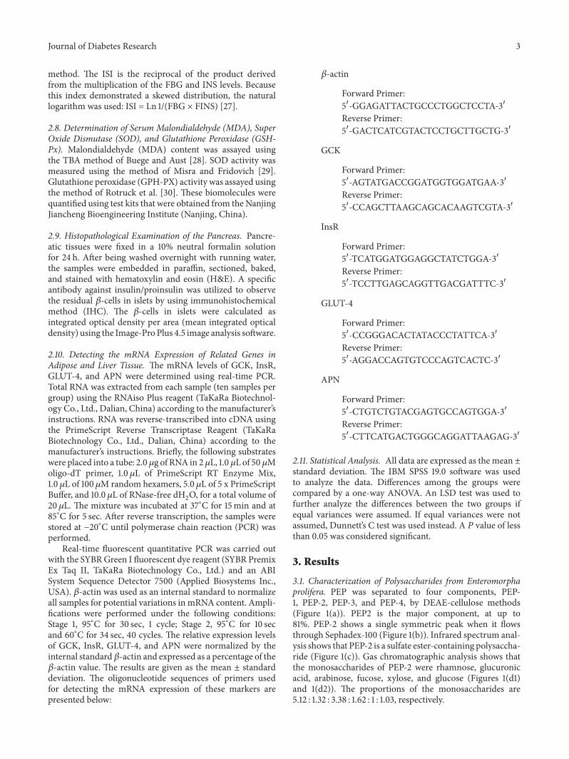
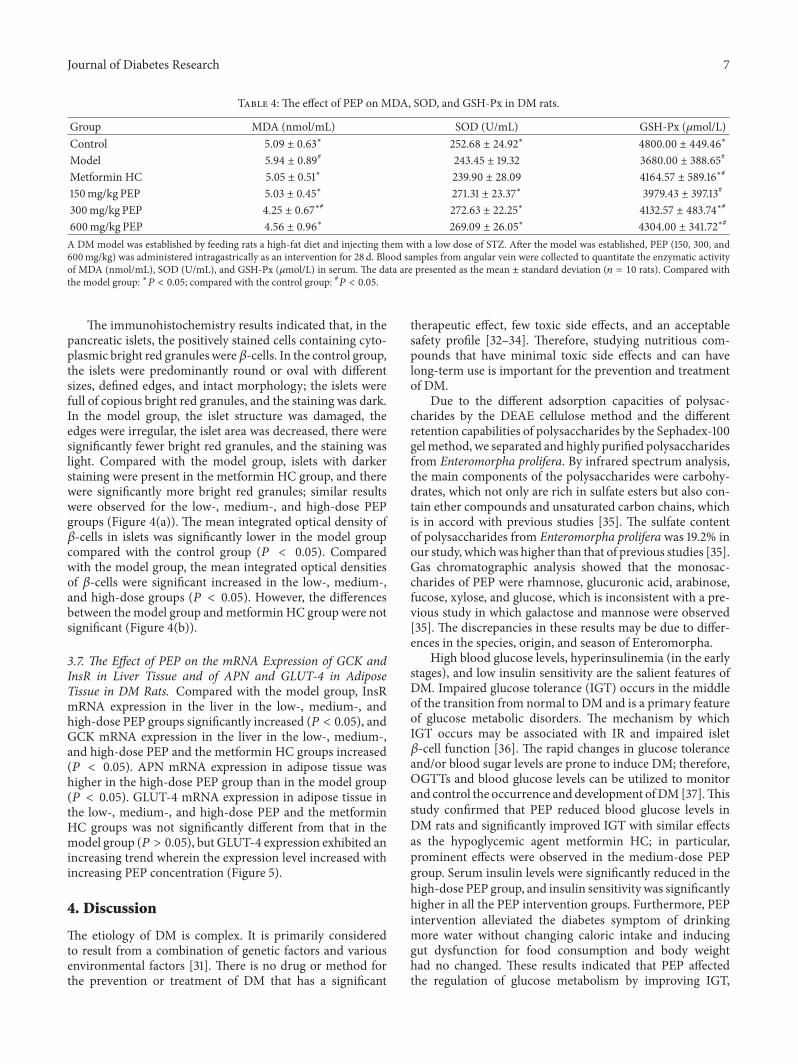