Research Article Photoluminescence Spectroscopic Study … · Research Article Photoluminescence...
6
Hindawi Publishing Corporation Journal of Spectroscopy Volume 2013, Article ID 312519, 5 pages http://dx.doi.org/10.1155/2013/312519 Research Article Photoluminescence Spectroscopic Study of BaMgAl 10 O 17 :Eu Phosphor Coated with CaF 2 via a Sol-Gel Process Feng Li, 1,2 Ying Yang, 2 Yang Song, 1 Wubao Wang, 3 Wei Yang, 3 and Bingya Yang 3 1 School of Material Science and Engineering, Xi’an University of Technology, Xi’an 710048, China 2 School of Physical Science and Technology, Nanjing University of Aeronautics and Astronautics, Nanjing 210016, China 3 IRICO Group Corporation, Xianyang 712000, China Correspondence should be addressed to Feng Li; fl[email protected] Received 30 June 2012; Accepted 21 January 2013 Academic Editor: Paola Luches Copyright © 2013 Feng Li et al. is is an open access article distributed under the Creative Commons Attribution License, which permits unrestricted use, distribution, and reproduction in any medium, provided the original work is properly cited. CaF 2 coatings on the surface of BaMgAl 10 O 17 :Eu (BAM) were prepared by a sol-gel process, and the optical properties and antithermal degradation properties were analyzed by photoluminescence spectra recorded under 254 nm and 147 nm excitation. e results indicate that BAM particles were successfully coated with CaF 2 and CaF 2 coatings show an interesting property to enhance the blue emission intensity of BAM. e optimum antithermal degradation properties were obtained at the weight ratio 0.4 wt% under 254 nm excitation and 0.3 wt% under 147 nm excitation, respectively. 1. Introduction BaMgAl 10 O 17 :Eu (BAM) is widely used in plasma display panels (PDPs) and lamps, due to its high luminescence effi- ciency and good chromaticity [1] and also used in other dis- plays and lighting devices, for example, white light-emitting diodes (WLED). However, luminance degradation of BAM, consisting of thermal degradation and lifetime degradation, restrains the application of BAM, especially in PDPs. In lamps, the degradation of BAM is mainly caused by thermal treatment during lamp manufacturing. In WLED, similar thermal degradation is also observed in BAM and Eu 2+ activated nitride-based phosphors [2]. ermal degradation occurs when the phosphors are heated to about 500–700 ∘ C in ambient atmosphere during the manufacturing of PDP and lamps, while in WLED thermal degradation is caused by the heat released by the operating devices. It is generally considered that the luminescent center Eu 2+ is oxidized to Eu 3+ [1–3]. However, some other potential mechanisms were proposed recently which are related with O 2− incorporated into the crystal lattice of BAM [4–6]. Lifetime degradation is caused by VUV radiation and ion sputtering during PDP operation, which can be explained by the formation of an amorphous surface layer [3]. One of the options to enhance thermal stability and ion resistance in BAM is the application of a closed particle coating [7]. Up to now, several inorganic materials have been reported to be selected as the coating materials such as SiO 2 [8, 9], SrO, MgO [7, 10], Al 2 O 3 [8, 11], AlPO 4 , and LaPO 4 [12] and so forth. However, most of them possess strong band gap absorption in the range of 140 to 200 nm, which would thereby cause the reduction of the phosphor efficiency. us the option is narrowed to metal fluorides which have a wide transparency range in VUV region and high secondary electron emission coefficient which can improve the pixel brightness [7, 13]. Metal fluorides can be deposited onto the phosphor surface by conventional precipitation or emulsion- assisted precipitation [14, 15]. However, this processes result in poor adherence of the coating to the phosphor [16]. More- over, CVD (chemical vapor deposition) and PVD (physical vapor deposition) would be good choices [17], but both are highly apparatus dependent, requiring elaborate instrumen- tation and careful air flow and temperature monitoring. us the cost of the phosphor fabrication process is increased. In our previous report [18], BAM coated with MgF 2 was prepared via a sol-gel process which is low cost, low apparatus dependent, and convenient to operate compared with the other reported methods. e coating percentage is easy to
Transcript of Research Article Photoluminescence Spectroscopic Study … · Research Article Photoluminescence...

Hindawi Publishing CorporationJournal of SpectroscopyVolume 2013, Article ID 312519, 5 pageshttp://dx.doi.org/10.1155/2013/312519
Research ArticlePhotoluminescence Spectroscopic Study of BaMgAl10O17:EuPhosphor Coated with CaF2 via a Sol-Gel Process
Feng Li,1,2 Ying Yang,2 Yang Song,1 Wubao Wang,3 Wei Yang,3 and Bingya Yang3
1 School of Material Science and Engineering, Xi’an University of Technology, Xi’an 710048, China2 School of Physical Science and Technology, Nanjing University of Aeronautics and Astronautics, Nanjing 210016, China3 IRICO Group Corporation, Xianyang 712000, China
Correspondence should be addressed to Feng Li; [email protected]
Received 30 June 2012; Accepted 21 January 2013
Academic Editor: Paola Luches
Copyright © 2013 Feng Li et al. This is an open access article distributed under the Creative Commons Attribution License, whichpermits unrestricted use, distribution, and reproduction in any medium, provided the original work is properly cited.
CaF2coatings on the surface of BaMgAl
10O17:Eu (BAM) were prepared by a sol-gel process, and the optical properties and
antithermal degradation properties were analyzed by photoluminescence spectra recorded under 254 nm and 147 nm excitation.The results indicate that BAM particles were successfully coated with CaF
2and CaF
2coatings show an interesting property to
enhance the blue emission intensity of BAM. The optimum antithermal degradation properties were obtained at the weight ratio0.4 wt% under 254 nm excitation and 0.3 wt% under 147 nm excitation, respectively.
1. Introduction
BaMgAl10O17:Eu (BAM) is widely used in plasma display
panels (PDPs) and lamps, due to its high luminescence effi-ciency and good chromaticity [1] and also used in other dis-plays and lighting devices, for example, white light-emittingdiodes (WLED). However, luminance degradation of BAM,consisting of thermal degradation and lifetime degradation,restrains the application of BAM, especially in PDPs. Inlamps, the degradation of BAM is mainly caused by thermaltreatment during lamp manufacturing. In WLED, similarthermal degradation is also observed in BAM and Eu2+activated nitride-based phosphors [2]. Thermal degradationoccurs when the phosphors are heated to about 500–700∘Cin ambient atmosphere during the manufacturing of PDPand lamps, while in WLED thermal degradation is causedby the heat released by the operating devices. It is generallyconsidered that the luminescent center Eu2+ is oxidized toEu3+ [1–3]. However, some other potential mechanisms wereproposed recently which are related with O2− incorporatedinto the crystal lattice of BAM [4–6]. Lifetime degradationis caused by VUV radiation and ion sputtering during PDPoperation, which can be explained by the formation of anamorphous surface layer [3].
One of the options to enhance thermal stability and ionresistance in BAM is the application of a closed particlecoating [7]. Up to now, several inorganic materials have beenreported to be selected as the coating materials such as SiO
2
[8, 9], SrO, MgO [7, 10], Al2O3[8, 11], AlPO
4, and LaPO
4
[12] and so forth. However, most of them possess strongband gap absorption in the range of 140 to 200 nm, whichwould thereby cause the reduction of the phosphor efficiency.Thus the option is narrowed to metal fluorides which have awide transparency range in VUV region and high secondaryelectron emission coefficient which can improve the pixelbrightness [7, 13]. Metal fluorides can be deposited onto thephosphor surface by conventional precipitation or emulsion-assisted precipitation [14, 15]. However, this processes resultin poor adherence of the coating to the phosphor [16]. More-over, CVD (chemical vapor deposition) and PVD (physicalvapor deposition) would be good choices [17], but both arehighly apparatus dependent, requiring elaborate instrumen-tation and careful air flow and temperature monitoring.Thusthe cost of the phosphor fabrication process is increased.
In our previous report [18], BAM coated with MgF2was
prepared via a sol-gel process which is low cost, low apparatusdependent, and convenient to operate compared with theother reported methods. The coating percentage is easy to

2 Journal of Spectroscopy
(a) (b)
Figure 1: SEM images of (a) raw BAM and (b) 0.4 wt% CaF2-coated BAM.
control as well.The results show that appropriateMgF2/BAM
ratio can significantly improve the thermal stability of BAMwithout serious deterioration of emission intensity. As it iswell known, CaF
2also has wide transparency range and high
secondary electron emission coefficient [19]. Thus in thispaper, CaF
2-coated BAMs were prepared using the sol-gel
process, and the luminescence properties and antithermaldegradation properties were also evaluated.
2. Experimental
The starting materials were TFA (CF3COOH, CP),
Ca(CH3COO)
2⋅4H2O (AR), and iso-C
3H7OH (AR).
BAMwas provided by IRICO Group Corporation. 0.005molCa(CH
3COO)
2⋅H2O was dissolved in 15mL of isopropanol
with the addition of 2mL of TFA and 2mL of distilled water.The solution was stirred for 2 h and then diluted to 300mLwith isopropanol. BAM phosphor particles were added tothe solution at the weight ratio CaF
2/BAM = 0.1–1.4 wt%
and stirred for 15min. Then the solution mixed with BAMwas dried at 80∘C for 24 h so that dry gel containing BAMsamples was obtained. After being calcined at 350∘C for15min, BAM particles coated with CaF
2were obtained. In
order to investigate the antithermal degradation properties,all samples were heat-treated at 600∘C for 30min.
X-ray photoelectron spectroscopy (XPS) was carried outon a MICROLAB VG 210 instrument for surface compo-nents analysis. Surface morphology was observed with ascanning electron microscope (SEM, Model JSM-5600LV).The photoluminescence properties were measured by anFLS920T spectrophotometer equipped with a VM504 vac-uummonochromator (ActonResearchCorporation) at roomtemperature.
3. Results and Discussion
The SEM images of coated (0.4 wt%) and uncoated BAMsare shown in Figure 1. The surfaces of pure BAM and CaF
2-
coated BAM are both smooth, although the coated samples’corners and edges are slightly less sharp. It is suggested thatthe phosphors may be covered by continuous CaF
2coatings,
695 690 685 680 675Binding energy (eV)
Inte
nsity
(a.u
.)
Figure 2: XPS spectrum of F1s.
Table 1: Binding energies of the elements of uncoated and coatedBAM phosphor surfaces.
Sample Ba3d5(eV)
Mg2p(eV)
O1s(eV)
Al2p(eV)
F1s(eV)
Ca2p3/2(eV)
BAM (uncoated) 780.41 49.94 531.08 74.08 — —BAM (coated) 780.41 50.00 531.09 74.08 685.0 347.3
which should be beneficial to prevent the access of oxygen tothe phosphors, rather than particles.
The XPS analysis of a series of samples shows that BAMphosphors were coated with CaF
2via this sol-gel process. As
an example, Figure 2 exhibits the F1s peak of the sample with0.4 wt% CaF
2. Table 1 shows the Ca2p3/2 and F1s checked in
the coated samples. It indicates that they are close to thatof CaF
2standard, in which Ca2p3/2 is 347.5 eV and F1s is
684.9 eV [20], respectively. Combined with the analysis ofSEM, it can be confirmed that the surfaces of BAMphosphorsare covered with CaF
2coatings.
The optical properties and antithermal degradation prop-erties of the coated samples were found to be highly depen-dent on the coating percentage. As it is shown in Line (a)

Journal of Spectroscopy 3
0 0.2 0.4 0.6 0.8 0.1 1.2 1.4CaF2/BAM (wt%)
0.7
0.75
0.8
0.85
0.9
0.95
1
1.05
1.1
1.15
Inte
nsity
(a.u
.)
(a)(b)
Figure 3: Emission intensities of CaF2-coated BAM phosphors as
a function of the weight ratio CaF2/BAM (a) before heat treatment
and (b) after 600∘C heat treatment (𝜆ex = 254 nm, 𝜆em = 450 nm).
400 425 450 475 500 525 550 575 600Wavelength (nm)
0
0.2
0.4
0.6
0.8
1
Nor
mal
ized
inte
nsity
(a.u
.)
0.8 wt% coatedRaw BAM
Figure 4: Emission spectra of 0.4 wt% CaF2-coated BAM and raw
BAM.
of Figure 3, the emission intensities of CaF2-coated BAMs
under 254 nm excitation show a maximum at 0.4 wt% (Linea) which is about 11% higher than that of uncoated BAMsbefore heat treatment. This phenomenon is rather differentfrom the cases of oxides or MgF
2-coated samples [14, 17] and
may be due to the reduction of the reflectivity of the excitationUV light from the surface of the BAM phosphor [9], causedby the CaF
2coating, whose refractive index is 1.32, smaller
than 1.38 of MgF2. This effect results in the less loss of UV
light passing through the coating layers. Thus the emissionintensity is thereby enhanced.
After heat treatment, the emission intensity at 0.8 wt%becomes the maximum (Figure 3, Line (b)), which is about10% higher than the uncoated counterparts. This may be
0 0.2 0.4 0.6 0.8 0.1 1.2 1.4CaF2/BAM (wt%)
(a)(b)
Inte
nsity
(a.u
.)
0.6
0.7
0.8
0.9
1
1.1
Figure 5: Photoluminescent intensities of CaF2-coated BAM phos-
phors as a function of the weight ratio CaF2/BAM (a) before heat
treatment and (b) after 600∘C heat treatment (𝜆ex = 147 nm, 𝜆em =450 nm).
because the coating could not cover the phosphor particlescompletely at lower weight ratios. Highweight ratios resultedin high reflectance and low transmission ratio, which causedlow luminescent intensity. These two factors are combinedto cause the optimum CaF
2/BAM ratio which is 0.8 wt% for
antithermal degradation property. The normalized emissionspectra of 0.4 wt% CaF
2-coated BAM and raw BAM are
presented in Figure 4, which clearly shows the emission bandshapes are similar.Neither obvious bandbroadening nor peakshift is observed.
As shown in Line (a) of Figure 5, when irradiated by147 nm, a maximum was observed at about 0.2 wt% beforeheat treatment. And the emission intensity at 0.2 wt% isabout 6% higher than that of the raw BAM. The increaseof the PL intensity is associated with the decrease of thereflectivity at the surface as mentioned above. After heattreatment, the maximum at about 0.6 wt% is 12% higher thanthe uncoated’s (Figure 5, Line (b)). This phenomenon maybe explained by the mechanism proposed in the discussionof Figure 3, that is, the coinfluence of the requirements oflow reflectance and coverage. The enhancement of thermalstability of CaF
2-coated BAMs is less efficient than that of
MgF2-coated samples prepared via the same sol-gel process
[18], whichmay be due to the higher porosity of CaF2coating
layers synthesized by this sol-gel process mentioned in [19].It is known that one of the most important factors
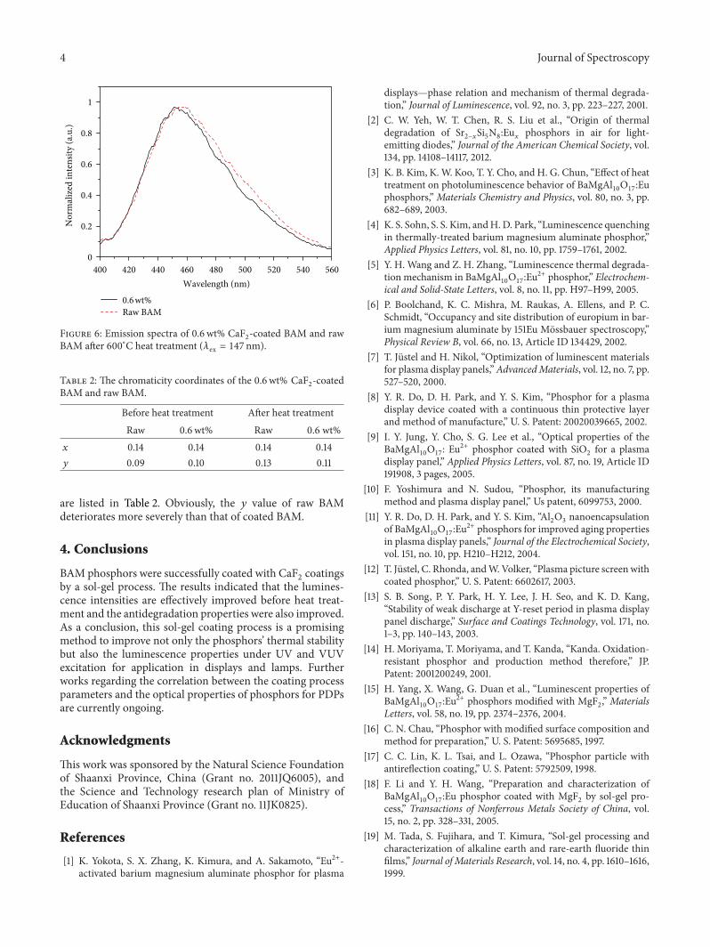
regarding BAM for application in PDPs is its color purity.Thenormalized emission spectra of 0.6 wt% CaF
2-coated BAM
and raw BAM after heat treatment are presented in Figure 6.The bandwidth of raw BAM is obviously bigger than that ofthe sample with 0.6 wt% CaF
2, which may be attributed to
themigration of Eu2+ frommirror planes to spinel blocks [5],indicating the more severe thermal degradation of raw BAM.Furthermore, the chromaticity coordinates of the samples
4 Journal of Spectroscopy
400 420 440 460 480 500 520 540 560Wavelength (nm)
0
0.2
0.4
0.6
0.8
1
Nor
mal
ized
inte
nsity
(a.u
.)
0.6 wt%Raw BAM
Figure 6: Emission spectra of 0.6 wt% CaF2-coated BAM and raw
BAM after 600∘C heat treatment (𝜆ex = 147 nm).
Table 2: The chromaticity coordinates of the 0.6 wt% CaF2-coated
BAM and raw BAM.
Before heat treatment After heat treatmentRaw 0.6wt% Raw 0.6 wt%
𝑥 0.14 0.14 0.14 0.14𝑦 0.09 0.10 0.13 0.11
are listed in Table 2. Obviously, the 𝑦 value of raw BAMdeteriorates more severely than that of coated BAM.
4. Conclusions
BAM phosphors were successfully coated with CaF2coatings
by a sol-gel process. The results indicated that the lumines-cence intensities are effectively improved before heat treat-ment and the antidegradation properties were also improved.As a conclusion, this sol-gel coating process is a promisingmethod to improve not only the phosphors’ thermal stabilitybut also the luminescence properties under UV and VUVexcitation for application in displays and lamps. Furtherworks regarding the correlation between the coating processparameters and the optical properties of phosphors for PDPsare currently ongoing.
Acknowledgments
This work was sponsored by the Natural Science Foundationof Shaanxi Province, China (Grant no. 2011JQ6005), andthe Science and Technology research plan of Ministry ofEducation of Shaanxi Province (Grant no. 11JK0825).
References
[1] K. Yokota, S. X. Zhang, K. Kimura, and A. Sakamoto, “Eu2+-activated barium magnesium aluminate phosphor for plasma
displays—phase relation and mechanism of thermal degrada-tion,” Journal of Luminescence, vol. 92, no. 3, pp. 223–227, 2001.
[2] C. W. Yeh, W. T. Chen, R. S. Liu et al., “Origin of thermaldegradation of Sr
2−𝑥Si5N8:Eu𝑥
phosphors in air for light-emitting diodes,” Journal of the American Chemical Society, vol.134, pp. 14108–14117, 2012.
[3] K. B. Kim, K.W. Koo, T. Y. Cho, and H. G. Chun, “Effect of heattreatment on photoluminescence behavior of BaMgAl
10O17:Eu
phosphors,”Materials Chemistry and Physics, vol. 80, no. 3, pp.682–689, 2003.
[4] K. S. Sohn, S. S. Kim, andH.D. Park, “Luminescence quenchingin thermally-treated barium magnesium aluminate phosphor,”Applied Physics Letters, vol. 81, no. 10, pp. 1759–1761, 2002.
[5] Y. H. Wang and Z. H. Zhang, “Luminescence thermal degrada-tion mechanism in BaMgAl
10O17:Eu2+ phosphor,” Electrochem-
ical and Solid-State Letters, vol. 8, no. 11, pp. H97–H99, 2005.[6] P. Boolchand, K. C. Mishra, M. Raukas, A. Ellens, and P. C.
Schmidt, “Occupancy and site distribution of europium in bar-ium magnesium aluminate by 151Eu Mossbauer spectroscopy,”Physical Review B, vol. 66, no. 13, Article ID 134429, 2002.
[7] T. Justel and H. Nikol, “Optimization of luminescent materialsfor plasma display panels,”AdvancedMaterials, vol. 12, no. 7, pp.527–520, 2000.
[8] Y. R. Do, D. H. Park, and Y. S. Kim, “Phosphor for a plasmadisplay device coated with a continuous thin protective layerand method of manufacture,” U. S. Patent: 20020039665, 2002.
[9] I. Y. Jung, Y. Cho, S. G. Lee et al., “Optical properties of theBaMgAl
10O17: Eu2+ phosphor coated with SiO
2for a plasma
display panel,” Applied Physics Letters, vol. 87, no. 19, Article ID191908, 3 pages, 2005.
[10] F. Yoshimura and N. Sudou, “Phosphor, its manufacturingmethod and plasma display panel,” Us patent, 6099753, 2000.
[11] Y. R. Do, D. H. Park, and Y. S. Kim, “Al2O3nanoencapsulation
of BaMgAl10O17:Eu2+ phosphors for improved aging properties
in plasma display panels,” Journal of the Electrochemical Society,vol. 151, no. 10, pp. H210–H212, 2004.
[12] T. Justel, C. Rhonda, andW.Volker, “Plasma picture screen withcoated phosphor,” U. S. Patent: 6602617, 2003.
[13] S. B. Song, P. Y. Park, H. Y. Lee, J. H. Seo, and K. D. Kang,“Stability of weak discharge at Y-reset period in plasma displaypanel discharge,” Surface and Coatings Technology, vol. 171, no.1–3, pp. 140–143, 2003.
[14] H. Moriyama, T. Moriyama, and T. Kanda, “Kanda. Oxidation-resistant phosphor and production method therefore,” JP.Patent: 2001200249, 2001.
[15] H. Yang, X. Wang, G. Duan et al., “Luminescent properties ofBaMgAl
10O17:Eu2+ phosphors modified with MgF
2,” Materials
Letters, vol. 58, no. 19, pp. 2374–2376, 2004.[16] C. N. Chau, “Phosphor with modified surface composition and
method for preparation,” U. S. Patent: 5695685, 1997.[17] C. C. Lin, K. L. Tsai, and L. Ozawa, “Phosphor particle with
antireflection coating,” U. S. Patent: 5792509, 1998.[18] F. Li and Y. H. Wang, “Preparation and characterization of
BaMgAl10O17:Eu phosphor coated with MgF
2by sol-gel pro-
cess,” Transactions of Nonferrous Metals Society of China, vol.15, no. 2, pp. 328–331, 2005.
[19] M. Tada, S. Fujihara, and T. Kimura, “Sol-gel processing andcharacterization of alkaline earth and rare-earth fluoride thinfilms,” Journal ofMaterials Research, vol. 14, no. 4, pp. 1610–1616,1999.

Journal of Spectroscopy 5
[20] V. I. Nefedov, Y. A. Buslaev, and Y. V. Kokunov, “X-ray electronstudy of internal levels of fluorides,” Zhurnal NeorganicheskoiKhimii, vol. 18, p. 931, 1973.

Submit your manuscripts athttp://www.hindawi.com
Hindawi Publishing Corporationhttp://www.hindawi.com Volume 2014
Inorganic ChemistryInternational Journal of
Hindawi Publishing Corporation http://www.hindawi.com Volume 2014
International Journal ofPhotoenergy
Hindawi Publishing Corporationhttp://www.hindawi.com Volume 2014
Carbohydrate Chemistry
International Journal of
Hindawi Publishing Corporationhttp://www.hindawi.com Volume 2014
Journal of
Chemistry
Hindawi Publishing Corporationhttp://www.hindawi.com Volume 2014
Advances in
Physical Chemistry
Hindawi Publishing Corporationhttp://www.hindawi.com
Analytical Methods in Chemistry
Journal of
Volume 2014
Bioinorganic Chemistry and ApplicationsHindawi Publishing Corporationhttp://www.hindawi.com Volume 2014
SpectroscopyInternational Journal of
Hindawi Publishing Corporationhttp://www.hindawi.com Volume 2014
The Scientific World JournalHindawi Publishing Corporation http://www.hindawi.com Volume 2014
Medicinal ChemistryInternational Journal of
Hindawi Publishing Corporationhttp://www.hindawi.com Volume 2014
Chromatography Research International
Hindawi Publishing Corporationhttp://www.hindawi.com Volume 2014
Applied ChemistryJournal of
Hindawi Publishing Corporationhttp://www.hindawi.com Volume 2014
Hindawi Publishing Corporationhttp://www.hindawi.com Volume 2014
Theoretical ChemistryJournal of
Hindawi Publishing Corporationhttp://www.hindawi.com Volume 2014
Journal of
Spectroscopy
Analytical ChemistryInternational Journal of
Hindawi Publishing Corporationhttp://www.hindawi.com Volume 2014
Journal of
Hindawi Publishing Corporationhttp://www.hindawi.com Volume 2014
Quantum Chemistry
Hindawi Publishing Corporationhttp://www.hindawi.com Volume 2014
Organic Chemistry International
ElectrochemistryInternational Journal of
Hindawi Publishing Corporation http://www.hindawi.com Volume 2014
Hindawi Publishing Corporationhttp://www.hindawi.com Volume 2014
CatalystsJournal of