Research Article - irjponline.com · phase was filtered through 0.2 mm millipore membrane filters...
9
Saba Shamim & Maryam Khan . Int. Res. J. Pharm. 2017, 8 (10) 29 INTERNATIONAL RESEARCH JOURNAL OF PHARMACY www.irjponline.com ISSN 2230 – 8407 Research Article PHYTOCHEMICAL SCREENING BY HIGH PERFORMANCE LIQUID CHROMATOGRAPHY (HPLC) AND ANTIMICROBIAL ACTIVITY OF DIFFERENT SOLVENT FRACTIONS OF ARECA NUTS AGAINST BACILLUS SUBTILIS BIOFILM Saba Shamim *, Maryam Khan Institute of Molecular Biology and Biotechnology (IMBB), The University of Lahore (UOL), Defence road campus, off Bhobatian chowk, Lahore, Pakistan *Corresponding Author Email: [email protected] Article Received on: 02/09/17 Approved for publication: 12/10/17 DOI: 10.7897/2230-8407.0810178 ABSTRACT Areca nut commonly known as Supari is in use since 15 th century. Plaque formation is one of the main reasons for deterioration of teeth. In this research work, the effect of areca nut on plaque formation was checked. For this, plaque forming microorganisms were isolated from healthy human teeth. A total of 100 samples were collected from individuals possessing healthy teeth. From these samples, five microorganisms were isolated which included Staphylcoccus sp., Streptococcus sp., Bacillus sp., Pseudomonas sp. and Klebsiella sp. Only Bacillus sp. was found common in teeth of all subjects. It was able to grow biofilm in the Luria Bertani (LB) broth after 24 hours of incubation at 37 °C. Bacillus sp. was revealed to be Bacillus subtilis by 16s rRNA sequencing (Macrogen®). The extracts of areca nuts were prepared in ethanol, methanol, chloroform and water. Both disc and well diffusion methods were employed to check the antimicrobial activity of these extracts. Areca nuts exhibited antimicrobial activity in all extracts except chloroform. High Performance Liquid Chromatography (HPLC) confirmed the presence on antimicrobial compounds i.e. quercetin and cinnamic acid. The biofilm growing ability of B. subtilis was interrupted up to 40 % by using 100 % ethanol and water extracts. For demolishing the established biofilm, 50 % of all extracts were found to be effective. It can be concluded that areca nut was effective for combating the biofilm formation by B. subtilis. It can be used in mouth wash formulations to get rid of plaque formation on teeth. Keywords: Bacillus subtilis, areca nuts, antimicrobial activity, biofilm formation, HPLC, plaque INTRODUCTION Areca nut known as supari in Pakistan is an important component of paan which was part of Mughal civilization back in 15th century. Areca nut is the seed of Areca catechu, an oriental palm of the Palmaceae family that grows in South and Southeast Asia, East Africa and parts of Tropical Pacific. The nut is not a fruit but a drupe and can be consumed in raw, processed, roasted, sun dried forms. Different forms of areca nut contribute to variance in taste and appearance 1 . It is considered to be the fourth most commonly used addictive and psychoactive substance preceded by tobacco, alcohol, and caffeine respectively. Approximately 10 % of the world’s population are involved in the consumption of areca nut. Its use is habitual in countries like India, Sri Lanka, Pakistan, Maldives, Bangladesh, Myanmar, Taiwan, Thailand, Indonesia, Cambodia, Vietnam, Philipines, Laos, and China 2 . Additionally, the practice of chewing pan masala with or without the use of tobacco is common in the subcontinent from thousands of years and currently an estimated 600 million people are inflicted in the consumption of betel quid with areca nut globally 3 . Normal oral microflora consists of many organisms reside in the mouth. Overproduction of microbes on the surfaces of teeth and gum are called plaques and it can lead to gingivitis, caries, septicemia, and endocarditis 4 . Various studies have demonstrated that the alkaloids extracted from areca nut contribute to the cytotoxic and genotoxic property and carcinogenicity to humans. Several reports have shown the association of chewing areca nuts with the development of various disorders, such as cardiovascular disease, metabolic syndrome, and hypertension 5 . Areca nut has been categorised as Group 1 human carcinogens by the World Health Organisation 6 . Areca nut has a wide range of adverse health effects reported in recent literature and various studies are further cementing this previously thought to be false notion. It is reported to be carcinogenic and to cancers of the pharynx, oesophagus, liver, oral cavity, and the uterus 7 . Pan Masala, also called betel quid, is a mixture of Acacia catechu with slaked lime, areca nut and other flavoring agents. It is widely available and used famously by inhabitants of the subcontinent. Betel chewing produces an increase in the blood pressure, a spike in the heart rate and body temperature and sweating. It also leads to an increase in the plasma concentrations of norepinephrine and epinephrine, which suggests that betel chewing affects the central and autonomic nervous systems 8 . It is reported to be hepatotoxic that results in an increase level of enzymes and disturbed lipid and carbohydrate metabolism. Additionally, it is harmful to kidneys with reported cases of increased creatinine 7 . This study was conducted to study the antimicrobial activity of areca nut on the formation of biofilms by oral flora and phytoconstituents of areca nuts by high performance liquid chromatography (HPLC). MATERIALS AND METHODS Chemicals All chemicals used in this study were of analytical grades. The solvents for fractionation were purchased from Merck, Germany. Microbiology media used were of Oxide, Thermofisher UK.
Transcript of Research Article - irjponline.com · phase was filtered through 0.2 mm millipore membrane filters...

Saba Shamim & Maryam Khan . Int. Res. J. Pharm. 2017, 8 (10)
29
INTERNATIONAL RESEARCH JOURNAL OF PHARMACY
www.irjponline.com
ISSN 2230 – 8407
Research Article
PHYTOCHEMICAL SCREENING BY HIGH PERFORMANCE LIQUID CHROMATOGRAPHY (HPLC)
AND ANTIMICROBIAL ACTIVITY OF DIFFERENT SOLVENT FRACTIONS OF ARECA NUTS AGAINST
BACILLUS SUBTILIS BIOFILM
Saba Shamim *, Maryam Khan
Institute of Molecular Biology and Biotechnology (IMBB), The University of Lahore (UOL), Defence road campus, off
Bhobatian chowk, Lahore, Pakistan *Corresponding Author Email: [email protected]
Article Received on: 02/09/17 Approved for publication: 12/10/17
DOI: 10.7897/2230-8407.0810178
ABSTRACT
Areca nut commonly known as Supari is in use since 15th century. Plaque formation is one of the main reasons for deterioration of teeth. In this research work, the effect of areca nut on plaque formation was checked. For this, plaque forming microorganisms were isolated from healthy human teeth. A
total of 100 samples were collected from individuals possessing healthy teeth. From these samples, five microorganisms were isolated which included
Staphylcoccus sp., Streptococcus sp., Bacillus sp., Pseudomonas sp. and Klebsiella sp. Only Bacillus sp. was found common in teeth of all subjects. It
was able to grow biofilm in the Luria Bertani (LB) broth after 24 hours of incubation at 37 °C. Bacillus sp. was revealed to be Bacillus subtilis by 16s
rRNA sequencing (Macrogen®). The extracts of areca nuts were prepared in ethanol, methanol, chloroform and water. Both disc and well diffusion
methods were employed to check the antimicrobial activity of these extracts. Areca nuts exhibited antimicrobial activity in all extracts except chloroform. High Performance Liquid Chromatography (HPLC) confirmed the presence on antimicrobial compounds i.e. quercetin and cinnamic acid.
The biofilm growing ability of B. subtilis was interrupted up to 40 % by using 100 % ethanol and water extracts. For demolishing the established
biofilm, 50 % of all extracts were found to be effective. It can be concluded that areca nut was effective for combating the biofilm formation by B. subtilis. It can be used in mouth wash formulations to get rid of plaque formation on teeth.
Keywords: Bacillus subtilis, areca nuts, antimicrobial activity, biofilm formation, HPLC, plaque
INTRODUCTION
Areca nut known as supari in Pakistan is an important component
of paan which was part of Mughal civilization back in 15th
century. Areca nut is the seed of Areca catechu, an oriental palm
of the Palmaceae family that grows in South and Southeast Asia,
East Africa and parts of Tropical Pacific. The nut is not a fruit but
a drupe and can be consumed in raw, processed, roasted, sun dried
forms. Different forms of areca nut contribute to variance in taste
and appearance1. It is considered to be the fourth most commonly
used addictive and psychoactive substance preceded by tobacco,
alcohol, and caffeine respectively. Approximately 10 % of the
world’s population are involved in the consumption of areca nut.
Its use is habitual in countries like India, Sri Lanka, Pakistan,
Maldives, Bangladesh, Myanmar, Taiwan, Thailand, Indonesia,
Cambodia, Vietnam, Philipines, Laos, and China2. Additionally,
the practice of chewing pan masala with or without the use of
tobacco is common in the subcontinent from thousands of years
and currently an estimated 600 million people are inflicted in the
consumption of betel quid with areca nut globally3. Normal oral
microflora consists of many organisms reside in the mouth.
Overproduction of microbes on the surfaces of teeth and gum are
called plaques and it can lead to gingivitis, caries, septicemia, and
endocarditis4. Various studies have demonstrated that the
alkaloids extracted from areca nut contribute to the cytotoxic and
genotoxic property and carcinogenicity to humans. Several
reports have shown the association of chewing areca nuts with the
development of various disorders, such as cardiovascular disease,
metabolic syndrome, and hypertension5. Areca nut has been
categorised as Group 1 human carcinogens by the World Health
Organisation6. Areca nut has a wide range of adverse health
effects reported in recent literature and various studies are further
cementing this previously thought to be false notion. It is reported
to be carcinogenic and to cancers of the pharynx, oesophagus,
liver, oral cavity, and the uterus7. Pan Masala, also called betel
quid, is a mixture of Acacia catechu with slaked lime, areca nut
and other flavoring agents. It is widely available and used
famously by inhabitants of the subcontinent. Betel chewing
produces an increase in the blood pressure, a spike in the heart
rate and body temperature and sweating. It also leads to an
increase in the plasma concentrations of norepinephrine and
epinephrine, which suggests that betel chewing affects the central
and autonomic nervous systems8. It is reported to be hepatotoxic
that results in an increase level of enzymes and disturbed lipid and
carbohydrate metabolism. Additionally, it is harmful to kidneys
with reported cases of increased creatinine7.
This study was conducted to study the antimicrobial activity of
areca nut on the formation of biofilms by oral flora and
phytoconstituents of areca nuts by high performance liquid
chromatography (HPLC).
MATERIALS AND METHODS
Chemicals
All chemicals used in this study were of analytical grades. The
solvents for fractionation were purchased from Merck, Germany.
Microbiology media used were of Oxide, Thermofisher UK.

Saba Shamim & Maryam Khan . Int. Res. J. Pharm. 2017, 8 (10)
30
Isolation of Plaque Forming Microorganisms
To isolate plaque forming microorganisms, dental swabs were
taken from subjects possessing healthy teeth. The subjects were
requested to rub the sterilize swab on plaque forming area of
tooth, after waking up in the morning before brushing their teeth.
Total 100 subjects were selected for this study (volunteer
university students, age group 20-30 years, males only, no
medical history of any disease, not in habit of any addiction like
smoking, paan chewing etc). The swabs were immediately
brought to Microbiology Laboratory for isolation of microbial
flora. The swabs were processed on Luria Bertani (LB) agar
plates till pure colonies were obtained. The Gram staining and
biochemical characterization were performed9,10. The bacterial
isolate found common in all samples was characterized on
molecular level by 16s rRNA sequencing (Macrogen®, South
Korea).
Extraction and Fractionation of Plant Material
The method of Saleem et al.,11 was followed with slight
modifications. Areca nuts were purchased from local market and
converted into powdered form. Powder was kept into clean dried
container for further procedure. Four different types of solvents
i.e. ethanol, methanol, chloroform and water were used for
preparation of areca nuts extract separately. The dry powder (30
grams) of areca nuts was mixed in 100 ml of respective solvent
and mixture was then kept in blue capped bottle at room
temperature for 4 days with occasional shaking. The filtrate was
filtered with Whatman no. 1 filter paper. To obtain the dried and
concentrated form, filtrate was processed at rotary evaporator
(Heidolph, Germany) at 55 °C and vaccum pump (V700, H-9230,
BUCHI, Switzerland) connected with refrigerated circulator
(DLSB 5/20, ZGSI, China). Finally the extract in all different
solvents was placed in refrigerator at 4 °C for further experiments.
Antimicrobial Activity of Extracts
Antimicrobial activity of extracts was checked by both well and
disc diffusion methods. It was performed by dissolving the dried
fraction areca nuts in different ratios in respective solvents
(ethanol, methanol, chloroform and water). Four different
concentrations of each solvent were prepared 100 mg/ml, 50
mg/ml, 25 mg/ml and 10 mg/ml respectively. Ciprofloxacin was
used as positive control, whereas 10 % respective solvent was
used as negative control.
Well Diffusion Method: Luria Bertani (LB) medium plates were
prepared. Pure colony of B. subtilis was swabbed on LB plates.
Wells were formed by pressing the backside of Pasteur pipette
into the agar medium. About 20 µl of each concentration of each
extract was added in wells. The plates were incubated at 37 °C for
24 hours. Zone of inhibition (mm) was measured after 24 hours.
The experiment was repeated with methanol, chloroform and
water extracts.
Disc Diffusion Method: For this, Luria Bertani (LB) medium
plates were prepared. B. subtilis pure colony was streaked on the
plate in the form of a mat. Filter paper was pinched to form discs
which were dipped in the extract, dried to remove extra extract
and placed on the plate. It was incubated for 24 hours at 37 ºC.
After 24 hours, the zone of inhibition (mm) was recorded12.
Biofilm Experiments
Biofilm Production by B. subtilis: B. subtilis (18 hours old)
culture (1 %) was inoculated in the wells of 96 well plate
containing 100 μl nutrient broth (Oxoid) followed by incubation
at 37 °C for 24 hours. The biofilm of bacterial growth was
observed and OD was taken at 600 nm. Biofilm production was
also confirmed in flask by giving the 1 % inoculum of B. subtilis
in 100 ml nutrient broth (Oxoid) followed by incubation at 37 °C
for 24 hours. After 24 hours, biofilm was observed, it was mixed
with the broth medium and OD was taken at 600 nm12.
Interruption of Biofilm Formation: The biofilm of B. subtilis
was grown in microtitre plate as mentioned above. After 24 hours,
100 µl of ethanol, methanol and water extracts was added in wells
and then placed in incubator for 24 hours. After one day, the
adhered bacterial growth was determined by following method:
100 µl 1 % crystal violet stain was added in each well. In the next
step, each well was emptied then washed three times with distilled
water. In the next step, 150 µl of methanol was added for 15
minutes. When methanol was completely dried, 160 µl of glacial
acetic acid was added to it. The biomass was then made ready for
microtiter reader. Readings were taken at 630 nm. The percentage
of inhibition was calculated13.
Disturbing the Established Biofilm: After incubation of the
plate the non-adherent cells were removed by washing it three
times with distilled water. Each concentration of extracts (100 µl)
was added to each well respectively, and then incubated for 24
hours at 37 °C. The disruption of biofilm was determined by using
crystal violet. Then the percentage of reduction in biofilm
structures was calculated12.
Quantification of Phytocomponents by HPLC
The phytocomponents of areca nuts were determined by HPLC.
Sample Preparation: Phenolic compounds were identified and
quantified from ethanol, methanol and water extracts of areca nuts
by reverse phase HPLC (Aligent HPLC system). Briefly, 25
milligrams of each extract was dissolved in 5 mL of 6 M HCl, 12
mL methanol with 8 mL of distilled water. The resultant solution
was incubated at 90 °C for 2 hours and filtered with 0.2 mm
millipore membrane filter before injection into HPLC column.
High Performance Liquid Chromatography: The HPLC
separation was performed using HPLC system with column 20
RBAX ECLIPSE, XDB-C18, (4.6 × 150 mm; 5μm, Agilent
USA), UV–VIS Spectra-Focus detector and injector-auto
sampler. The isocratic mobile phase, consisting of A (water: AA-
94:6, pH=2.27), B (ACN, 100%), The flow of mobile phase was
adjusted as 0-15 min. (15 % B), 15-30 min. (45 % B), 30-45 min.
(100 % B) at a flow-rate of 1 mL/ min. Prior to use, the mobile
phase was filtered through 0.2 mm millipore membrane filters
and degassed by sonication in an ultrasonic bath. Detection
wavelength was set at 280 nm and the column temperature was
maintained at room temperature (37 °C) with injection volume of
10 μL.
RESULTS
Isolation and Identification of Plaque Forming
Microorganisms
On the basis of Gram staining and biochemical characterization,
five microorganisms including Staphylococcus sp., Streptococcus
sp., Bacillus sp., Pseudomonas sp. and Klebsiella sp. were
isolated from 100 subjects. Only Bacillus sp. was found common
in teeth of all subjects. The 16s rRNA sequencing revealed it
Bacillus subtilis. It was selected for further experimental work.
Antimicrobial Activity of Areca Nuts
Disc diffusion method showed more prominent zones as
compared to well diffusion method (Table 1, Figure 2).
Saba Shamim & Maryam Khan . Int. Res. J. Pharm. 2017, 8 (10)
31
Chloroform did not show any zone of inhibition (results not
shown here).
HPLC
HPLC chromatogram of ethanol, methanol and aqueous extracts
are given in Figure 3. The phenolic acids which are represented
by each peak, their retention time, area in mV.s and % are given
(Table 2, Figure 3). Among the various components, quercetin
and cinnamic acid were found to possessed antimicrobial
properties.
Biofilm Experiments
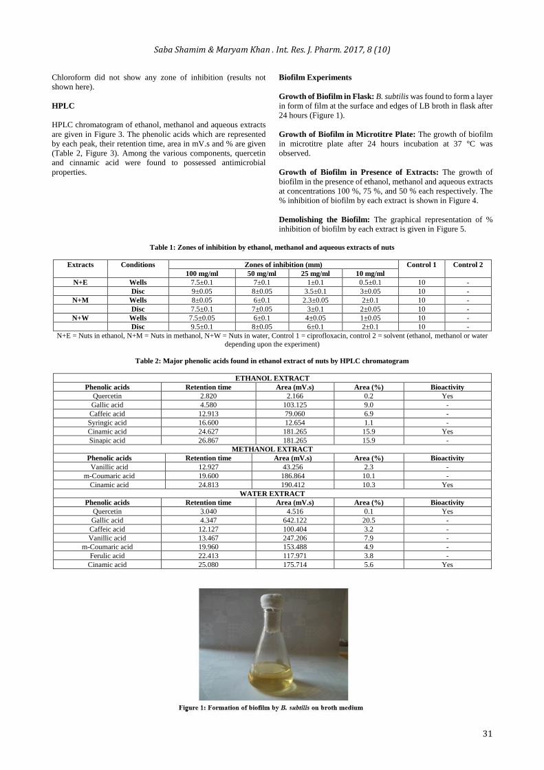
Growth of Biofilm in Flask: B. subtilis was found to form a layer
in form of film at the surface and edges of LB broth in flask after
24 hours (Figure 1).
Growth of Biofilm in Microtitre Plate: The growth of biofilm
in microtitre plate after 24 hours incubation at 37 °C was
observed.
Growth of Biofilm in Presence of Extracts: The growth of
biofilm in the presence of ethanol, methanol and aqueous extracts
at concentrations 100 %, 75 %, and 50 % each respectively. The
% inhibition of biofilm by each extract is shown in Figure 4.
Demolishing the Biofilm: The graphical representation of %
inhibition of biofilm by each extract is given in Figure 5.
Table 1: Zones of inhibition by ethanol, methanol and aqueous extracts of nuts
Extracts Conditions Zones of inhibition (mm) Control 1 Control 2
100 mg/ml 50 mg/ml 25 mg/ml 10 mg/ml
N+E Wells 7.5±0.1 7±0.1 1±0.1 0.5±0.1 10 -
Disc 9±0.05 8±0.05 3.5±0.1 3±0.05 10 -
N+M Wells 8±0.05 6±0.1 2.3±0.05 2±0.1 10 -
Disc 7.5±0.1 7±0.05 3±0.1 2±0.05 10 -
N+W Wells 7.5±0.05 6±0.1 4±0.05 1±0.05 10 -
Disc 9.5±0.1 8±0.05 6±0.1 2±0.1 10 -
N+E = Nuts in ethanol, N+M = Nuts in methanol, N+W = Nuts in water, Control 1 = ciprofloxacin, control 2 = solvent (ethanol, methanol or water
depending upon the experiment)
Table 2: Major phenolic acids found in ethanol extract of nuts by HPLC chromatogram
ETHANOL EXTRACT
Phenolic acids Retention time Area (mV.s) Area (%) Bioactivity
Quercetin 2.820 2.166 0.2 Yes
Gallic acid 4.580 103.125 9.0 -
Caffeic acid 12.913 79.060 6.9 -
Syringic acid 16.600 12.654 1.1 -
Cinamic acid 24.627 181.265 15.9 Yes
Sinapic acid 26.867 181.265 15.9 -
METHANOL EXTRACT
Phenolic acids Retention time Area (mV.s) Area (%) Bioactivity
Vanillic acid 12.927 43.256 2.3 -
m-Coumaric acid 19.600 186.864 10.1 -
Cinamic acid 24.813 190.412 10.3 Yes
WATER EXTRACT
Phenolic acids Retention time Area (mV.s) Area (%) Bioactivity
Quercetin 3.040 4.516 0.1 Yes
Gallic acid 4.347 642.122 20.5 -
Caffeic acid 12.127 100.404 3.2 -
Vanillic acid 13.467 247.206 7.9 -
m-Coumaric acid 19.960 153.488 4.9 -
Ferulic acid 22.413 117.971 3.8 -
Cinamic acid 25.080 175.714 5.6 Yes

Saba Shamim & Maryam Khan . Int. Res. J. Pharm. 2017, 8 (10)
32

Saba Shamim & Maryam Khan . Int. Res. J. Pharm. 2017, 8 (10)
33

Saba Shamim & Maryam Khan . Int. Res. J. Pharm. 2017, 8 (10)
34

Saba Shamim & Maryam Khan . Int. Res. J. Pharm. 2017, 8 (10)
35

Saba Shamim & Maryam Khan . Int. Res. J. Pharm. 2017, 8 (10)
36
DISCUSSION
Dental plaque is a community of microorganisms in form of
biofilm. Tooth provides solid support which microorganisms use
to bind with each other as well as with the tooth surface to form a
film14, 15. If this biofilm is not removed on daily basis, it will lead
to caries formation16. Various microorganisms are known to
cause this biofilm formation e.g. Streptococcus mutans,
Lactobacillus casei17. It is known that mouth inhabits 200-300
bacterial species16 (Loesche, 1996). In this study, five
microorganisms were isolated from healthy teeth of 100
individuals. They included Staphylococcus sp., Streptococcus sp.,
Bacillus sp., Pseudomonas sp. and Klebsiella sp. Streptococcus
sp. were also reported by Li et al.,17. B. subtilis was found in all
teeth of subjects. On this basis, it was selected for further
experiments. B. subtilis is Gram positive, motile, rod shaped and
spore forming bacterium18. Areca nut is considered an active and
abundant source of bioactive compounds as compared to its other
plant parts19. The antibacterial mode of action of areca nuts in
betel quid was also explained by the fact that during betel quid
chewing, polyphenols of areca nuts get oxidized which results in
the formation of superoxide anion (O2.-). The presence of lime
greatly increased the pH of the mouth during chewing process.
This superoxide anion gets converted to H2O2 which further
generates hydroxyl radicals. These radicals are toxic to microbial
flora20, 21. Kenney et al., (1975) explained that alteration of pH
and oxidation-reduction potential of oral region causes death of
Gram positive oral flora due to which Gram negative bacteria
caused dental caries thus, leads to deterioration of teeth in
general22.
Previous workers used both disc and well diffusion methods
methods to check the antimicrobial activity. Chin et al., 22
employed well diffusion method whereas Saleem et al.,11 used
disc diffusion method. In this study, both disc and well diffusion
methods were used and compared. Overall, disc diffusion method
showed more zones as compared to well diffusion method. In
Figure 2, disc method showed more zones of inhibition as
compared to well method. It was also observed that with
increasing nuts concentration in ethanol, enhanced zone was
observed. Similar was the case with methanol and water extracts.
Overall, ethanol extract showed more zone followed by water
then methanol. No zone was observed with chloroform (results
not shown here). It showed that chloroform did not extract the
bioactive compound from nuts. Anthikat and Michael,23 observed
100 % growth inhibition of Gram positive bacteria at 16 μg/ ml
areca nuts concentration. Rahman et al.,24 also reported
antimicrobial activity of areca nuts from ethanol and water
extracts which was in accordance with our results. The mode of
antibacterial action of nut extract is the immediate precipitation
of bacterial proteins thus, inhibiting the signals molecules by
denaturing the bacterial proteins22. When areca nuts are mixed
with other ingredients to form betel quid, then decrease in
microbial population was observed25. The antibacterial activity of
areca nuts found in this study, confirmed the previous literature 22, 26.
Here HPLC chromatogram of areca nuts in ethanol, methanol and
water extracts showed variety of phenolic acids namely quercetin,
gallic acid, caffeic acid, syringic acid, cinnamic acid, sinapic acid,
vanillic acid, coumaric acid and ferulic acid (Table 2, Figure 3).
They were responsible for antibacterial, antihelminthic,
antifungal, anti-inflammatory and antioxidative properties.
Quercetin is a flavonoid. Flavonoids are natural bioactive
compounds found in plants27. The bioactivity of quercetin is due
to disruption of cell membrane and formation of complexes with
soluble and extracellular proteins resulting in inactivation of
proteins28. The antibacterial activity of quercetin against 11
microorganisms was reported earlier29 which confirms our
results. In this study, quercetin was found in ethanol and water
extracts but not in methanol extract although it showed
antimicrobial activity. Cinnamic acid was found in all three
extracts. Cinnamic acid is already reported to possess
antibacterial properties30. It can be concluded that the bioactivity
of methanolic extract was due to cinnamic acid in our study
(Table 2).
B. subtilis has ability to form floating biofilm or pellicle at liquid-
air interface18, 31 which is in agreement with this study (Figure 1).
Bacterial biofilm formation is an important topic since last
decade32 as it provides shelter to bacteria residing inside and helps
bacteria to become resistant to drugs and antibiotics. Biofilms
dwelling bacteria are considered 1000 times more resistant to
antibiotics than free floating cells31. Due to this reason, medical
treatment often fails and disease condition persists. Many factors
are responsible for biofilm resistance like expression of multidrug
efflux pumps, type IV secretion systems, restricted diffusion of
antibiotics in biofilm matrix, decreased permeability, and action
of antibiotic modifying enzymes33. An antibiofilm agent of plant
origin is a hot topic these days as they are considered safe and
have no side effects32. In this study, all three extracts inhibited the
biofilm formation of B. subtilis (Figure 4). The 100 % ethanol
extract inhibited 40 % biofilm growth. The methanol extract
inhibited 30 % whereas much inhibition was observed with water
extract i.e. more than 60 % (Figure 4). Similarly, ethanol,
methanol and water extracts demolished the established biofilm
i.e. about 40 % growth inhibition of biofilm was observed with
all extracts. The ability to demolish biofilm increased with
increasing concentration of extract (Figure 5). Biofilm
experiments showed that ethanol, methanol and water extracts of
areca nuts possessed the antibacterial and antibiofilm ability.
Further research can help in getting deep insight into the
molecular mechanisms that are responsible for antibiofilm ability
of areca nuts. Further research in this area can help in developing
antibiofilm agent from areca nuts.
CONCLUSION
Areca nuts possess antimicrobial activity against B. subtilis
biofilms. There is a need to check its antimicrobial activity
against other microorganisms before it can be used in the
formulation of mouth wash.
ACKNOWLEDGEMENT
The authors are grateful to Macrogen® for providing 16s rRNA
services. We would like to pay our gratitude to Prof. Dr. M. H.
Qazi Director of Institute of Molecular Biology and
Biotechnology (IMBB), The University of Lahore (UOL), for
providing fully quipped Microbiology laboratory facilities for
carrying out the research work.
REFERENCES
1. Patidar KA, Parwani R, Wanjari SP, Patidar AP. Various
terminologies associated with areca nut and tobacco chewing:
a review. J Oral Maxillofac Pathol 2015; 19(1):69-76.
2. Mirza SS, Shafique K, Vart P, Arain MI. Areca nut chewing
and dependency syndrome: is the dependence comparable to
smoking? A cross-sectional study. Subst Abuse Treat Prev
Policy 2011; 6:23-8.
3. Adhikari A, Hazra AK, Sur TK. Detection of arecoline by
simple high-performance thin-layer chromatographic method
in Indian nontobacco pan masala. J Adv Pharm Technol Res
2015; 6(4):195-99.

Saba Shamim & Maryam Khan . Int. Res. J. Pharm. 2017, 8 (10)
37
4. Salako NO, Rotimi V, Philip L, Haider HA, Hamdan HM.
The prevalence and antibiotic sensitivity of oral viridans
streptococci in healthy children and children with disabilities
in Kuwait. Spec Care Dent 2007; 27(2):67-72.
5. Liu Y-C, Chen C-J, Lee M-R, Li M, Hsieh W-T, Chung J-G,
Ho, H-C. Peroxidase as the major protein constituent in areca
nut and identification of its natural substrates. Evid Based
Complement Alternat Med 2013; 2013:1-12.
6. Paulino YC, Novotny R, Miller MJ, Murphy SP. Areca
(betel) nut chewing practices in Micronesian populations.
Hawaii J Pub Hlth, 2011; 3(1):19-29.
7. Garg A, Chaturvedi P, Mishra A, Datta S. A review on
harmful effects of pan masala. Indian J Cancer 2015;
52(4):663-66.
8. Chu NS. Effects of betel chewing on the central and
autonomic nervous systems. J Biomed Sci 2001; 8(3):229-36.
9. Cheesbrough M(a). Culturing bacterial pathogens. In: District
laboratory practice in tropical countries – part 2, Cambridge
University Press; 2000. p. 45-62.
10. Cheesbrough M(b). Biochemical tests to identify bacteria. In:
District laboratory practice in tropical countries – part 2,
Cambridge University Press; 2000. p. 63-70
11. Saleem A, Younas U, Muhammad G, Jabbar A, Manan A,
Shamim S, Altaf S. Phytochemicals screening by FTIR
spectroscopy and antimicrobial activity of different solvent
fractions from Murraya koenigii L. shoots. Int Res J Pharm
2016; 7(4):30-8.
12. Mohsenipour Z, Hassanshahian M. The effects of Allium
sativum extracts in biofilm formation and activities of six
pathogenic bacteria. Jundishapur J Microbiol 2015; 8(8): 1-7.
13. O’Toole GA, Kolter R. Flagellar and twitching motility are
necessary for Pseudomonas aeruginosa biofilm development.
Mol Microbiol 1998; 30(2):295-304.
14. Marsh PD. Dental plaque as a microbial biofilm. Caries
Res 2004; 38:204-11.
15. Marsh PD. Dental plaque as a biofilm and a microbial
community - implications for health and disease. BMC Oral
Hlth 2006; 6(Suppl 1):S14.
16. Loesche WJ. Microbiology of dental decay and periodontal
disease. In: Baron S, editor. Chapter 99: Medical
Microbiology. 4th ed. Galveston (TX): University of Texas
Medical Branch at Galveston; 1996.
17. Li Y, Tang N, Aspiras MB, Lau PCY, Lee JH, Ellen RP,
Cvitkovitch DG. A quorum-sensing signaling system
essential for genetic competence in Streptococcus mutans is
involved in biofilm formation. J Bacteriol 2002; 184:2699-
708.
18. Lemon KP, Am E, Vlamakis HC, Aquilar C, Kolter R.
Biofilm development with an emphasis on Bacillus subtilis.
Curr Top Microbiol Immunol 2008; 322:1-16.
19. Anjali S, Rao AR. Modulatory influence of areca nut on
antioxidant 2(3)-tert-butyl-1-4-hydroxynisole-induced
hepatic detoxification system and antioxidant defence
mechanism in mice. Cancer Lett 1995; 91:107-114.
20. Nair UJ, Floyd RA, Nair J, Bussachini V, Friesen M, Bartsch
H. Formation of reactive oxygen species and of 8-hydroxy
deoxyguanosine in DNA in vitro with betel quid ingredients.
Chem Biol Interact 1987; 63(2):157-69.
21. IARC. Betel quid and areca nut chewing and some areca nut
derived nitrosamines. IARC monogr Eval Carcinog Risks
Hum 2004; 85:1-334.
22. Chin AA, Fernandez CD, Sanchez RB, Santos BMS,
Tolentino RF, Masangkay FR. Antimicrobial performance of
ethanolic extract of Areca catechu L seeds against mixed-oral
flora from tooth scum and gram negative laboratory isolates.
Int J Res Ayurv Pharm 2013; 4(6):876-80.
23. Anthikat RRN, Michael A. Study on the areca nut for its
antimicrobial properties. J Young Pharma 2009; 1(1):42-5.
24. Rahman MA, Sultana P, Islam MS, Mahmud MT, Rashid
MMO, Hossen F. Comparative antimicrobial activity of areca
catechu nut extracts using different extracting solvents.
Bangladesh J Microbiol 2014; 31(1-2):19-23.
25. Ghanwate N, Thakare P. Antimicrobial and synergistic
activity of ingredients of betel quid on oral and enteric
pathogens. Biosci Discov 2012; 3(10):47-51.
26. de Miranda CM, van Wyk CW, van Der Biji P, Basson NJ.
The effect of areca nut on samilvary and seleceted oral
microorganisms. Int Dent J 1996; 46:350-56.
27. Woźnicka E, Kuźniar A, Nowak D, Nykiel E, Kopacz M,
Gruszecka J, Golec K. Comparative study on the antibacterial
activity of some flavonoids and their sulfonic derivatves.
Acta Poloniae Pharmaceutica-Drug Res 2013; 70(3):567-71.
28. Chusnie TP, Lamb AJ. Antimicrobial activity of flavonoids.
Int J Antimicrob Agents 2005; 26(5):343-56.
29. Shu Y, Liu Y, Li L, Feng J, Lou B, Zhou X, Wu H.
Antibacterial activity of quercetin on oral infectious
pathogens. Afr J Microbiol Res 2011; 5(30):5358-61.
30. Sova M. Antioxidant and antimicrobial activities of cinnamic
acid derivatives. Mini Rev Med Chem 2012; 12(8):749-67.
31. Mielich-Süss B, Lopez D. Molecular mechanisms involved in
Bacillus subtilis biofilm formation. Environ Microbiol 2015;
17(3):555-65.
32. Sánchez E, Morales CR, Castillos S, Leos-Rivas C, García-
Becerra and Martínez DMO. Antibacterial and antibiofilm
activity of methanolic plant extracts against nosocomial
microorganism. Evid Based Complement Alternat Med 2016;
2016:1-8.
33. Alekshun MN, Levy SB. Molecular mechanisms of
antibacterial multidrug resistance. Cell 2007; 128(6):1037-
50.
Cite this article as:
Saba Shamim and Maryam Khan. Phytochemical screening by
high performance liquid chromatography (HPLC) and
antimicrobial activity of different solvent fractions of Areca nuts
against Bacillus subtilis biofilm. Int. Res. J. Pharm.
2017;8(10):29-37 http://dx.doi.org/10.7897/2230-8407.0810178
Source of support: Nil, Conflict of interest: None Declared
Disclaimer: IRJP is solely owned by Moksha Publishing House - A non-profit publishing house, dedicated to publish quality research, while every effort has been taken to verify the accuracy of the content published in our Journal. IRJP cannot accept any responsibility or liability for the site content and articles published. The views expressed in articles by our contributing authors are not necessarily those of IRJP editor or editorial board members.