RESEARCH ARTICLE Open Access Transcriptomic profiling of ... · 5ABiTEP GmbH, Glienicker Weg 185,...
13
RESEARCH ARTICLE Open Access Transcriptomic profiling of Bacillus amyloliquefaciens FZB42 in response to maize root exudates Ben Fan 1,2 , Lilia C Carvalhais 2 , Anke Becker 3 , Dmitri Fedoseyenko 4 , Nicolaus von Wirén 4 and Rainer Borriss 2,5* Abstract Background: Plant root exudates have been shown to play an important role in mediating interactions between plant growth-promoting rhizobacteria (PGPR) and their host plants. Most investigations were performed on Gram-negative rhizobacteria, while much less is known about Gram-positive rhizobacteria. To elucidate early responses of PGPR to root exudates, we investigated changes in the transcriptome of a Gram-positive PGPR to plant root exudates. Results: Bacillus amyloliquefaciens FZB42 is a well-studied Gram-positive PGPR. To obtain a comprehensive overview of FZB42 gene expression in response to maize root exudates, microarray experiments were performed. A total of 302 genes representing 8.2% of the FZB42 transcriptome showed significantly altered expression levels in the presence of root exudates. The majority of the genes (261) was up-regulated after incubation of FZB42 with root exudates, whereas only 41 genes were down-regulated. Several groups of the genes which were strongly induced by the root exudates are involved in metabolic pathways relating to nutrient utilization, bacterial chemotaxis and motility, and non-ribosomal synthesis of antimicrobial peptides and polyketides. Conclusions: Here we present a transcriptome analysis of the root-colonizing bacterium Bacillus amyloliquefaciens FZB42 in response to maize root exudates. The 302 genes identified as being differentially transcribed are proposed to be involved in interactions of Gram-positive bacteria with plants. Background Plant growth-promoting rhizobacteria (PGPR) are gener- ally referred to as a heterogeneous group of bacteria which colonize the rhizoplane and/or rhizosphere and stimulate plant growth [1,2]. PGPR have been commer- cially exploited as biofertilizers to improve the yield of crops. Some PGPR have also been successfully used as biocontrol agents to prevent plant diseases caused by phytopathogens, especially some soil-borne diseases [3-5]. The investigations on the interactions between PGPR and their host plants can not only contribute to our understanding of eukaryote-prokaryote relationships, but also have fundamental implications for designing new strategies to promote agricultural plant production. In recent years, there is increasing evidence that plant root exudates play a key role in plant-microbe interac- tions [6-10]. Root exudates consist of an enormous range of compounds, among which some can attract beneficial associative bacteria to overcome stress situa- tions [8]. On the other hand, root exudates contain low molecular-weight carbon such as sugars and organic acids that primarily act as energy sources for rhizobac- teria and shape bacterial communities in the rhizosphere [11]. To date, however, it remains unclear how root exu- dates exert differential effects on rhizobacteria and which mechanisms or pathways make rhizobacteria re- sponsive to plant root exudates. Transcriptome analyses are an efficient approach to study host-microbe interactions at a wider scale. So far, the use of this approach to analyse bacterial gene expres- sion has been extensively used to study pathogenic microbes infecting their host [12]. Only a few studies were performed with beneficial PGPR [13-15]. Several * Correspondence: [email protected] 2 Institut für Biologie Bakteriengenetik, Humboldt Universität Berlin, Chausseestrasse 117, D-10115 Berlin, Germany 5 ABiTEP GmbH, Glienicker Weg 185, D-12489 Berlin, Germany Full list of author information is available at the end of the article © 2012 Fan et al.; licensee BioMed Central Ltd. This is an Open Access article distributed under the terms of the Creative Commons Attribution License (http://creativecommons.org/licenses/by/2.0), which permits unrestricted use, distribution, and reproduction in any medium, provided the original work is properly cited. Fan et al. BMC Microbiology 2012, 12:116 http://www.biomedcentral.com/1471-2180/12/116
Transcript of RESEARCH ARTICLE Open Access Transcriptomic profiling of ... · 5ABiTEP GmbH, Glienicker Weg 185,...
Fan et al. BMC Microbiology 2012, 12:116http://www.biomedcentral.com/1471-2180/12/116
RESEARCH ARTICLE Open Access
Transcriptomic profiling of Bacillusamyloliquefaciens FZB42 in response to maizeroot exudatesBen Fan1,2, Lilia C Carvalhais2, Anke Becker3, Dmitri Fedoseyenko4, Nicolaus von Wirén4 and Rainer Borriss2,5*
Abstract
Background: Plant root exudates have been shown to play an important role in mediating interactions betweenplant growth-promoting rhizobacteria (PGPR) and their host plants. Most investigations were performed onGram-negative rhizobacteria, while much less is known about Gram-positive rhizobacteria. To elucidate earlyresponses of PGPR to root exudates, we investigated changes in the transcriptome of a Gram-positive PGPR toplant root exudates.
Results: Bacillus amyloliquefaciens FZB42 is a well-studied Gram-positive PGPR. To obtain a comprehensive overviewof FZB42 gene expression in response to maize root exudates, microarray experiments were performed. A total of302 genes representing 8.2% of the FZB42 transcriptome showed significantly altered expression levels in thepresence of root exudates. The majority of the genes (261) was up-regulated after incubation of FZB42 with rootexudates, whereas only 41 genes were down-regulated. Several groups of the genes which were strongly inducedby the root exudates are involved in metabolic pathways relating to nutrient utilization, bacterial chemotaxis andmotility, and non-ribosomal synthesis of antimicrobial peptides and polyketides.
Conclusions: Here we present a transcriptome analysis of the root-colonizing bacterium Bacillus amyloliquefaciensFZB42 in response to maize root exudates. The 302 genes identified as being differentially transcribed are proposedto be involved in interactions of Gram-positive bacteria with plants.
BackgroundPlant growth-promoting rhizobacteria (PGPR) are gener-ally referred to as a heterogeneous group of bacteriawhich colonize the rhizoplane and/or rhizosphere andstimulate plant growth [1,2]. PGPR have been commer-cially exploited as biofertilizers to improve the yield ofcrops. Some PGPR have also been successfully used asbiocontrol agents to prevent plant diseases caused byphytopathogens, especially some soil-borne diseases[3-5]. The investigations on the interactions betweenPGPR and their host plants can not only contribute toour understanding of eukaryote-prokaryote relationships,but also have fundamental implications for designingnew strategies to promote agricultural plant production.
* Correspondence: [email protected] für Biologie Bakteriengenetik, Humboldt Universität Berlin,Chausseestrasse 117, D-10115 Berlin, Germany5ABiTEP GmbH, Glienicker Weg 185, D-12489 Berlin, GermanyFull list of author information is available at the end of the article
© 2012 Fan et al.; licensee BioMed Central LtdCommons Attribution License (http://creativecreproduction in any medium, provided the or
In recent years, there is increasing evidence that plantroot exudates play a key role in plant-microbe interac-tions [6-10]. Root exudates consist of an enormousrange of compounds, among which some can attractbeneficial associative bacteria to overcome stress situa-tions [8]. On the other hand, root exudates contain lowmolecular-weight carbon such as sugars and organicacids that primarily act as energy sources for rhizobac-teria and shape bacterial communities in the rhizosphere[11]. To date, however, it remains unclear how root exu-dates exert differential effects on rhizobacteria andwhich mechanisms or pathways make rhizobacteria re-sponsive to plant root exudates.Transcriptome analyses are an efficient approach to
study host-microbe interactions at a wider scale. So far,the use of this approach to analyse bacterial gene expres-sion has been extensively used to study pathogenicmicrobes infecting their host [12]. Only a few studieswere performed with beneficial PGPR [13-15]. Several
. This is an Open Access article distributed under the terms of the Creativeommons.org/licenses/by/2.0), which permits unrestricted use, distribution, andiginal work is properly cited.
Fan et al. BMC Microbiology 2012, 12:116 Page 2 of 13http://www.biomedcentral.com/1471-2180/12/116
genes from Pseudomonas aeruginosa involved in metab-olism, chemotaxis and type II secretion were identifiedto respond to sugar-beet root exudates [13]. In anotherstudy, it has been suggested that the availability of par-ticular metabolites in root exudates, especially aminoacids and aromatic compounds, support Pseudomonasputida to colonize the rhizosphere [14]. Rhizobium legu-minosarum was grown in the rhizospheres of its host-legume pea and two other non-host plants, alfalfa andsugar-beet. Although numerous sugar and putative com-plex carbohydrate transport systems are induced in therhizosphere, they are less important carbon sources thanorganic acids. A common core of rhizosphere-inducedgenes was identified [15].So far, studies on the impact of root exudates on
PGPR, have been conducted with Gram-negativebacteria, mainly Azospirillum and Pseudomonas spp.[16,17]. Related studies performed with Gram-positivePGPR are still missing. Owing to differences in lifestyleand physiology, Gram-positive and Gram-negative rhizo-bacteria may use distinct mechanisms when interactingwith plants. Due to their ability to produce durableendo-spores, bacilli are now preferred in manufacturingbiofertilizer formulations [18], however, their successfulapplication is still hampered by a lack of knowledgeabout factors determining interactions between plantsand those bacteria, especially root colonization is farfrom being completely understood.Bacillus amyloliquefaciens FZB42 is a plant root-
colonizing Gram-positive PGPR. A series of studies haselucidated several aspects of this rhizobacterium, particu-larly the molecular basis of its plant growth-promotingactivity, which is mainly based on the production of sec-ondary metabolites suppressing competitive microbialpathogens occurring in the plant rhizosphere, the
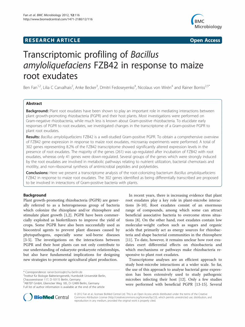
Figure 1 Composition and concentration of the maize root exudates.HPLC. Organic acids, amino acids, and carbohydrates were quantified and tincluded in the graph. Proline, a known constituent of maize root exudate,only with primary amino groups.
secretion of the plant growth hormone auxin, and thesynthesis of volatiles stimulating plant growth andinduced systemic resistance (ISR) [19-21]. In the case ofGram-positive PGPR, however, it is still not clear howthey maneuver their gene expression when exposed toplant-derived compounds. To address this question, thecommercially established FZB42 wild type strain fromBacillus amyloliqufaciens was tested in this study for itstranscriptomic responses to maize root exudates using atwo-color DNA microarray system.
Results and discussionComposition of maize root exudatesMaize root exudates were collected from axenic hydro-ponic cultures and analysed by HPLC for organic acids,amino acids, and oligosaccharides, which have been pre-viously reported to be among the major ingredients inroot exudates [8,22-24].Among the compounds detected, in particular organic
acids such as malic acid, malonic acid, succinic acid andtrans-aconitic acid, were present at highest concentra-tions (Figure 1). Corroborating an earlier report [25], wefound that lactic acid was a main constituent of maizeroot exudates. A variety of amino acids was alsodetected. Glucose and melibiose were the most promin-ent sugars occurring in root exudates. According to thisanalysis, most low-molecular weight organic carbonappeared to be present in the form of organic acids.
Overall changes in gene expression in response to rootexudatesIn the rhizosphere, root exudates may occur at high con-centrations in certain microenvironments, e.g. in vicinityof root tips [26], but their concentration in specificniches of the environment is unknown. Therefore, the
Exudates collected from the roots of maize seedlings were assayed byhose which had a concentration of >0.1 μmol g-1(dry weight) werewas not detected since the derivatization reagent (OPA) used reacts

Fan et al. BMC Microbiology 2012, 12:116 Page 3 of 13http://www.biomedcentral.com/1471-2180/12/116
choice of a physiologically relevant concentration of exu-dates to be used for microarray experiments can only betentative. Based on a previous study on changes in theproteomics of FZB42 [27], three exudate concentrations(0.25 g l-1, 0.5 g l-1 and 1.0 g l-1) were applied to liquidcultures of FZB42, and bacterial cells were harvested forRNA extraction at two growth stages (OD600 = 1.0 andOD600 = 3.0). For simplicity, the two population densitieswere referred to as OD1.0 and OD3.0 throughout thispaper, respectively. A concentration of 0.25 g l-1 was suf-ficient to result in a significant response of FZB42 tran-scriptome. When bacteria were cultured at OD3.0 thenumber of up-regulated genes gradually decreased withincreasing root exudate concentration, suggesting thatsome compounds need to occur at lower abundance toinduce gene expression, or that gene transcription ingeneral may be suppressed at high concentrations ofsome exudates components (Figure 2). More transcriptswere significantly altered (q ≤ 0.01) at the transition tothe stationary growth (OD 3.0) than at the exponentialgrowth (OD1.0) (Figure 2), suggesting that OD 3.0 was asampling point which reflected more clearly the effect ofroot exudates on FZB42 than OD1.0. For these reasons,the exudate concentration of 0.25 g l-1 and the OD3.0for harvesting of cells were used for all subsequentmicroarray experiments.Six independent experiments were performed and the
genes whose transcription fulfilled the condition ofyielding a q value not greater than 0.01 (q ≤ 0.01) and a
Figure 2 Number of FZB42 genes altered in transcription in responsedensities. Maize root exudates were supplemented in three concentrationRNA was prepared from the bacterial cells harvested at two optical densitietranscription (q≤ 0.01 and fold change ≥1.5) by presence of root exudates
fold change not less than 1.5 (FCH ≥ 1.5) were regardedas being significantly influenced by root exudates. Atotal of 302 genes, representing 8.2% of the FZB42 tran-scriptome, were significantly regulated in their transcriptlevels by the applied root exudates (see supplementalmaterial Additional file 1: Tables S1, S2, and S3). Themajority of these genes (261 genes) was up-regulated,whereas only 41 genes were down-regulated (Figure 3).Although most of the regulated genes have been func-tionally annotated, a significant proportion (~23%)remained of unknown function, among which 19 geneswere unique for FZB42. In addition, 44 genes (~15%)encoded either hypothetical proteins or proteins withputative functions (Figure 3). The distribution in variousfunctional categories of all the gene with known (189genes) or putative (44 genes) products are summarizedin Figure 4.
Validation of microarray result by real-time PCRNine up-regulated genes with different levels of foldchanges in expression (1.5 ~ 5.2 fold) were chosen to beevaluated by quantitative real-time PCR. All these geneswere confirmed to be significantly up-regulated in thepresence of root exudates (Figure 5). The fold change ofeach gene revealed by real-time PCR was similar to thatobtained in the microarray experiments (Figure 5). Insummary, the real-time PCR suggested that the micro-array data were reliable.
to root exudates at different exudate concentrations and cells (0.25 mg/ml, 0.5 mg/ml and 1.0 mg/ml) to FZB42 cultures and totals (OD600 = 1.0 and OD600 = 3.0). Genes significantly altered inare represented in the figure.

Figure 3 Overview of groups of the 302 genes altered intranscription by root exudates. A total of 302 genes weresignificantly altered (q≤ 0.01 and fold change ≥1.5) in transcriptionby the maize root exudates. “Up” indicates genes that wereup-regulated in presence root exudates, while “down” the ones thatwere down-regulated by the root exudates. The genes encoding aproduct with known or unknown function and those encoding ahypothetical protein were indicated. The number of genes of eachsection and their percentage is depicted.
Fan et al. BMC Microbiology 2012, 12:116 Page 4 of 13http://www.biomedcentral.com/1471-2180/12/116
The regulated genes with known functionAmong the 302 genes with significantly altered expres-sion by root exudates, 189 were annotated with knownfunctions. These were categorized into various classes[28], such as cell envelope and cellular processes, inter-mediary metabolism, information pathway and otherfunctions . In these categories, three groups (Table 1)contained the largest numbers of genes and at least onethird of the genes within these groups had a fold changeof ≥2.0. This suggests that these three groups of geneswere strongly affected by root exudates:i) The transcription of 46 genes involved in carbon
and nitrogen utilization was altered in response to rootexudates, with 43 of them being up-regulated. These 46genes were involved in different aspects of the metabol-ism of carbohydrates, amino acids and related metabo-lites. To obtain a more comprehensive understanding oftheir relevance in the metabolic context, the genes weremapped into the KEGG pathway and a representation ofmetabolic pathways was constructed (Figure 6). A totalof 12 genes encoding enzymes involved in the Embden-Meyerhof-Parnas (EMP) pathway (including pgi encod-ing for glucose-6-phosphate isomerase) and the TCAcycle were significantly up-regulated. These genes cov-ered almost the entire glycolysis and TCA pathway.
Nearly a quarter of the genes with altered transcription(46 out of 189) were involved in uptake or utilization ofnutrients. This observation corroborated that root exu-dates serve as energy sources in the interaction betweenroots and rhizobacteria.Among the up-regulated genes, glvA, glvC and glvR
showed the highest fold change (glvA: 5.2-fold up-regu-lated, glvC: 2.5-fold up-regulated, glvR: 4.4-fold up-regu-lated). The enhancement of glvA expression was alsovalidated by real-time PCR as well as by the proteomicdata (unpublished). The three genes comprise the glv op-eron (glvA-glvR-glvC), which is responsible for maltosedissimilation and positively regulated by maltose [29].The significant up-regulation of these genes indicatedthat maltose was present in the exudates, which wasconfirmed by the HPLC analysis (Figure 1).The genes involved in inositol metabolism (iolA, iolB,
iolC, iolD, iolE, iolF, iolG, iolI, iolS) were also up-regu-lated, mainly with a fold change of ≥2.0 (Figure 6). Ex-cept iolS, which is involved in the regulation of inositolcatabolism, the other eight genes are members of the ioloperon. The increased transcription of iolA and iolD wasfurther confirmed by real-time PCR whereas the en-hancement of iolB and iolL was validated by a proteo-mics approach (unpublished data). The activation ofnine genes indicated the presence of inositol in the exu-dates, which has also been verified by HPLC.ii) A second group of genes with a higher fold change
were those associated with sensing, chemotaxis, motilityand biofilm formation (Table 2). These processes arecrucial for bacterial colonization of roots. The recogni-tion of signals released from roots and rhizobacteria isthe first step of the establishment of a mutual cross-talk[30]. Once plant signals have been perceived, bacteriamove towards the plant root to establish in the rhizo-sphere [31-34]. Bacterial motility in the rhizosphereinvolves several processes such as chemotaxis, flagella-driven motility, swarming, and production of surfactants[35-38]. The observed transcriptional changes of genesrequired for chemotaxis (cheC, cheD) and motility (hag,fliD, fliP and flgM) indicated that root exudates containcompounds that induce attraction of FZB42 cells toroots.Biofilm formation has been documented to be involved
in directing or modulating efficient colonization byPGPR [39,40]. Biofilms can also provide the plant rootsystem with a protective barrier against attack of patho-genic microbes [35]. Two B. amyloliquefaciens genesinvolved in biofilm formation, ycmA and luxS, wereenhanced by maize root exudates (Table 2, Additionalfile 1: Table S1). The gene luxS, required for synthesisof the quorum-sensing signaling molecule autoinducer-2 (AI-2) [41], is involved in biofilm formation ofpathogenic Streptococcus species [42-44] and the

Figure 4 Distribution in various functional categories of the genes altered in transcription by root exudates. Among the 302 genesaltered in transcription by maize root exudates at OD3.0, those with known (189 genes) or putative (44 genes) products were classified accordingto their function. The percentage of each group is indicated.
Fan et al. BMC Microbiology 2012, 12:116 Page 5 of 13http://www.biomedcentral.com/1471-2180/12/116
probiotic B. subtilis natto [45]. The gene ycmA hasalso been indentified to be involved in facilitating bio-film formation [46,47]. The increased transcription ofluxS and ycmA indicated that biofilm formation ofFZB42 could be enhanced by some compoundspresent in root exudates.
Figure 5 Fold-change of differentially expressed genes selected for vPCR of the selected genes were determined using the software REST. Threechanges obtained in microarray analysis were shown in parenthesis below25th and the 75th percentile. The lines in the boxes represent the medianobservations.
iii) The third functional group with the highest num-ber of genes induced by root exudates was associatedwith the non-ribosomal synthesis of secondary metabo-lites with antimicrobial action (Table 3). Producing sec-ondary metabolites suppressing deleterious microbes inthe rhizosphere is an established mechanism of
alidation by Real-time PCR. The fold changes revealed by real-timerepeats were performed for each gene. For comparison, the foldeach specific gene. The boxes represent the distance between thegene expression. Whiskers represent the minimum and maximum

Table 1 Functional categories* of the FZB42 genessignificantly regulated by the maize root exudates andwith known functions
Classification code_Functional category Nr. of thegenes included
1_cell envelope and cellular processes 58
1.7_ Cell division 6
1.1_ Cell wall 5
1.4_ Membrane bioenergetics 7
1.5_ Mobility and chemotaxis 6
1.3_ Sensors (signal transduction) 2
1.6_ Protein secretion 5
1.8_ Sporulation 7
1.1_ Transformation/competence 2
1.2_ Transport/binding proteins and lipoproteins 18
2_intermediary metabolism 59
2.1_Metabolism of carbohydrates andrelated molecules
34
2.2_ Metabolism of amino acids andrelated molecules
12
2.5_ Metabolism of coenzymes and prosthetic groups 4
2.4_ Metabolism of lipids 5
2.3_ Metabolism of nucleotides and nucleic acids 4
3_information pathways 45
3.3_ DNA recombination 1
3.1_ DNA replication 3
3.8_ Protein modification 2
3.7_ Protein synthesis 20
3.6_ RNA modification 1
3.5_ RNA synthesis 18
4_other functions 27
4.1_ Adaptation to atypical conditions 6
4.2_ Detoxification 4
4.6_ Miscellaneous 3
4.4_ Phage-related functions 1
4.3_ Antibiotic production 13
In total 189
The categories in which more than one third of the genes had a fold changeof ≥2.0 were highlighted in bold text (Refer to experiment “Response to RE”:E-MEXP-3421). *The genes were categorized according to [28].
Fan et al. BMC Microbiology 2012, 12:116 Page 6 of 13http://www.biomedcentral.com/1471-2180/12/116
biocontrol adopted by B. amyloliquefaciens FZB42 onplants [19,48,49]. The majority of the induced genes aredevoted to the synthesis of two polyketide antibiotics,bacillaene and difficidin. Some components in the exu-dates could stimulate the production of these two anti-biotics, which have been demonstrated to be able toprotect orchard trees from fire blight disease caused byErwinia amylovora [49].Another two genes, mlnH and fenE, were also in-
duced, which are known to participate in non-ribosmal
biosynthesis of macrolactin and fengycin, respectively.Macrolactin, a polyketide product found in FZB42,has activity against some Gram-positive bacteria [50],while fengycin can act against phytopathogenic fungiin a synergistic manner with bacillomycin D [19,51].In addition, two genes encoding surfactin synthe-
tase were also activated by root exudates (Table 3).Surfactin is one of Bacillus cyclic lipopeptides, dis-playing antiviral and antibacterial activities. In Arabi-dopsis it has been shown that the ability of Bacillusto synthesize surfactin can reduce the invasion ofPseudomonas syringae [30]. although it is not yetclear whether the protective effect resulted directlyfrom the antibacterial activity of surfactin or from itsbiofilm-related properties. Surfactin is cruciallyinvolved in the motility of Bacillus by reducing sur-face tensions [36,37,52] and contributing to biofilmformation on Arabidopsis roots [30]. It has also beendemonstrated that surfactin production of FZB42 wasenhanced when colonizing the duckweed plant Lemnaminor [21]. It can be expected that up-regulation ofsrfAC and srfAD may contribute to the protectiverole of surfactin against plant pathogens.
The regulated genes with putative functionAmong the 302 genes significantly altered in tran-scription by root exudates, 44 were annotated to en-code a putative enzyme or a hypothetical protein.Similar to the genes with known function, these 44genes fell into three categories: metabolism of carbo-hydrates and related molecules, metabolism of aminoacids and related molecules, and transport/bindingproteins and lipoproteins (Additional file 1: Table S2).Some of the 44 genes were closely associated withplant-microbe interactions. For example, the tran-scription of ydjL, nowadays renamed bdhA, encodingacetoin reductase/butanediol dehydrogenase [53], was1.5-fold enhanced by root exudates. 2, 3-Butanediol isa volatile organic compound released by PGPR andable to promote significantly plant growth [54]. Theexpression of the gene epsE, residing in a 15-geneoperon epsA-O, was also enhanced by root exudates.EpsE is involved in formation of biofilm by arrestingflagellar rotation of cells embedded in biofilm matrix[55]. Another activated gene was dfnY, which encodes ahypothetical protein. Like other induced genes known tobe involved in antibiotic production such as dfnF, dfnG,dfnI and dfnJ (Table 3), dfnY is part of the gene cluster re-sponsible for synthesis of the polyketide antibiotic diffici-din. It is worth mentioning that antibiotic production isenergetically very costly and its strict control is a clear evo-lutionary advantage.In contrast to a few genes significantly altered during
the exponential phase (OD1.0), hundreds of genes were
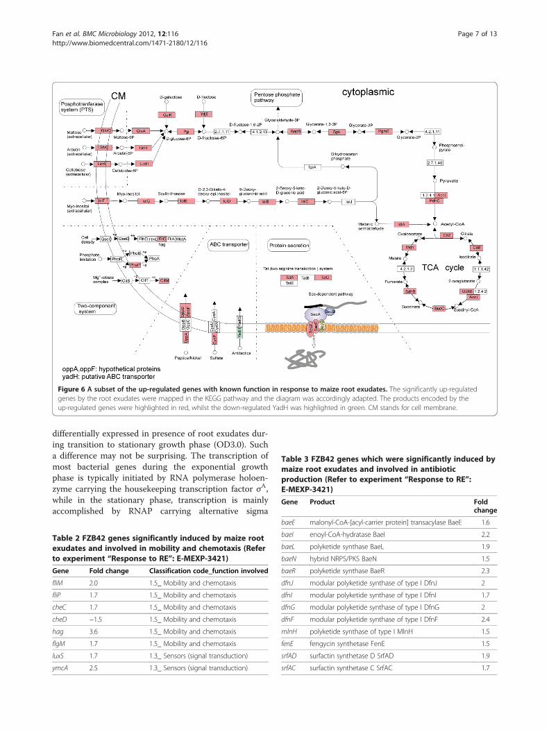
Figure 6 A subset of the up-regulated genes with known function in response to maize root exudates. The significantly up-regulatedgenes by the root exudates were mapped in the KEGG pathway and the diagram was accordingly adapted. The products encoded by theup-regulated genes were highlighted in red, whilst the down-regulated YadH was highlighted in green. CM stands for cell membrane.
Table 3 FZB42 genes which were significantly induced bymaize root exudates and involved in antibioticproduction (Refer to experiment “Response to RE”:E-MEXP-3421)
Gene Product Foldchange
Fan et al. BMC Microbiology 2012, 12:116 Page 7 of 13http://www.biomedcentral.com/1471-2180/12/116
differentially expressed in presence of root exudates dur-ing transition to stationary growth phase (OD3.0). Sucha difference may not be surprising. The transcription ofmost bacterial genes during the exponential growthphase is typically initiated by RNA polymerase holoen-zyme carrying the housekeeping transcription factor σA,while in the stationary phase, transcription is mainlyaccomplished by RNAP carrying alternative sigma
Table 2 FZB42 genes significantly induced by maize rootexudates and involved in mobility and chemotaxis (Referto experiment “Response to RE”: E-MEXP-3421)
Gene Fold change Classification code_function involved
fliM 2.0 1.5_ Mobility and chemotaxis
fliP 1.7 1.5_ Mobility and chemotaxis
cheC 1.7 1.5_ Mobility and chemotaxis
cheD −1.5 1.5_ Mobility and chemotaxis
hag 3.6 1.5_ Mobility and chemotaxis
flgM 1.7 1.5_ Mobility and chemotaxis
luxS 1.7 1.3_ Sensors (signal transduction)
ymcA 2.5 1.3_ Sensors (signal transduction)
baeE malonyl-CoA-[acyl-carrier protein] transacylase BaeE 1.6
baeI enoyl-CoA-hydratase BaeI 2.2
baeL polyketide synthase BaeL 1.9
baeN hybrid NRPS/PKS BaeN 1.5
baeR polyketide synthase BaeR 2.3
dfnJ modular polyketide synthase of type I DfnJ 2
dfnI modular polyketide synthase of type I DfnI 1.7
dfnG modular polyketide synthase of type I DfnG 2
dfnF modular polyketide synthase of type I DfnF 2.4
mlnH polyketide synthase of type I MlnH 1.5
fenE fengycin synthetase FenE 1.5
srfAD surfactin synthetase D SrfAD 1.9
srfAC surfactin synthetase C SrfAC 1.7
Fan et al. BMC Microbiology 2012, 12:116 Page 8 of 13http://www.biomedcentral.com/1471-2180/12/116
factors allowing to adapt to a permanently changing en-vironment. The extracytoplasmic-function (ECF) sigmafactor W was enhanced in presence of root-exudate(Figure 5). SigW is known as being expressed in earlystationary growth-phase and induced by various cell wallantibiotics, alkaline shock, and other stresses affectingthe cell envelope. It controls a large “antibiosis” reguloninvolved in mediating resistance to various antibioticsincluding fosfomycin and the antibiotic peptides sublan-cin and SdpC [56]. It has been observed that manyvirulence-associated factors influence the colonization,persistence and spreading mechanisms of the humanpathogen Streptococcus pyogenes in a growth phase-dependent manner [57-59]. Likewise, rhizobacteria mayemploy an early stationary phase-related mechanism tofavor expression of those genes that mediate rhizospherecompetence.
Effect of soil extractTo simulate in part the conditions that bacteria experi-ence in the rhizosphere soil, 10% soil extract was addedto the culture media. Additional microarray experimentswere performed in a similar way as before to investigatethe effect of the soil extract on gene expression ofFZB42. The result showed that no gene was significantlyup-regulated by the soil extract during exponentialgrowth phase of OD1.0, whereas five genes wererepressed in the presence of the soil extract at OD3.0(Table 4). This negligible number of genes that were dif-ferentially transcribed indicates that the supplement of asoil extract did not have major effects on gene transcrip-tion under the growth conditions used.
Effect of exudates prepared from maize plants colonizedby FZB42Typically, most root exudates studied were collectedfrom plants grown in axenic systems. The release of rootexudates is not only determined by the plant species, butalso by plant age, physiological status, and the biotic en-vironment that plants thrive including the rhizospheremicroflora that influence the composition and quantity ofroot exudates [60-66]. It was reported that P. aeruginosaproduces N-acyl homoserine lactone (AHL) signaling
Table 4 FZB42 genes repressed by soil extract at OD3.0 (Refe
Gene Fold change Product
ypeQ −2.6 hypothetical protein YpeQ
yurV −2.4 iron-sulfur cofactor synthesis protein nifU h
iolS −2.2 inositol utilization protein S (IolS)
yaaA −2.0 conserved hypothetical protein YaaA
ahpF −2.0 alkyl hydroperoxide reductase (large subundehydrogenase AhpF
compounds that induce changes in the root exudation ofMedicago truncatula [67]. Exudate compounds that arespecifically induced or repressed by rhizobacteria may inturn affect bacterial gene expression. Such an effect can-not be demonstrated using root exudates collected froma gnotobiotic system, therefore, a batch of “interactionexudates (IE)” was collected from maize roots which werepreviously inoculated with FZB42.The transcriptional responses of FZB42 to the IE were
compared with responses to the root exudates (RE) col-lected from axenic culture. No significant differences(q ≤ 0.01 and FCH ≥ 1.5) were found between the effectof IE and RE at OD1.0, while four genes were differen-tially expressed at OD3.0 (Additional file 2: Table S5).When a less stringent selection filter was applied(q ≤ 0.05 and FCH ≥ 1.5), a total of nine genes were dif-ferentially expressed (Additional file 2: Table S5). Thefour genes, significantly enhanced in presence of FZB42at maize roots, encode enzymes involved in the degrad-ation of macromolecules or cellular compounds, such asggt, nprE, clpP, RBAM00438 (ycsN). Among all fourgenes, expression of the ggt gene was found mostenhanced, bearing a fold change of 2.2 in presence ofthe rhizobacterium (Additional file 2: Table S5). GGT, γ-glutamyltranspeptidase (GGT) (EC 2.3.2.2) catalyzes thehydrolysis of γ-glutamyl compounds, such as glutathione(GSH), and the transfer of γ-glutamyl moieties to aminoacids and peptides. The nprE gene, which is mainlyexpressed during early stationary phase, encodes extra-cellular neutral protease involved in degradation of pro-teins and peptides. The peptidase ClpP, encoded by theclpP gene, can associate with the ATPases ClpC, ClpE,and ClpX, thereby forming a substrate specific channelfor several regulatory proteins directing spore formationor genetic competence in bacilli. RBAM00438 is a mem-ber of the aldo-keto reductases (AKRs) superfamily ofsoluble NAD(P)(H) oxidoreductases whose chief purposeis to reduce aldehydes and ketones to primary and sec-ondary alcohols. At present, it remains questionable ifthose gene products are linked with any specific processtriggered by the IE. The number of the genes obtainedwas much less than expected. We conclude that possibledifferences between the transcriptome responses to these
r to experiment “Response to SE”: E-MEXP-3551)
Function involved
unknown
omolog YurV miscellaneous
metabolism of carbohydrates and related molecules
unknown
it) and NADH detoxification

Fan et al. BMC Microbiology 2012, 12:116 Page 9 of 13http://www.biomedcentral.com/1471-2180/12/116
two exudate samples are either very rare or too subtle tobe revealed sufficiently by two-color microarrays.One drawback of the present investigation is that some
effects of the root exudates may have been masked bycomponents of the 1 C medium and therefore did notresult in altered gene expression. On the other hand,using 0.25 mg exudates per ml medium, some compo-nents in the exudates may have been diluted to a level atwhich they no longer show detectable effect on bacterialgene expression. It has been reported that the rhizo-sphere is a very heterogeneous soil volume, with someregions being “hotspots” of root exudation and bacterialcolonization. In natural environments, bacterial popula-tions are likely to be exposed to different concentrationof exudates along the root axis [68,69].It needs to be mentioned that the exudates used in this
study were a pooled mixture of the samples collectedwithin seven days from maize roots (see Methods). Ithas not yet been described to which extent the compos-ition of root exudates is affected by the developmentalstage of a plant [70] and therefore the presented bacter-ial responses cannot be assigned to a particular physio-logical state of the host plant. This question may beaddressed by performing bacterial transcriptome ana-lyses in response to exudates collected at different timepoints during plant development. Such an approach maybe helpful to elucidate the progression of the plant-bacteria association during the plant development.In summary, this microarray work reflects the interac-
tions between a Gram-positive rhizobacterium and itshost plant in a genome-scale perspective. Critical targetgenes and pathways for further investigations of theinteraction were identified. Given the limited reports ontranscriptomic analysis of rhizobacteria in response totheir host plants [13-15], the results provided a valuableinsight into PGPR behaviour in the rhizosphere. About10% of the total number of genes were found up-regulated in presence of root exudate during transitionto stationary growth phase. In addition to the findingscorroborating previous transcriptome analyses per-formed in Gram-negative bacteria, we could demon-strate that presence of root exudate induced expressionof numerous genes involved in non-ribosomal synthesisof secondary metabolites with antifungal and antibacter-ial action. We hypothesize that competitive colonizationat plant root surfaces by FZB42 might be supported byenhanced synthesis of antimicrobial compounds.
ConclusionsUsing the data from six independent micro array experi-ments, differentially transcribed genes of the PGPR B.amyloliquefaciens FZB42 were identified and theirknown or putative functions were related to their asso-ciative behavior with regard to interactions with maize
roots. A large group of genes specifically expressed sug-gested that root exudates serve primarily as a source ofcarbon and energy for FZB42. Another group of genessignificantly induced by plant root exudates encode thenon-ribosomal synthesis of antimicrobial secondarymetabolites. It is possible that enhanced synthesis ofantimicrobial compounds might suppress the competingphytopathogenic organisms growing within the plantrhizosphere. However, direct evidence for occurrence ofthose compounds in vicinity of plant rhizosphereremains to be accomplished. The addition of soilextracts to the growth medium showed no major effecton gene expression of FZB42. Similarly, the resultsobtained with the “interaction exudates” collected fromthe maize roots inoculated with FZB42 did not indicatealtered effects on gene expression compared with that ofcommon root exudates collected in the gnotobioticsystem.
MethodsRoot exudates collection and analysisMaize seeds (Saaten-Union, Germany) were surface-sterilized and germinated as described previously [21].Root exudates were collected from the maize seedlingsgrown in an axenic system with sterile water (1:1 dis-tilled water and tap water, v/v). Forty germinated seedsharboring a main root of at least 2 cm length were trans-ferred into test tubes filled with 2 ml of autoclavedwater, with the maize seeds being placed just above thewater surface. The tubes were kept under sterile condi-tions and maintained in a plant growth room (16-hlight/8-h dark) at 24°C for 8 days. In the first two days,water was supplemented to the tubes, and seedlingswere pulled to a higher position to ensure that the maizeseeds were always above the water surface as the rootselongated. From the third day on, the water containingthe exudates was collected and the tubes were refilledwith sterile water. Sampling was performed every dayuntil the eighth day after transferring the seedlings. Eachcollection were kept separate, from which a 100 μL ali-quot was taken and spread on a solid LB media to checkfor contamination. The contaminated samples werediscarded.To collect the “interaction exudates (IE)”, the germi-
nated maize seeds were inoculated with FZB42 asdescribed previously [21] before transferring the testtubes. Afterward the maize was grown and the exudateswere prepared in the same way as described above.The collected exudates were pooled, freeze-dried and
stored at −20°C. Before use, the lyophilized exudateswere weighted, and dissolved in a certain volume of dis-tilled water. The obtained exudates solution was centri-fuged to remove any insoluble constituents. Thesupernatant was filter-sterilized and the resulting stock
Fan et al. BMC Microbiology 2012, 12:116 Page 10 of 13http://www.biomedcentral.com/1471-2180/12/116
exudates were stored in dark at −80°C. The final concen-tration of the exudates in the culture vessel was gener-ally adjusted to 0.25 g L-1. Chemical analysis of the rootexudates was performed as described previously [71]:amino acids were determined using a Shimadzu HPLCsystem. 40 μL samples were derivatized with 160 μl OPA(o-phthaldialdehyde) reagent and 20 μL of the resultingmixture were injected and separated on a GROM-SILOPA-3 column using solvent gradient elution by solventA (25 mM phosphate buffer pH 7.2 with 0.75% tetra-hydrofuran) and solvent B (methanol to acetonitrile to25 mM phosphate buffer 7.2 [35 : 15 : 50/v : v : v]). Gra-dient profile: 0–2 min, 0% B; 2–10 min, 0%-50% B; 10–15 min, 50–60% B; 15–20 min, 60–100% B; 20–25 min,100% B; 25–26 min, 100%-0% B; 26–35 min, 0% B. Theflow-rate was 1 mL min-1. Subsequent fluorescence de-tection of the derivatives was performed at an excitationwavelength of 330 nm and 450 nm. Organic acids weredetermined by means of ion chromatography (DionexIonPac AS 11 HC column) using a gradient rangingfrom 4 mM to 80 mM KOH. Organic acids were identi-fied by comparison of retention time with known stan-dards. Sugars were determined by GC-TOF-MS. Alyophilized 75 μL aliquot of root exudates was dissolvedin 50 mL methoxyamine hydrochloride in dry pyrididineand derivatized for 2 h at 37°C followed by 30 min. treat-ment with 50 μL N-methyl-N-trifluoroacetamide at 37°C. A volume of 1 μL was injected into the GC column.
Microarray designThe Bam4kOLI microarray was designed based on thesequenced complete genome of B. amyloliquefaciensFZB42 [27] (Additional file 3: Table S6). The array con-tained 3931 50-70mer oligonucleotides representingpredicted protein-encoding genes and a set of smallnon-coding RNA genes of FZB42. In addition, the arrayincluded stringency controls with 71%, 80% and 89%identity to the native sequences of five genes, dnaA,rpsL, rpsO, rpsP, and rpmI, to monitor the extent ofcross hybridization. The array also contained alien DNAoligonucleotides for four antibiotic resistance genes(Emr, Cmr, Nmr and Spcr) and eight spiking controls aswell as one empty control (nothing spotted). All oligo-nucleotide probes were printed in four replicates. Micro-arrays were produced and processed as describedpreviously [72].Oligonucleotides were designed using the Oligo De-
signer software (Bioinformatics Resource Facility, CeBi-Tec, Bielefeld University). Melting temperatures of theoligonucleotides were calculated based on %GC andoligo length, ranging from 73°C to 83°C (optimal 78°C).Salt concentration was set to 0.1 M. QGramMatch wasused to analyse uniqueness of the oligos.
Experimental designThe experiment designs of FZB42 in response to variousconditions are summarized in Additional file 3: Table S6.Independent experiments were used as biological repli-cates. In all comparisons dye-swap were carried out tominimize the effect of dye biases.1 C medium (0.7%w/v pancreatic digest of casein,
0.3%w/v papain digest of soya flour, 0.5%w/v NaCl)containing 0.1% glucose was used in all experiments. Ex-cept the controls of the experiment “Response to SE”(Additional file 3: Table S6), 10% soil extract was alsosupplemented in the media. Soil extract was prepared byextracting 500 g dried, fertile garden soil with one litredistilled water for 2 hrs and autoclaving. After coolingdown, the supernatant was filtered with 0.22 μm Nucle-pore unit and then stored at 4°C until use.
Total RNA preparationOne overnight colony of FZB42 was inoculated into 1 Cmedium plus 0.1% glucose and then shaken at 210 rpmat 24°C. After 14 hours the obtained preculture was usedto inoculate a new 1 C medium (containing 0.1% glu-cose) plus the corresponding solution to be studied(maize root exudates, soil extract, or interaction exu-dates. See Additional file 3: Table S6). The main cultureswere grown at 24°C until they reached late exponentialgrowth phase (OD 1.0) and/or the transition to station-ary phase (OD3.0, see Additional file 4: Figure S1).The FZB42 cells of OD1.0 or OD3.0 were harvested
for preparation of total RNA. A volume of 15 ml of theculture was mixed with 7.5 ml “killing buffer” (20 mMTris–HCl, 5 mM MgCl2, 20 mM NaN3, pH 7.5) andthen centrifuged at 5,000 rpm for 3 minutes at roomtemperature. The pellet was washed once more with1 ml “killing buffer” and then immediately frozen in li-quid nitrogen. The frozen cell pellets were stored at−80°C until RNA isolation.Isolation of RNA was performed using the Nucleo
SpinW RNA L (Macherey Nagel) according to the manu-facturer’s instructions. The isolated RNA was addition-ally digested with DNaseI to avoid possible trace DNAcontamination. After ethanol precipitation RNA pelletswere resuspended in 300 μl RNase-free water. The con-centration of total RNA was spectrophotometricallydetermined, whereas its quality was checked on a 1.5%RNA agarose gel in 1 ×MEN buffer (20 mM MOPS;1 mM EDTA, 5 mM NaAc; pH7.0) with 16%formaldehyde.
Synthesis of labeled cDNA, hybridization and imageacquisitionSynthesis of first-strand cDNA, microarray hybridizationand image acquisition were performed in CeBiTec, theCenter for Biotechnology at Bielefeld University. Briefly,
Fan et al. BMC Microbiology 2012, 12:116 Page 11 of 13http://www.biomedcentral.com/1471-2180/12/116
aminoallyl-modified first-strand cDNAs were synthe-sized by reverse transcription according to DeRisi et al[73]. and then coupled with Cy3- and Cy5-N-hydroxy-succinimidyl ester dyes (GE Healthcare, Little Chalfont,UK). After hybridization using the HS4800 hybridizationstation (Tecan Trading AG, Switzerland), slides werescanned with a pixel size of 10 μm using the LSReloaded microarray scanner (Tecan Trading AG,Switzerland).
Data processingThe microarray data obtained was analysed by using theEMMA 2.8.2 software [74]. The mean signal intensity(Ai) was calculated for each spot using the formula Ai =log2(RiGi)
0.5 [75]. Ri = Ich1(i)−Bgch1(i) and Gi = Ich2(i)−Bgch2(i), where Ich1(i) or Ich2(i) is the intensity of a spot inchannel 1 or channel 2, and Bgch1(i) or Bgch2(i) is thebackground intensity of a spot in channel1 or channel 2,respectively. The log2 value of the ratio of signal inten-sities (Mi) was calculated for each spot using the for-mula Mi = log2(Ri/Gi). Spots were flagged as “empty” ifR ≤ 0.5 in both channels, where R= (signal mean–back-ground mean)/background standard deviation [76]. Theraw data were normalized by the method of LOWESS(locally weighted scattered plot smoothing). A signifi-cance test was performed by the method of false discov-ery rate (FDR) control and the adjusted p-value definedby FDR was called q-value [77,78].An arbitrary cutoff, fold change (FCH) greater than
1.5, was applied to the genes with a q-value of ≤0.01.Only those genes which meet both filter conditions(q ≤ 0.01 & FCH ≥ 1.5) were regarded to be significantlydifferentially expressed.
Real-time PCRThe first-strand cDNA was obtained by reverse tran-scription with RevertAidTM Premium Reverse Tran-scriptase (Fermentas, St. Leon-Rot, Germany), usingrandom hexamers as primers. Oligonucleotide primerswere designed by the software PrimerExpress and listedin supplemental materials (Additional files 1: Table S4).Real-time PCR was performed with SYBRW Green PCRMaster Mix kit (Carlsbad, California, USA) using 7500Fast Real-Time PCR System (Carlsbad, California, USA)according to the manufacturers’ instructions. As an in-ternal control, the housekeeping gene gyrA was used asits expression was not significantly altered in all micro-array experiments. Three technical replicates were car-ried out for each target gene. Quantification wasanalysed based on the threshold cycle (Ct) values asdescribed by Pfaffl [79].The raw data of the Micro-array experiments,
described here, are available in the ArrayExpress data-base under the accession numbers: E-MEXP-3421, E-
MEXP-3550, E-MEXP-3551, E-MEXP-3553, E-MEXP-3554, respectively (see also Additional file 3: Table S6).
Additional files
Additional file 1: Table S1. The genes of FZB42 with known functionwhose transcriptions were significantly altered in response to maizeroot exudates at OD3.0 (Refer to experiment “Response to RE”:E-MEXP-3421). Table S2: The genes of FZB42 with putative function orencoding hypothetical protein whose transcriptions were significantlyaltered in response to maize root exudates at OD3.0 (Refer to experiment“Response to RE”: E-MEXP-3421). Table S3: The genes of FZB42 withunknown function whose transcriptions were significantly altered inresponse to maize root exudates at OD3.0 (Refer to experiment “Responseto RE”: E-MEXP-3421). Table S4: The primers used for real-time PCR.
Additional file 2: Table S5. Differentially expressed genes of FZB42in response to IE compared with those to RE (Refer to experiment“IE <> RE”: E-MEXP-3553). The genes highlighted were those with a qvalue of ≤0.01.
Additional file 3: Table S6. Microarray experimental design anddata bank accession.
Additional file 4: Figure S2. Growth of FZB42 at 24°C undercontinuous shaking (220 rpm/min.) in medium 1 C supplementedwith sterilized 10% soil extract prepared by extracting of 500 g (dryweight) compost soil with 1 L distilled water. Cells were sampledduring exponential growth (OD600 = 1.0) and during transition tostationary growth phase. The time of sampling in the transition phase (O.D.600 = 3.0) is indicated by the red arrow.
Authors’ contributionsBF carried out the main experiments and data analysis and wrote themanuscript draft. LCC performed complementary experiments and revisedthe manuscript. AB designed the array and was responsible for thehybridization experiments. DF performed the metabolite analysis of rootexudates. NvW revised the manuscript. RB guided experimental design andwrote the final version of the manuscript. All authors read and approved thefinal manuscript.
AcknowledgementsThe financial support for FB by the Priority Academic Development Programof Jiangsu Higher Education Institutions and the National Natural ScienceFoundation of China (No. 31100081) and the German Academic ExchangeService (DAAD) is gratefully acknowledged, as well as, the financial supportgiven to RB in-frame of the competence network Genome Research onBacteria (GenoMikPlus, GenoMikTransfer) and of the Chinese-Germancollaboration program by the German Ministry for Education and Research(BMBF). This study was further supported by the EU-FP6-funded projectRHIBAC. We are very indebted to Birgit Baumgarth, ComputationalGenomics, Center for Biotechnology (CeBiTec), Bielefeld University forperforming later hybridisation experiments and support in data processing.We also would like to thank Anne Pohlmann for the excellent assistance inthe real-time experiments.
Author details1Institute of Forest Protection, Nanjing Forestry University, Longpan Road159, 210037 Nanjing, China. 2Institut für Biologie Bakteriengenetik, HumboldtUniversität Berlin, Chausseestrasse 117, D-10115 Berlin, Germany. 3MolekulareGenetik, Institut für Biologie III, Albert-Ludwigs-Universität Freiburg,Schänzlestrasse 1, D-79104 Freiburg, Germany. 4Leibniz Institute for PlantGenetics and Crop Plant Research, Corrensstr. 3, 06466 Gatersleben,Germany. 5ABiTEP GmbH, Glienicker Weg 185, D-12489 Berlin, Germany.
Received: 1 April 2012 Accepted: 31 May 2012Published: 21 June 2012
References1. Lugtenberg BJJ, Kamilova F: Plant-growth-promoting rhizobacteria. Annu
Rev Microbiol 2009, 63:541–556.
Fan et al. BMC Microbiology 2012, 12:116 Page 12 of 13http://www.biomedcentral.com/1471-2180/12/116
2. Kloepper JW, Schroth MN: Plant growth-promoting rhizobacteria onradishes. In Proc of the 4th Internat Conf on Plant Pathogenic Bacter. Angers,France: INRA; 1978.
3. Domenech J, Reddy MS, Kloepper JW, Ramos B, Gutierrez-Manero J:Combined application of the biological product LS213 with Bacillus,Pseudomonas or Chryseobacterium for growth promotion and biologicalcontrol of soil-borne diseases in pepper and tomato. BioControl 2006,51(2):245–258.
4. Alabouvette C, Olivain C, Migheli Q, Steinberg C: Microbiological control ofsoil-borne phytopathogenic fungi with special emphasis on wilt-inducing Fusarium oxysporum. New Phytol 2009, 184(3):529–544.
5. Dessaux Y, Ryan PR, Thomashow LS, Weller DM: Rhizosphere engineeringand management for sustainable agriculture. Plant Soil 2009,321(1–2):363–383.
6. Somers E, Vanderleyden J, Srinivasan M: Rhizosphere bacterial signalling: alove parade beneath our feet. Crit Rev Microbiol 2004, 30(4):205–240.
7. Oger P, Petit A, Dessaux Y: Genetically engineered plants producingopines alter their biological environment. Nat Biotech 1997,15(4):369–372.
8. Rudrappa T, Czymmek KJ, Pare PW, Bais HP: Root-secreted malic acidrecruits beneficial soil bacteria. Plant Physiol 2008, 148(3):1547–1556.
9. Micallef SA, Shiaris MP, Colon-Carmona A: Influence of Arabidopsisthaliana accessions on rhizobacterial communities and natural variationin root exudates. J Exp Bot 2009, 60(6):1729–1742.
10. Badri DV, Vivanco JM: Regulation and function of root exudates. Plant CellEnviron 2009, 32(6):666–681.
11. Shi S, Richardson AE, O'Callaghan M, DeAngelis KM, Jones EE, Stewart A,Firestone MK, Condron LM: Effects of selected root exudate componentson soil bacterial communities. FEMS Microbiol Ecol 2011, 77(3):600–610.
12. Diehn M, Relman DA: Comparing functional genomic datasets: lessonsfrom DNA microarray analyses of host-pathogen interactions. Curr OpinMicrobiol 2001, 4(1):95–101.
13. Mark GL, Dow JM, Kiely PD, Higgins H, Haynes J, Baysse C, Abbas A, Foley T,Franks A, Morrissey J, et al: Transcriptome profiling of bacterial responsesto root exudates identifies genes involved in microbe-plant interactions.Proc Natl Acad Sci U S A 2005, 102(48):17454–17459.
14. Matilla M, Espinosa-Urgel M, Rodriguez-Herva J, Ramos J, Ramos-GonzalezM: Genomic analysis reveals the major driving forces of bacterial life inthe rhizosphere. Genome Biol 2007, 8(9):R179.
15. Ramachandran VK, East AK, Karunakaran R, Downie JA, Poole PS: Adaptationof Rhizobium leguminosarum to pea, alfalfa and sugar beet rhizospheresinvestigated by comparative transcriptomics. Genome Biol 2011,12(10):R106.
16. Bashan Y, Holguin G, de-Bashan LE: Azospirillum-plant relationships:physiological, molecular, agricultural, and environmental advances(1997–2003). Can J Microbiol 2004, 50(8):521–577.
17. Steenhoudt O, Vanderleyden J: Azospirillum, a free-living nitrogen-fixingbacterium closely associated with grasses: genetic, biochemical andecological aspects. FEMS Microbiol Rev 2000, 24(4):487–506.
18. Elizabeth ABE, Jo H: Biocontrol of plant disease: a (Gram-) positiveperspective. FEMS Microbiol Lett 1999, 171(1):1–9.
19. Chen XH, Koumoutsi A, Scholz R, Borriss R: More than anticipated -production of antibiotics and other secondary metabolites by Bacillusamyloliquefaciens FZB42. J Mol Microbiol Biotechnol 2009, 16(1–2):14–24.
20. Idris EE, Iglesias DJ, Talon M, Borriss R: Tryptophan-dependent productionof indole-3-acetic acid (IAA) affects level of plant growth promotion byBacillus amyloliquefaciens FZB42. Mol Plant Microbe Interact 2007,20(6):619–626.
21. Fan B, Chen XH, Budiharjo A, Bleiss W, Vater J, Borriss R: Efficientcolonization of plant roots by the plant growth promoting bacteriumBacillus amyloliquefaciens FZB42, engineered to express greenfluorescent protein. J Biotechnol 2011, 151(4):303–311.
22. Lugtenberg BJJ, Dekkers LC, Bloemberg GV: Molecular determinants ofrhizosphere colonization by Pseudomonas. Annu Rev Phytopathol 2001,39:461–490.
23. Lugtenberg BJJ, Dekkers LC: What makes Pseudomonas bacteriarhizosphere competent? Environ Microbiol 1999, 1(1):9–13.
24. Simons M, van der Bij AJ, Brand I, de Weger LA, Wijffelman CA, LugtenbergBJ: Gnotobiotic system for studying rhizosphere colonization by plantgrowth-promoting Pseudomonas bacteria. Mol Plant Microbe Interact 1996,9(7):600–607.
25. Kraffczyk I, Trolldenier G, Beringer H: Soluble root exudates of maize:Influence of potassium supply and rhizosphere microorganisms. Soil BiolBiochem 1984, 16(4):315–322.
26. Dennis PG, Miller AJ, Hirsch PR: Are root exudates more important thanother sources of rhizodeposits in structuring rhizosphere bacterialcommunities? FEMS Microbiol Ecol 2010, 72(3):313–327.
27. Chen XH, Koumoutsi A, Scholz R, Eisenreich A, Schneider K, Heinemeyer I,Morgenstern B, Voss B, Hess WR, Reva O, et al: Comparative analysis of thecomplete genome sequence of the plant growth-promoting bacteriumBacillus amyloliquefaciens FZB42. Nat Biotechnol 2007, 25(9):1007–1014.
28. Moszer I, Jones LM, Moreira S, Fabry C, Danchin A: SubtiList: the referencedatabase for the Bacillus subtilis genome. Nucleic Acids Res 2002,30(1):62–65.
29. Yamamoto H, Serizawa M, Thompson J, Sekiguchi J: Regulation of the glvoperon in Bacillus subtilis: YfiA (GlvR) is a positive regulator of theoperon that is repressed through CcpA and cre. J Bacteriol 2001,183(17):5110–5121.
30. Bais HP, Fall R, Vivanco JM: Biocontrol of Bacillus subtilis against infectionof Arabidopsis roots by Pseudomonas syringae is facilitated by biofilmformation and surfactin production. Plant Physiol 2004, 134(1):307–319.
31. de Weert S, Vermeiren H, Mulders IH, Kuiper I, Hendrickx N, Bloemberg GV,Vanderleyden J, De Mot R, Lugtenberg BJ: Flagella-driven chemotaxistowards exudate components is an important trait for tomato rootcolonization by Pseudomonas fluorescens. Mol Plant Microbe Interact 2002,15(11):1173–1180.
32. De Weert S, Kuiper I, Lagendijk EL, Lamers GE, Lugtenberg BJ: Role ofchemotaxis toward fusaric acid in colonization of hyphae of Fusariumoxysporum f. sp. radicis-lycopersici by Pseudomonas fluorescens WCS365.Mol Plant Microbe Interact 2004, 17(11):1185–1191.
33. O'Sullivan DJ, O'Gara F: Traits of fluorescent Pseudomonas spp. involvedin suppression of plant root pathogens. Microbiol Rev 1992, 56(4):662–676.
34. Walsh UF, Morrissey JP, O'Gara F: Pseudomonas for biocontrol ofphytopathogens: from functional genomics to commercial exploitation.Curr Opin Biotechnol 2001, 12(3):289–295.
35. Ongena M, Jacques P: Bacillus lipopeptides: versatile weapons for plantdisease biocontrol. Trends Microbiol 2008, 16(3):115–125.
36. Raaijmakers JM, de Bruijn I, de Kock MJ: Cyclic lipopeptide production byplant-associated Pseudomonas spp.: diversity, activity, biosynthesis, andregulation. Mol Plant Microbe Interact 2006, 19(7):699–710.
37. Daniels R, Vanderleyden J, Michiels J: Quorum sensing and swarmingmigration in bacteria. FEMS Microbiol Rev 2004, 28(3):261–289.
38. Capdevila S, Martinez-Granero FM, Sanchez-Contreras M, Rivilla R, Martin M:Analysis of Pseudomonas fluorescens F113 genes implicated in flagellarfilament synthesis and their role in competitive root colonization.Microbiology 2004, 150(Pt 11):3889–3897.
39. Combes-Meynet E, Pothier JF, Moenne-Loccoz Y, Prigent-Combaret C: ThePseudomonas secondary metabolite 2,4-diacetylphloroglucinol is a signalinducing rhizoplane expression of Azospirillum genes involved in plant-growth promotion. Mol Plant Microbe Interact 2010, 24(2):271–284.
40. Ramey BE, Koutsoudis M, Bodman SBv, Fuqua C: Biofilm formation inplant-microbe associations. Curr Opin Microbiol 2004, 7(6):602–609.
41. Surette MG, Miller MB, Bassler BL: Quorum sensing in Escherichia coli,Salmonella typhimurium, and Vibrio harveyi: a new family of genesresponsible for autoinducer production. Proc Natl Acad Sci U S A 1999,96(4):1639–1644.
42. Heilmann C, Schweitzer O, Gerke C, Vanittanakom N, Mack D, Gotz F:Molecular basis of intercellular adhesion in the biofilm-formingStaphylococcus epidermidis. Mol Microbiol 1996, 20(5):1083–1091.
43. Gotz F: Staphylococcus and biofilms. Mol Microbiol 2002, 43(6):1367–1378.44. Huang Z, Meric G, Liu Z, Ma R, Tang Z, Lejeune P: luxS-based quorum-
sensing signaling affects Biofilm formation in Streptococcus mutans.J Mol Microbiol Biotechnol 2009, 17(1):12–19.
45. Lombardia E, Rovetto AJ, Arabolaza AL, Grau RR: A LuxS-dependent cell-to-cell language regulates social behavior and development in Bacillussubtilis. J Bacteriol 2006, 188(12):4442–4452.
46. Branda SS, Gonzalez-Pastor JE, Dervyn E, Ehrlich SD, Losick R, Kolter R:Genes involved in formation of structured multicellular communities byBacillus subtilis. J Bacteriol 2004, 186(12):3970–3979.
47. Kearns DB, Chu F, Branda SS, Kolter R, Losick R: A master regulator forbiofilm formation by Bacillus subtilis. Mol Microbiol 2005,55(3):739–749.
Fan et al. BMC Microbiology 2012, 12:116 Page 13 of 13http://www.biomedcentral.com/1471-2180/12/116
48. Chen XH, Koumoutsi A, Scholz R, Schneider K, Vater J, Sussmuth R, Piel J,Borriss R: Genome analysis of Bacillus amyloliquefaciens FZB42 reveals itspotential for biocontrol of plant pathogens. J Biotechnol 2009,140(1–2):27–37.
49. Chen XH, Scholz R, Borriss M, Junge H, Mogel G, Kunz S, Borriss R: Difficidinand bacilysin produced by plant-associated Bacillus amyloliquefaciens areefficient in controlling fire blight disease. J Biotechnol 2009,140(1–2):38–44.
50. Schneider K, Chen XH, Vater J, Franke P, Nicholson G, Borriss R, SussmuthRD: Macrolactin is the polyketide biosynthesis product of the pks2cluster of Bacillus amyloliquefaciens FZB42. J Nat Prod 2007,70(9):1417–1423.
51. Koumoutsi A, Chen XH, Henne A, Liesegang H, Hitzeroth G, Franke P, VaterJ, Borriss R: Structural and functional characterization of gene clustersdirecting nonribosomal synthesis of bioactive cyclic lipopeptides inBacillus amyloliquefaciens strain FZB42. J Bacteriol 2004,186(4):1084–1096.
52. Leclere V, Marti R, Bechet M, Fickers P, Jacques P: The lipopeptidesmycosubtilin and surfactin enhance spreading of Bacillus subtilis strainsby their surface-active properties. Arch Microbiol 2006, 186(6):475–483.
53. Nicholson WL: The Bacillus subtilis ydjL (bdhA) gene encodes acetoinreductase/2,3-butanediol dehydrogenase. Appl Environ Microbiol 2008,74(22):6832–6838.
54. Ryu CM, Farag MA, Hu CH, Reddy MS, Wei HX, Pare PW, Kloepper JW:Bacterial volatiles promote growth in Arabidopsis. Proc Natl Acad SciU S A 2003, 100(8):4927–4932.
55. Blair KM, Turner L, Winkelman JT, Berg HC, Kearns DB: A molecular clutchdisables flagella in the Bacillus subtilis biofilm. Science 2008,320(5883):1636–1638.
56. Mascher T, Hachmann AB, Helmann JD: Regulatory overlap and functionalredundancy among Bacillus subtilis extracytoplasmic function sigmafactors. J Bacteriol 2007, 189(19):6919–6927.
57. Kreikemeyer B, McIver KS, Podbielski A: Virulence factor regulation andregulatory networks in Streptococcus pyogenes and their impact onpathogen-host interactions. Trends Microbiol 2003, 11(5):224–232.
58. Beyer-Sehlmeyer G, Kreikemeyer B, Horster A, Podbielski A: Analysis of thegrowth phase-associated transcriptome of Streptococcus pyogenes. Int JMed Microbiol 2005, 295(3):161–177.
59. Chaussee MA, Dmitriev AV, Callegari EA, Chaussee MS: Growth phase-associated changes in the transcriptome and proteome of Streptococcuspyogenes. Arch Microbiol 2008, 189(1):27–41.
60. Wieland G, Neumann R, Backhaus H: Variation of microbial communities insoil, rhizosphere, and rhizoplane in response to crop species, soil type,and crop development. Appl Environ Microbiol 2001, 67(12):5849–5854.
61. Buyer JS, Roberts DP, Russek-Cohen E: Soil and plant effects on microbialcommunity structure. Can J Microbiol 2002, 48(11):955–964.
62. Kowalchuk GA, Buma DS, de Boer W, Klinkhamer PG, van Veen JA: Effects ofabove-ground plant species composition and diversity on the diversityof soil-borne microorganisms. Antonie van Leeuwenhoek 2002,81(1–4):509–520.
63. Broeckling CD, Broz AK, Bergelson J, Manter DK, Vivanco JM: Root exudatesregulate soil fungal community composition and diversity. Appl EnvironMicrobiol 2008, 74(3):738–744.
64. Kuzyakov Y, Raskatov A, Kaupenjohann M: Turnover and distribution ofroot exudates of Zea mays. Plant Soil 2003, 254(2):317–327.
65. Yang CH, Crowley DE: Rhizosphere microbial community structure inrelation to root location and plant iron nutritional status. Appl EnvironMicrobiol 2000, 66(1):345–351.
66. Wang Y, Ohara Y, Nakayashiki H, Tosa Y, Mayama S: Microarray analysis ofthe gene expression profile induced by the endophytic plant growth-promoting rhizobacteria, Pseudomonas fluorescens FPT9601-T5 inArabidopsis. Mol Plant Microbe Interact 2005, 18(5):385–396.
67. Mathesius U, Mulders S, Gao M, Teplitski M, Caetano-Anolles G, Rolfe BG,Bauer WD: Extensive and specific responses of a eukaryote to bacterialquorum-sensing signals. Proc Natl Acad Sci U S A 2003, 100(3):1444–1449.
68. Dennis PG, Miller AJ, Hirsch PR: Are root exudates more important thanother sources of rhizodeposits in structuring rhizosphere bacterialcommunities? FEMS Microbiol Ecol, 72(3):313–327.
69. Kuzyakov Y: Priming effects: Interactions between living and deadorganic matter. Soil Biol Biochem 2010, 42(9):1363–1371.
70. Haichar FZ, Marol C, Berge O, Rangel-Castro JI, Prosser JI, Balesdent J,Heulin T, Achouak W: Plant host habitat and root exudates shape soilbacterial community structure. ISME J 2008, 2(12):1221–1230.
71. Carvalhais LC, Dennis PG, Fedoseyenko D, Hajirezaei MR, Borriss R, vonWiren N: Root exudation of sugars, amino acids, and organic acids bymaize as affected by nitrogen, phosphorus, potassium, and irondeficiency. Journal of Plant Nutrition and Soil Science 2011, 174(1):3–11.
72. Brune I, Becker A, Paarmann D, Albersmeier A, Kalinowski J, Puhler A,Tauch A: Under the influence of the active deodorant ingredient 4-hydroxy-3-methoxybenzyl alcohol, the skin bacterium Corynebacteriumjeikeium moderately responds with differential gene expression. JBiotechnol 2006, 127(1):21–33.
73. DeRisi JL, Iyer VR, Brown PO: Exploring the metabolic and genetic controlof gene expression on a genomic scale. Science 1997, 278(5338):680–686.
74. Dondrup M, Albaum SP, Griebel T, Henckel K, Junemann S, Kahlke T, KleindtCK, Kuster H, Linke B, Mertens D, et al: EMMA 2–a MAGE-compliant systemfor the collaborative analysis and integration of microarray data. BMCBioinforma 2009, 10:50.
75. Dudoit S, Yang YH, Callow MJ, Speed TP: Statistical methods foridentifying differentially expressed genes in replicated cDNA microarrayexperiments. Stat Sin 2002, 12(1):111–139.
76. Serrania J, Vorholter FJ, Niehaus K, Puhler A, Becker A: Identification ofXanthomonas campestris pv. campestris galactose utilization genes fromtranscriptome data. J Biotechnol 2008, 135(3):309–317.
77. Benjamini Y, Hochberg Y: Controlling the False Discovery Rate: A Practicaland Powerful Approach to Multiple Testing. Journal of the Royal StatisticalSociety Series B (Methodological) 1995, 57(1):289–300.
78. Roberts PC, El-Gewely MR: Gene expression microarray data analysisdemystified. Biotechnol Annu Rev 2008, 14:29–61.
79. Pfaffl MW: A new mathematical model for relative quantification inreal-time RT-PCR. Nucleic Acids Res 2001, 29(9):e45.
doi:10.1186/1471-2180-12-116Cite this article as: Fan et al.: Transcriptomic profiling of Bacillusamyloliquefaciens FZB42 in response to maize root exudates. BMCMicrobiology 2012 12:116.
Submit your next manuscript to BioMed Centraland take full advantage of:
• Convenient online submission
• Thorough peer review
• No space constraints or color figure charges
• Immediate publication on acceptance
• Inclusion in PubMed, CAS, Scopus and Google Scholar
• Research which is freely available for redistribution
Submit your manuscript at www.biomedcentral.com/submit