Genotyping: A Tool for Controlling Classical Scrapie Revised October 2008.

RESEARCH ARTICLE Open Access
The natural atypical scrapie phenotype ispreserved on experimental transmission andsub-passage in PRNP homologous sheepMarion M Simmons1*, Timm Konold1, Lisa Thurston1, Susan J Bellworthy1, Melanie J Chaplin2, S Jo Moore1
Abstract
Background: Atypical scrapie was first identified in Norwegian sheep in 1998 and has subsequently beenidentified in many countries. Retrospective studies have identified cases predating the initial identification of thisform of scrapie, and epidemiological studies have indicated that it does not conform to the behaviour of aninfectious disease, giving rise to the hypothesis that it represents spontaneous disease.However, atypical scrapie isolates have been shown to be infectious experimentally, through intracerebral inocula-tion in transgenic mice and sheep. The first successful challenge of a sheep with ‘field’ atypical scrapie from anhomologous donor sheep was reported in 2007.
Results: This study demonstrates that atypical scrapie has distinct clinical, pathological and biochemicalcharacteristics which are maintained on transmission and sub-passage, and which are distinct from other strains oftransmissible spongiform encephalopathies in the same host genotype.
Conclusions: Atypical scrapie is consistently transmissible within AHQ homozygous sheep, and the diseasephenotype is preserved on sub-passage.
BackgroundAtypical scrapie was first identified in Norwegian sheepin 1998 [1], and has subsequently been identified inmany European countries [2] and elsewhere around theworld, including North America [3] and the FalklandIslands [4]. Retrospective studies have identified casespredating this initial identification [5,6] and epidemiolo-gical studies have indicated that this form of scrapiedoes not conform to the behaviour of an infectious dis-ease [7]. In many cases, atypical scrapie does not co-exist with classical scrapie (e.g. on individual farms, butalso at a national level, such as in Portugal and the Falk-land Islands [8]), although some field cases originatefrom flocks which have also reported classical scrapie,and it has occurred in a closed research flock with ende-mic classical disease [9]. It has also been reported in aresearch flock in which detailed biosecurity measureshave been maintained, founded with sheep from a coun-try free of classical scrapie [10]. This has led to
proposals [2,10,11] that this form of scrapie is in fact aspontaneous disease which may have existed undetectedfor a long time.Despite lack of evidence for infectivity in the epide-
miological data, atypical scrapie isolates have beenshown to be infectious experimentally, through intracer-ebral inoculation into ovinised transgenic mice [12] andsheep [13].The interactions of host and infecting strain on ovine
transmissible spongiform encephalopathies (TSEs) areknown to be complex, and have a profound effect onthe resulting phenotype of disease. Following experi-mental challenges distinct ‘strain’ characteristics can beidentified [14], but when naturally-occurring disease isstudied it is clear that host prion protein gene (PRNP)genotype can have a significant effect on the resultingphenotype [15], although a single genotype may supportmore than one phenotype [15,16]. The preferred geno-type ‘target’ range of atypical scrapie is different to thatof classical scrapie [17,18] but there is some overlap insusceptible genotypes.* Correspondence: [email protected]
1Department of Pathology, Veterinary Laboratories Agency Weybridge, NewHaw, Addlestone KT15 3NB, UK
Simmons et al. BMC Veterinary Research 2010, 6:14http://www.biomedcentral.com/1746-6148/6/14
© 2010 Simmons et al; licensee BioMed Central Ltd. This is an Open Access article distributed under the terms of the CreativeCommons Attribution License (http://creativecommons.org/licenses/by/2.0), which permits unrestricted use, distribution, andreproduction in any medium, provided the original work is properly cited.

In contrast to the widespread variation in pathologyand immunopathology characteristics seen in classicalscrapie [15], the pathology observed in atypical casesappears to be much more consistent, regardless of geno-type [1,19-21].The first successful challenge of an AHQ/AHQ sheep
with ‘field’ atypical scrapie from an homologous donorsheep was reported in 2007 [13]. This paper providesdata on further homologous AHQ/AHQ transmissions,including sub-passage of the initial case, and comparesthis with similar data from field cases of classical scra-pie, and experimental bovine spongiform encephalopa-thy (BSE) in the same genotype.
ResultsFull details of all the animals presented in this paper aregiven in Table 1. In addition to the AHQ/AHQ animalsintracerebrally challenged with atypical scrapie (cases 1-9),data is presented on the experimental classical scrapiepositive control animals which were also intracerebrallychallenged (cases 18-20).The scale of such studies, and the limited availability
of both inoculum and recipient sheep precludes a studydesign which includes every genotype/strain/route ofchallenge combination. To enable us to assess the abilityof a single PRNP genotype to support various TSEs withdistinct pathological characteristics, data is presented forcomparison from AHQ/AHQ animals orally challengedwith BSE (cases 10-14) [22-24] and field cases of classi-cal scrapie which have arisen naturally in AHQ/AHQsheep (cases 15-17) [25,26].Animals challenged with field atypical scrapie suc-
cumbed to disease from 378 - 1057 days post challenge(mean 751 days), which compares with 126, 537 and605 days for the one VRQ/VRQ and two ARQ/VRQclassical scrapie intracerebrally inoculated controlsrespectively.
ClinicalFull details of the clinical signs displayed by each animalare given in Table 1. Unlike classical scrapie and ovineBSE, atypical scrapie did not appear to present with anysigns of pruritus. Ataxia was observed in all atypicalscrapie cases. Four of nine atypical cases displayed signsof an asymmetric lesion distribution in the brain, suchas circling to one side and a unilateral absent menaceresponse. This was however not consistently displayedin all animals inoculated with the same material.
Pathology and immunohistochemistryBoth the vacuolation profiles and immunopathologicalcharacteristics were very consistent within the animalschallenged with atypical scrapie, and distinct from the
other AHQ/AHQ animals where disease was attributa-ble to other strains.Full vacuolation profiles could only be obtained for
four of the five primary transmission cases, and onesub-passage animal. Vacuolation (Figures 1a, b) in theatypical cases was absent in the brainstem, and mostpronounced in the cerebellar and cerebral cortices, ascompared to the BSE and classical scrapie cases inwhich brainstem lesions were predominant. In the thala-mic and frontal areas the main difference between theclassical scrapie and BSE profiles related to the relativeintensity of vacuolation, which was greater in the classi-cal scrapie cases.Immunohistochemistry (IHC; Figure 2) shows that
immunolabelling patterns are also distinct, with boththe type of labelling and the neuroanatomical distribu-tion being different for the atypical scrapie compared tothe other strains. However, the atypical scrapie patternremains the same on primary versus secondary passage(Figure 3), and is consistent with the IHC patternsdescribed in natural disease [20].Western immunoblotting (WB; Figure 4) shows that
the banding pattern associated with atypical scrapie[1,27] is present in all the atypical challenged cases, andis consistent regardless of whether it is primary or sec-ondary passage (Figure 4). The blot of the field casewhich was examined in the context of a retrospectivereview of natural disease [28] was consistent with classi-cal scrapie. The BSE cases were blotted within the remitof their source project (Chaplin and Bellworthy, unpub-lished data), and conform to the blot pattern (low mole-cular mass of the unglycosylated band with antibody6H4 and a weak P4 antibody reaction) associated withexperimental BSE in sheep.No immunolabelling was seen in any lymphoid tissue
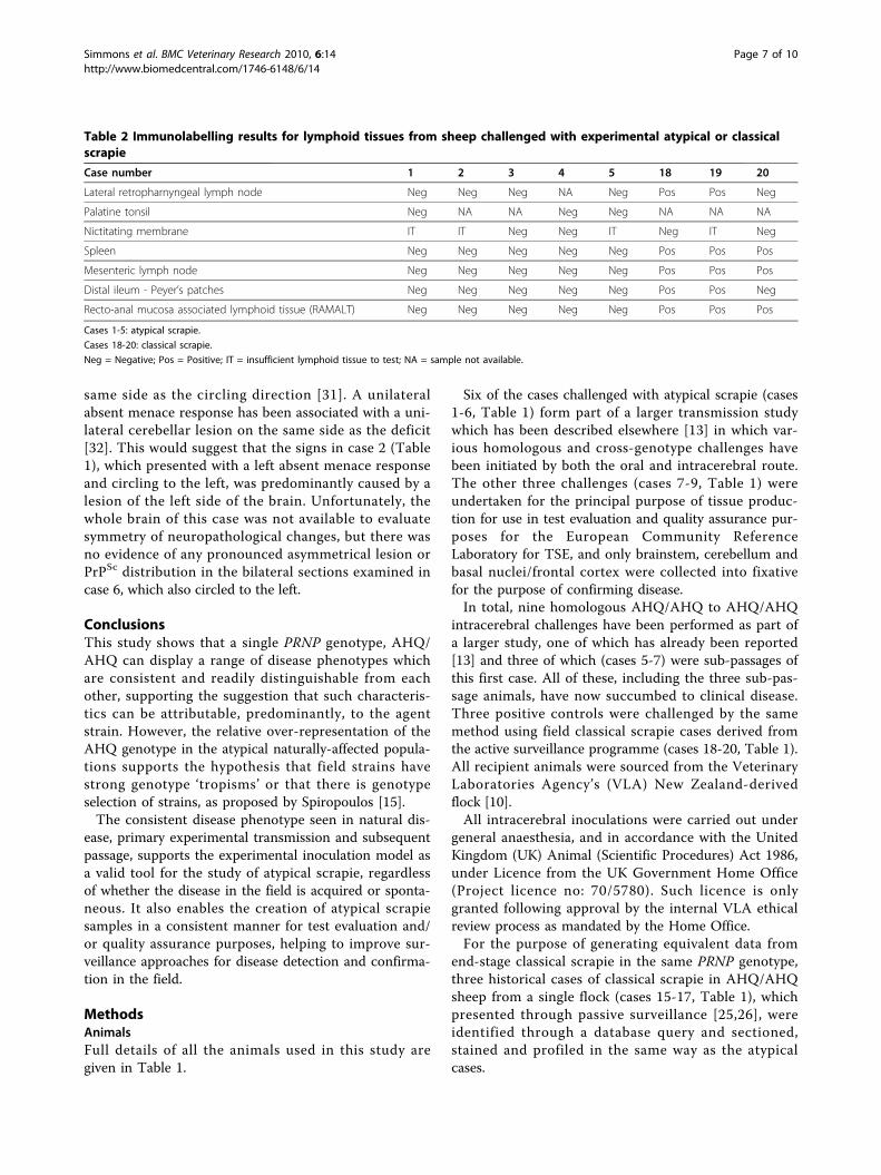
from the atypical challenged sheep, despite widespreadinvolvement of the equivalent tissues in the positiveclassical scrapie controls and the BSE-challenged sheep(Table 2).
DiscussionThis data confirms the experimental transmissibilityof atypical scrapie, and the stability of disease phenotype -clinical, biochemical and pathological - on PRNPhomologous experimental transmission in AHQ/AHQanimals. The mean incubation time of these cases (751days) is approximately 50% of the average age at deathin field cases with atypical scrapie [20]. This observationthat intracerebral challenge shortens the incubation per-iod is consistent with the substantial reductions in incu-bation period which are seen in the classical scrapiecontrols which are 90% (for VRQ/VRQ) and 60% (forVRQ/ARQ) less than the average age at onset of field
Simmons et al. BMC Veterinary Research 2010, 6:14http://www.biomedcentral.com/1746-6148/6/14
Page 2 of 10

Table 1 Individual case data of genotype, breed, inoculum, route of inoculation, incubation periods and clinical signs
Caseno
Breed/genotype Challenged with Route Age atchallenge(months)
Ip (days) Clinical signs
1* Cheviot AHQ/AHQ
AHQ -atypicalpassive (PG1347/04) Portland
Intracerebral 5.5 378 Behavioural changes (confusion, separation from others),compulsive behaviour (spontaneous nibbling, circlingclockwise), mild hind limb ataxia, weight loss, normalmenace response, no signs of pruritus
2 Cheviot AHQ/AHQ
As above Intracerebral 5.5 1057 Compulsive behaviour (circling anti-clockwise), absentmenace response in the left eye, head tremor, generalataxia, no weight loss, no signs of pruritus
3 Cheviot AHQ/AHQ
Active surveillance- breed notknown
Intracerebral 5.5 878 Behavioural change (nervousness), bilateral absent menaceresponse, head tremor, general ataxia, minor weight loss
4 Cheviot AHQ/AHQ
Same case asabove
Intracerebral 6 990 Behavioural changes (confusion, separation from others),bilateral absent menace response, head tremor, generalataxia, no weight loss, no signs of pruritus
5 Cheviot AHQ/AHQ
Subpassage ofmaterial fromcase 1
Intracerebral 3 667 No behavioural changes, bilateral absent menace response,general ataxia, no weight loss, no signs of pruritus
6 Cheviot AHQ/AHQ
Subpassage ofmaterial fromcase 1
Intracerebral 3 747 Compulsive behaviour (circling anti-clockwise), bilateralabsent menace response, head tremor, general ataxia &hypermetria, minor weight loss, no signs of pruritus
7 Cheviot AHQ/AHQ
Subpassage ofmaterial fromcase 1
Intracerebral 3 671 Behavioural changes (confusion), bilateral absent menaceresponse, head tremor, general ataxia, minor weight loss,no signs of pruritus
8 Cheviot AHQ/AHQ
(PG519/06) AHQ/AHQ passive case- Cheviot
Intracerebral 3 607 Behaviour changes (dullness, teeth grinding), normalmenace response, head tremor, general ataxia, weight loss,no signs of pruritus
9 Cheviot AHQ/AHQ
(PG519/06) AHQ/AHQ passive case- Cheviot
Intracerebral 3 833 Behavioural changes (confusion, separation from others),compulsive behaviour (circling anti-clockwise), normalmenace response, general ataxia, minor weight loss, nosigns of pruritus
10 Cheviot AHQ/AHQ
Bovine BSE oral 4 573 Abnormal behaviour (confusion), signs of pruritus (frequentrubbing, positive scratch test, wool loss), mild ataxia
11 Cheviot AHQ/AHQ
Bovine BSE oral 4.5 595 Head tremor, signs of pruritus (frequent rubbing, wool loss,positive scratch test), minor weight loss
12 Cheviot AHQ/AHQ
Bovine BSE oral 4.5 617 Signs of pruritus (wool loss), weight loss
13 Cheviot AHQ/AHQ
Bovine BSE oral 8 642 Abnormal behaviour (separation from others), normalmenace response, head tremor, signs or pruritus (frequentrubbing), mild ataxia, weight loss
14 Cheviot AHQ/AHQ
Bovine BSE oral 4.5 664 Abnormal behaviour (teeth grinding), signs of pruritus(frequent rubbing, wool loss), weight loss
15 British milksheepX Finn DorsetAHQ/AHQ
Classical scrapie Natural Notapplicable
9 years atclinical onset
Abnormal behaviour (teeth grinding), ataxia, no signs ofpruritus
16 British milksheepX Finn DorsetAHQ/AHQ
Classical scrapie Natural Notapplicable
5 years atclinical onset
No behavioural abnormalities, mild ataxia, pruritus (positivescratch test)
17 British milksheepX Finn DorsetAHQ/AHQ
Classical scrapie Natural Notapplicable
3 years 9months atclinical onset
No behavioural abnormalities, mild ataxia, signs of pruritus(frequent rubbing)
18 Cheviot ARQ/VRQ
Classical scrapie Intracerebral 10 537 Abnormal behaviour (nervousness, separation from others),normal menace response, head & body tremor, signs ofpruritus (positive scratch test, rubbing with nibble reflex),ataxia, weight loss
Simmons et al. BMC Veterinary Research 2010, 6:14http://www.biomedcentral.com/1746-6148/6/14
Page 3 of 10

cases (Ortiz-Pelaez, personal communication) in thesegenotypes.There was a slight shortening of mean incubation per-
iod between the initial experimentally challenged sheep(790 days) and those which succumbed following sub-passage (695 days), but the group sizes are too small todraw any robust conclusions from this observation.
The wide range of incubation periods seen cannot bereadily interpreted due to the very small numbers ofanimals in this study (a necessary restriction due to thevery limited amount of suitable donor material, and lim-ited number of recipients of a suitable genotype). It isinteresting to note that the outliers (cases 1 and 2,Table 1) received the same inoculum. All donor inocula
Table 1: Individual case data of genotype, breed, inoculum, route of inoculation, incubation periods and clinical signs(Continued)
19 Dorset ARQ/VRQ Classical scrapie Intracerebral 10 605 Abnormal behaviour (nervousness), normal menaceresponse, head tremor, signs of pruritus (rubbing withnibble reflex), mild ataxia, weight loss
20 Dorset VRQ/VRQ Classical scrapie Intracerebral 11 126 Abnormal behaviour (dullness), absent menace response inthe eye, signs of pruritus (positive scratch test, frequentrubbing), hind limb ataxia, minor weight loss
Ip = incubation period.
* This case previously reported [13].
Figure 1 Vacuolation profiles for atypical scrapie, classical scrapie and BSE in AHQ/AHQ sheep. See Table 1 for details of the cases, andTable 3 for area code key, and mean ± SD for each data point. a) The profiles for the classical scrapie (pink, cases 15-17) and ovine BSE (blue,cases 10-14) cases are similar, particularly in terms of brainstem involvement, whereas the atypical scrapie profile (green, cases 1-9) has distinctivepeaks in the cerebellum (area 12) and frontal cortex (area 20). b) The vacuolation profiles for experimental atypical scrapie cases subdivided bypassage. The profile from the original challenged animals (green, cases 1-4 and 8-9) is very similar to that in the animals that succumbed todisease following sub-passage (red, cases 5-7).
Simmons et al. BMC Veterinary Research 2010, 6:14http://www.biomedcentral.com/1746-6148/6/14
Page 4 of 10

give similar incubation periods in ovinised transgenic(Tg338) mice when inoculated as part of a separatestudy (Spiropoulos, personal communication), althoughsome difference in titre is likely to be masked by thesensitivity of these mice. In one titration of an atypicalscrapie isolate the mean incubation period did not
change more than 10% over the first three log dilutions(Spiropoulos and Simmons, unpublished data).The absence of any visible lymphoreticular system
(LRS) involvement in the experimentally challenged ani-mals is consistent with what has been observed in nat-ural cases of atypical scrapie [13] and cannot be
Figure 2 PrPSc deposition pattern (PDP) maps for atypical scrapie, classical scrapie, and BSE in AHQ/AHQ sheep. Note that, incomparison to classical scrapie and ovine BSE, the atypical scrapie cases have relatively sparse immunolabelling in the obex and rostral midbrain,widespread and prominent immunolabelling in the cerebellar cortex and white matter, sparse immunolabelling in the hypothalamus, andprominent immunolabelling in the neocortex and cortical white matter.
Simmons et al. BMC Veterinary Research 2010, 6:14http://www.biomedcentral.com/1746-6148/6/14
Page 5 of 10

attributed to the experimental route of exposure, sincethe classical scrapie controls challenged by the sameroute had widespread labelling of disease-associated PrP(PrPSc) in the lymphoid tissues.The clinical presentation of atypical scrapie cases was
different to classical scrapie or BSE in sheep: atypicalscrapie did not appear to cause evident pruritus, whereasthe scrapie and BSE cases described here usually pre-sented with pruritus. Compulsive behaviour, such as cir-cling, was only observed in atypical scrapie, and an
impaired menace response was considerably more fre-quent in atypical scrapie cases. The difference in the clin-ical picture does not appear to be confined to thegenotype or the route of inoculation since similar signsin the absence of pruritus have been observed in natu-rally affected atypical scrapie cases of various genotypes[1,10,29], whereas pruritus is frequent in naturallyaffected classical scrapie cases [26] or sheep orally chal-lenged or naturally infected with the BSE agent underexperimental settings [24]. Ataxia with hypermetria, headtremor and an absent menace response, which were pre-dominantly observed in sheep with atypical scrapie, aresigns indicative of a dysfunction of the cerebellum [30].This concurs with the lesion and PrPSc immunolabellingpattern distribution in atypical scrapie, which is particu-larly prominent in the cerebellum. However, the laterali-sation of clinical signs (circling to one side or aunilaterally deficient menace response) seen in atypicalscrapie is unusual and to the authors’ knowledge has notbeen described elsewhere in ovine BSE or classical scra-pie. It is unlikely that the intracerebral route was respon-sible for the apparent lateralisation of clinical signsbecause it was not observed in ovine cases of BSE [24]and classical scrapie inoculated by the same route (cases18-20, Table 1). Compulsive circling in the absence ofvestibular signs like head tilt and nystagmus is suggestiveof an asymmetric lesion of the forebrain, usually on the
Figure 3 Immunolabelling in atypical scrapie cases. Immunolabelling characteristics in atypical scrapie are unchanged in natural disease (a-c),and primary (d-f) and secondary (g-i) passage. Note the characteristic three-band pattern in the neocortex (a, d, g); prominent immunolabellingin the molecular and granular cell layers of the cerebellum (b, e, h); prominent fine granular immunolabelling in the basal nuclei (c, f, i).
1 2 3 4 5 6 7 8 9 10
20.1 kDa29 kDa
Figure 4 Western immunoblot of caudal medulla of theatypical scrapie cases. WB using monoclonal antibody Sha31,showing the consistency of blot characteristics between field case,experimental and sub-passaged atypical scrapie. Lanes 1 and 10Biotinylated markers. Lane 2 Case 4. Lane 3 Case 8 (the sub-passageof Case 4). Lanes 4 and 5 Cases 7 and 5, both sub-passages of Case1 (not shown). Lane 6 Field case atypical scrapie positive control.Lane 7 Field case classical scrapie control. Lane 8 Bovine BSEpositive control. Lane 9 Scrapie negative control.
Simmons et al. BMC Veterinary Research 2010, 6:14http://www.biomedcentral.com/1746-6148/6/14
Page 6 of 10
same side as the circling direction [31]. A unilateralabsent menace response has been associated with a uni-lateral cerebellar lesion on the same side as the deficit[32]. This would suggest that the signs in case 2 (Table1), which presented with a left absent menace responseand circling to the left, was predominantly caused by alesion of the left side of the brain. Unfortunately, thewhole brain of this case was not available to evaluatesymmetry of neuropathological changes, but there wasno evidence of any pronounced asymmetrical lesion orPrPSc distribution in the bilateral sections examined incase 6, which also circled to the left.
ConclusionsThis study shows that a single PRNP genotype, AHQ/AHQ can display a range of disease phenotypes whichare consistent and readily distinguishable from eachother, supporting the suggestion that such characteris-tics can be attributable, predominantly, to the agentstrain. However, the relative over-representation of theAHQ genotype in the atypical naturally-affected popula-tions supports the hypothesis that field strains havestrong genotype ‘tropisms’ or that there is genotypeselection of strains, as proposed by Spiropoulos [15].The consistent disease phenotype seen in natural dis-
ease, primary experimental transmission and subsequentpassage, supports the experimental inoculation model asa valid tool for the study of atypical scrapie, regardlessof whether the disease in the field is acquired or sponta-neous. It also enables the creation of atypical scrapiesamples in a consistent manner for test evaluation and/or quality assurance purposes, helping to improve sur-veillance approaches for disease detection and confirma-tion in the field.
MethodsAnimalsFull details of all the animals used in this study aregiven in Table 1.
Six of the cases challenged with atypical scrapie (cases1-6, Table 1) form part of a larger transmission studywhich has been described elsewhere [13] in which var-ious homologous and cross-genotype challenges havebeen initiated by both the oral and intracerebral route.The other three challenges (cases 7-9, Table 1) wereundertaken for the principal purpose of tissue produc-tion for use in test evaluation and quality assurance pur-poses for the European Community ReferenceLaboratory for TSE, and only brainstem, cerebellum andbasal nuclei/frontal cortex were collected into fixativefor the purpose of confirming disease.In total, nine homologous AHQ/AHQ to AHQ/AHQ
intracerebral challenges have been performed as part ofa larger study, one of which has already been reported[13] and three of which (cases 5-7) were sub-passages ofthis first case. All of these, including the three sub-pas-sage animals, have now succumbed to clinical disease.Three positive controls were challenged by the samemethod using field classical scrapie cases derived fromthe active surveillance programme (cases 18-20, Table 1).All recipient animals were sourced from the VeterinaryLaboratories Agency’s (VLA) New Zealand-derivedflock [10].All intracerebral inoculations were carried out under
general anaesthesia, and in accordance with the UnitedKingdom (UK) Animal (Scientific Procedures) Act 1986,under Licence from the UK Government Home Office(Project licence no: 70/5780). Such licence is onlygranted following approval by the internal VLA ethicalreview process as mandated by the Home Office.For the purpose of generating equivalent data from
end-stage classical scrapie in the same PRNP genotype,three historical cases of classical scrapie in AHQ/AHQsheep from a single flock (cases 15-17, Table 1), whichpresented through passive surveillance [25,26], wereidentified through a database query and sectioned,stained and profiled in the same way as the atypicalcases.
Table 2 Immunolabelling results for lymphoid tissues from sheep challenged with experimental atypical or classicalscrapie
Case number 1 2 3 4 5 18 19 20
Lateral retropharnyngeal lymph node Neg Neg Neg NA Neg Pos Pos Neg
Palatine tonsil Neg NA NA Neg Neg NA NA NA
Nictitating membrane IT IT Neg Neg IT Neg IT Neg
Spleen Neg Neg Neg Neg Neg Pos Pos Pos
Mesenteric lymph node Neg Neg Neg Neg Neg Pos Pos Pos
Distal ileum - Peyer’s patches Neg Neg Neg Neg Neg Pos Pos Neg
Recto-anal mucosa associated lymphoid tissue (RAMALT) Neg Neg Neg Neg Neg Pos Pos Pos
Cases 1-5: atypical scrapie.
Cases 18-20: classical scrapie.
Neg = Negative; Pos = Positive; IT = insufficient lymphoid tissue to test; NA = sample not available.
Simmons et al. BMC Veterinary Research 2010, 6:14http://www.biomedcentral.com/1746-6148/6/14
Page 7 of 10
Similarly, five cases of experimental BSE in sheep(orally challenged [22-24], cases 10-14, Table 1) wereavailable to us for comparison, and sections were simi-larly prepared and scored.
Clinical monitoringA neurological examination was conducted when animalhusbandry staff suspected clinical disease (for moredetails, see [13]).
Pathology and immunohistochemistryWhole brain was removed from each animal and hemi-sected longitudinally. One half of the brain was placedinto 10% formal saline for histology, and the other halfstored at -80°C. Prompted by the clinical observations inone case (case 6, Table 1) an additional section of cerebel-lum and cerebrum of the ‘fresh’ half of the brain was takenand fixed for comparison of lesion intensity and distribu-tion with the other half of the brain. A range of lymphoidtissues were also collected from the experimental group.All brain tissue was routinely fixed, processed into
paraffin wax, sectioned and stained with haematoxylin
and eosin as described in detail elsewhere [16]. Fulldetails of the areas profiled are given in Table 3.Immunohistochemical detection of PrPSc was per-
formed using mouse monoclonal antibody 2G11 (InstitutPourquier, Montpellier, France), raised against ovine PrPpeptide sequence 146-R154 R171-182.Tissue sections were de-waxed and rehydrated routinely.
Epitope demasking was performed by immersion of sec-tions for 30 minutes in undiluted formic acid, then washedin running tap water for 15 minutes, followed by autoclav-ing at 121°C in citrate buffer pH 6.1 (8.8 mM tri-sodiumcitrate dihydrate, 1.3 mM citric acid in 2 litres purifiedwater). Endogenous peroxidase was blocked using 3%hydrogen peroxide (100 vol) in methanol, and washingbuffer used throughout the procedure was tris bufferedsaline, supplemented with 0.2% tween20 (TBST). Primaryantibody was applied at dilution of 1/400 for 1 hour atroom temperature, with immunodetection performedusing biotinylated goat anti mouse and avidin-biotin-per-oxidase-complex (Vector Elite, Burlingame, USA) techni-que using diaminobenzidine chromogen prepared inMcIlvane’s citrate buffer. Sections were counterstained
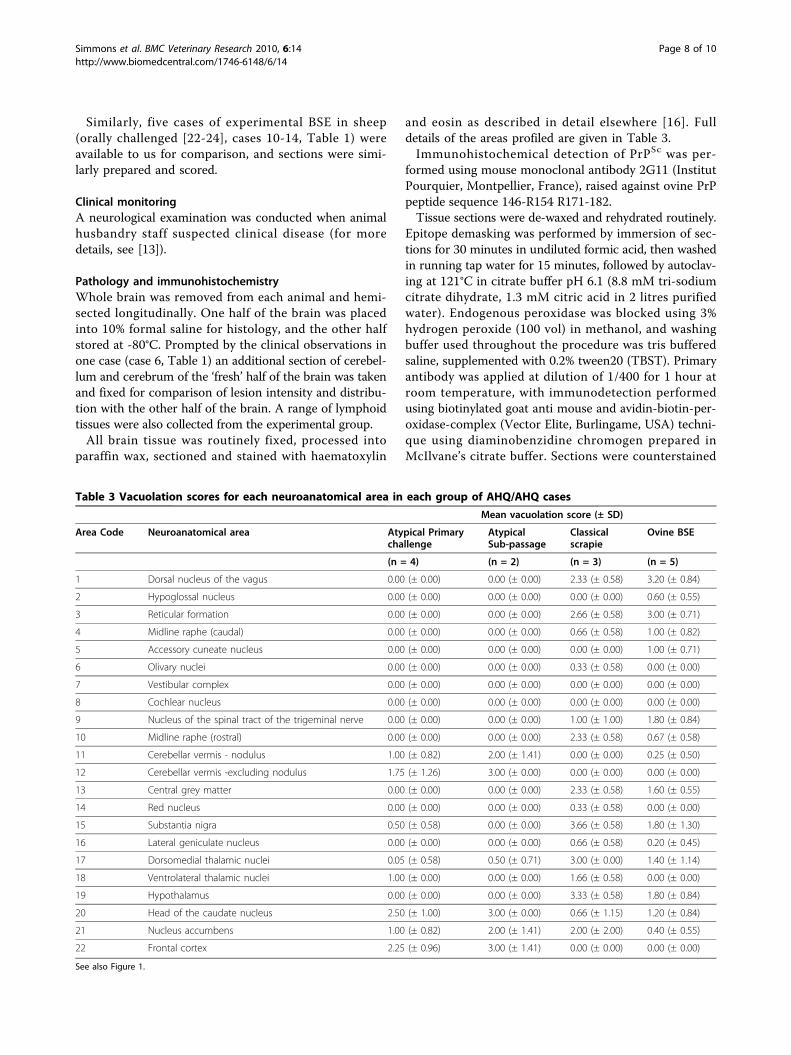
Table 3 Vacuolation scores for each neuroanatomical area in each group of AHQ/AHQ cases
Mean vacuolation score (± SD)
Area Code Neuroanatomical area Atypical Primarychallenge
AtypicalSub-passage
Classicalscrapie
Ovine BSE
(n = 4) (n = 2) (n = 3) (n = 5)
1 Dorsal nucleus of the vagus 0.00 (± 0.00) 0.00 (± 0.00) 2.33 (± 0.58) 3.20 (± 0.84)
2 Hypoglossal nucleus 0.00 (± 0.00) 0.00 (± 0.00) 0.00 (± 0.00) 0.60 (± 0.55)
3 Reticular formation 0.00 (± 0.00) 0.00 (± 0.00) 2.66 (± 0.58) 3.00 (± 0.71)
4 Midline raphe (caudal) 0.00 (± 0.00) 0.00 (± 0.00) 0.66 (± 0.58) 1.00 (± 0.82)
5 Accessory cuneate nucleus 0.00 (± 0.00) 0.00 (± 0.00) 0.00 (± 0.00) 1.00 (± 0.71)
6 Olivary nuclei 0.00 (± 0.00) 0.00 (± 0.00) 0.33 (± 0.58) 0.00 (± 0.00)
7 Vestibular complex 0.00 (± 0.00) 0.00 (± 0.00) 0.00 (± 0.00) 0.00 (± 0.00)
8 Cochlear nucleus 0.00 (± 0.00) 0.00 (± 0.00) 0.00 (± 0.00) 0.00 (± 0.00)
9 Nucleus of the spinal tract of the trigeminal nerve 0.00 (± 0.00) 0.00 (± 0.00) 1.00 (± 1.00) 1.80 (± 0.84)
10 Midline raphe (rostral) 0.00 (± 0.00) 0.00 (± 0.00) 2.33 (± 0.58) 0.67 (± 0.58)
11 Cerebellar vermis - nodulus 1.00 (± 0.82) 2.00 (± 1.41) 0.00 (± 0.00) 0.25 (± 0.50)
12 Cerebellar vermis -excluding nodulus 1.75 (± 1.26) 3.00 (± 0.00) 0.00 (± 0.00) 0.00 (± 0.00)
13 Central grey matter 0.00 (± 0.00) 0.00 (± 0.00) 2.33 (± 0.58) 1.60 (± 0.55)
14 Red nucleus 0.00 (± 0.00) 0.00 (± 0.00) 0.33 (± 0.58) 0.00 (± 0.00)
15 Substantia nigra 0.50 (± 0.58) 0.00 (± 0.00) 3.66 (± 0.58) 1.80 (± 1.30)
16 Lateral geniculate nucleus 0.00 (± 0.00) 0.00 (± 0.00) 0.66 (± 0.58) 0.20 (± 0.45)
17 Dorsomedial thalamic nuclei 0.05 (± 0.58) 0.50 (± 0.71) 3.00 (± 0.00) 1.40 (± 1.14)
18 Ventrolateral thalamic nuclei 1.00 (± 0.00) 0.00 (± 0.00) 1.66 (± 0.58) 0.00 (± 0.00)
19 Hypothalamus 0.00 (± 0.00) 0.00 (± 0.00) 3.33 (± 0.58) 1.80 (± 0.84)
20 Head of the caudate nucleus 2.50 (± 1.00) 3.00 (± 0.00) 0.66 (± 1.15) 1.20 (± 0.84)
21 Nucleus accumbens 1.00 (± 0.82) 2.00 (± 1.41) 2.00 (± 2.00) 0.40 (± 0.55)
22 Frontal cortex 2.25 (± 0.96) 3.00 (± 1.41) 0.00 (± 0.00) 0.00 (± 0.00)
See also Figure 1.
Simmons et al. BMC Veterinary Research 2010, 6:14http://www.biomedcentral.com/1746-6148/6/14
Page 8 of 10
using Mayer’s haematoxylin, then routinely dehydrated,cleared and mounted in dibutylphthalate in xylene (DPX),before examination by light microscopy.Vacuolation and immunohistochemistry profiles were
created using standard subjective methods as previouslydescribed [16,20] in which the severity of vacuolarlesions, or the type of PrP immunolabelling, is assessedin a standard range of precise neuroanatomical areas.Some modifications were made to the original method[20] to accommodate the range of morphological PrPSc
immunolabelling types seen across the three TSEstrains. Globular and punctuate labelling types, whichhave only been observed in atypical scrapie cases[2,20,33], were not included in the calculation of theaverage scores for each area. Therefore average PrPdeposition pattern scores (0 = negative to 6 = strongpositive) for each area were calculated using only theremaining 12 immunolabelling types (intraneuronal,intraglial, intra-astrocytic, (fine) granular, stellate, linear,perineuronal, plaque-like, subpial, (sub)ependymal, peri-vascular, vascular plaques).Tissues from the LRS of each challenged animal were
examined by IHC, using the same method describedabove. The effects of the intracerebral route of challengewere controlled for by examination of the LRS frompositive control VRQ/VRQ and ARQ/VRQ animals thathad been challenged intracerebrally with classical scrapie(cases 18-20, Table 1).
Western ImmunoblotFresh brain samples were subjected to the TeSeE Univer-sal WB (Bio-Rad Cat No: 355 1169 Marnes-la-Coquette,France).A 0.35 g tissue sample from each case was ribolysed,
purified, Proteinase K treated and PrPSc concentrated fol-lowing the kit instructions and reagents supplied. Sam-ples were heated for 4 minutes at 100°C and15 μl of eachsample was loaded in single lanes onto pre-cast 12% bis-tris gels (Criterion, Bio-Rad) and electrophoresed for50 minutes at 200 V. The proteins were then transferredonto PVDF membranes (115 V for 60 minutes) andblocked (Bio-Rad blocking solution) for 40 minutesat room temperature. They were probed with the kitprimary antibody for 30 minutes at room temperature.The membranes were washed, incubated for 20 min-
utes in Bio-Rad secondary antibody at room tempera-ture, washed again and the membranes were incubatedwith ECL substrate (GE Healthcare, Amersham, UK) for45-60 seconds. The signal was detected with the Fluor-SMultiImager (Bio-Rad).Molecular mass markers were included at either end
of the gel. A single lane each of a known UK classicalscrapie, known UK bovine BSE and a known UK atypi-cal scrapie case were included for profile comparisons.
Frozen material for the WB component of the studywas only available from the experimental challenges inthe current study. One of the scrapie cases was blottedas part of the retrospective study previously reported[28]. The other two cases were sampled before WB wasa routine approach for TSE diagnosis, so no blot data isavailable from these animals.All five ovine BSE cases were blotted using the VLA-
Hybrid Western method [28].
AcknowledgementsThe authors would like to thank Derek Clifford and the ASU animal carestaff, and the technical teams in the Neuropathology, Histopathology andMolecular Pathogenesis and Genetics Workgroups for their excellenttechnical support. Sheep were provided by Dr Hugh Simmons (through theUK Department for the Environment, Food and Rural Affairs (Defra) ProjectSE1931). Some clinical information for the BSE-challenged sheep was kindlyprovided by ADAS staff at Defra Drayton. We thank Dr Martin Jeffrey, VLALasswade, for access to the BSE-challenged sheep brains.This work is funded by Defra (project SE1847) and the European Union,through its support of the Community Reference Laboratory functions.
Author details1Department of Pathology, Veterinary Laboratories Agency Weybridge, NewHaw, Addlestone KT15 3NB, UK. 2Molecular Pathogenesis and GeneticsDepartment, Veterinary Laboratories Agency Weybridge, New Haw,Addlestone KT15 3NB, UK.
Authors’ contributionsMMS led the project, analysed the data and drafted the manuscript. TKperformed the clinical examinations and drafted the manuscript. LT led thetechnical support team and SJB managed the project which provided theBSE data. MJC performed the Western immunoblotting. SJM performed theIHC mapping and drafted the manuscript. All authors read and approvedthe final manuscript.
Received: 26 October 2009 Accepted: 10 March 2010Published: 10 March 2010
References1. Benestad SL, Sarradin P, Thu B, Schönheit J, Tranulis MA, Bratberg B: Cases
of scrapie with unusual features in Norway and designation of a newtype, Nor98. Vet Rec 2003, 153:202-208.
2. Benestad SL, Arsac JN, Goldmann W, Nöremark M: Atypical/Nor98 scrapie:properties of the agent, genetics, and epidemiology. Vet Res 2008, 39:19.
3. Loiacono CM, Thomsen BV, Hall SM, Kiupel M, Sutton D, O’Rourke K, Barr B,Anthenill L, Keane D: Nor98 scrapie identified in the United States. J VetDiagn Invest 2009, 21:454-463.
4. Epstein V, Pointing S, Halfacre S: Atypical scrapie in the Falkland Islands.Vet Rec 2005, 157:667-668.
5. Bruce ME, Nonno R, Foster J, Goldmann W, Di Bari M, Esposito E,Benestad SL, Hunter N, Agrimi U: Nor98-like sheep scrapie in the UnitedKingdom in 1989. Vet Rec 2007, 160:665-666.
6. Webb PR, Powell L, Denyer M, Marsh S, Weaver C, Simmons MM, Johns E,Sheehan J, Horsfield P, Lyth C, Wilson C, Long A, Cawthraw S, Saunders GC,Spencer YI: A retrospective immunohistochemical study reveals atypicalscrapie has existed in the United Kingdom since at least 1987. J VetDiagn Invest 2009, 21:826-829.
7. McIntyre KM, Del Rio Vilas V, Gubbins S: No temporal trends in theprevalence of atypical scrapie in British sheep, 2002-2006. BMC Vet Res2008, 4:13.
8. EFSA: Opinion of the scientific panel on biological hazards on therequest from the European Commission on classification of atypicaltransmissible spongiform encephalopathy (TSE) cases in smallruminants. EFSA J 2005, 276:1-30.
Simmons et al. BMC Veterinary Research 2010, 6:14http://www.biomedcentral.com/1746-6148/6/14
Page 9 of 10

9. Foster J, Toovey L, McKenzie C, Chong A, Parnham D, Drummond D,Hunter N: Atypical scrapie in a sheep in a closed UK flock with endemicclassical natural scrapie. Vet Rec 2008, 162:723-724.
10. Simmons HA, Simmons MM, Spencer YI, Chaplin MJ, Povey G, Davis A,Ortiz-Pelaez A, Hunter N, Matthews D, Wrathall AE: Atypical scrapie insheep from a UK research flock which is free from classical scrapie. BMCVet Res 2009, 5:8.
11. Baron T, Biacabe AG, Arsac JN, Benestad S, Groschup MH: Atypicaltransmissible spongiform encephalopathies (TSEs) in ruminants. Vaccine2007, 25:5625-5630.
12. Le Dur A, Beringue V, Andréoletti O, Reine F, Lai TL, Baron T, Bratberg B,Vilotte JL, Sarradin P, Benestad SL, Laude H: A newly identified type ofscrapie agent can naturally infect sheep with resistant PrP genotypes.Proc Natl Acad Sci USA 2005, 102:16031-16036.
13. Simmons MM, Konold T, Simmons HA, Spencer YI, Lockey R, Spiropoulos J,Everitt S, Clifford D: Experimental transmission of atypical scrapie tosheep. BMC Vet Res 2007, 3:20.
14. González L, Martin S, Begara-McGorum I, Hunter N, Houston F, Simmons M,Jeffrey M: Effects of agent strain and host genotype on PrPaccumulation in the brain of sheep naturally and experimentallyaffected with scrapie. J Comp Pathol 2002, 126:17-29.
15. Spiropoulos J, Casalone C, Caramelli M, Simmons MM:Immunohistochemistry for PrPSc in natural scrapie reveals patternswhich are associated with the PrP genotype. Neuropathol Appl Neurobiol2007, 33:398-409.
16. Ligios C, Jeffrey M, Ryder SJ, Bellworthy SJ, Simmons MM: Distinction ofscrapie phenotypes in sheep by lesion profiling. J Comp Pathol 2002,127:45-57.
17. Moum T, Olsaker I, Hopp P, Moldal T, Valheim M, Moum T, Benestad SL:Polymorphisms at codons 141 and 154 in the ovine prion protein geneare associated with scrapie Nor98 cases. J Gen Virol 2005, 86:231-235.
18. Saunders GC, Cawthraw S, Mountjoy SJ, Hope J, Windl O: PrP genotypes ofatypical scrapie cases in Great Britain. J Gen Virol 2006, 87:3141-3149.
19. Gavier-Widén D, Nöremark M, Benestad S, Simmons M, Renström L,Bratberg B, Elvander M, af Segerstad CH: Recognition of the Nor98 variantof scrapie in the Swedish sheep population. J Vet Diagn Invest 2004,16:562-567.
20. Moore SJ, Simmons M, Chaplin M, Spiropoulos J: Neuroanatomicaldistribution of abnormal prion protein in naturally occurring atypicalscrapie cases in Great Britain. Acta Neuropathol 2008, 116:547-559.
21. Onnasch H, Gunn HM, Bradshaw BJ, Benestad SL, Bassett HF: Two Irishcases of scrapie resembling Nor98. Vet Rec 2004, 155:636-637.
22. Bellworthy SJ, Dexter G, Stack M, Chaplin M, Hawkins SAC, Simmons MM,Jeffrey M, Martin S, González L, Hill P: Natural transmission of BSEbetween sheep within an experimental flock. Vet Rec 2005, 157:206.
23. Bellworthy SJ, Dexter G, Stack M, Chaplin M, Hawkins SAC, Simmons MM,Jeffrey M, Martin S, González L, Martin S, Hill P: Oral transmission of BSE toVRQ/VRQ sheep in an experimental flock. Vet Rec 2008, 162:130-131.
24. Konold T, Bone G, Vidal-Diez A, Tortosa R, Davis A, Dexter G, Hill P,Jeffrey M, Simmons MM, Chaplin MJ, Bellworthy SJ, Berthelin-Baker C:Pruritus is a common feature in sheep infected with the BSE agent. BMCVet Res 2008, 4:16.
25. Hoinville LJ, Tongue SC, Wilesmith JW: Evidence for maternal transmissionof scrapie in naturally affected flocks. Prev Vet Med 2010, 93:121-128.
26. Konold T, Bone G, Ortiz-Pelaez A, Tortosa R, Clifford D, Dexter G,Simmons MM, Spiropoulos J, Berthelin-Baker CF: Associations of clinicalsigns and prion protein genotypes in British sheep with scrapie. DtschTierärztl Wochenschr 2009, 116:380-388.
27. Gretzschel A, Buschmann A, Langeveld J, Groschup MH: Immunologicalcharacterization of abnormal prion protein from atypical scrapie cases insheep using a panel of monoclonal antibodies. J Gen Virol 2006,87:3715-3722.
28. Stack M, Jeffrey M, Gubbins S, Grimmer S, González L, Martin S, Chaplin M,Webb P, Simmons M, Spencer Y, Bellerby P, Hope J, Wilesmith J,Matthews D: Monitoring for bovine spongiform encephalopathy in sheepin Great Britain, 1998-2004. J Gen Virol 2006, 87:2099-2107.
29. Konold T, Davis A, Bone G, Bracegirdle J, Everitt S, Chaplin M, Saunders GC,Cawthraw S, Simmons MM: Clinical findings in two cases of atypicalscrapie in sheep: a case report. BMC Vet Res 2007, 3:2.
30. Mayhew IG: Incoordination of the head and limbs: cerebellar diseases.Large animal neurology Chichester, UK: Wiley-Blackwell, 2 2008, 143-146.
31. Mayhew IG: Disorders of behaviour. Large animal neurology Chichester, UK:Wiley-Blackwell, 2 2008, 77-82.
32. De Lahunta A, Glass E: Visual system. Veterinary neuroanatomy and clinicalneurology St. Louis, USA: Saunders-Elsevier, 3 2009, 389-432.
33. Nentwig A, Oevermann A, Heim D, Botteron C, Zellweger K, Drögemüller C,Zurbriggen A, Seuberlich T: Diversity in neuroanatomical distribution ofabnormal prion protein in atypical scrapie. PLoS Pathog 2007, 3:e82.
doi:10.1186/1746-6148-6-14Cite this article as: Simmons et al.: The natural atypical scrapiephenotype is preserved on experimental transmission and sub-passagein PRNP homologous sheep. BMC Veterinary Research 2010 6:14.
Submit your next manuscript to BioMed Centraland take full advantage of:
• Convenient online submission
• Thorough peer review
• No space constraints or color figure charges
• Immediate publication on acceptance
• Inclusion in PubMed, CAS, Scopus and Google Scholar
• Research which is freely available for redistribution
Submit your manuscript at www.biomedcentral.com/submit
Simmons et al. BMC Veterinary Research 2010, 6:14http://www.biomedcentral.com/1746-6148/6/14
Page 10 of 10