RESEARCH ARTICLE Open Access Quantitative Interactor ...
13
RESEARCH ARTICLE Open Access Quantitative Interactor Screening with next- generation Sequencing (QIS-Seq) identifies Arabidopsis thaliana MLO2 as a target of the Pseudomonas syringae type III effector HopZ2 Jennifer D Lewis 1,3 , Janet Wan 1 , Rachel Ford 1 , Yunchen Gong 2 , Pauline Fung 2 , Hardeep Nahal 2 , Pauline W Wang 1,2 , Darrell Desveaux 1,2*† and David S Guttman 1,2*† Abstract Background: Identification of protein-protein interactions is a fundamental aspect of understanding protein function. A commonly used method for identifying protein interactions is the yeast two-hybrid system. Results: Here we describe the application of next-generation sequencing to yeast two-hybrid interaction screens and develop Quantitative Interactor Screen Sequencing (QIS-Seq). QIS-Seq provides a quantitative measurement of enrichment for each interactor relative to its frequency in the library as well as its general stickiness (non-specific binding). The QIS-Seq approach is scalable and can be used with any yeast two-hybrid screen and with any next- generation sequencing platform. The quantitative nature of QIS-Seq data make it amenable to statistical evaluation, and importantly, facilitates the standardization of experimental design, data collection, and data analysis. We applied QIS-Seq to identify the Arabidopsis thaliana MLO2 protein as a target of the Pseudomonas syringae type III secreted effector protein HopZ2. We validate the interaction between HopZ2 and MLO2 in planta and show that the interaction is required for HopZ2-associated virulence. Conclusions: We demonstrate that QIS-Seq is a high-throughput quantitative interactor screen and validate MLO2 as an interactor and novel virulence target of the P. syringae type III secreted effector HopZ2. Keywords: Next-generation sequencing, yeast two-hybrid, high-throughput screening, Arabidopsis, Pseudomonas syringae, type III effector, MLO2, HopZ Background The Gram-negative bacterial pathogen Pseudomonas syr- ingae uses a type III secretion system (T3SS) to translo- cate type III effector (T3SE) proteins into the cytoplasm of plant cells. The primary function of these T3SEs is believed to be the suppression of plant immunity [1-5]. Some plant hosts are able to respond to this challenge via effector-triggered immunity (ETI), a defense response that is elicited when a plant resistance (R) pro- tein recognizes a specific effector protein either through direct interaction, or indirectly via the action of the T3SE on its host targets [6,7]. The pathogen may respond by acquiring a new effector protein to suppress this recognition or by diversifying away from recognition [7,8]. Thus, the pathogen and host each endeavor to gain the upper hand, resulting in a co-evolutionary arms race. There are ~60 T3SE families identified in P. syringae, yet a majority of these remain functionally uncharacter- ized. A key to ascribing functions to these virulence pro- teins will be the identification of their host target proteins. In addition, since many T3SEs have evolved to suppress plant immunity, they can be used as probes to identify important components of resistance signaling pathways. * Correspondence: [email protected]; [email protected] † Contributed equally 1 Department of Cell and Systems Biology, University of Toronto, Toronto, ON, M5S 3B2, Canada Full list of author information is available at the end of the article Lewis et al. BMC Genomics 2012, 13:8 http://www.biomedcentral.com/1471-2164/13/8 © 2012 Lewis et al; licensee BioMed Central Ltd. This is an Open Access article distributed under the terms of the Creative Commons Attribution License (http://creativecommons.org/licenses/by/2.0), which permits unrestricted use, distribution, and reproduction in any medium, provided the original work is properly cited.
Transcript of RESEARCH ARTICLE Open Access Quantitative Interactor ...
RESEARCH ARTICLE Open Access
Quantitative Interactor Screening with next-generation Sequencing (QIS-Seq) identifiesArabidopsis thaliana MLO2 as a target of thePseudomonas syringae type III effector HopZ2Jennifer D Lewis1,3, Janet Wan1, Rachel Ford1, Yunchen Gong2, Pauline Fung2, Hardeep Nahal2, Pauline W Wang1,2,Darrell Desveaux1,2*† and David S Guttman1,2*†
Abstract
Background: Identification of protein-protein interactions is a fundamental aspect of understanding proteinfunction. A commonly used method for identifying protein interactions is the yeast two-hybrid system.
Results: Here we describe the application of next-generation sequencing to yeast two-hybrid interaction screensand develop Quantitative Interactor Screen Sequencing (QIS-Seq). QIS-Seq provides a quantitative measurement ofenrichment for each interactor relative to its frequency in the library as well as its general stickiness (non-specificbinding). The QIS-Seq approach is scalable and can be used with any yeast two-hybrid screen and with any next-generation sequencing platform. The quantitative nature of QIS-Seq data make it amenable to statistical evaluation,and importantly, facilitates the standardization of experimental design, data collection, and data analysis. Weapplied QIS-Seq to identify the Arabidopsis thaliana MLO2 protein as a target of the Pseudomonas syringae type IIIsecreted effector protein HopZ2. We validate the interaction between HopZ2 and MLO2 in planta and show thatthe interaction is required for HopZ2-associated virulence.
Conclusions: We demonstrate that QIS-Seq is a high-throughput quantitative interactor screen and validate MLO2as an interactor and novel virulence target of the P. syringae type III secreted effector HopZ2.
Keywords: Next-generation sequencing, yeast two-hybrid, high-throughput screening, Arabidopsis, Pseudomonassyringae, type III effector, MLO2, HopZ
BackgroundThe Gram-negative bacterial pathogen Pseudomonas syr-ingae uses a type III secretion system (T3SS) to translo-cate type III effector (T3SE) proteins into the cytoplasmof plant cells. The primary function of these T3SEs isbelieved to be the suppression of plant immunity [1-5].Some plant hosts are able to respond to this challengevia effector-triggered immunity (ETI), a defenseresponse that is elicited when a plant resistance (R) pro-tein recognizes a specific effector protein either through
direct interaction, or indirectly via the action of theT3SE on its host targets [6,7]. The pathogen mayrespond by acquiring a new effector protein to suppressthis recognition or by diversifying away from recognition[7,8]. Thus, the pathogen and host each endeavor togain the upper hand, resulting in a co-evolutionary armsrace.There are ~60 T3SE families identified in P. syringae,
yet a majority of these remain functionally uncharacter-ized. A key to ascribing functions to these virulence pro-teins will be the identification of their host targetproteins. In addition, since many T3SEs have evolved tosuppress plant immunity, they can be used as probes toidentify important components of resistance signalingpathways.
* Correspondence: [email protected]; [email protected]† Contributed equally1Department of Cell and Systems Biology, University of Toronto, Toronto,ON, M5S 3B2, CanadaFull list of author information is available at the end of the article
Lewis et al. BMC Genomics 2012, 13:8http://www.biomedcentral.com/1471-2164/13/8
© 2012 Lewis et al; licensee BioMed Central Ltd. This is an Open Access article distributed under the terms of the Creative CommonsAttribution License (http://creativecommons.org/licenses/by/2.0), which permits unrestricted use, distribution, and reproduction inany medium, provided the original work is properly cited.
The HopZ family of T3SE proteins is an evolutionarydiverse family that is part of the YopJ T3SE superfamilyfound in animal and plant pathogens [9,10]. The HopZfamily of P. syringae is composed of three distinct allelefamilies (HopZ1, HopZ2 and HopZ3), while HopZ1 alsohas three closely-related allele sub-families (HopZ1a,HopZ1b and HopZ1c). HopZ1a is most similar to theancestral HopZ allele and is recognized by the ZAR1resistance protein in Arabidopsis [9,11,12]. Althoughclosely related to HopZ1a, HopZ1b is only weaklyrecognized, and HopZ1c is not recognized in Arabidop-sis [11]. HopZ2 and HopZ3 are more similar to YopJsuperfamily members found in other phytopathogensand were likely acquired by P. syringae via horizontalgene transfer [9]. Both HopZ2 and HopZ3 have beendemonstrated to enhance P. syringae growth on Arabi-dopsis [11,13]. Overall, the HopZ family displaysremarkable functional diversification in Arabidopsis withmembers able to enhance bacterial virulence whileothers trigger ETI. Therefore, the targets of this T3SEfamily will likely include critical components of hostimmunity.The yeast two-hybrid (Y2H) system is a powerful tool
to query protein-protein interactions [14,15]. Althoughseveral modifications of this method have been devel-oped, they all involve using a bait protein of interest toidentify interacting prey proteins. In general, this can bedone by using a bait to systematically test specific preyclones, or alternatively using a bait to identify interact-ing proteins from a pooled library of prey clones. Theformer method has been applied extensively in high-throughput fashion to generate high quality protein-pro-tein interactome maps [16,17]; nevertheless, the cover-age of these interactome maps is relatively low and istypically limited to model organisms, which have highquality libraries of cloned open reading frames (ORFs).If an ORF library is not available, a widely used alterna-tive is to screen a cDNA library; however, this approachcarries the ascertainment and representation biases asso-ciated with all cDNA library methods. Additionally,screening of these biased libraries is typically limited bythe throughput that candidate interactors can besequenced.Recently, Vidal and colleagues have established a fra-
mework to generate and assess high-throughput Y2Hscreens and established the first protocol for next-gen-eration sequencing of protein-protein interactomes[18,19]. Their Stitch-seq method employs PCR to conca-tenate the sequences of putatively interacting bait andprey proteins so that they comprise a single ampliconfor downstream next-generation sequencing [19]. PCRstitching is done via common priming sites locateddownstream of both the bait and prey sequences. Theirprotocol was specifically designed for Y2H assays using
Gateway sequences and clones, but can be generalizedto other vectors and a wide variety of interaction assays.While unquestionably promising, there are some poten-tial limitations to this approach. The first relates to thesize of the Stitch-seq concatenated amplicon, which issubstantially larger than read lengths produced by cur-rent next-generation genome sequencers. As the authorsnote, in principle this obstacle can be overcome as readlengths improve or through paired-end sequencing, butthe short reads generated by many next-gen sequencersmay prove difficult to associate with a specific genewhen working with random cDNA libraries generatedfrom organisms with limited genomic resources.Another potential limitation arises from the long lengthsof the stitched PCR products, which encompass the baitORF, a linker, and the prey ORF, and the need for tworounds of PCR, which may result in PCR biases thatinfluence the recovery of candidate interactors.Here we describe Quantitative Interaction Screen
Sequencing (QIS-Seq), which couples split-ubiquitinyeast two-hybrid screening with Illumina next-genera-tion sequencing to rapidly identify interacting partnersof a bait of interest. We employed this high-throughputand quantitative interactor screen to identify host targetsof the HopZ family of T3SE proteins, and then demon-strate that these targets include components of plantinnate immunity in plants. All members of the HopZT3SE family (except for HopZ3) are membrane-asso-ciated [11,20], and as such, we used the split-ubiquitinyeast two-hybrid screen that utilizes a membrane-asso-ciated bait protein [21] in order to enrich for physiologi-cally relevant interactors. We used this approach toidentify MLO2 as an interactor of HopZ2, and con-firmed this interaction in vivo by bimolecular fluores-cence microscopy (BiFC). MLO2 has a characterizedrole in powdery mildew resistance, but had not pre-viously been shown to contribute to P. syringae growth.We demonstrate that MLO2 contributes to resistanceagainst P. syringae in Arabidopsis and is required forHopZ2 virulence function.
ResultsEvaluation of the cDNA prey libraryOur cDNA prey library was commercially made (Nor-clone Biotech Laboratories, Ontario) from RNAextracted from uninfected 4-5 week old Arabidopsisrosettes, as well as plants infiltrated with a virulentpathogen (P. syringae pv. tomato DC3000, PtoDC3000),a non-virulent strain lacking the T3SS apparatus(PtoDC3000 ΔhrcC), and an avirulent strain translocat-ing a T3SE recognized by an Arabidopsis R protein(PtoDC3000 with AvrRpm1, which is recognized byRPM1). Although it is common to amplify primarycDNA libraries after their initial construction, this step
Lewis et al. BMC Genomics 2012, 13:8http://www.biomedcentral.com/1471-2164/13/8
Page 2 of 13

can potentially introduce representation biases that mayinfluence the interactor screen. We amplified the pri-mary library by semi-solid amplification as this methodis believed to reduce overall amplification bias [22]. Wefirst sequenced the primary and secondary cDNA preylibraries to assess representation and bias arising fromthe initial library construction and subsequent amplifica-tion. Amplified DNA was sequenced on an IlluminaGA-IIx using standard protocols at the University ofToronto Centre for the Analysis of Genome Evolutionand Function (CAGEF). Sequencing of the primary- andamplified libraries yielded 59.8 M and 5.8 M readsrespectively, which read-mapped to ~11 K Arabidopsisgenes (4,119 M and 213 M bases of data, Additional file1). The cDNAs ranged from ~40 nt up to ~2,800 nt,with most not being full length since they were
generated by random hexamer priming (Additional file2A, B). A scatterplot of the hits/locus for the twolibraries revealed very high congruence (R2 = 0.96, Addi-tional file 2C), indicating that very little bias was intro-duced during the amplification process.To further evaluate the range of genes represented in
the library, we analyzed the gene ontology (GO) annota-tions of the cDNAs recovered (Figure 1A). Many biolo-gical processes were represented including metabolism,response to stress or stimulus, development, transportand signal transduction, although unknown was themost common (42%). The subcellular localization ofmany of the cDNAs was also unknown; however, lociwere identified in virtually every cellular compartmentincluding 13% associated with membranes. Genes withknown molecular functions included those involved in
Figure 1 Characterization of cDNA library and HopZ2C/A putative interactors. A. Percentage of primary cDNA library (left) and HopZ2C/A
interactors (right) encoding proteins belonging to gene ontology (GO) terms for biological processes, molecular functions and cellularcomponents. * indicates categories that are missing for HopZ2C/A interactors. B. Percentage of genes in library that are upregulated in responseto biotic stress (from bacteria, oomycetes or elicitors of innate immunity).
Lewis et al. BMC Genomics 2012, 13:8http://www.biomedcentral.com/1471-2164/13/8
Page 3 of 13

protein binding, hydrolase activity, nucleic acid metabo-lism, transcription factors, transporters, and kinases. Wefurther examined publicly available microarray dataavailable through the CAGEF Bio-Array Resource (BAR,http://bar.utoronto.ca, [23]) to determine whether locirepresented in our library were differentially regulatedin responses to biotic stress from bacteria (P. syringae),oomycetes (Botrytis cinerea, Phytophthora infestans,Golonivomyces orontii), or elicitors of innate immunity(harpins, lipopolysaccharides, and an oomycete elicitorNPP1). 38% of genes were upregulated more than 2-foldin response to biotic stress while 46% did not respond
to biotic stress (Figure 1B). A further 16% did not haveprobe sets to detect transcriptional changes arising frombiotic stress. None of our library loci were downregu-lated in response to biotic stress.
Quantitative Interaction Screen Sequencing (QIS-Seq)Since most members of the HopZ family of T3SE pro-teins are membrane-associated by myristoylation [11],we adapted the split-ubiquitin yeast two-hybrid systemthat was developed for transmembrane bait proteins(Figure 2A and Additional file 3) [21]. Based on mem-brane-association studies of K-Ras, we constructed a
Figure 2 Schematic of QIS-Seq protocol. A. Outline of QIS-Seq screening protocol described in text. B. Example of enrichment calculationshowing the effect of varying the recovery of a bait of interest (HopZ), the non-specific control (luciferase), and the frequency in the library. Thecounts and enrichment score are heat-mapped with high numbers in blue to low numbers in yellow.
Lewis et al. BMC Genomics 2012, 13:8http://www.biomedcentral.com/1471-2164/13/8
Page 4 of 13

bait vector with a C-terminal prenylation signal andpolybasic sequence in order to stably associate the pro-tein with membranes (Additional file 4) [24,25].We screened the Arabidopsis prey library by cloning
catalytic mutants of all five of the HopZ alleles, as wellas the Arabidopsis R protein ZAR1 that recognizesHopZ1a into our membrane-tethered bait vector (Figure2A,Additional files 3, 4). We used the catalytic mutantsof the HopZ T3SEs in order to prevent processing ofputative substrates and potentially stabilize transientinteractions that would occur with the active enzyme.This method has been termed inactive catalytic domaincapture (ICDC) and has proven successful for stabilizingenzyme-substrate interactions [26,27]. We also screenedthe prey library with HopF2Pto, which is a P. syringaeT3SE that is unrelated, both with respect to functionand sequence, to the HopZ family, as well as luciferaseas a non-specific protein control since it should not spe-cifically interact with any Arabidopsis proteins. Wetransformed our baits of interest by lithium acetate/PEGtransformation and screened them on plates with drop-out media lacking histidine, one of the interactionreporter genes. Approximately 2000 colonies of eachbait were collected and then restreaked twice on drop-out media lacking tryptophan in order to preferentiallyretain the prey plasmid and lose the bait plasmidthereby reducing the complexity of the plasmid pool fornext-generation sequencing [28]. The colonies were har-vested en masse, digested with lyticase, and then plas-mids were purified using alkaline lysis. Finally, the prey-plasmid inserts were amplified with vector-specific pri-mers using low-cycle PCR to reduce amplification biasand the PCR product was Illumina sequenced. Each baitprovided 4.7 M to 33.6 M quality reads (176 M to 2,544M bases of data, Additional File 1) which were read-mapped to the Arabidopsis reference using Novoalign(http://Novocraft.com), which performs base-qualityaware, global alignments with affine gap penalties usingfull implementation of the Needleman-Wunsch algo-rithm. The number of reads per Arabidopsis codingsequence were converted to reads per million in orderto normalize across samples.We then assessed for overall enrichment of each can-
didate interactor by scaling the number of hits observedbetween our bait of interest and each candidate interac-tor relative to the frequency of those candidates in theprimary library. The candidates’ general ‘stickiness’ wasalso assessed by the number of times it was recoveredusing luciferase, our non-specific bait protein. Specifi-cally, enrichment was calculated as (T3SE-luciferase)/(T3SE+luciferase+library) *100, where each term isscaled as the number of mapped-reads per million (Fig-ure 2B). This enrichment measure scales from 0 to 100with higher values corresponding to those candidate
interactors that do not bind luciferase (are not sticky)and are rare in the library (Additional file 5).
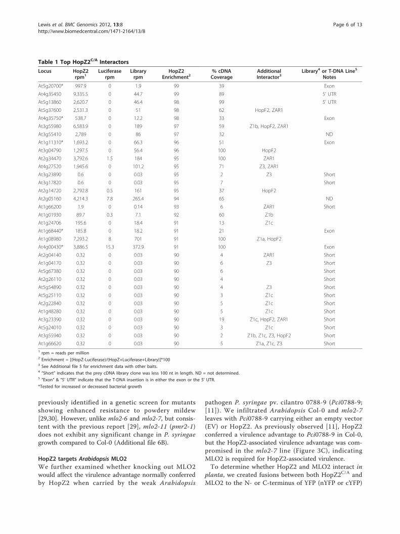
Functional analysis of HopZ2-interactorsWe elected to focus our initial functional study onHopZ2 because it can promote P. syringae growth inArabidopsis and also since a preliminary analysis of thedata provided the most interesting candidate interactors.Our enrichment analysis of HopZ2C229A (hereafterHopZ2C/A) interactors identified several highly overre-presented and specific candidate interactors (Table 1and Additional file 5). HopZ2C/A interactors wereenriched for membrane-associated proteins (28%HopZ2C/A vs. 13% cDNA library; Figure 1A) as well asproteins associated with responses to stress relative tothe prey library (17% HopZ2C/A vs. 7% cDNA library;Figure 1A). Based on our sequencing of the cDNA preylibrary, we could also assess the percent cDNA coverageof each HopZ2C/A interactor in the prey library. Forfunctional analyses we focused on the HopZ2C/A inter-actors that had: (1) an enrichment value > 90% (33 loci),(2) were represented by clones > 33 amino acids in thecDNA prey library (ie. cDNA > 100 nucleotides) (18/33loci) and (3) were specific to HopZ2C/A (i.e. not presentwith an enrichment score of > 50% in other baits tested)(11/33 loci). We hypothesized that these genes wouldinclude HopZ2 virulence targets and that their disrup-tion would alter P. syringae growth.We measured P. syringae growth in Arabidopsis lines
carrying T-DNA insertions for each HopZ2C/A specificinteractor to determine if the candidate HopZ2 interact-ing proteins played any role in P. syringae disease orresistance. We focused on interactors for which therewere confirmed homozygous T-DNA insertion linesavailable and that were predicted to have the T-DNAinsertion in an exon of the gene, and thus be more likelyto interrupt the protein (5/33 loci, Table 1). We assayedfor changes in immunity by infiltrating the T-DNA inser-tion lines with the virulent pathogen PtoDC3000 andevaluating bacterial growth after three days. Insertions ingenes At4g35750, At5g20700, At4g00430 and At1g68440,showed no difference in PtoDC3000 growth compared tothe wild-type Col-0; however, an interruption in geneAt1g11310 (line mlo2-7), encoding MLO2 showed a ten-fold decrease in PtoDC3000 growth (Figure 3A). Tofurther assess if this locus plays a role in resistance ofArabidopsis to PtoDC3000 we tested an additional T-DNA insertion line in At1g11310 (mlo2-6, Figure 3B).Bacterial growth was reduced by approximately 10-foldin mlo2-6 compared to Col-0 wildtype (Additional file6A) indicating that the mlo2 mutation increases resis-tance to PtoDC3000.The mlo2-11 (pmr2-1) mutant of Arabidopsis, which is
a point mutant (D287N) in MLO2 Figure 3B), was
Lewis et al. BMC Genomics 2012, 13:8http://www.biomedcentral.com/1471-2164/13/8
Page 5 of 13
previously identified in a genetic screen for mutantsshowing enhanced resistance to powdery mildew[29,30]. However, unlike mlo2-6 and mlo2-7, but consis-tent with the previous report [29], mlo2-11 (pmr2-1)does not exhibit any significant change in P. syringaegrowth compared to Col-0 (Additional file 6B).
HopZ2 targets Arabidopsis MLO2We further examined whether knocking out MLO2would affect the virulence advantage normally conferredby HopZ2 when carried by the weak Arabidopsis
pathogen P. syringae pv. cilantro 0788-9 (Pci0788-9;[11]). We infiltrated Arabidopsis Col-0 and mlo2-7leaves with Pci0788-9 carrying either an empty vector(EV) or HopZ2. As previously observed [11], HopZ2conferred a virulence advantage to Pci0788-9 in Col-0,but the HopZ2-associated virulence advantage was com-promised in the mlo2-7 line (Figure 3C), indicatingMLO2 is required for HopZ2-associated virulence.To determine whether HopZ2 and MLO2 interact in
planta, we created fusions between both HopZ2C/A andMLO2 to the N- or C-terminus of YFP (nYFP or cYFP)
Table 1 Top HopZ2C/A Interactors
Locus HopZ2rpm1
Luciferaserpm
Libraryrpm
HopZ2Enrichment2
% cDNACoverage
AdditionalInteractor3
Library4 or T-DNA Line5
Notes
At5g20700* 997.9 0 1.9 99 39 Exon
At4g35450 9,335.5 0 44.7 99 89 5’ UTR
At5g13860 2,620.7 0 46.4 98 99 5’ UTR
At5g37600 2,531.3 0 51 98 62 HopF2, ZAR1
At4g35750* 538.7 0 12.2 98 33 Exon
At3g55980 6,583.9 0 189 97 59 Z1b, HopF2, ZAR1
At3g55410 2,789 0 86 97 32 ND
At1g11310* 1,693.2 0 66.3 96 51 Exon
At3g04790 1,297.5 0 56.4 96 100 HopF2
At2g34470 3,792.6 1.5 184 95 100 ZAR1
At4g27520 1,945.6 0 101.2 95 71 Z3, ZAR1
At3g23890 0.6 0 0.03 95 2 Z3 Short
At3g17820 0.6 0 0.03 95 7 Short
At2g14720 2,792.8 0.5 161 95 37 HopF2
At2g05160 4,214.3 7.8 265.4 94 65 ND
At1g66200 1.9 0 0.14 93 6 ZAR1 Short
At1g01930 89.7 0.3 7.1 92 60 Z1b
At1g24706 195.6 0 18.4 91 13 Z1c
At1g68440* 185.8 0 18.2 91 21 Exon
At1g08980 7,293.2 8 701 91 100 Z1a, HopF2
At4g00430* 3,886.5 15.3 372.9 91 100 Exon
At2g04140 0.32 0 0.03 90 4 ZAR1 Short
At1g04170 0.32 0 0.03 90 6 Z3 Short
At5g67380 0.32 0 0.03 90 6 Short
At2g26110 0.32 0 0.03 90 4 Short
At5g54890 0.32 0 0.03 90 4 Z3 Short
At5g25110 0.32 0 0.03 90 3 Z1c Short
At2g22840 0.32 0 0.03 90 5 Z1c Short
At1g48280 0.32 0 0.03 90 5 Z1c Short
At3g23390 0.32 0 0.03 90 19 Z1c, HopF2, ZAR1 Short
At5g24010 0.32 0 0.03 90 3 Z1c Short
At3g55940 0.32 0 0.03 90 2 Z1b, Z1c, Z3, HopF2 Short
At1g66620 0.32 0 0.03 90 5 Z1a, Z1c, Z3 Short1 rpm = reads per million2 Enrichment = [(HopZ-Luciferase)/(HopZ+Luciferase+Library)]*1003 See Additional file 5 for enrichment data with other baits.4 “Short” indicates that the prey cDNA library clone was less 100 nt in length. ND = not determined.5 “Exon” & “5’ UTR” indicate that the T-DNA insertion is in either the exon or the 5’ UTR.
*Tested for increased or decreased bacterial growth
Lewis et al. BMC Genomics 2012, 13:8http://www.biomedcentral.com/1471-2164/13/8
Page 6 of 13

in a glucocorticoid-inducible conditional expression vec-tor. We used a partial clone of MLO2 beginning atamino acid residue 281 of the full-length protein andcontaining the 4th, 5th, 6th and 7th transmembranedomains as well as the C-terminal cytosolic tail of theprotein (MLO2Δ1-280), corresponding to the fragment ofthe clone in our cDNA prey library (Figure 3B).We infiltrated equivalent optical densities of Agrobac-
terium carrying HopZ2::nYFP, HopZ2C/A::nYFP orHopZ1c::nYFP with MLO2Δ1-280::cYFP, as well as thereciprocal combination. We used HopZ1c as a negativecontrol because it did not interact with MLO2 in ouryeast two-hybrid screening (Additional file 5, data notshown). Protein expression was induced by spraying theplants with dexamethasone post-infiltration. 72 and 96hours after dexamethasone application we observedbright fluorescence in leaf sections co-infiltrated withHopZ2::nYFP or HopZ2C/A::nYFP and MLO2Δ1-280::cYFP, as well as the reciprocal combination (Figure 4;Additional file 7A). No fluorescence was observed withHopZ1c::nYFP and MLO2Δ1-280::cYFP (or the reciprocalcombination) at these time points. The interactionbetween MLO2Δ1-280 and HopZ2 localized to the
Figure 3 Disruption of MLO2 compromises P. syringaevirulence. A. The virulent pathogen PtoDC3000 was pressure-infiltrated into the leaves of Arabidopsis Col-0 or T-DNA insertionlines in putative interactors of HopZ2C/A. An insertion in At1g11310(mlo2-7) results in increased resistance to PtoDC3000. * indicatessignificant difference from Col-0 by Fisher’s Protected LeastSignificant Difference (PLSD) test. Error bars indicate one standarddeviation of the mean. B. Schematic showing the MLO2 gene andprotein structure with the point of insertion for the T-DNA insertionlines and the point mutant. The left side shows the gene withintrons represented as lines and exons represented as boxes. MLO2has two splice forms which differ at the 3’ end of the gene. Theright side shows the transmembrane structure of the MLO2 protein.mlo2-6 and mlo2-7 are T-DNA insertion lines while mlo2-11 is apoint mutant. MLOΔ1-280 indicates the clone identified in the cDNA
library screening. C. The weak pathogen Pci0788-9 carrying theempty vector (Ev) or HopZ2 was pressure-infiltrated into the leavesof Arabidopsis Col-0 or mlo2-7. Statistical significance wasdetermined as stated in part A.
Figure 4 HopZ2C/A, but not HopZ1c, interacts with MLO2Δ1-280
in planta by bimolecular fluorescence microscopy. Agrobacteriumcarrying HopZ2C/A::nYFP or MLO2Δ1-280::cYFP (or the reciprocalcombination) were mixed at equivalent optical densities andpressure-infiltrated into the leaves of N. benthamiana. Expression ofthe proteins was induced by 20 μM dexamethasone. Sections of leaftissue were imaged with a Leica SP5 confocal scanning microscope72-96 hours post-induction. The close-up images in the secondcolumn show the reticulate pattern reminiscent of the endoplasmicreticulum. The scale bar indicates 100 μm.
Lewis et al. BMC Genomics 2012, 13:8http://www.biomedcentral.com/1471-2164/13/8
Page 7 of 13

periphery of the cell suggestive of the plasma membraneas well as reticulate network reminiscent of the endoplas-mic reticulum (ER; Figure 4). This localization patternwas also observed when an MLO2Δ1-280::YFP fusion wastransiently expressed in N. benthamiana (Additional file7B).
DiscussionWe developed QIS-Seq, a quantitative, high-throughputyeast two-hybrid screening protocol combined with Illu-mina next-generation sequencing, to identify putativeinteracting proteins with the HopZ family of type IIIeffector proteins. QIS-Seq provides many significantadvances over traditional interactor screens: (1) it elimi-nates the need to individually sequence each interactingcolony while at the same time vastly increasing thenumber of candidates interrogated; (2) the results arequantitative and therefore amenable to statistical analy-sis; (3) the method explicitly evaluates the enrichmentof each interactor relative to both its presence in theprey library as well as its general (non-specific) sticki-ness; (4) sequencing of the prey library provides a heretounprecedented ability to evaluate the cDNA library forcomplexity and completeness; (5) it is amenable to anytype of yeast two-hybrid screen; (6) it is amenable toany type of next-generation sequencing; (7) it is comple-tely scalable and therefore applicable to experiments runin a very small, multiplex format, to very large auto-mated, high-throughput screens; and (8) the quantitativenature of the data also enhances the method’s ‘portabil-ity’ among laboratories.A number of these points are worth elaborating. The
ability to interrogate putative interactors relative to theirpresences in the prey library (points 3 and 4) is particu-larly critical when not working with well-establishedmodel systems. One of the great benefits of next-genera-tion sequencing is the ability to more easily study non-model systems. By definition, these systems have fewestablished genomic resources, such as normalizedcDNA libraries. The in silico normalization provided byQIS-Seq facilitates the use of any prey library, regardlessof its means of preparation. For example, tissue, cell,age or stage-specific libraries could be rapidly con-structed and tested without the need for tedious andsample consuming normalization steps.Portability in the context mentioned in point 8, means
that standards can be established for experimentaldesign, data collection, and data analysis, which willallow experimental results to be comparable amonglaboratories. Examples of such portability standardsincluded the MIAME (Minimum Information About aMicroarray Experiment) [31] and MIGS (MinimumInformation about a Genome Sequence) specifications[32]. Another benefit of these standards is that it
encourages the development of data repositories andmeta-analysis tools such as the Bio-Array Resource [23]for microarray data.A potential criticism of QIS-Seq is its cost-effective-
ness, since the cost of next-generation sequencing is nottrivial. Currently, it cost between US$1000-US$4000 fora single channel of Illumina next-generation data(depending on the specific protocol and platform).While we sequenced to quite high coverage in thisproof-of-principle study, this depth is not generallyrequired, and we found that 5 million reads were morethan adequate. Since the current Illumina HiSeq2000platform currently produces over 100 million reads perlane, it should be possible to multiplex as many as 20samples per channel. Importantly, bar-codes can bedirectly incorporated onto the primers used to amplifythe prey-plasmid inserts, thereby permitting the poolingof independent samples prior to Illumina sample prep.Early pooling of bar-coded samples means that only onesample prep is required for all pooled samples, and con-sequently, while the cost for a single channel of Illuminadata may be US$2500, the cost per sample (if multiplex-ing 20 samples per channel) would only be US$125.This price is substantially less than the cost for Sangersequencing 100 clones, and the cost will only continueto drop as the next-generation sequencing technologyimproves.Since we had previously shown that HopZ2 confers a
virulence advantage to P. syringae in Arabidopsis, wetherefore rationalized that we could use HopZ2 as aprobe for the identification of innate immunity compo-nents. By conducting QIS-seq screens on all members ofthe HopZ family, we were able to identify proteins thatinteract specifically with HopZ2. These HopZ2 interac-tors were enriched for membrane-associated proteins aswell as proteins from genes induced during stressresponses, including Arabidopsis MLO2. The lack ofinteraction between MLO2 and the other HopZ familymembers suggests that the HopZ family has diversifiedto target different host proteins.MLO2 has seven transmembrane domains with an
extracellular N-terminus and an intracellular C-terminusand is localized to the plasma membrane [33]. HopZ2 isnormally present at the plasma membrane and would beideally localized to interact with MLO2 [11]. Our analy-sis identified a partial clone of MLO2 starting just priorto the fourth transmembrane domain and including theentire intracellular C-terminus (MLO2Δ1-280; Figure 3B).Using MLO2Δ1-280 in BiFC analyses, we demonstratedthat HopZ2 and MLO2 interact directly in planta. How-ever, the observed fluorescence complementation loca-lized to a reticulate structure reminiscent of the ER aswell as the plasma membrane. This localization was alsoobserved with MLO2Δ1-280::YFP (Additional file 7B),
Lewis et al. BMC Genomics 2012, 13:8http://www.biomedcentral.com/1471-2164/13/8
Page 8 of 13

suggesting that MLO2Δ1-280 may be partiallymislocalized.The MLO gene was first identified by map-based clon-
ing in barley from mutants that were resistant to thepowdery mildew fungal pathogen Blumeria (formerlyErysiphe) graminis f. sp. hordei (Bgh) [34]. However,mlo-based resistance in crop species has been employedby plant breeders for decades [35]. As in barley, Arabi-dopsis mlo2 confers increased resistance to a powderymildew fungal pathogen, Golonivomyces (formerly Ery-siphe) orontii [30]. However, it has been reported (withdata not shown) that P. syringae growth did not signifi-cantly differ in mlo2 compared to Col-0 [29,30]. Vogeland colleagues [29] evaluated symptom production inmlo2 (originally called pmr2) point mutants when infil-trated or sprayed with PtoDC3000, while Consonni andcolleagues [30] tested bacterial growth and symptomproduction from PtoDC3000 in a T-DNA insertion line(SAIL_878_H12; mlo2-5) that is inserted towards theend of the 6th exon (Figure 3B). Our growth assays withPtoDC3000 in the mlo2-11 (pmr2-1) point mutant con-firmed that it did not exhibit increased resistance to P.syringae (Additional file 6B). While mlo2-11 (pmr2-1),which has a D287N point mutation, in the intracellularloop between the third and fourth transmembranedomains [30], confers increased resistance to powderymildew, it does not appear to be sufficient to conferincreased resistance to P. syringae, suggesting thatMLO2 differentially contributes to immunity againstthese distinct pathogens. In contrast, unlike Consonniand colleagues we did observe a significant decrease inPtoDC3000 bacterial growth in two independent Salk T-DNA lines in the MLO2 gene (mlo2-6 and mlo2-7),although we did not test their T-DNA insertion line(mlo2-5). Our results suggest that MLO2 negatively con-tributes to resistance against P. syringae in Arabidopsis,and are consistent with the proposed role of MLO2 as anegative regulator of defenses against oomycetepathogens.Previous work has shown that MLO is relocalized to a
lipid raft-like domain in the plasma membrane uponpathogen attack [36]. MLO2 has also been shown tonegatively regulate PEN1-dependent vesicular traffickingto regions of the plasma membrane associated withpathogen entry [7,37,38]. PEN1 is a syntaxin that hasbeen associated with aberrant non-host resistance to thefungal barley pathogen Bgh, and is likely part of aSNARE complex involved in vesicular trafficking ofdefense components [37,39]. When PEN1 is recruited tosites of pathogen attack, it contributes to the rapid for-mation of papillae, an important component of theinnate immunity [38,39]. Our data in conjunction withthe prior studies suggest that pathogens may stabilizeMLO2 or cause its accumulation at the plasma
membrane in order to suppress PEN1-dependent secre-tion of defense components.There is precedence in the literature for P. syringae
T3SEs to target negative regulators of plant immunity.The absence of RIN4 in rin4 rps2 plants compromisesplant immunity whereas RIN4 overexpression enhancesimmunity [40]. Interestingly, at least four unrelatedT3SEs have been demonstrated to target RIN4, poten-tially to enhance its role as a negative regulator of plantimmunity [40-44]. Plant vesicular trafficking pathwaysare also targeted by P. syringae T3SEs. The T3SEHopM1 induces the degradation of AtMIN7, an ARFguanine exchange factor (GEF) that is involved in vesi-cular trafficking [45]. Similarly, HopZ2 may stabilizeMLO2 in order to prevent the secretion of defense com-ponents to the regions of pathogen attack.
ConclusionsOverall we have demonstrated that QIS-Seq provides apowerful new approach to identify protein interactionsusing next-generation sequencing. We used thisapproach to identify Arabidopsis MLO2 as a virulencetarget of the P. syringae T3SE HopZ2. Since HopZ2 (aswell as other P. syringae T3SEs) is membrane localizedwe used the split-ubiquitin yeast two-hybrid system forinteraction screening [11,21,46]. However, QIS-Seq isapplicable to any sequencing-based yeast two-hybridscreening method. Furthermore, this approach can beapplied to both ORF as well as cDNA libraries.Although we sequenced the interactors of individualbaits separately, the use of barcodes will allow thesequencing of pooled baits while maintaining the asso-ciations between interacting pairs. This approach willincrease the number of baits that can be screened perexperiment and decrease the cost of screening individualbaits. In addition, as the costs of next-generationsequencing experiments continue to drop, QIS-Seq pro-mises to become a cost-effective alternative to tradi-tional yeast two-hybrid screening approaches.
MethodsBacterial strains and routine culture conditionsEscherichia coli and Agrobacterium tumefaciens weregrown in Luria-Bertani broth, and Pseudomonas syringaewas grown in King’s broth (KB). Antibiotics were usedat the following concentrations: for E. coli, 50 μg/mLkanamycin, 100 μg/mL ampicillin; for A. tumefaciens:100 μg/mL kanamycin, 100 μg/mL rifamcipin; and for P.syringae: 50 μg/mL kanamycin, 50 μg/mL rifamcipinand 50 μg/mL cycloheximide.
Plant growth conditionsArabidopsis plants were grown with 9 h of light (~130microeinsteins m-2 s-1) and 16 h of darkness at 22°C in
Lewis et al. BMC Genomics 2012, 13:8http://www.biomedcentral.com/1471-2164/13/8
Page 9 of 13

Sunshine #1 soil supplemented with 20:20:20 fertilizer.Nicotiana benthamiana plants were grown with 9 h oflight (~130 microeinsteins m-2 s-1) and 16 h of darknessat 22°C in Sunshine #1 soil supplemented with 20:20:20fertilizer and osmocute.
CloningPfu polymerase (Fermentas) was used for all cloning andall constructs were confirmed by sequencing. For thesplit-ubiquitin constructs, bait genes were amplified byPCR to contain an in-frame HA epitope, a polybasicregion (K6 or K8) and a CAAX box, as well as appropri-ate unique restriction sites. The bait-HA-K6-CAAXgenes were cloned into the pBT3-N vector (DualsystemsBiotech) using SfiI. The bait-HA-K8-CAAX genes werecloned into the pTLB-1 vector (gift of Dr. Igor Stagljar,University of Toronto) using NcoI. The orientation ofeach gene in the vector was confirmed.For the split-YFP constructs, the HopZ genes or the 3’
end of MLO2 were amplified by PCR to contain an in-frame HA epitope and appropriate unique restrictionsites. All of the genes for the split-YFP system werecloned using XhoI and StuI into pBD-nYFP or pBD-cYFP. pBD-nYFP and pBD-cYFP were modified frompTA7002 [47] to add an HA tag and the N- or C-termi-nus of YFP between the StuI and SpeI sites. The N-ter-minus of YFP encompasses residues 1-155 while the C-terminus of YFP includes residues 156 to the stopcodon.The constructs used for plant infectivity assays were
previously described [11]. In brief, the HopZ allele isexpressed under its native promoter and contains an in-frame HA tag.
cDNA libraryFive week old Arabidopsis rosette leaves were infiltratedby hand with a needleless syringe with P. syringae pv.tomato DC3000 (PtoDC3000), PtoDC3000 carryingAvrRpm1 or the ΔhrcC mutant of PtoDC3000 at anoptical density of 0.1 (~5 × 10⁷ CFU/mL) at 600 nm.Infiltrated leaves were harvested at 4 hpi (PtoDC3000,PtoDC3000 carrying AvrRpm1, or PtoDC3000 ΔhrcC)or 12 hpi (PtoDC3000, PtoDC3000 ΔhrcC). Uninfiltratedleaves were harvested at 4 pi and 12 hpi. RNA wasextracted using Trizol (Invitrogen). mRNA was clonedinto the pPR3-N vector (Dualsystems Biotech) using theSfiI sites (Norclone Biotech Laboratories, Ontario) withthe NubG at the N-terminus of the prey proteins. Thelibrary contained ~2.3 × 10⁹ clones, with an average sizeof 1.2 kB and was 90% recombinant. Amplification ofthe library was carried out by the semi-solid method[22]. 0.5 μL of the primary library in E. coli strain DH5awas inoculated into 2× LB broth with 0.3% Seaprepagarose (FMC, Rockland) and 100 μg/mL ampicillin.
The inoculated cultures were then incubated in a water-ice bath for 1 hour. Subsequently, the inoculated cul-tures were incubated at 30°C for 44 hours without shak-ing. To sequence the primary and secondary libraries,low-cycle PCR amplification was carried out with pPR3-N vector-specific primers and a high-fidelity Taq/proof-reading polymerase mix (Fermentas, Burlington). Thispool of DNA was sheared and prepared for Illuminasequencing by standard methods.
Yeast two-hybrid screeningHopZ1aC/A, HopZ1bC/A, HopZ1cC/A, HopZ2C/A, andZAR1CC were expressed under the weak CYC1 promo-ter in the pBT3-N vector, while HopZ3C/A wasexpressed under the strong TEF1 promoter in thepTLB-1 vector. AP-4 yeast [48] carrying the bait con-struct were transformed using the PEG/LiAc method. Inbrief, yeast carrying the bait construct were subculturedin 300 mL SD-Leu overnight to an optical density of 0.6at 600 nm. Yeast were washed twice in sterile H2O andresuspended in 1.5 mL. Transformations were per-formed with 1 μg of cDNA library, 200 μL of yeast cellsand 600 μL of PEG/LiAc (50% PEG, 120 mM LiAc, 10μL 10 mg/mL boiled salmon sperm DNA) by the heatshock method at 42°C for 45 min. Yeast were washedtwice in sterile H2O and plated on SD-LeuTrp and SD-LeuTrpHis + 3-amino-1,2,4-triazole (3-AT). Interactingcolonies were identified by growth on SD-LeuTrpHis +3-AT. The appropriate amount of 3-AT was determinedfor each bait by testing for growth when transformedwith the positive control pFur4-NubI and a lack ofgrowth with pFur4-NubG [48]. Screening was performeduntil ~2000 interacting colonies were identified. Colo-nies were restreaked twice on SD-Trp to preferentiallylose the bait plasmid [10] and grown at 28°C. Prior toplasmid isolation, colonies were restreaked onto SD-Trpand grown at 28°C. Yeast were harvested en masse inSD-Trp and pelleted at 1000 g for 5 min. The pellet (~5 g) was washed in 0.1 M NaPO4 pH 7.4 and 1.2 M sor-bitol and resuspended in 7.1 mL of lyticase buffer [0.1M NaPO4 pH 7.4, 1.2 M sorbitol, 500 μL lyticase(Sigma), 50 μL of 10 mg/mL RNAseA]. 25 KU of lyti-case was resuspended in 0.01 M NaPO4 pH 7.4 and 50%glycerol and used immediately. Yeast were incubated inthe lyticase buffer overnight at 37°C. The Qiagen alka-line lysis spin kit was used to extract the plasmid DNA.The lyticase buffer was used instead of buffer P1.Volumes of buffers P2 and N3 were scaled up to thetypical Qiagen proportions based on the volume of theresuspended pellet (1 vol lyticase buffer: 1 vol P2: 1.4vol N3). Lysis in the P2 buffer was done for 15 min atroom temperature and 15 min at 65°C. Buffer N3 waschilled prior to use. After addition of buffer N3, theyeast were incubated on ice for 20 min. Yeast were
Lewis et al. BMC Genomics 2012, 13:8http://www.biomedcentral.com/1471-2164/13/8
Page 10 of 13

pelleted at 14000 rpm for 30 min at 4°C. The superna-tant was removed and cleared again by centrifugation at14000 rpm for 15 min at 4°C. The supernatant wasloaded onto multiple Qiagen spin columns to purify theplasmid DNA. The columns were washed with bufferPB and buffer PE. Plasmid DNA was eluted with 50 μLof buffer EB after a 1 min incubation. A second elutionwas performed with 35 μL of buffer EB after a 1 minincubation. To sequence the putative interactors, low-cycle PCR amplification was carried out with pPR3-Nvector-specific primers and a high-fidelity Taq/proof-reading polymerase mix (Fermentas, Burlington). Thispool of DNA was sheared and prepared for Illuminasequencing by standard methods.
Illumina SequencingIllumina sequencing was performed either with 37 cyclesingle reads or 72 cycle paired-end reads (Additional file1) following the manufacturer’s protocol on an IlluminaGAIIx and pipelined using the GA pipeline v1.4.
BioinformaticsIllumina reads were mapped to Arabidopsis gene modelsdownloaded from NCBI, using a short read mappingtool novoalign (novocraft.com). From the mapping data,the number of mapped reads and the total length ofmapped regions for each gene were determined with inhouse scripts. Read numbers per gene were further nor-malized as reads per million (rpm) within each sampleand compared among the samples. The enrichment of aspecific interactor with a bait of interest was determinedby considering the number of reads obtained with thebait and luciferase and normalizing against the abun-dance of reads for luciferase, the bait and the library(Additional file 5). The percentage of the mapped lengthwas calculated using length of mapped regions and thetheoretical length of the gene model. Gene Ontologyterms for Arabidopsis genes were downloaded from theTAIR website, and assigned to the genes in the cDNAlibrary. Up- or down-regulation of each gene inresponse to biotic stress was determined from microar-ray data available through the CAGEF Bio-ArrayResource (BAR, http://bar.utoronto.ca, [23]. Biotic stresstreatments in the BAR included inoculation with viru-lent, avirulent and non-host P. syringae, inoculationwith oomycetes (Botrytis cinerea, Phytophthora infestans,Golonivomyces orontii) and inoculation with elicitors ofinnate immunity (harpins, lipopolysaccharides, and anoomycete elicitor NPP1).
Genotyping of T-DNA insertion linesThe following T-DNA insertion lines were used:SALK_060284C (At5g20700), SALK_082464C(At4g35750), SALK_079850C (At1g11310, mlo2-7),
SALK_050191C (At1g11310, mlo2-6), SALK_024490C(At1g68440), SAIL_808_A10 (At4g00430). GenomicDNA was extracted from a leaf of 5-6 week old Arabi-dopsis plants. Primers were designed using the iSct fea-ture in the SIGnAL database. Primer sequences areavailable upon request. PCR-based genotyping wasemployed to determine the homozygosity or heterozyg-osity of the individuals. PCR products were sequencedusing Big Dye Terminator 3.1 on an ABI 3730 geneticanalyzer.
Infectivity assaysFor infiltration, P. syringae was resuspended to an opti-cal density of 0.1 (~5 × 10⁷ CFU/mL) at 600 nm anddiluted to a concentration of 1 × 10⁵ CFU/mL forgrowth curves. Plants were infiltrated by hand with aneedleless syringe, as previously described [49]. Fourdisks (1 cm2) were harvested, ground in 10 mM MgCl2,and plated on KB with rifampicin and cycloheximide ondays 0 and 3 for colony counting.
StatisticsFor growth assays, 8-10 plants were used for day 3counts. Significance was determined using Fisher’s Pro-tected Least Significant Difference (PLSD) on the day 3count data. Error bars indicate one standard deviationof the mean.
Agrobacterium transient expression assays and BiFCFive-milliliter A. tumefaciens GV2260 cultures weregrown overnight at 28°C in Luria-Bertani broth withkanamycin and rifampicin. The next day, the cultureswere washed twice in induction medium (50 mM MESpH 5.6, 0.5% (w/v) glucose, 1.7 mM NaH2PO4, 20 mMNH4Cl, 1.2 mM MgSO4, 2 mM KCl, 17 μM FeSO4, 70μM CaCl2, 200 μM acetosyringone) [50], and 3.75 mLwas inoculated into 35 mL fresh induction medium togrow overnight. The following day, cultures were spundown, washed twice in 10 mM MES pH 5.6 with 200μM acetosyringone and resuspended to an optical den-sity of 0.4 at 600 nm. The culture containing theMLO2Δ1-280-cYFP plasmid was mixed in equal volumeswith a culture containing the HopZ1c-nYFP, HopZ2-nYFP or HopZ2C/A-nYFP plasmid. The culture contain-ing the MLO2Δ1-280-nYFP plasmid was mixed in equalvolumes with a culture containing the HopZ1c-cYFP,HopZ2-cYFP or HopZ2C/A-cYFP plasmid. The under-side of the leaves of 5- to 7-week-old N. benthamianaplants were infiltrated by hand with a needleless syringe.Plants were sprayed with 20 μM dexamethasone (Sigma)1-2 hours after inoculation. Sections of leaves wereimaged with a Leica SP5 microscope using Leica soft-ware at 24 hours (YFP fluorescence) or 72-96 hours(BiFC) post-dexamethasone induction.
Lewis et al. BMC Genomics 2012, 13:8http://www.biomedcentral.com/1471-2164/13/8
Page 11 of 13

Additional material
Additional file 1: Illumina sequencing of cDNA libraries andinteractors. Table showing the prey cDNA library used for the yeasttwo-hybrid screening, the number of Illumina cycles, the number ofquality clusters, and the number of bases for each bait or the cDNAlibrary.
Additional file 2: Characterization of the Arabidopsis cDNA libraryused in this study. A. Histogram shows the number of Arabidopsisgenes on log scale plotted against the length of the cDNAs present inthe cDNA library. There are no genes present in the 2300-2499 or 2600-2699 ranges. There is one gene present in the 2500-2599 and 2700-2799ranges. B. Histogram shows the number of genes on log scale plottedagainst the percent coverage of the cDNAs in the cDNA library. C.Scatter plot of the genes present in the primary and amplified(secondary) libraries. The R2 value of 0.9567 shows a high congruencebetween the primary unamplified and secondary amplified libraries.
Additional file 3: Schematic of the split-ubiquitin yeast two hybridapproach. The N-terminus of the bait protein is compromised of theLexA-VP16 transcription factor (VP16), followed by the C-terminus ofubiquitin (Cub), the bait protein of interest, an HA epitope tag, a C-terminal polybasic region (K6 or K8) and CAAX box. We added the K6 (orK8) and CAAX box sequences to non-transmembrane bait proteins inorder to anchor them to membranes and minimize autoactivation. Atthe N-terminus of the prey proteins is the N-terminal half of ubiquitinwith an isoleucine to glycine mutation, which reduces non-specificassociation with Cub (NubG) [21]. The prey proteins may be membrane-associated or cytoplasmic (not shown). In the case of interactionbetween a bait and prey, the Cub and Nub portions of ubiquitin arebrought into close proximity and reconstitute the full ubiquitin protein. Aubiquitin-specific protease (the “blades”) cleaves the VP16 transcriptionfactor, allowing it to translocate to the nucleus and activate the HISreporter gene.
Additional file 4: Bait and prey vectors used in QIS-Seq. The bait:HA:K6:CAAX was cloned into the SfiI sites of the pBT3-N vector, in framewith an N-terminal LexA-VP16 (VP16) and the C-terminus of ubiquitin(Cub). Bait protein expression is driven by the weak CYC1 promoter.Stronger bait expression was achieved by cloning the bait:HA:K8:CAAXinto the NcoI site of the pTLB-1 vector, in frame with an N-terminal VP16and Cub (not shown). The prey cDNAs were cloned into the SfiI sites ofthe pPR3-N vector, in frame with an N-terminal ubiquitin (the N-terminusof ubiquitin with an isoleucine to glycine mutation, NubG) [21]. Preyprotein expression is driven by the weak CYC1 promoter.
Additional file 5: Rank list of top interactors with HopZ2 and theirenrichment with other HopZ alleles, an unrelated T3SE HopF2 andZAR1. Table indicates the percentage enrichment with each tested baitfor the top HopZ2 interactors in Arabidopsis.
Additional file 6: Disruption of MLO2 compromises P. syringaevirulence. A. The virulent pathogen PtoDC3000 was pressure-infiltratedinto the leaves of Arabidopsis Col-0 or the mlo2-6 T-DNA insertion line. *indicates significant difference from Col-0 by Fisher’s PLSD test. Error barsindicate one standard deviation of the mean. B. The virulent pathogenPtoDC3000 was pressure-infiltrated into the leaves of Arabidopsis Col-0 orthe mlo2-11 (pmr2-1) point mutant. Error bars indicate one standarddeviation of the mean.
Additional file 7: HopZ2 interacts with MLO2Δ1-280 in planta bybimolecular fluorescence microscopy and MLOΔ1-280 localizes to areticulate network. A. Agrobacterium carrying HopZ2::nYFP or MLO2Δ1-280::cYFP were mixed at equivalent optical densities and pressure-infiltrated into the leaves of N. benthamiana. Expression of the proteinswas induced by 20 μM dexamethasone. Sections of leaf tissue wereimaged with a Leica SP5 confocal scanning microscope 72-96 hourspost-induction. The scale bar indicates 100 μm. B. Agrobacterium carryingMLO2Δ1-280::YFP was pressure-infiltrated into the leaves of N.benthamiana. Expression of the proteins was induced by 20 μMdexamethasone. Sections of leaf tissue were imaged with a Leica SP5confocal scanning microscope 24 hours post-induction. The scale barindicates 100 μm. Fluorescence is observed in a reticular patternreminiscent of the endoplasmic reticulum.
AbbreviationsETI: effector-triggered immunity; MLO: powdery mildew resistance gene O;Pci: Pseudomonas syringae pv. cilantro; Pto: Pseudomonas syringae pv. tomato;R: resistance; RIN4: RPM1-interacting protein 4; T3SE: type III secretedeffector; T3SS: type III secretion system; ZAR: HopZ-activated resistance
AcknowledgementsWe are grateful to Mike Wilton for infiltrating the plants, preparing the RNAused in the cDNA library construction and sharing the QIS-Seq data forHopF2Pto; Dr. Igor Stagljar for split-ubiquitin yeast two-hybrid vectors andyeast strain; the Arabidopsis Biological Research Center (ABRC) for providingseed stocks. This research was supported by NSERC discovery grants to DDand DSG a Canada Research Chair in Plant-Microbe Systems Biology (DD) orComparative Genomics (D.S.G.); the Centre for the Analysis of GenomeEvolution and Function (DD and DSG); and an NSERC undergraduateresearch award (JW).
Author details1Department of Cell and Systems Biology, University of Toronto, Toronto,ON, M5S 3B2, Canada. 2Centre for the Analysis of Genome Evolution andFunction, University of Toronto, Toronto, ON, M5S 3B2, Canada. 3Plant GeneExpression Center, USDA, 800 Buchanan St., Albany, CA, 94710, USA.
Authors’ contributionsJDL, DD and DSG conceived the experiment; JDL performed theexperiments; JW and RF assisted with the split-ubiquitin yeast two-hybridscreening; Y.G. performed the bioinformatics analysis; PF ran the IlluminaGAII; HN analyzed microarray data from the BAR; PW provided critical inputfor GAII sequencing; JDL, YG, DD and DSG analyzed the data; JDL, DD andDSG wrote the manuscript. All authors read and approved the finalmanuscript.
Received: 16 September 2011 Accepted: 9 January 2012Published: 9 January 2012
References1. Espinosa A, Alfano JR: Disabling surveillance: bacterial type III secretion
system effectors that suppress innate immunity. Cell Microbiol 2004,6:1027-1040.
2. Grant SR, Fisher EJ, Chang JH, Mole BM, Dangl JL: Subterfuge andmanipulation: type III effector proteins of phytopathogenic bacteria.Annu Rev Microbiol 2006, 60:425-449.
3. Block A, Li GY, Fu ZQ, Alfano JR: Phytopathogen type III effectorweaponry and their plant targets. Curr Opin Plant Biol 2008, 11:396-403.
4. Boller T, He SY: Innate immunity in plants: an arms race between patternrecognition receptors in plants and effectors in microbial pathogens.Science 2009, 324:742-744.
5. Lewis JD, Desveaux D, Guttman DS: The targeting of plant cellularsystems by injected type III effector proteins. Semin Cell Dev Biol 2009,20:1055-1063.
6. Dangl JL, Jones JDG: Plant pathogens and integrated defence responsesto infection. Nature 2001, 411:826-833.
7. Jones JDG, Dangl JL: The plant immune system. Nature 2006, 444:323-329.8. McCann HC, Guttman DS: Evolution of the type III secretion system and
its effectors in plant-microbe interactions. New Phytol 2008, 177:33-47.9. Ma WB, Dong FFT, Stavrinides J, Guttman DS: Type III effector
diversification via both pathoadaptation and horizontal transfer inresponse to a coevolutionary arms race. PLoS Genet 2006, 2:e209.
10. Lewis JD, Lee A, Ma W, Zhou H, Guttman DS, Desveaux D: The YopJsuperfamily in plant-associated bacteria. Mol Plant Path 2011, 12:928-937.
11. Lewis JD, Abada W, Ma WB, Guttman DS, Desveaux D: The HopZ family ofPseudomonas syringae type III effectors require myristoylation forvirulence and avirulence functions in Arabidopsis thaliana. J Bacteriol2008, 190:2880-2891.
12. Lewis JD, Wu R, Guttman DS, Desveaux D: Allele-specific virulenceattenuation of the Pseudomonas syringae HopZ1a type III effector viathe Arabidopsis ZAR1 resistance protein. PLoS Genet 2010, 6:e1000894.
Lewis et al. BMC Genomics 2012, 13:8http://www.biomedcentral.com/1471-2164/13/8
Page 12 of 13

13. Vinatzer BA, Teitzel GM, Lee MW, Jelenska J, Hotton S, Fairfax K, Jenrette J,Greenberg JT: The type III effector repertoire of Pseudomonas syringaepv. syringae B728a and its role in survival and disease on host and non-host plants. Mol Microbiol 2006, 62:26-44.
14. Fields S, Song OK: A novel genetic system to detect protein-proteininteractions. Nature 1989, 340:245-246.
15. Walhout AJM, Vidal M: High-throughput yeast two-hybrid assays forlarge-scale protein interaction manning. Methods 2001, 24:297-306.
16. Yu HY, Braun P, Yildirim MA, Lemmens I, Venkatesan K, Sahalie J, Hirozane-Kishikawa T, Gebreab F, Li N, Simonis N, Hao T, Rual JF, Dricot A, Vazquez A,Murray RR, Simon C, Tardivo L, Tam S, Svrzikapa N, Fan CY, de Smet AS,Motyl A, Hudson ME, Park J, Xin XF, Cusick ME, Moore T, Boone C,Synder M, Roth RP, Barabasi AL, Tavernier J, Hill DE, Vidal M: High-qualitybinary protein interaction map of the yeast interactome network. Science2008, 322:104-110.
17. Dreze M, Monachello D, Lurin C, Cusick ME, Hill DE, Vidal M, Braun P: High-quality binary interactome mapping. Methods Enzymol 2010, 470:281-315.
18. Venkatesan K, Rual JF, Vazquez A, Stelzl U, Lemmens I, Hirozane-Kishikawa T,Hao T, Zenkner M, Xin XF, Goh KI, Yildirim MA, Simonis N, Heinzmann K,Gebreab F, Sahalie JM, Cevik S, Simon C, de Smet AS, Dann E, Smolyar A,Vinayagam A, Yu HY, Szeto D, Borick H, Dricot A, Klitgord N, Murray RR,Lin C, Lalowski M, Timm J, Rau K, Boone C, Braun P, Cusick ME, Roth FP,Hill DE, Tavernier J, Wanker EE, Barabasi AL, Vidal M: An empiricalframework for binary interactome mapping. Nat Methods 2009, 6:83-90.
19. Yu HY, Tardivo L, Tam S, Weiner E, Gebreab F, Fan C, Svrzikapa N, Hirozane-Kishikawa T, Rietman E, Yang X, Sahalie J, Salehi-Ashtiani K, Hao T,Cusick ME, Hill DE, Roth RP, Braun P, Vidal M: Next-generation sequencingto generate interactome datasets. 2011, 8:478-480.
20. Zhou HB, Morgan RL, Guttman DS, Ma WB: Allelic variants of thePseudomonas syringae type III effector HopZ1 are differentiallyrecognized by plant resistance systems. Mol Plant-Microbe Interact 2009,22:176-189.
21. Stagljar I, Korostensky C, Johnsson N, te Heesen S: A genetic system basedon split-ubiquitin for the analysis of interactions between membraneproteins in vivo. Proc Natl Acad Sci USA 1998, 95:5187-5192.
22. Kriegler M: Expression cloning. Gene transfer and expression: a laboratorymanual New York: Stockton Press; 1990, 131-132.
23. Toufighi K, Brady SM, Austin R, Ly E, Provart NJ: The botany array resource:e-Northerns, expression angling, and promoter analyses. Plant J 2005,43:153-163.
24. Hancock JF, Paterson H, Marshall CJ: A polybasic domain or palmitoylationis required in addition to the CAAX motif to localize p21ras to theplasma membrane. Cell 1990, 63:133-139.
25. Yeung T, Gilbert GE, Shi J, Silvius J, Kapus A, Grinstein S: Membranephosphatidylserine regulates surface charge and protein localization.Science 2008, 319:210-213.
26. Overall CM, Tam EM, Kappelhoff R, Connor A, Ewart T, Morrison CJ,Puente X, Lopez-Otin C, Seth A: Protease degradomics: massspectrometry discovery of protease substrates and the CLIP-CHIP, adedicated DNA microarray of all human proteases and inhibitors. BiolChem 2004, 385:493-504.
27. Blanchetot C, Chagnon M, Dube N, Halle A, Tremblay ML: Substrate-trapping techniques in the identification of cellular PTP targets. Methods2005, 35:44-53.
28. Zhang Z, MooYoung M, Chisti Y: Plasmid stability in recombinantSaccharomyces cerevisiae. Biotechnol Adv 1996, 14:401-435.
29. Vogel J, Somerville S: Isolation and characterization of powdery mildew-resistant Arabidopsis mutants. Proc Natl Acad Sci USA 2000, 97:1897-1902.
30. Consonni C, Humphry ME, Hartmann HA, Livaja M, Durner J, Westphal L,Vogel J, Lipka V, Kemmerling B, Schulze-Lefert P, Somerville SC, Panstruga R:Conserved requirement for a plant host cell protein in powdery mildewpathogenesis. Nature Genet 2006, 38:716-720.
31. Dodds PN, Rathjen JP: Plant immunity: towards an integrated view ofplant-pathogen interactions. Nat Rev Genet 2010, 11:539-548.
32. Block A, Alfano JR: Plant targets for Pseudomonas syringae type IIIeffectors: virulence targets or guarded decoys? Curr Opin Microbiol 2011,14:39-46.
33. Devoto A, Piffanelli P, Nilsson I, Wallin E, Panstruga R, von Heijne G,Schulze-Lefert P: Topology, subcellular localization, and sequencediversity of the Mlo family in plants. J Biol Chem 1999, 274:34993-35004.
34. Buschges R, Hollricher K, Panstruga R, Simons G, Wolter M, Frijters A, vanDaelen R, van der Lee T, Diergaarde P, Groenendijk J, Topsch S, Vos P,Salamini F, Schulze-Lefert P: The barley Mlo gene: a novel control elementof plant pathogen resistance. Cell 1997, 88:695-705.
35. Humphry M, Consonni C, Panstruga R: mlo-based powdery mildewimmunity: silver bullet or simply non-host resistance? Mol Plant Pathol2006, 7:605-610.
36. Bhat RA, Miklis M, Schmelzer E, Schulze-Lefert P, Panstruga R: Recruitmentand interaction dynamics of plant penetration resistance components ina plasma membrane microdomain. Proc Natl Acad Sci USA 2005,102:3135-3140.
37. Collins NC, Thordal-Christensen H, Lipka V, Bau S, Kombrink E, Qiu JL,Huckelhoven R, Stein M, Freialdenhoven A, Somerville SC, Schulze-Lefert P:SNARE-protein-mediated disease resistance at the plant cell wall. Nature2003, 425:973-977.
38. Assaad FF, Qiu JL, Youngs H, Ehrhardt D, Zimmerli L, Kalde M, Wanner G,Peck SC, Edwards H, Ramonell K, Somerville SC, Thordal-Christensen H: ThePEN1 syntaxin defines a novel cellular compartment upon fungal attackand is required for the timely assembly of papillae. Mol Biol Cell 2004,15:5118-5129.
39. Meyer D, Pajonk S, Micali C, O’Connell R, Schulze-Lefert P: Extracellulartransport and integration of plant secretory proteins into pathogen-induced cell wall compartments. Plant J 2009, 57:986-999.
40. Kim HS, Desveaux D, Singer AU, Patel P, Sondek J, Dangl JL: ThePseudomonas syringae effector AvrRpt2 cleaves its C-terminally acylatedtarget, RIN4, from Arabidopsis membranes to block RPM1 activation. ProcNatl Acad Sci USA 2005, 102:6496-6501.
41. Mackey D, Holt BF, Wiig A, Dangl JL: RIN4 interacts with Pseudomonassyringae type III effector molecules and is required for RPM1-mediatedresistance in Arabidopsis. Cell 2002, 108:743-754.
42. Axtell MJ, Chisholm ST, Dahlbeck D, Staskawicz BJ: Genetic and molecularevidence that the Pseudomonas syringae type III effector protein AvrRpt2is a cysteine protease. Mol Microbiol 2003, 49:1537-1546.
43. Mackey D, Belkhadir Y, Alonso JM, Ecker JR, Dangl JL: Arabidopsis RIN4 is atarget of the type III virulence effector AvrRpt2 and modulates RPS2-mediated resistance. Cell 2003, 112:379-389.
44. Wilton M, Subramaniam R, Elmore J, Felsensteiner C, Coaker G, Desveaux D:The type III effector HopF2(Pto) targets Arabidopsis RIN4 protein topromote Pseudomonas syringae virulence. Proc Natl Acad Sci USA 2010,107:2349-2354.
45. Nomura K, DebRoy S, Lee YH, Pumplin N, Jones J, He SY: A bacterialvirulence protein suppresses host innate immunity to cause plantdisease. Science 2006, 313:220-223.
46. Nimchuk Z, Marois E, Kjemtrup S, Leister RT, Katagiri F, Dangl JL: Eukaryoticfatty acylation drives plasma membrane targeting and enhancesfunction of several type III effector proteins from Pseudomonas syringae.Cell 2000, 101:353-363.
47. Aoyama T, Chua NH: A glucocorticoid-mediated transcriptional inductionsystem in transgenic plants. Plant J 1997, 11:605-612.
48. Iyer K, Burkle L, Auerback D, Thaminy S, Dinkel M, Engels K, Stagljar I:Utilizing the split-ubiquitin membrane yeast two-hybrid system toidentify protein-protein interactions of integral membrane proteins. SciSTKE 2005, 2005:I3.
49. Katagiri F, Thilmony R, He SY: The Arabidopsis thaliana-Pseudomonassyringae interaction. In The Arabidopsis Book doi/101199/tab 0039. Editedby: Somerville CR, Meyerowitz EM. Rockville: American Society of PlantBiologists; 2002:.
50. Sessa G, D’Ascenzo M, Martin GB: Thr38 and Ser198 are Ptoautophosphorylation sites required for the AvrPto-Pto-mediatedhypersensitive response. Embo J 2000, 19:2257-2269.
doi:10.1186/1471-2164-13-8Cite this article as: Lewis et al.: Quantitative Interactor Screening withnext-generation Sequencing (QIS-Seq) identifies Arabidopsis thalianaMLO2 as a target of the Pseudomonas syringae type III effector HopZ2.BMC Genomics 2012 13:8.
Lewis et al. BMC Genomics 2012, 13:8http://www.biomedcentral.com/1471-2164/13/8
Page 13 of 13