RESEARCH ARTICLE Open Access Mifepristone prevents ...
16
RESEARCH ARTICLE Open Access Mifepristone prevents repopulation of ovarian cancer cells escaping cisplatin-paclitaxel therapy Carlos D Gamarra-Luques 1,2 , Alicia A Goyeneche 1 , Maria B Hapon 1,3 and Carlos M Telleria 1* Abstract Background: Advanced ovarian cancer is treated with cytoreductive surgery and combination platinum- and taxane-based chemotherapy. Although most patients have acute clinical response to this strategy, the disease ultimately recurs. In this work we questioned whether the synthetic steroid mifepristone, which as monotherapy inhibits the growth of ovarian cancer cells, is capable of preventing repopulation of ovarian cancer cells if given after a round of lethal cisplatin-paclitaxel combination treatment. Methods: We established an in vitro approach wherein ovarian cancer cells with various sensitivities to cisplatin or paclitaxel were exposed to a round of lethal doses of cisplatin for 1 h plus paclitaxel for 3 h. Thereafter, cells were maintained in media with or without mifepristone, and short- and long-term cytotoxicity was assessed. Results: Four days after treatment the lethality of cisplatin-paclitaxel was evidenced by reduced number of cells, increased hypodiploid DNA content, morphological features of apoptosis, DNA fragmentation, and cleavage of caspase-3, and of its downstream substrate PARP. Short-term presence of mifepristone either enhanced or did not modify such acute lethality. Seven days after receiving cisplatin-paclitaxel, cultures showed signs of relapse with escaping colonies that repopulated the plate in a time-dependent manner. Conversely, cultures exposed to cisplatin-paclitaxel followed by mifepristone not only did not display signs of repopulation following initial chemotherapy, but they also had their clonogenic capacity drastically reduced when compared to cells repopulating after cisplatin-paclitaxel. Conclusions: Cytostatic concentrations of mifepristone after exposure to lethal doses of cisplatin and paclitaxel in combination blocks repopulation of remnant cells surviving and escaping the cytotoxic drugs. Background Ovarian cancer is the most lethal gynecologic disease [1]. Because early detection biomarkers are not yet available and the symptomatology is vague, the disease is usually diagnosed at a late stage when growths have extended within the peritoneal cavity [2-4]. At this point, patients usually undergo cytoreductive surgery followed by plat- inum plus taxane-based chemotherapy [1,3]. The response to this regime is successful with disease remission in at least 70% of the cases; however, the majority of first responders will relapse within 18 months with a platinum- resistant disease [3-6]. Unfortunately, there is no current agreed maintenance therapy following the initial cisplatin- paclitaxel chemotherapy regimen [5,6], and the median survival time for patients after recurrence is only approxi- mately two years [7]. Cisplatin was adopted as primary chemotherapy sched- ule in the 1970s in association with cyclophospha- mide [5]. In the 1990s a microtubule stabilizer, paclitaxel, was shown to potentiate cisplatin-based therapy in ovar- ian cancer patients with better efficacy than cisplatin- cyclophosphamide [8,9]. Since these clinical trials, only minor variations in the standard chemotherapeutic schedule for ovarian cancer patients have been imple- mented. For instance, in the early 2000s it was demon- strated that carboplatin was equality effective as cisplatin in association with paclitaxel but with much less nephro- toxicity, and better tolerability and quality of life than cisplatin [10]. Thus, cisplatin and paclitaxel, and later carboplatin plus paclitaxel, have been broadly accepted as first-line chemotherapy for advanced epithelial ovarian cancer. Another improvement in overall survival was * Correspondence: [email protected] 1 Division of Basic Biomedical Sciences, Sanford School of Medicine of The University of South Dakota, 414 East Clark Street, Vermillion, SD, USA Full list of author information is available at the end of the article © 2012 Gamarra-Luques et al.; licensee BioMed Central Ltd. This is an Open Access article distributed under the terms of the Creative Commons Attribution License (http://creativecommons.org/licenses/by/2.0), which permits unrestricted use, distribution, and reproduction in any medium, provided the original work is properly cited. Gamarra-Luques et al. BMC Cancer 2012, 12:200 http://www.biomedcentral.com/1471-2407/12/200
Transcript of RESEARCH ARTICLE Open Access Mifepristone prevents ...

Gamarra-Luques et al. BMC Cancer 2012, 12:200http://www.biomedcentral.com/1471-2407/12/200
RESEARCH ARTICLE Open Access
Mifepristone prevents repopulation of ovariancancer cells escaping cisplatin-paclitaxel therapyCarlos D Gamarra-Luques1,2, Alicia A Goyeneche1, Maria B Hapon1,3 and Carlos M Telleria1*
Abstract
Background: Advanced ovarian cancer is treated with cytoreductive surgery and combination platinum- andtaxane-based chemotherapy. Although most patients have acute clinical response to this strategy, the diseaseultimately recurs. In this work we questioned whether the synthetic steroid mifepristone, which as monotherapyinhibits the growth of ovarian cancer cells, is capable of preventing repopulation of ovarian cancer cells if givenafter a round of lethal cisplatin-paclitaxel combination treatment.
Methods: We established an in vitro approach wherein ovarian cancer cells with various sensitivities to cisplatin orpaclitaxel were exposed to a round of lethal doses of cisplatin for 1 h plus paclitaxel for 3 h. Thereafter, cells weremaintained in media with or without mifepristone, and short- and long-term cytotoxicity was assessed.
Results: Four days after treatment the lethality of cisplatin-paclitaxel was evidenced by reduced number of cells,increased hypodiploid DNA content, morphological features of apoptosis, DNA fragmentation, and cleavage ofcaspase-3, and of its downstream substrate PARP. Short-term presence of mifepristone either enhanced or did notmodify such acute lethality. Seven days after receiving cisplatin-paclitaxel, cultures showed signs of relapse withescaping colonies that repopulated the plate in a time-dependent manner. Conversely, cultures exposed tocisplatin-paclitaxel followed by mifepristone not only did not display signs of repopulation following initialchemotherapy, but they also had their clonogenic capacity drastically reduced when compared to cellsrepopulating after cisplatin-paclitaxel.
Conclusions: Cytostatic concentrations of mifepristone after exposure to lethal doses of cisplatin and paclitaxel incombination blocks repopulation of remnant cells surviving and escaping the cytotoxic drugs.
BackgroundOvarian cancer is the most lethal gynecologic disease [1].Because early detection biomarkers are not yet availableand the symptomatology is vague, the disease is usuallydiagnosed at a late stage when growths have extendedwithin the peritoneal cavity [2-4]. At this point, patientsusually undergo cytoreductive surgery followed by plat-inum plus taxane-based chemotherapy [1,3]. The responseto this regime is successful with disease remission in atleast 70% of the cases; however, the majority of firstresponders will relapse within 18 months with a platinum-resistant disease [3-6]. Unfortunately, there is no currentagreed maintenance therapy following the initial cisplatin-paclitaxel chemotherapy regimen [5,6], and the median
* Correspondence: [email protected] of Basic Biomedical Sciences, Sanford School of Medicine of TheUniversity of South Dakota, 414 East Clark Street, Vermillion, SD, USAFull list of author information is available at the end of the article
© 2012 Gamarra-Luques et al.; licensee BioMeCreative Commons Attribution License (http://distribution, and reproduction in any medium
survival time for patients after recurrence is only approxi-mately two years [7].Cisplatin was adopted as primary chemotherapy sched-
ule in the 1970s in association with cyclophospha-mide [5]. In the 1990s a microtubule stabilizer, paclitaxel,was shown to potentiate cisplatin-based therapy in ovar-ian cancer patients with better efficacy than cisplatin-cyclophosphamide [8,9]. Since these clinical trials, onlyminor variations in the standard chemotherapeuticschedule for ovarian cancer patients have been imple-mented. For instance, in the early 2000s it was demon-strated that carboplatin was equality effective as cisplatinin association with paclitaxel but with much less nephro-toxicity, and better tolerability and quality of life thancisplatin [10]. Thus, cisplatin and paclitaxel, and latercarboplatin plus paclitaxel, have been broadly acceptedas first-line chemotherapy for advanced epithelial ovariancancer. Another improvement in overall survival was
d Central Ltd. This is an Open Access article distributed under the terms of thecreativecommons.org/licenses/by/2.0), which permits unrestricted use,, provided the original work is properly cited.

Gamarra-Luques et al. BMC Cancer 2012, 12:200 Page 2 of 16http://www.biomedcentral.com/1471-2407/12/200
achieved by the adoption of intraperitoneal delivery ofthese drugs when compared with intravenous adminis-tration [11]. However, data worldwide concur that in thepast 20 years there has been little change in the 5-yearsurvival rates post-diagnosis of patients with ovariancancer [1].Our laboratory provided evidence that the synthetic
steroid mifepristone is effective as a single agent in vitroand in vivo blocking the growth of human epithelialovarian cancer cells [12]. When used at concentrationslikely to be achieved in vivo in humans [13-16], mifepris-tone inhibited cell growth by inducing G1 cell cyclearrest associated with inhibition of DNA synthesis,downregulation of the transcription factor E2F1 neededfor S phase progression, and inhibition of the activity ofcyclin dependent kinase 2 [12,17], which is critical topromote G1 to S phase transition [18]. We also reportedthat the growth inhibitory effect of mifepristone in ovar-ian cancer cells does not require the expression of cog-nate progesterone receptors [19], and is independent ofp53 functionality and platinum sensitivity [20], makingmifepristone an even more interesting chemotherapeuticcandidate for ovarian cancer as the majority of tumors inrelapsing patients are platinum resistant and p53 mutant[7]. Finally, we have shown in ovarian cancer cells thatmifepristone potentiates the lethality of otherwise sub-lethal doses of cisplatin, and synergizes with cisplatingrowth inhibiting ovarian cancer cells of different geneticbackgrounds and platinum sensitivities [21].In this work we set out to study whether mifepristone
has the capacity to block repopulation or regrowth ofovarian cancer cells escaping front-line cisplatin plus pac-litaxel chemotherapy. We report that although ovariancancer cells were initially severely damaged by cisplatin-paclitaxel, the cultures eventually recovered due to theproliferation of escape cells. Such cell repopulation, none-theless, was blunted by the chronic presence of clinicallyrelevant doses of mifepristone.
MethodsCell lines, culture conditions and treatmentsThe human ovarian carcinoma cell lines OV2008, A2780,and IGROV-1 were obtained in 2003 from Dr. StephenHowell (University of California, San Diego) [22]. Thecells were maintained in RPMI 1640 (Mediatech, Herndon,VA) supplemented with 5% or 10% (OV2008 or A2780/IGROV-1, respectively) heat inactivated FBS (Atlanta Bio-logicals, Lawrencenville, GA), 10 mM HEPES (Mediatech),4 mM L-glutamine (Mediatech), 1 mM sodium pyruvate(Mediatech), 100 IU penicillin (Mediatech), and 100 μg/mlstreptomycin (Mediatech). SK-OV-3 ovarian cancer cellswere obtained from the American Type Culture Collection(ATCC, Manassas, VA) and were routinely maintainedin RPMI 1640 (Mediatech) supplemented with 10% FBS
(Atlanta Biologicals), 10 mM HEPES (Mediatech), 4 mM L-glutamine (Mediatech), 0.45% D (+) glucose (Sigma Chem-ical Company, St. Louis, MO), 1 mM sodium pyruvate(Mediatech), 1 X non-essential amino acids (Mediatech),100 IU penicillin (Mediatech), 100 μg/ml streptomycin(Mediatech), and 0.01 mg/ml human insulin (Roche, In-dianapolis, IN) as we previously described [12,17,19,20]. Allcell lines were cultured at 37°C in a humidified atmospherein the presence of 5% CO2.The stock of cisplatin (cis-diamminedichloroplatinum
II) (cisplatin; Sigma) was a 3 mM solution in 0.9% NaCl.Cells were exposed to cisplatin for only 1 h; thereafter, themedium was replaced with fresh cisplatin-free medium.The stock of paclitaxel (Sigma) was a 100 μM solution inDMSO. Cells were exposed to paclitaxel for 3 h; thereafter,the medium was replaced with fresh paclitaxel-freemedium. Treatment of cells with mifepristone (Sigma) wasdone from a 20 mM stock solution in DMSO, which wasmaintained at −20°C. The maximal concentration ofDMSO in medium was less than 0.02% (v/v).
Cell proliferationTriplicate cultures were trypsinized, pelleted by centrifu-gation at 500 g for 5 min, and washed with PBS. The cellswere resuspended in ViaCount reagent (Guava Tech-nologies, Hayward, CA) and studied using the GuavaViaCount application in the Guava EasyCyte Mini micro-capillary cytometer (Guava Technologies) as we previ-ously reported [20]. For data presentation purposes,when ‘growth’ is indicated, controls are considered 100%,whereas when ‘relative growth’ is stated, the number ofcells at the beginning of the experiment was consideredas 1.
Phase contrast microscopyAlong the various treatment paradigms, cells maintainedin 6-well plates where observed and photographed usinga Zeiss Axiovert M200 inverted microscope with a phasecontrast objective (Carl Zeiss, Thornwood, NY).
Determination of sub-G1 DNA content and cell cyclestagesAfter treatment, cells were trypsinized, pelleted by cen-trifugation at 500 g for 5 min, washed with PBS, andfixed with 4% paraformaldehyde. Cells were once againwashed with PBS and pelleted by centrifugation at 500 gfor 5 min. Then, approximately 100,000–200,000 cellswere resuspended in 200 μl of cell cycle buffer [3.8 mMsodium citrate (Sigma), 7 U/ml RNase A (Sigma), 0.1%(v/v) Triton X-100 (Sigma), and 0.05 mg/ml propidiumiodide (Sigma)], at a concentration of 500–1000 cells/μl.Cells were studied for the capacity of their DNA to bindpropidium iodide utilizing the Guava EasyCyte Minimicrocapillary cytometer and the cell cycle application of
Gamarra-Luques et al. BMC Cancer 2012, 12:200 Page 3 of 16http://www.biomedcentral.com/1471-2407/12/200
the CytoSoft 4.1 software (Guava Technologies). In somecircumstances, to calculate changes in hypodiploid DNAcontent induced by treatment, we expressed the data asspecific sub-G1 as follows: specific sub-G1 = [100* (trea-ted sub-G1 – control sub-G1)/(100 – control sub-G1)].
DNA fragmentationFloating and adherent cells were pelleted and digestedovernight at 50°C in a buffer composed of 100 mM NaCl,10 mM Tris HCl (pH 8.0), 25 mM EDTA (pH 8.0), 0.5%SDS and 0.1 mg/ml proteinase K (Life Technologies, Rock-ville, MD). The genomic DNA was extracted from thedigested cells with phenol/chloroform/isoamyl alcohol(25:24:1, v/v/v), precipitated, and digested for 60 min at37°C with 1 μg/ml ribonuclease (deoxyribonuclease-free;Roche, Indianapolis, IN). After extraction and precipita-tion, an equal amount of DNA for each sample (2 μg) wasseparated by electrophoresis on a 2.5% agarose gel, im-pregnated with SYBR Gold nucleic acid gel stain (Molec-ular Probes, Eugene, OR) and photographed with theAmersham Typhoon Fluorescence imaging system (Amer-sham Biosciences Corp., Piscataway, NJ). A 100 bp DNAladder (Promega, Madison, WI) was utilized to determinethe size of the fragments of DNA.
SDS-PAGE and western blottingCells were scraped, pelleted, washed twice with PBS, andlysed by the addition of two volumes of radioimmuno-precipitation assay buffer (RIPA) containing 50 mM Tris-HCl (pH 7.4), 150 mM NaCl, 1% NP-40 (Sigma), 0.25%sodium deoxycholate (Sigma), 1 mM EDTA, 1 mMPMSF (Sigma), 1 μg/ml pepstatin (Sigma), 1 mM ortho-vanadate (Sigma) and 1 mM sodium fluoride (Sigma).Cells were disrupted by passing them through a 21 gaugeneedle, and gently rocked on ice for 30 min. Lysates werecentrifuged at 16,000 g for 15 min at 4°C, and the super-natant was considered the whole cell extract, which wasassayed for protein content by using the bicinchoninicacid method (BCA; Pierce, Rockford, IL). Equivalentamounts of protein (50 μg) per point were loaded in 12%(w/v) acrylamide gels, subjected to SDS-PAGE and trans-ferred to PVDF membranes. The blots were blocked in5% (v/v) nonfat milk in TBS containing 0.1% (v/v) Tween20 (T). Blots were then probed overnight with primaryantibodies against poly (ADP-ribose) polymerase (PARP)(#9542; 1:1000; Cell Signaling Technologies, Danvers, MA)or caspase caspase-3 (#9662; 1:1000; Cell Signaling). Themembranes were washed 3×5 min in TBS-T and incubatedwith 1:10,000 dilution of peroxidase-conjugate secondaryantibody (#111-035-003; Jackson ImmunoResearch Labora-tories, West Grove, PA) for 30 min at room temperature.The blots were again washed, developed by chemilumines-cence, and exposed to radiographic film. Blots were alsoprobed with an antibody directed against glyceraldehyde-3-
phosphate dehydrogenase (GAPDH) (ab9485; 1:10,000;Abcam, Cambridge, MA) to control for protein loading.
Analysis of drug interactionTo characterize the pharmacological impact of addingmifepristone to the standard cisplatin-paclitaxel combi-nation chemotherapy, we used the CalcuSyn software(Biosoft, Cambridge, UK). This program utilizes thecombination index (CI) as a method for quantifying drugcytotoxic synergism based on the mass-action law asdesigned by Chou and Talalay [23,24]. Synergism isdefined as a more than expected additive effect, and an-tagonism is defined as a less than expected additive ef-fect. Percent growth inhibition was used as a variable forthe dose–response analyses, and the CI was calculatedutilizing as ‘effect level’ or ‘fraction affected’ level thepercent growth inhibition divided by 100. For drug inter-action purposes, the combination cisplatin-paclitaxel wasconsidered as one variable. Cells were exposed to variousdoses of cisplatin in the range of 2–20 μM, paclitaxel inthe range of 0.5-100 nM, and mifepristone in the rangeof 5–20 μM. When combinations cisplatin-paclitaxel andcisplatin-paclitaxel-mifepristone were tested, cisplatinwas fixed at 20 μM, mifepristone at 10 μM, and pacli-taxel was varied in the range of 0.5-100 nM. To the spe-cified drug association, CI = 0.9-1.1 denotes an additiveeffect, CI = 0.7-0.9 indicates slight synergism, CI = 0.3-0.7indicates strong synergism, whereas CI >1.1 indicatesantagonism. Drug interaction among the combinationcisplatin-paclitaxel with mifepristone for the differentcell lines was expressed as normalized isobolograms forthe most relevant combination data point leading to syn-ergism. The median dose (Dm) of each single drug andthe dose reduction index (DRI) were also calculated. Dmvalues indicate median-effect dose or concentration,which is usually depicted as IC50. DRI is a measure ofhow much the dose of each drug can be reduced to ob-tain any given biological effect when compared with thedoses for each drug alone. Although a DRI> 1 is benefi-cial, it does not necessarily indicate synergism; howeverit is important from a clinical standpoint where dose-reduction predicts reduced toxicity toward the host whileretaining therapeutic efficacy.
Clonogenic survival assayTwenty one days after challenge with the drugs, 500viable cells were placed in 6-well plates and cultured for7 days until colonies were large enough to be clearly dis-cerned. At this point, the medium was aspirated; thedishes were washed twice with PBS, fixed with 100%methanol for 30 min, and stained with a filtered solutionof 0.5% (w/v) crystal violet (Sigma) for 10 min. The wellswere then washed with tap water and dried at roomtemperature. The colonies, defined as groups of ≥30

Gamarra-Luques et al. BMC Cancer 2012, 12:200 Page 4 of 16http://www.biomedcentral.com/1471-2407/12/200
cells, were scored manually with the aid of a Nikon Dia-phot inverted microscope (Nikon, Garden City, NY).Clonogenic survival was expressed as the number of col-onies formed during the different treatment paradigms.
ResultsExposure of ovarian cancer cells to cisplatin and paclitaxelinduces substantial growth inhibition and lethality thatare either unaffected or enhanced by chronic presence ofa cytostatic dose of mifepristone after removal of thecytotoxic agentsTo study whether mifepristone is capable of improvingthe efficacy of cisplatin when the platinating agent iscombined with paclitaxel, we set up a preclinical in vitromodel system using exposure times and concentrationranges known to cause lethality [25,26]. We exposedovarian cancer cells of different genetic backgrounds(OV2008, A2780, IGROV-1, and SK-OV-3) to 20 μM cis-platin for 1 h, and/or 100 nM paclitaxel for 3 h. After re-moval of the cytotoxic drugs, the cells were maintainedin media with or without a cytostatic, 10 μM mainte-nance dose of mifepristone. The acute cytotoxicity of cis-platin, paclitaxel or the combination cisplatin-paclitaxelwas evidenced by the reduction in cell density andincreased lethality observed 4 days after treatment. Interms of inhibition of cell growth (Figure 1A), cisplatinalone reduced cell density remarkably in OV2008,A2780, and IGROV-1 cells, but was slightly less effectivein SK-OV-3 cells. Paclitaxel was highly toxic to A2780and SK-OV-3 cells, but less so to IGROV-1 and OV2008cells. Mifepristone monotherapy, at the dose utilized,had a mild cytostatic effect in OV2008, A2780 andIGROV-1 cells, but no evident effect on SK-OV-3 cells.Cells treated with cisplatin and paclitaxel were largelyaffected in their growth capacity regardless of their gen-etic backgrounds, thus confirming in vitro their com-bined efficacy shown in the clinic. The chronic presenceof mifepristone following the acute exposure to cisplatinand paclitaxel did not interfere with the action of thechemotherapeutic drugs. Instead, in some cases, mifep-ristone seemed to enhance the effect of the combinationcisplatin-paclitaxel (see below).We next studied the killing capacity of the various
treatment paradigms by assessing the hypo-diploid DNAcontent, DNA fragmentation, and caspase-3 activation, allknown markers of cisplatin-paclitaxel-induced toxicity[4,27-33]. Cisplatin alone was lethal to all cell lines asassessed by hypodiploid DNA content (Figure 1B), frag-mentation of the DNA (Figure 1C) and cleavage of exe-cuter of apoptosis, caspase-3, and of its downstreamsubstrate PARP (Figure 1D). Paclitaxel alone induced letha-lity to all cell lines except to OV2008 cells (Figure 1B-D).As expected, based on our previous studies [12,17,19,20],cells receiving mifepristone monotherapy did not display
any evidence of cellular damage at the concentrationsused. The combination cisplatin-paclitaxel was lethal toall cell lines, irrespective of their genetic backgrounds(Figure 1B-D). The chronic presence of mifepristone fol-lowing cisplatin-paclitaxel did not interfere with the tox-icity caused by the standard drugs. On the contrary, forinstance in IGROV-1 cells, the presence of mifepristoneenhanced cisplatin-paclitaxel toxicity as reflected byspecific sub-G1 DNA content (Figure 1B) and caspase-3activation (Figure 1D). Collectively, results in Figure 1confirm that in combination, cisplatin and paclitaxel arehighly efficient in growth inhibiting and/or killing ovar-ian cancer cells regardless of their genetic makeup, anddemonstrate that a follow-up, chronic, non-toxic dose ofmifepristone does not interfere with the primary toxicityof cisplatin-paclitaxel.
Time-course of cell growth and cell death-related eventsoccurring following cytotoxic cisplatin-paclitaxel therapyin cells subjected or not to the chronic presence ofmifepristoneWe utilized OV2008 to study in further detail the kinet-ics of the effect of cisplatin, paclitaxel, mifepristone ortheir combination. We treated cells with cisplatin for1 h, paclitaxel for 3 h, then removed the drugs, andexposed the cultures to fresh media containing or notcontaining mifepristone. We assessed cell number, viabil-ity, cell cycle distribution and morphology of the cultureon days 2 and 4 following treatment. During the experi-ment we did not replace the media in order to assess thefate of the total cellular mass, thus documenting theoverall process of toxicity associated with the simulta-neous presence of adherent and non-adherent cells; wehave evidence that mifepristone has a long lasting effectin culture (data not shown). Cells receiving only vehiclegrew significantly from day 2 to day 4, had a healthymorphology and over 90% viability with cells distributedamong the different phases of the cell cycle (Figure 2A-H).As anticipated in previous experiments, paclitaxel aloneslightly affected cell growth by day 4 and caused a smallincrease in sub-G1 DNA content, yet without detrimentof viability. Cells receiving cisplatin alone did not grow,declined their viability and displayed morphology withmajor cellular damage, and DNA content with abundanthypodiploidism and hyperploidism. Mifepristone alone wasmildly cytostatic and did not cause either loss of viability orincrease in hypodiploid DNA content. Cells receiving thecombination paclitaxel-cisplatin showed reduced number,reduced viability, and signs of extended toxicity in theirmorphology and at the level of DNA distribution, havingcells with hypodiploidism or hyperploidism. The tripletcisplatin-paclitaxel-mifepristone displayed the largesttoxicity, with the maximal reduction in viability, andincrease in sub-diploid DNA content, while preserving

Figure 1 (See legend on next page.)
Gamarra-Luques et al. BMC Cancer 2012, 12:200 Page 5 of 16http://www.biomedcentral.com/1471-2407/12/200

(See figure on previous page.)Figure 1 Short-term cytotoxicity of cisplatin (CDDP) and paclitaxel (PTX) towards ovarian cancer cells is unchanged or enhanced bymifepristone (MF). (A) Number of ovarian cancer cells measured 4 days after exposure to 20 μM CDDP for 1 h, 100 nM PTX for 3 h, 10 μM MFfor 4 days, the combination of CDDP plus PTX, or CDDP plus PTX followed by MF. (B) Hypo-diploid DNA content calculated for the same experimentalgroups depicted in (A). (C) A similar experiment was done in which all floating and adherent cells were pelleted, total DNA isolated, subjected toagarose electrophoresis, stained with SYBR Gold nuclei acid stain, and photographed with the Amersham Typhoon Fluorescence imaging system. A100 base pair (bp) marker was run in parallel. (D) In a similar experiment as in (C), whole protein extracts were obtained and separated byelectrophoresis, and immunoblots were probed with the indicated antibodies. The housekeeping gene GAPDH was used as protein loading control.
Gamarra-Luques et al. BMC Cancer 2012, 12:200 Page 6 of 16http://www.biomedcentral.com/1471-2407/12/200
the hyperploidism also observed in cells receiving cisplatinalone or cisplatin plus paclitaxel. For all the groups studiedrespectively on days 2 and 4, Additional file 1: Figures S1and Additional file 2: Figure S2 show the histograms of theviability (panel A) and DNA distribution (panel B) as mea-sured by flow cytometry.
Cisplatin-paclitaxel combination therapy is efficient in theshort term causing substantial cellular damage, yetculture repopulation ensues with time; such repopulationcan be prevented by the presence of mifepristoneOV2008, A2780, IGROV-1 and SK-OV-3 cells treated with20 μM cisplatin and 100 nM paclitaxel for 1 h and 3 h re-spectively, despite the apparent efficacy of the treatment interms of cytotoxicity within the first 4 days following drugremoval (Figures 1 and 2), escaped, eventually recurred, andrepopulated the culture plate. We documented the escape/repopulation phenomenon following cytotoxic therapy inOV2008, A2780, IGROV-1 and SK-OV-3 cell lines. The cellcultures were photographed on day 7 following cisplatin-paclitaxel or cisplatin-paclitaxel followed by chronic expos-ure to mifepristone (Figure 3). Repopulating escape cellsafter cisplatin-paclitaxel show similar morphologies thanuntreated cells in their exponential phase of growth(Figure 3, middle and left panels). However, when 10 μMmifepristone was present in the culture media followingthe removal of the cytotoxic drugs, almost no colonieswere observed in OV2008, A2780 and IGROV-1 cultures,while the anti-repopulation efficacy of mifepristone wasonly modest in SK-OV-3 cells, which nonetheless displayedenlarged morphology (Figure 3, right panel).We analysed the phenomenon of repopulation in further
detail in OV2008 that were observed on days 6 and 8 fol-lowing initial treatment with cisplatin-paclitaxel, or cis-platin-paclitaxel followed by mifepristone. For thisexperiment we did not replace the culture media for the8 days the experiment lasted in order to document all cellu-lar events taking place. Due to the long term culture, ascontrols, we used cells that were plated at lower density toavoid growth arrest due to cell contact inhibition. Figure 4Ashows that untreated cells, despite the lack of media change,were able to significantly growth, and to display an overallhealthy morphology (Figure 4B), although an increase inhypodiploid DNA content consistent with a certain degreeof cellular damage can be measured (Figure 4F). Culturesreceiving cisplatin plus paclitaxel had an increased cellular
density when compared to the culture of origin (Figure 4C).The culture shows coexistence of pockets of cells with ap-parent normal morphology emerging among heavilydamaged cells (Figure 4D). This damage is also reflected inthe elevated hypodiploid DNA content of cells receiving cis-platin-paclitaxel and studied 6 or 8 days after initial treat-ment (Figure 4F and G). Yet a population of cells pre-treated with cisplatin-paclitaxel is entering the cell cycle asreflected by the increase in the percentage of cellular parti-cles allocated to the G1 phase on day 8 (Figure 4G) whencompared to the same culture 2 days earlier (Figure 4F).Cultures receiving cisplatin-paclitaxel followed by chronicexposure to 10 μM mifepristone, in contrast, show reducednumber of cells on day 8 following cisplatin-paclitaxelexposure, when compared to the number of cells originallyplated (Figure 4C). The culture displays more than 60% ofparticles with hypo-diploid DNA content consistent withapoptosis (Figure 4F), and there is coexistence of large,heavily vacuolated cells with small and likely dead cells,without the presence of healthy pockets of repopulatingcells (Figure 4E). The effect of mifepristone was furtheremphasized when used at a higher concentration followingpaclitaxel-cisplatin (Additional file 3: Figure S3).In summary, data presented in Figures 1–4 suggest
that despite cisplatin alone or in combination with pacli-taxel is/are efficient in killing ovarian cancer cells, thereare cells that escape the cytotoxic challenge. These es-cape cells do not die. Instead they are capable of repopu-lating the culture plate. This phenomenon, nonetheless,is abrogated by the presence of mifepristone.
The blockage of repopulation by mifepristone of cellsescaping cisplatin-paclitaxel is synergistic from apharmacological standpointTo quantify the added efficacy of mifepristone whenblocking cell repopulation following cytotoxic cisplatin-paclitaxel therapy, we evaluated the growth inhibitionproperties of the drugs 7 days following the acute chal-lenge with cisplatin for 1 h, paclitaxel for 3 h, cisplatinfor 1 h and paclitaxel for 3 h, or the combination ofacute cisplatin-paclitaxel challenge followed or not bychronic exposure to 10 μM mifepristone. Single agentIC50s (concentrations needed to inhibit cellular growthby 50%) ranged from 18.2 to more than 170 nM forpaclitaxel, from 3.59 to more than 14 μM for cisplatin,and from 6.8 to 15.8 μM for mifepristone (Additional file 4:

Figure 2 Time-course kinetics of cytotoxicity triggered by cisplatin (CDDP) and paclitaxel (PTX) followed or not followed bymifepristone (MF) in OV2008 cells. Number of cells (A and E), viability (B and F), distribution within the cell cycle (C and G) and phase contrastimages (D and H) were obtained 2 days (A through D) or 4 days (E through H) following initial exposure to CDDP, PTX, MF, the combinationCDDP-PTX, or the triplet CDDP-PTX-MF. Scale bar, 120 μm.
Gamarra-Luques et al. BMC Cancer 2012, 12:200 Page 7 of 16http://www.biomedcentral.com/1471-2407/12/200

Figure 3 Ovarian cancer cells escaping cisplatin (CDDP)-paclitaxel (PTX) therapy repopulate the culture, a phenomenon that is limitedby the chronic presence of mifepristone (MF). Depicted are images obtained by phase contrast microscopy 7 days after initial exposure tovehicle, CDDP-PTX, of the triplet CDDP-PTX followed by chronic presence of MF. Scale bar, 200 μm.
Gamarra-Luques et al. BMC Cancer 2012, 12:200 Page 8 of 16http://www.biomedcentral.com/1471-2407/12/200
Table S1). Cisplatin was more cytotoxic to OV2008, A2780and IGROV-1 cells, all considered platinum sensitive [22],when compared to SK-OV-3 cells that were obtained froma patient resistant to clinically achievable concentrations ofcisplatin, and are considered semi-resistant in vitro [32]. Interms of paclitaxel response, OV2008 were less sensitive,whereas A2780, IGROV-1 and SK-OV-3 were more sensi-tive to concentrations lower than 100 nM, confirming pre-vious reports [4,34-36]. All cell lines were growth inhibitedby the cytostatic agent mifepristone, confirming our previ-ous results [12,17,20].To study whether the presence of mifepristone poten-
tiated the therapeutic efficacy of cisplatin, paclitaxel or thecombination of cisplatin-paclitaxel, we studied cell growthin the presence of increasing concentrations of cisplatin(Figure 5A), paclitaxel (Figure 5B) or the combination ofa fixed dose of cisplatin with varying doses of paclitaxel
(Figure 5C). Parallely, we cultured cells with similar doses ofcisplatin and paclitaxel but adding a fixed, 10 μM concen-tration of mifepristone to the culture media. Data show thatpresence of mifepristone decreased the concentration of cis-platin needed to achieve the IC50 (shown by a dashed line)in OV2008, A2780 and IGROV-1, but not in SK-OV-3 cells(Figure 5A). Adding mifepristone to cells cultured withvarying doses of paclitaxel reduced largely the IC50s also inOV2008, A2780, and IGROV-1, but not in SK-OV-3 cells(Figure 5B). Finally, when mifepristone was added to a fixedconcentration of cisplatin and varying doses of paclitaxel,the potentiation of growth inhibition induced by the pres-ence of mifepristone was clearly observed in OV2008 andIGROV-1 cells, but less so in A2780 and SK-OV-3 cells(Figure 5C).To determine whether the nature of the potentiation
induced in some of the ovarian cancer cell lines by
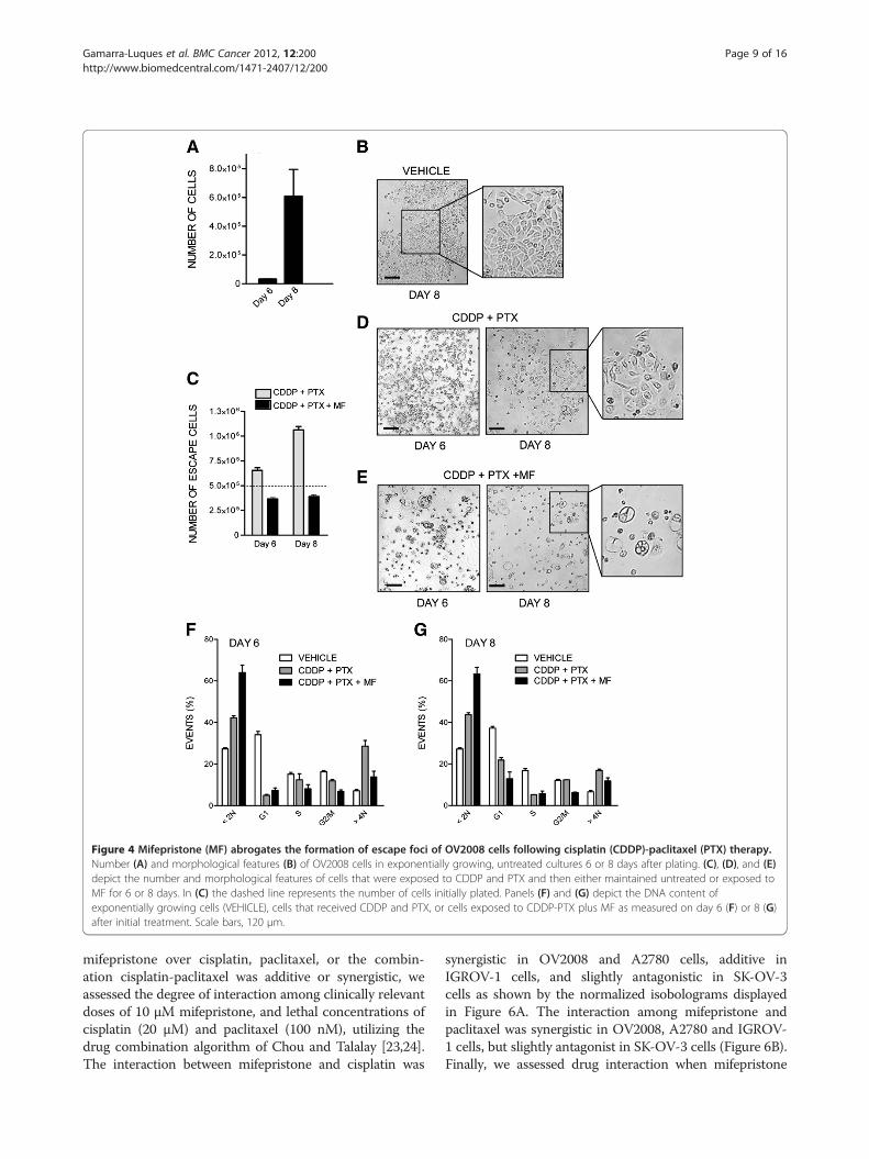
Figure 4 Mifepristone (MF) abrogates the formation of escape foci of OV2008 cells following cisplatin (CDDP)-paclitaxel (PTX) therapy.Number (A) and morphological features (B) of OV2008 cells in exponentially growing, untreated cultures 6 or 8 days after plating. (C), (D), and (E)depict the number and morphological features of cells that were exposed to CDDP and PTX and then either maintained untreated or exposed toMF for 6 or 8 days. In (C) the dashed line represents the number of cells initially plated. Panels (F) and (G) depict the DNA content ofexponentially growing cells (VEHICLE), cells that received CDDP and PTX, or cells exposed to CDDP-PTX plus MF as measured on day 6 (F) or 8 (G)after initial treatment. Scale bars, 120 μm.
Gamarra-Luques et al. BMC Cancer 2012, 12:200 Page 9 of 16http://www.biomedcentral.com/1471-2407/12/200
mifepristone over cisplatin, paclitaxel, or the combin-ation cisplatin-paclitaxel was additive or synergistic, weassessed the degree of interaction among clinically relevantdoses of 10 μM mifepristone, and lethal concentrations ofcisplatin (20 μM) and paclitaxel (100 nM), utilizing thedrug combination algorithm of Chou and Talalay [23,24].The interaction between mifepristone and cisplatin was
synergistic in OV2008 and A2780 cells, additive inIGROV-1 cells, and slightly antagonistic in SK-OV-3cells as shown by the normalized isobolograms displayedin Figure 6A. The interaction among mifepristone andpaclitaxel was synergistic in OV2008, A2780 and IGROV-1 cells, but slightly antagonist in SK-OV-3 cells (Figure 6B).Finally, we assessed drug interaction when mifepristone

Figure 5 Mifepristone (MF) enhances the therapeutic efficacy of cisplatin (CDDP) and paclitaxel (PTX) as shown by comparative doseresponse curves. Cells were treated with CDDP for 1 h (A and C), PTX for 3 h (B and C), and exposed or not exposed to 10 μM MF for 7 days(A-C). Cell growth (A and B) was calculated considering controls as 100%. In (C), relative growth was assessed by considering 1 the number of cells atthe beginning of the experiment. The dashed lines represent 50% of the growth depicted by control, untreated cells after 7 days of incubation.
Gamarra-Luques et al. BMC Cancer 2012, 12:200 Page 10 of 16http://www.biomedcentral.com/1471-2407/12/200
was added to the combination cisplatin-paclitaxel. In thiscontext, the mixture cisplatin-paclitaxel behaves as thirddrug to the cells and, consequently, from the mathematicalstandpoint it was considered as just one treatment or drug.Addition of mifepristone to the cisplatin-paclitaxel mix-ture had a therapeutic advantage in terms of growth inhib-ition in all cell lines studied as suggested by combinationindexes in the range of synergism (CI = 0.3–0.7) or strongsynergism (CI = 0.1–0.3) (Figure 6C). The therapeutic ad-vantage of adding mifepristone to the cisplatin-paclitaxelchemotherapeutic schedule is further reinforced by thecalculation of the dose reduction index (DRI). Additionalfile 5: Table S2 shows the DRI that indicate how manyfolds the dose of cisplatin, paclitaxel, or mifepristone,within the scenario of a synergistic combination, may bereduced at a given effect level compared with the dose ofeach drug alone, and in order to achieve the IC50. The DRIwere positive for cisplatin when combined with
mifepristone or with paclitaxel/mifepristone, for paclitaxelwhen combined with mifepristone or cisplatin-mifepris-tone, and for mifepristone, when combined with paclitaxelor paclitaxel-mifepristone. All DRI> 1 are relevant from aclinical perspective because dose-reduction suggest thatsimilar efficacy with lower toxicity may be achieved.
Mifepristone blocks time-dependent repopulation andabolishes clonogenic survival of ovarian cancer cells thatescape cisplatin-paclitaxel cytotoxic therapyThe regrowth of cells escaping cisplatin-paclitaxel tox-icity was time-dependent with a marked increase in cul-ture density as measured on days 7, 14 or 21 followinginitial chemotherapy (Figure 7). However, chronic expo-sure to a low cytostatic concentration of mifepristoneblunted such cellular escape in all days studied. Culturesof OV2008, A2780 and IGROV-1 cells exposed to mife-pristone following cisplatin-paclitaxel show a cellular

Figure 6 Normalized isobolograms depicting the pharmacological interaction of mifepristone (MF) when it follows cisplatin (CDDP)and/or paclitaxel (PTX) as calculated from the comparative dose response curves depicted in Figure 5. (A) Interaction between CDDP andMF. (B) Interaction among PTX and MF. (C) Interaction between MF and the combination CDDP-PTX, which was considered as ‘one treatment’ forthe calculation purposes. The normalized isobolograms were obtained using CalcuSyn software. The depicted combination indexes (CI) in theisobolograms (*) were calculated using the doses of 20 μM CDDP, 100 nM PTX and 10 μM MF; n = 3.
Gamarra-Luques et al. BMC Cancer 2012, 12:200 Page 11 of 16http://www.biomedcentral.com/1471-2407/12/200
density lower than the cellular density originally plated(Figure 7, dashed line). Mifepristone, however, was lessefficacious in preventing escape of SK-OV-3 cells.To determine whether there is an added long-term
lethal imprint by mifepristone in cells pre-treated withcisplatin-paclitaxel, we subjected OV2008, IGROV-1 andSK-OV-3 cells that remained viable 21 days after treat-ment initiation, to a clonogenic survival assay. Resultsshown in Figure 8 reveal that escape cells followingcisplatin-paclitaxel therapy have a growth advantage whencompared to the cells of origin in the OV2008 andIGROV-1 cell lines. SK-OV-3, to which mifepristone didnot appear to offer much advantage in terms of blockingrepopulation assessed as cellularity in culture (Figure 7),showed a slightly reduced clonogenic survival in cellsprimed with cisplatin-paclitaxel, and a much more reducedclonogenic capacity in cells primed with cisplatin-paclitaxelbut followed by chronic mifepristone maintenance therapy.The A2780 cells that escaped cisplatin-paclitaxel toxicity
were not able to be studied in a clonogenic survival assayas they behave differently than the original A2780 cells,showing a live population of cells arranged in clusters thatsurvive without adherence, thus invalidating the natureof the clonogenic survival assay that is based on countingadherent colonies (data not shown). When however totalnumber of cells (either adherent of non-adherent) wherecounted in the clonogenic plate, the number of escapeA2780 cells originally treated with cisplatin-paclitaxel andfollowed by mifepristone was highly reduced when com-pared to the number of cells escaping cisplatin-paclitaxel(data not shown), further confirming the phenomenonobserved in the other cell lines in which mifepristone notonly prevented escape of cells following cisplatin-paclitaxeltherapy, but also impinged a long-term lethal influence.
DiscussionOvarian cancer is known as a silent killer due to its latedetection and high mortality. Despite that much research

Figure 7 The long-term repopulation of escape cells followingcisplatin (CDDP)-paclitaxel (PTX) cytotoxic therapy is abrogatedor limited by mifepristone (MF) in OV2008 (A), A2780 (B),IGROV-1 (C) and SK-OV-3 (D) cells. Cells were exposed to 20 μMCDDP for 1 h, 100 nM PTX for 3 h and either chronically exposed ornot exposed to 10 μM MF. Number of cells was assessed after one,two or three weeks of initial treatment. The dashed lines representthe relative number of cells originally plated before subjecting theculture to the various treatment schedules.
Figure 8 Mifepristone (MF) diminishes the clonogenic survivalof escape cells repopulating after cisplatin (CDDP)-paclitaxel(PTX) cytotoxicity. Viable cells from a control culture, or fromprevious experiment as collected on day 21 after initial treatment,were subjected to a clonogenic survival assay. The number ofpositive colonies formed after one week of plating was assessed andthe data were expressed relative to the clonogenicity elicited byuntreated cells.
Gamarra-Luques et al. BMC Cancer 2012, 12:200 Page 12 of 16http://www.biomedcentral.com/1471-2407/12/200
has been done to improve the quality and quantity of lifeof ovarian cancer patients, the overall survival for thisdisease has remained stagnant at 30% since the introduc-tion of the dual cisplatin-paclitaxel therapy in the mid-1990s. Efforts are geared at finding manners to diagnosethe disease early, discovering target therapies, and im-proving the efficacy of current chemotherapeutic agents.We here provide evidence that when ovarian cancer cellsare subjected to clinically relevant exposure times of cis-platin and paclitaxel, and to supra-pharmacological dosesof the drugs –i.e. doses exceeding the maximal plasmaconcentrations that limit their tolerability, reported to bein the range of 50–100 nM for paclitaxel, and of 6–10 μMfor cisplatin [37-40]—the therapy is initially successful, yetthere are cells that eventually escape toxicity and relapse.Such relapse after remission can be, however, efficientlyprevented by adding clinically relevant concentrations ofthe synthetic steroid mifepristone [13-16].We modelled in vitro a scenario in which we utilized
supra-pharmacological doses of cisplatin and paclitaxel[25,26] to maximize the cytotoxicity of the drugs, butlimited their effect to clinically relevant times. Thus, cellswere exposed to cisplatin in the range of 0–20 μM foronly 1 h, to paclitaxel in the range of 0–100 nM for only3 h, and chronically to 10 μM mifepristone, likely to be
maintained in vivo [13-16]. Using this in vitro approach,we demonstrate that although the combination cisplatin-paclitaxel used at high doses is very efficacious in killinga large proportion of ovarian cancer cells encompassingbroad genetic backgrounds, there are cells that escapethe therapy and repopulate the culture following anotherwise apparent successful chemotherapeutic round.The initial efficacy of the therapy with cisplatin and
paclitaxel for ovarian cancer was amply validated andtranslated to the clinic many years ago [8,9]. However,the molecular mechanisms whereby cisplatin and pacli-taxel alone or in combination cause cell toxicity necessi-tate further elucidation. Cisplatin displays cytotoxicitytargeting the cytoplasm and the nucleus. In the cyto-plasm, cisplatin interacts with a wide number of sub-strates tilting the redox balance to oxidative stress,which facilitates DNA damage [41]. Cisplatin also causesdirect mitochondrial dysfunction [42] and endoplasmicreticulum stress [43]. In the nucleus, cisplatin bindsDNA leading to the generation of DNA-DNA inter- andintra-strand adducts. These lesions cause distortions inthe DNA that can be recognized by multiple repair path-ways. When the extent of damage is limited, cisplatinadducts induce an arrest in the S and G2 phases of the

Gamarra-Luques et al. BMC Cancer 2012, 12:200 Page 13 of 16http://www.biomedcentral.com/1471-2407/12/200
cell cycle to allow the DNA repair mechanisms to re-es-tablish DNA integrity and prevent abortive or abnormalmitosis. In contrast, if DNA damage is beyond repair,cells embark into a delayed death pathway [41,44-49].Paclitaxel, on the other hand, acts by binding to intracel-lular β-tubulin leading to microtubule stabilization andG2-M arrest; thereafter, the cells may either die by apop-tosis or necrosis immediately after the mitotic arrest, orfollowing an aberrant mitotic exit into a G1-like multi-nucleated state [50]. Paclitaxel-induced cell death is, atleast in part, mediated through the degradation of Bcl-2[51]. In contrast to cisplatin, sensitivity to paclitaxel isindependent of the p53 tissue expression status [52,53],while cisplatin-resistant cells retain sensitivity to pacli-taxel [27,36]. There is no doubt of the initial efficacy ofthe combination cisplatin-paclitaxel; their synergisticpharmacological interaction has been amply proven[28,29]. In our work we substantiated that adding mife-pristone after cisplatin-paclitaxel does not interfere withthe efficacy of the dual combination, but rather, in somecases enhances it.Dual cisplatin-paclitaxel therapy is followed by dis-
ease remission; however, it rarely provides cure as thedisease eventually relapses after remaining dormant asminimal residual for over a year [6]. Such therapeutic fail-ure is recapitulated in our in vitro toxicity model system inwhich scarce, yet critical cells escape cisplatin-paclitaxeltherapy and regrow as demonstrated by their increase innumber and by having a clonogenic survival capacity evensuperior than that of cells never treated with cisplatin-paclitaxel. When mifepristone was added after the initialtoxic cisplatin-paclitaxel combination, repopulation wasprevented and the clonogenic capacity of the remainingcells in culture was minimal.The repopulation of cancer cells that escape or re-
lapse after chemo- or radiotherapy accounts for thelack of long-term success of current cancer therapy.Repopulation of tumor cells was defined in 2005 byKim and Tannock [54] as ‘the continuing proliferation,sometimes at an accelerated pace, of surviving tumorcells with the capacity to regenerate the tumor that canoccur during a course of chemotherapy or fractionatedradiotherapy.’ The mechanism of repopulation of es-cape cells, however, is less understood: some recentstudies provide insights into it. For instance, cancercells develop the capacity to escape DNA damagecaused by pharmacological doses of platinum-basedtherapy via reverse polyploidy (a.k.a ‘neosis’ [55-57]),leading to the formation of diploid, rapidly proliferat-ing cells with increased platinum resistance [58]. Suchdiploid descendants are formed upon reactivation ofmeiosis-specific genes from a polyploid genome [59,60],and in association with the formation of sub-nuclei thatbecome degraded by autophagy [61]. Thus, a possibility
exists that mifepristone blocks repopulation of escape cellsby preventing reverse polyploidy. Supporting this hypoth-esis, we observed that OV2008 cultures that retain certainviability in between courses of cisplatin exposure, showgiant cells together with a nascent population of small cells[62] that may originate from the likely polyploid, giantprogenitors. Cultures treated with mifepristone after cis-platin do not show this small pool of repopulating cells; in-stead, they display an overall reduced number of cells,with predominance of a giant phenotype that ends upcommitting suicide as marked by cleaved PARP positivity[62]. Similar mechanism may be taking place in cells re-populating after cisplatin-paclitaxel combination therapy,because it is known that the driving force behind this ther-apy is cisplatin, not paclitaxel [44]. Additionally, withincultures repopulating after cisplatin-paclitaxel we observeda population of cells with hyperploid DNA content that isreduced parallel to cell repopulation and increased per-centage of G1 cells. In cultured treated with cisplatin-paclitaxel plus mifepristone, however, such hyperploidpopulation disappears in favor of hypodiploid DNA con-tent consisting with cells dying by apoptosis instead ofreturning to the cell cycle.Two other survival mechanisms that may explain re-
population after escape to chemotherapy could be thetarget of mifepristone interference. Firstly, mifepristonecan block the release of survival factors from dying cells,because a recent study demonstrated that cells that aredying as a consequence of the chemotherapy, releasechemical mediators (prostaglandins) that promote thegrowth of still surviving cells; more importantly, thismechanism requires caspase-3 activity in the dying cells[63], suggesting that caspase-3 has paradoxical functions,on one hand driving apoptotic cell death and on theother promoting the release of survival factors [64]. Sec-ondly, there is also a possibility that mifepristone blocksthe growth of scarce tumor initiating cells with the cap-acity to regenerate the culture and that may remain inculture because are resistant to cisplatin-paclitaxel incontrast to the bulk of differentiated cancer cells thatsuccumb to the chemotherapy [65]. A genetic evolutionstudy of high-grade serous ovarian adenocarcinomassuggests that resistance to cisplatin may develop frompre-existing minor clones that remain as minimal re-sidual disease and become enriched after initial chemo-therapy [66]. Within this scenario, mifepristone mayblock the repopulation of cells that never responded tocisplatin-paclitaxel therapy since, as we have shown, thedrug has similar growth inhibition potency in platinumsensitive and platinum resistant ovarian cancer cells [20].We provide proof-of-principle that the escape process
following cytotoxic therapy can be abrogated by a chronicexposure to mifepristone. Long-term (months to years) ofdaily administration of mifepristone is feasible and

Gamarra-Luques et al. BMC Cancer 2012, 12:200 Page 14 of 16http://www.biomedcentral.com/1471-2407/12/200
clinically well tolerated [67]. Other synthetic steroid agentswith similar structure than mifepristone containing adimethylaminophenyl substitution at the 11-β positionhave been developed [68]. We demonstrated that, similarto mifepristone, the related steroids ORG-31710 andCDB-2914 block ovarian cancer cell growth in associationwith inhibition of the activity of cyclin dependent kinase 2[17]. It warrants investigation whether these agents areequivalent or more efficient than mifepristone when usedto block repopulation following cisplatin-paclitaxeltherapy.
ConclusionsUsing an in vitro model of recurrence after exposure ofovarian cancer cells to supra-pharmacological doses of cis-platin and paclitaxel, and for clinically relevant exposuretimes, we demonstrated that a clinically relevant dose ofthe synthetic steroid mifepristone significantly improvestreatment efficacy by reducing the number and clonogenicsurvival capacity of escape cells. Thus, mifepristone couldbe used for chronic, non-toxic maintenance therapy fol-lowing cytotoxic standard cisplatin-paclitaxel chemother-apy to improve treatment efficacy by abrogating relapse ofcells escaping cisplatin-paclitaxel.
Additional files
Additional file 1: Figure S1. Histograms representing the viability(upper panel) and DNA content (lower panel) of OV2008 cells assessed bymicrocytometric analysis 2 days following treatment with paclitaxel (PTX),mifepristone (MF), cisplatin (CDDP), CDDP-PTX, or the triplet CDDP-PTX-MF. FSC, forward scatter.
Additional file 2: Figure S2. Histograms representing the viability(upper panel) and DNA content (lower panel) of OV2008 cells assessed bymicrocytometric analysis 4 days following treatment with paclitaxel (PTX),mifepristone (MF), cisplatin (CDDP), CDDP-PTX, or the triplet CDDP-PTX-MF. FSC, forward scatter.
Additional file 3: Figure S3. Phase contrast images obtained fromOV2008 cultures 4, 6, or 8 days following exposure to 20 μM cisplatin(CDDP) for 1 h, the doublet combination of 20 μM CDDP for 1 h and 100nM paclitaxel (PTX) for 3 h, or the triplet combination of 20 μM CDDP for1 h, 100 nM PTX for 3 h, and 20 μM mifepristone (MF) for the entire timein culture.
Additional file 4: Table S1. Concentrations of cisplatin (CDDP),paclitaxel (PTX), or mifepristone (MF) that inhibit growth by 50% (IC50s) inovarian cancer cells.
Additional file 5: Table S2. Dose reduction index values (DRI) forcisplatin (CDDP), paclitaxel (PTX) and mifepristone (MF) in ovarian cancercells.
Competing interestsThe authors declare that there is no conflict of interest that could influencethe impartiality of the research reported.
AcknowledgementsThis study was supported by National Cancer Institute Grant K22CA121991,ARRA Supplement K22CA121991-S1, and R15CA164622 and in part by aSouth Dakota Board of Regents competitive research grant award (SDBOR/USD 2011-10-06). CGL and MBH were recipients of a visiting scholarship fromthe National Council for Scientific and Technical Research (CONICET),
Argentina. We are indebted to Nahuel Telleria for the edition of themanuscript.
Author details1Division of Basic Biomedical Sciences, Sanford School of Medicine of TheUniversity of South Dakota, 414 East Clark Street, Vermillion, SD, USA. 2Presentaddress: Institute of Histology and Embryology of Cuyo, National Council forScientific and Technical Research (CONICET), Mendoza, Argentina. 3Presentaddress: Institute of Medicine and Experimental Biology of Cuyo, NationalCouncil for Scientific and Technical Research (CONICET), Mendoza, Argentina.
Authors' contributionsConceived and designed the experiments: CGL AAG CMT. Performedexperiments CGL AAG MBH. Analyzed the data: CGL AAG MBH CMT.Contributed reagents/materials/analysis tools: CMT. Wrote the paper: CGLCMT. All authors read and approved the final manuscript.
Received: 22 March 2012 Accepted: 29 May 2012Published: 29 May 2012
References1. Vaughan S, Coward JI, Bast RC Jr, Berchuck A, Berek JS, Brenton JD, Coukos
G, Crum CC, Drapkin R, Etemadmoghadam D, Friedlander M, Gabra H, KayeSB, Lord CJ, Lengyel E, Levine DA, McNeish IA, Menon U, Mills GB, NephewKP, Oza AM, Sood AK, Stronach EA, Walczak H, Bowtell DD, Balkwill FR:Rethinking ovarian cancer: recommendations for improving outcomes.Nat Rev Cancer 2011, 11(10):719–725.
2. Lengyel E: Ovarian cancer development and metastasis. Am J Pathol 2010,177(3):1053–1064.
3. Bast RC Jr, Hennessy B, Mills GB: The biology of ovarian cancer: newopportunities for translation. Nat Rev Cancer 2009, 9(6):415–428.
4. Gubbels JA, Claussen N, Kapur AK, Connor JP, Patankar MS: The detection,treatment, and biology of epithelial ovarian cancer. J Ovarian Res 2010,3:8.
5. Agarwal R, Kaye SB: Ovarian cancer: strategies for overcoming resistanceto chemotherapy. Nat Rev Cancer 2003, 3(7):502–516.
6. Yap TA, Carden CP, Kaye SB: Beyond chemotherapy: targeted therapies inovarian cancer. Nat Rev Cancer 2009, 9(3):167–181.
7. Ozols RF: Systemic therapy for ovarian cancer: current status and newtreatments. Sem Oncol 2006, 33(2 Suppl 6):S3–S11.
8. McGuire WP, Hoskins WJ, Brady MF, Kucera PR, Partridge EE, Look KY, Clarke-Pearson DL, Davidson M: Cyclophosphamide and cisplatin compared withpaclitaxel and cisplatin in patients with stage III and stage IV ovariancancer. N Engl J Med 1996, 334(1):1–6.
9. Piccart MJ, Bertelsen K, James K, Cassidy J, Mangioni C, Simonsen E, Stuart G,Kaye S, Vergote I, Blom R, Grimshaw R, Atkinson RJ, Swenerton KD, Trope C,Nardi M, Kaern J, Tumolo S, Timmers P, Roy JA, Lhoas F, Lindvall B, Bacon M,Birt A, Andersen JE, Zee B, Paul J, Baron B, Pecorelli S: Randomizedintergroup trial of cisplatin-paclitaxel versus cisplatin-cyclophosphamidein women with advanced epithelial ovarian cancer: three-year results.J Natl Cancer Inst 2000, 92(9):699–708.
10. Ozols RF, Bundy BN, Greer BE, Fowler JM, Clarke-Pearson D, Burger RA,Mannel RS, DeGeest K, Hartenbach EM, Baergen R: Phase III trial ofcarboplatin and paclitaxel compared with cisplatin and paclitaxel inpatients with optimally resected stage III ovarian cancer: a GynecologicOncology Group study. J Clin Oncol 2003, 21(17):3194–3200.
11. Armstrong DK, Bundy B, Wenzel L, Huang HQ, Baergen R, Lele S, CopelandLJ, Walker JL, Burger RA: Intraperitoneal cisplatin and paclitaxel in ovariancancer. N Engl J Med 2006, 354(1):34–43.
12. Goyeneche AA, Caron RW, Telleria CM: Mifepristone inhibits ovarian cancercell growth in vitro and in vivo. Clin Cancer Res 2007, 13(11):3370–3379.
13. Heikinheimo O, Lahteenmaki PL, Koivunen E, Shoupe D, Croxatto H,Luukkainen T, Lahteenmaki P: Metabolism and serum binding of RU 486in women after various single doses. Hum Reprod 1987, 2(5):379–385.
14. Heikinheimo O: Pharmacokinetics of the antiprogesterone RU 486 inwomen during multiple dose administration. J Steroid Biochem 1989, 32(1A):21–25.
15. Shoupe D, Mishell DR Jr, Lahteenmaki P, Heikinheimo O, Birgerson L,Madkour H, Spitz IM: Effects of the antiprogesterone RU 486 in normalwomen. I. Single-dose administration in the midluteal phase. Am J ObstGynecol 1987, 157(6):1415–1420.

Gamarra-Luques et al. BMC Cancer 2012, 12:200 Page 15 of 16http://www.biomedcentral.com/1471-2407/12/200
16. Kawai S, Nieman LK, Brandon DD, Udelsman R, Loriaux DL, Chrousos GP:Pharmacokinetic properties of the antiglucocorticoid andantiprogesterone steroid RU 486 in man. J Pharmacol Exp Ther 1987, 241(2):401–406.
17. Goyeneche AA, Seidel EE, Telleria CM: Growth inhibition induced byantiprogestins RU-38486, ORG-31710, and CDB-2914 in ovarian cancercells involves inhibition of cyclin dependent kinase 2. Invest New Drugs2012, 30:967–980.
18. Conradie R, Bruggeman FJ, Ciliberto A, Csikasz-Nagy A, Novak B, WesterhoffHV, Snoep JL: Restriction point control of the mammalian cell cycle viathe cyclin E/Cdk2:p27 complex. FEBS J 2010, 277(2):357–367.
19. Tieszen CR, Goyeneche AA, Brandhagen BN, Ortbahn CT, Telleria CM:Antiprogestin mifepristone inhibits the growth of cancer cells ofreproductive and non-reproductive origin regardless of progesteronereceptor expression. BMC Cancer 2011, 11(1):207.
20. Freeburg EM, Goyeneche AA, Seidel EE, Telleria CM: Resistance to cisplatindoes not affect sensitivity of human ovarian cancer cell lines tomifepristone cytotoxicity. Cancer Cell Int 2009, 9:4.
21. Gamarra-Luques CD, Telleria CM: Enhancement of the lethality ofplatinum-based therapy by antiprogestin mifepristone in ovarian cancer.Cancer Epidemiol Bio Prev 2010, 19(10):Supplement 1, A113.
22. Katano K, Kondo A, Safaei R, Holzer A, Samimi G, Mishima M, Kuo YM,Rochdi M, Howell SB: Acquisition of resistance to cisplatin is accompaniedby changes in the cellular pharmacology of copper. Cancer Res 2002, 62(22):6559–6565.
23. Chou TC: Drug combination studies and their synergy quantificationusing the Chou-Talalay method. Cancer Res 2010, 70(2):440–446.
24. Chou TC: Theoretical basis, experimental design, and computerizedsimulation of synergism and antagonism in drug combination studies.Pharmacol Rev 2006, 58(3):621–681.
25. Mansouri A, Ridgway LD, Korapati AL, Zhang Q, Tian L, Wang Y, Siddik ZH,Mills GB, Claret FX: Sustained activation of JNK/p38 MAPK pathways inresponse to cisplatin leads to Fas ligand induction and cell death inovarian carcinoma cells. J Biol Chem 2003, 278(21):19245–19256.
26. Karlsson MO, Molnar V, Freijs A, Nygren P, Bergh J, Larsson R:Pharmacokinetic models for the saturable distribution of paclitaxel. DrugMetab Dispos 1999, 27(10):1220–1223.
27. Jones NA, Turner J, McIlwrath AJ, Brown R, Dive C: Cisplatin- and paclitaxel-induced apoptosis of ovarian carcinoma cells and the relationshipbetween bax and bak up-regulation and the functional status of p53.Mol Pharmacol 1998, 53(5):819–826.
28. Jekunen AP, Christen RD, Shalinsky DR, Howell SB: Synergistic interactionbetween cisplatin and taxol in human ovarian carcinoma cells in vitro. BrJ Cancer 1994, 69(2):299–306.
29. Gately DP, Sharma A, Christen RD, Howell SB: Cisplatin and taxol activatedifferent signal pathways regulating cellular injury-induced expression ofGADD153. Br J Cancer 1996, 73(1):18–23.
30. Hayakawa J, Ohmichi M, Kurachi H, Ikegami H, Kimura A, Matsuoka T,Jikihara H, Mercola D, Murata Y: Inhibition of extracellular signal-regulatedprotein kinase or c-Jun N-terminal protein kinase cascade, differentiallyactivated by cisplatin, sensitizes human ovarian cancer cell line. J BiolChem 1999, 274(44):31648–31654.
31. Sorenson CM, Barry MA, Eastman A: Analysis of events associated with cellcycle arrest at G2 phase and cell death induced by cisplatin. J Natl CancerInst 1990, 82(9):749–755.
32. Ormerod MG, O’Neill C, Robertson D, Kelland LR, Harrap KR: cis-Diamminedichloroplatinum(II)-induced cell death through apoptosis insensitive and resistant human ovarian carcinoma cell lines. CancerChemother Pharmacol 1996, 37(5):463–471.
33. Selvakumaran M, Pisarcik DA, Bao R, Yeung AT, Hamilton TC: Enhancedcisplatin cytotoxicity by disturbing the nucleotide excision repairpathway in ovarian cancer cell lines. Cancer Res 2003, 63(6):1311–1316.
34. Sain N, Krishnan B, Ormerod MG, De Rienzo A, Liu WM, Kaye SB, Workman P,Jackman AL: Potentiation of paclitaxel activity by the HSP90 inhibitor 17-allylamino-17-demethoxygeldanamycin in human ovarian carcinoma celllines with high levels of activated AKT. Mol Cancer Ther 2006, 5(5):1197–1208.
35. Wang Y, Niu XL, Qu Y, Wu J, Zhu YQ, Sun WJ, Li LZ: Autocrine productionof interleukin-6 confers cisplatin and paclitaxel resistance in ovariancancer cells. Cancer Lett 2010, 295(1):110–123.
36. Judson PL, Watson JM, Gehrig PA, Fowler WC Jr, Haskill JS: Cisplatin inhibitspaclitaxel-induced apoptosis in cisplatin-resistant ovarian cancer cell
lines: possible explanation for failure of combination therapy. Cancer Res1999, 59(10):x2425–2432.
37. Ohtsu T, Sasaki Y, Tamura T, Miyata Y, Nakanomyo H, Nishiwaki Y, Saijo N:Clinical pharmacokinetics and pharmacodynamics of paclitaxel: a 3-hourinfusion versus a 24-hour infusion. Clin Cancer Res 1995, 1(6):599–606.
38. Rowinsky EK, Jiroutek M, Bonomi P, Johnson D, Baker SD: Paclitaxel steady-state plasma concentration as a determinant of disease outcome andtoxicity in lung cancer patients treated with paclitaxel and cisplatin. ClinCancer Res 1999, 5(4):767–774.
39. Kurata T, Tamura T, Shinkai T, Ohe Y, Kunitoh H, Kodama T, Kakinuma R,Matsumoto T, Kubota K, Omatsu H, Nishiwaki Y, Saijo N: Phase I andpharmacological study of paclitaxel given over 3 h with cisplatin foradvanced non-small cell lung cancer. Jpn J Clin Oncol 2001, 31(3):93–99.
40. Urien S, Lokiec F: Population pharmacokinetics of total and unboundplasma cisplatin in adult patients. Br J Clin Pharmacol 2004, 57(6):756–763.
41. Galluzzi L, Senovilla L, Vitale I, Michels J, Martins I, Kepp O, Castedo M,Kroemer G: Molecular mechanisms of cisplatin resistance. Oncogene 2012,31(15):1869–1883.
42. Cullen KJ, Yang Z, Schumaker L, Guo Z: Mitochondria as a critical target ofthe chemotheraputic agent cisplatin in head and neck cancer. J BioenergBiomembr 2007, 39(1):x43–50.
43. Mandic A, Hansson J, Linder S, Shoshan MC: Cisplatin induces endoplasmicreticulum stress and nucleus-independent apoptotic signaling. J BiolChem 2003, 278(11):9100–9106.
44. Kelland L: The resurgence of platinum-based cancer chemotherapy. NatRev Cancer 2007, 7(8):573–584.
45. Cepeda V, Fuertes MA, Castilla J, Alonso C, Quevedo C, Perez JM:Biochemical mechanisms of cisplatin cytotoxicity. Anticancer Agents MedChem 2007, 7(1):3–18.
46. Rabik CA, Dolan ME: Molecular mechanisms of resistance and toxicityassociated with platinating agents. Cancer Treat Rev 2007, 33(1):9–23.
47. Jung Y, Lippard SJ: Direct cellular responses to platinum-induced DNAdamage. Chem Rev 2007, 107(5):1387–1407.
48. Wang D, Lippard SJ: Cellular processing of platinum anticancer drugs. NatRev Drug Discov 2005, 4(4):307–320.
49. Fayad W, Brnjic S, Berglind D, Blixt S, Shoshan MC, Berndtsson M, OlofssonMH, Linder S: Restriction of cisplatin induction of acute apoptosis to asubpopulation of cells in a three-dimensional carcinoma culture model.Int J Cancer 2009, 125(10):2450–2455.
50. Abal M, Andreu JM, Barasoain I: Taxanes: microtubule and centrosometargets, and cell cycle dependent mechanisms of action. Curr Cancer DrugTargets 2003, 3(3):193–203.
51. Haldar S, Chintapalli J, Croce CM: Taxol induces bcl-2 phosphorylation anddeath of prostate cancer cells. Cancer Res 1996, 56(6):1253–1255.
52. Debernardis D, Sire EG, De Feudis P, Vikhanskaya F, Valenti M, Russo P,Parodi S, D’Incalci M, Broggini M: p53 status does not affect sensitivity ofhuman ovarian cancer cell lines to paclitaxel. Cancer Res 1997, 57(5):870–874.
53. Wahl AF, Donaldson KL, Fairchild C, Lee FY, Foster SA, Demers GW, GallowayDA: Loss of normal p53 function confers sensitization to Taxol byincreasing G2/M arrest and apoptosis. Nat Med 1996, 2(1):72–79.
54. Kim JJ, Tannock IF: Repopulation of cancer cells during therapy: animportant cause of treatment failure. Nat Rev Cancer 2005, 5(7):516–525.
55. Erenpreisa J, Cragg MS: Cancer: a matter of life cycle? Cell Biol Int 2007, 31(12):1507–1510.
56. Illidge TM, Cragg MS, Fringes B, Olive P, Erenpreisa JA: Polyploid giant cellsprovide a survival mechanism for p53 mutant cells after DNA damage.Cell Biol Int 2000, 24(9):621–633.
57. Sundaram M, Guernsey DL, Rajaraman MM, Rajaraman R: Neosis: a noveltype of cell division in cancer. Cancer Biol Ther 2004, 3(2):207–218.
58. Puig PE, Guilly MN, Bouchot A, Droin N, Cathelin D, Bouyer F, Favier L,Ghiringhelli F, Kroemer G, Solary E, Martin F, Chauffert B: Tumor cells canescape DNA-damaging cisplatin through DNA endoreduplication andreversible polyploidy. Cell Biol Int 2008, 32(9):1031–1043.
59. Kalejs M, Ivanov A, Plakhins G, Cragg MS, Emzinsh D, Illidge TM, Erenpreisa J:Upregulation of meiosis-specific genes in lymphoma cell lines followinggenotoxic insult and induction of mitotic catastrophe. BMC Cancer 2006,6:6.
60. Salmina K, Jankevics E, Huna A, Perminov D, Radovica I, Klymenko T, IvanovA, Jascenko E, Scherthan H, Cragg M, Erenpreisa J: Up-regulation of theembryonic self-renewal network through reversible polyploidy in
Gamarra-Luques et al. BMC Cancer 2012, 12:200 Page 16 of 16http://www.biomedcentral.com/1471-2407/12/200
irradiated p53-mutant tumour cells. Exp Cell Res 2010, 316(13):2099–2112.61. Erenpreisa J, Salmina K, Huna A, Kosmacek EA, Cragg MS, Ianzini F, Anisimov
AP: Polyploid tumour cells elicit paradiploid progeny throughdepolyploidizing divisions and regulated autophagic degradation. CellBiol Int 2011, 35(7):687–695.
62. Freeburg EM, Goyeneche AA, Telleria CM: Mifepristone abrogatesrepopulation of ovarian cancer cells in between courses of cisplatintreatment. Int J Oncol 2009, 34(3):743–755.
63. Huang Q, Li F, Liu X, Li W, Shi W, Liu FF, O’Sullivan B, He Z, Peng Y, Tan AC,Zhou L, Shen J, Han G, Wang XJ, Thorburn J, Thorburn A, Jimeno A, RabenD, Bedford JS, Li CY: Caspase 3-mediated stimulation of tumor cellrepopulation during cancer radiotherapy. Nat Med 2011, 17(7):860–866.
64. Galluzzi L, Kepp O, Kroemer G: Caspase-3 and prostaglandins signal fortumor regrowth in cancer therapy. Oncogene 2012,31(23):2805–2808.
65. Kvinlaug BT, Huntly BJ: Targeting cancer stem cells. Expert Opin Ther Targets2007, 11(7):915–927.
66. Cooke SL, Ng CK, Melnyk N, Garcia MJ, Hardcastle T, Temple J, Langdon S,Huntsman D, Brenton JD: Genomic analysis of genetic heterogeneity andevolution in high-grade serous ovarian carcinoma. Oncogene 2010, 29(35):4905–4913.
67. Grunberg SM, Weiss MH, Russell CA, Spitz IM, Ahmadi J, Sadun A, Sitruk-Ware R: Long-term administration of mifepristone (RU486): clinicaltolerance during extended treatment of meningioma. Cancer Invest 2006,24(8):727–733.
68. Benagiano G, Bastianelli C, Farris M: Selective progesterone receptormodulators 1: use during pregnancy. Expert Opin Pharmacother 2008, 9(14):2459–2472.
doi:10.1186/1471-2407-12-200Cite this article as: Gamarra-Luques et al.: Mifepristone preventsrepopulation of ovarian cancer cells escaping cisplatin-paclitaxeltherapy. BMC Cancer 2012 12:200.
Submit your next manuscript to BioMed Centraland take full advantage of:
• Convenient online submission
• Thorough peer review
• No space constraints or color figure charges
• Immediate publication on acceptance
• Inclusion in PubMed, CAS, Scopus and Google Scholar
• Research which is freely available for redistribution
Submit your manuscript at www.biomedcentral.com/submit