Research Article Molecular Epidemiology of Enteric...
6
Research Article Molecular Epidemiology of Enteric Adenovirus Gastroenteritis in under-Five-Year-Old Children in Iran Anahita Sanaei Dashti, 1 Pedram Ghahremani, 2 Tayebeh Hashempoor, 3 and Abdollah Karimi 2 1 Shiraz HIV/AIDS Research Center, Shiraz University of Medical Sciences, Shiraz 7193613311, Iran 2 Pediatric Infectious Diseases Research Center, Shahid Beheshti University of Medical Sciences, Tehran, Iran 3 Professor Alborzi Clinical Microbiology Research Center, Namazi Hospital, Shiraz University of Medical Sciences, Shiraz 7193613311, Iran Correspondence should be addressed to Anahita Sanaei Dashti; anahita [email protected] Received 8 August 2015; Revised 25 October 2015; Accepted 26 October 2015 Academic Editor: Greger Lindberg Copyright © 2016 Anahita Sanaei Dashti et al. is is an open access article distributed under the Creative Commons Attribution License, which permits unrestricted use, distribution, and reproduction in any medium, provided the original work is properly cited. Background. Acute gastroenteritis is one of the major sources of morbidity and mortality among young children in developed and developing countries. e aim of this study was to determine the prevalence of human adenovirus- (HAdV-) 40 and HAdV-41 in children hospitalized with gastroenteritis in five different health centers of Iran. Methods. In a cross-sectional epidemiological study, we studied 2682 fecal specimens that were collected from children under the age of 5 years in five educational and therapeutic pediatric centers in Iran from February 2012 to February 2013. Samples were tested for HAdV-40 and HAdV-41, using a specific pair of primers in polymerase chain reaction (PCR) method. Results. HAdV-40 and HAdV-41 were detected in 132 (5.18%) of the patients with diarrhea. A significantly higher prevalence of HAdV-40 and HAdV-41 (58.3%) was observed in children under 12 months of age, compared to other age groups. e male to female ratio was 1.7. Conclusion. e results of this study demonstrated that HAdV- 40 and HAdV-41 could be considered etiological agents for acute gastroenteritis among children in Iran. e PCR as a rapid test may increase the chance for a relatively mild course of the disease followed by a complete recovery and avoiding administration of unnecessary antibiotics. 1. Introduction Acute gastroenteritis is a very common disease that causes a significant mortality in developing countries. Worldwide, gastroenteritis affects 3 to 5 million children each year [1] and accounts for 1.5 to 2.5 million deaths per year or 12% of all deaths among children less than 5 years of age [1–3]. ough it seldom causes death in developed countries, it puts a heavy burden on the health care system [4], as it accounts for 10% of all hospital admissions for children under the age of 5 years. Viruses are the most important etiologic causes respon- sible for approximately 70% of the episodes of acute gas- troenteritis in children [4]. Worldwide, rotavirus is still the most common virus causing this disease [5, 6], followed by adenovirus types 40 and 41, astrovirus, and calicivirus [6, 7]. e rate of enteric adenovirus 40 and 41 varies from 1–8% in developed countries to 2–31% in developing countries [5, 7], but the prevalence is increased in immunocompromised patients [8]. Human adenoviruses (HAdVs) are one of the major causes of a number of different clinical syndromes including gastroenteritis, respiratory disease, conjunctivitis, hemor- rhagic cystitis, and exanthema. ey comprise 51 different serotypes (HAdV-1 to HAdV-51) grouped into 6 species, A to F. e enteric serotypes that are mostly associated with gastroenteritis are Ad-40 and Ad-41 which belong to species F. Enteric adenoviruses associated with protracted diarrhea which may contribute to infant dehydration and malnutrition in developing countries [4, 5] spread predominantly by the fecal-oral route [3, 4]. Usually, aſter an incubation period of 8 to 10 days, periodic diarrhea occurs, with low grade fever, vomiting, abdominal pains, and dehydration [3, 6]. Hindawi Publishing Corporation Gastroenterology Research and Practice Volume 2016, Article ID 2045697, 5 pages http://dx.doi.org/10.1155/2016/2045697
-
Upload
phungtuong -
Category
Documents
-
view
215 -
download
0
Transcript of Research Article Molecular Epidemiology of Enteric...

Research ArticleMolecular Epidemiology of Enteric Adenovirus Gastroenteritisin under-Five-Year-Old Children in Iran
Anahita Sanaei Dashti,1 Pedram Ghahremani,2
Tayebeh Hashempoor,3 and Abdollah Karimi2
1Shiraz HIV/AIDS Research Center, Shiraz University of Medical Sciences, Shiraz 7193613311, Iran2Pediatric Infectious Diseases Research Center, Shahid Beheshti University of Medical Sciences, Tehran, Iran3Professor Alborzi Clinical Microbiology Research Center, Namazi Hospital, Shiraz University of Medical Sciences,Shiraz 7193613311, Iran
Correspondence should be addressed to Anahita Sanaei Dashti; anahita [email protected]
Received 8 August 2015; Revised 25 October 2015; Accepted 26 October 2015
Academic Editor: Greger Lindberg
Copyright © 2016 Anahita Sanaei Dashti et al. This is an open access article distributed under the Creative Commons AttributionLicense, which permits unrestricted use, distribution, and reproduction in any medium, provided the original work is properlycited.
Background. Acute gastroenteritis is one of the major sources of morbidity and mortality among young children in developed anddeveloping countries. The aim of this study was to determine the prevalence of human adenovirus- (HAdV-) 40 and HAdV-41in children hospitalized with gastroenteritis in five different health centers of Iran. Methods. In a cross-sectional epidemiologicalstudy, we studied 2682 fecal specimens that were collected from children under the age of 5 years in five educational and therapeuticpediatric centers in Iran from February 2012 to February 2013. Samples were tested for HAdV-40 andHAdV-41, using a specific pairof primers in polymerase chain reaction (PCR)method.Results. HAdV-40 andHAdV-41 were detected in 132 (5.18%) of the patientswith diarrhea. A significantly higher prevalence of HAdV-40 and HAdV-41 (58.3%) was observed in children under 12 months ofage, compared to other age groups.Themale to female ratio was 1.7. Conclusion. The results of this study demonstrated that HAdV-40 and HAdV-41 could be considered etiological agents for acute gastroenteritis among children in Iran. The PCR as a rapid testmay increase the chance for a relatively mild course of the disease followed by a complete recovery and avoiding administration ofunnecessary antibiotics.
1. Introduction
Acute gastroenteritis is a very common disease that causesa significant mortality in developing countries. Worldwide,gastroenteritis affects 3 to 5million children each year [1] andaccounts for 1.5 to 2.5 million deaths per year or 12% of alldeaths among children less than 5 years of age [1–3].
Though it seldom causes death in developed countries,it puts a heavy burden on the health care system [4], as itaccounts for 10% of all hospital admissions for children underthe age of 5 years.
Viruses are the most important etiologic causes respon-sible for approximately 70% of the episodes of acute gas-troenteritis in children [4]. Worldwide, rotavirus is still themost common virus causing this disease [5, 6], followed byadenovirus types 40 and 41, astrovirus, and calicivirus [6, 7].The rate of enteric adenovirus 40 and 41 varies from 1–8%
in developed countries to 2–31% in developing countries [5,7], but the prevalence is increased in immunocompromisedpatients [8].
Human adenoviruses (HAdVs) are one of the majorcauses of a number of different clinical syndromes includinggastroenteritis, respiratory disease, conjunctivitis, hemor-rhagic cystitis, and exanthema. They comprise 51 differentserotypes (HAdV-1 to HAdV-51) grouped into 6 species, Ato F. The enteric serotypes that are mostly associated withgastroenteritis are Ad-40 and Ad-41 which belong to speciesF.
Enteric adenoviruses associated with protracted diarrheawhichmay contribute to infant dehydration andmalnutritionin developing countries [4, 5] spread predominantly by thefecal-oral route [3, 4]. Usually, after an incubation period of8 to 10 days, periodic diarrhea occurs, with low grade fever,vomiting, abdominal pains, and dehydration [3, 6].
Hindawi Publishing CorporationGastroenterology Research and PracticeVolume 2016, Article ID 2045697, 5 pageshttp://dx.doi.org/10.1155/2016/2045697
2 Gastroenterology Research and Practice
In a survey done in Taiwan, clinical features of enteric ad-enoviruses types 40 and 41 in children were diarrhea (96.9%),fever (54.7%), vomiting (45.3%), mild dehydration (43.8%),symptoms of upper respiratory tract infection (21.9%), andabdominal pain (12.5%) [9]. Long-lasting diarrhea (mean 10.8days) was a predominant symptom of enteric adenoviruses incomparison to rotavirus [10]. In another comparative study,adenovirus gastroenteritis differed from rota- and astrovirusinfections by subacute onset, less frequent vomiting, morefrequent development of mild and moderate dehydration,and abdominal pains and distension [11].
These viruses exist year-round in all parts of the worldbut are most prevalent during spring, early summer, andmidwinter in temperate climates [3, 5].
The present studywas performed to identify the predomi-nant adenovirus serotypes and their epidemiologic character-istics in infants and children with acute gastroenteritis fromfive different pediatric therapy centers in Iran.
2. Methods and Materials
The study was carried out from February 2012 to February2013 on 2682 stool samples, obtained within 48 hours ofadmission to the hospitals of children under 5 years, sufferingfrom acute gastroenteritis, from five different cities of Iran:Tehran, Shiraz, Mashhad, Tabriz, and Bandar Abbas. All thepatients were examined according to the criteria describedearlier for the number of episodes and the duration ofvomiting and diarrhea, associated symptoms, and the extentof dehydration and treatment [12]. Patients with acquired orcongenital immune deficiency were excluded from the study.The fecal specimens were stored at −70∘C immediately forlater virological tests. Informed consents were obtained fromparents before collecting the samples. The Ethics Committeeof Shahid Beheshti University of Medical Sciences approvedthe study.Demographic datawere collected by questionnairesincluding sex, age, and living place. The specimens werestored at −80∘C until processing to detect viral antigens.
The viral nucleic acid was extracted from stool suspen-sions by using the Bioneer kit, according to the manufac-turer’s instructions (Bioneer, Korea).
The extracted DNA was used as a template for amplifica-tion of HAdV-hexon gene. A 261 bp fragment was amplifiedusing specific sense and antisense primers (Ref) including 5-GCCACCGATACGTACTTCAGCCTG-3 and 5-GGCAGT-GCCGGAGTAGGGTTTAAA-3, respectively [13]. Briefly,the extracted DNA was amplified by standard PCR usingprimers recognizing the HAdV-hexon region. The reactionmixture consisted of 0.2mM dNTP, 1.5mM MgCl2, 1 U ofTaq DNA polymerase (Fermentas, Lithuania), PCR-buffer 1x(Fermentas, Lithuania), and 1mM of each primer. The PCRconditions consisted of 30 cycles of 4min at 94∘C, 30 s at94∘C, 62 s at 60∘C, 60 s at 72∘C, and a final extension cycleof 72∘C for 7min. PCR products were loaded on 1% agarosegel along with 100 bpmolecular size DNA ladder (Fermentas,Lithuania).
Descriptive analysis including frequencies and nonpara-metric tests including chi-square and t-test were performed.
0
10
20
30
40
50
60
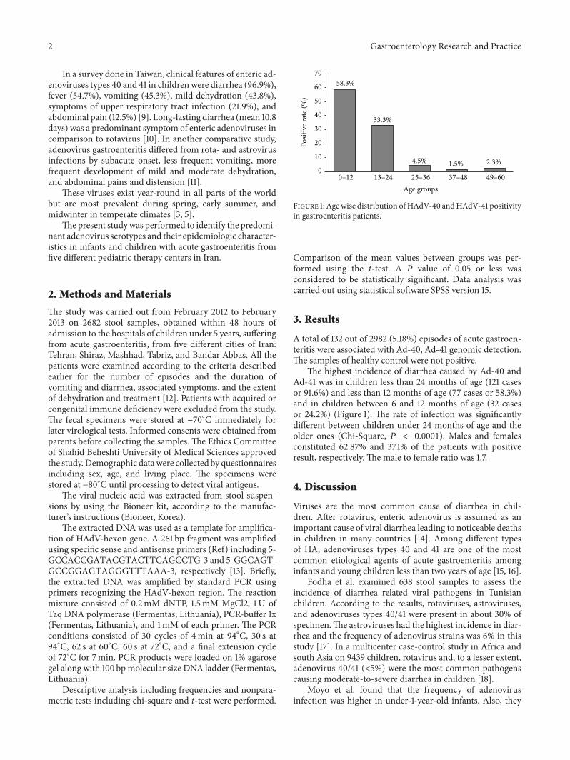
7058.3%
33.3%
4.5% 1.5% 2.3%
Posit
ive r
ate (
%)
Age groups0–12 13–24 25–36 37–48 49–60
Figure 1: Agewise distribution ofHAdV-40 andHAdV-41 positivityin gastroenteritis patients.
Comparison of the mean values between groups was per-formed using the t-test. A 𝑃 value of 0.05 or less wasconsidered to be statistically significant. Data analysis wascarried out using statistical software SPSS version 15.
3. Results
A total of 132 out of 2982 (5.18%) episodes of acute gastroen-teritis were associated with Ad-40, Ad-41 genomic detection.The samples of healthy control were not positive.
The highest incidence of diarrhea caused by Ad-40 andAd-41 was in children less than 24 months of age (121 casesor 91.6%) and less than 12 months of age (77 cases or 58.3%)and in children between 6 and 12 months of age (32 casesor 24.2%) (Figure 1). The rate of infection was significantlydifferent between children under 24 months of age and theolder ones (Chi-Square, 𝑃 < 0.0001). Males and femalesconstituted 62.87% and 37.1% of the patients with positiveresult, respectively. The male to female ratio was 1.7.
4. Discussion
Viruses are the most common cause of diarrhea in chil-dren. After rotavirus, enteric adenovirus is assumed as animportant cause of viral diarrhea leading to noticeable deathsin children in many countries [14]. Among different typesof HA, adenoviruses types 40 and 41 are one of the mostcommon etiological agents of acute gastroenteritis amonginfants and young children less than two years of age [15, 16].
Fodha et al. examined 638 stool samples to assess theincidence of diarrhea related viral pathogens in Tunisianchildren. According to the results, rotaviruses, astroviruses,and adenoviruses types 40/41 were present in about 30% ofspecimen.The astroviruses had the highest incidence in diar-rhea and the frequency of adenovirus strains was 6% in thisstudy [17]. In a multicenter case-control study in Africa andsouth Asia on 9439 children, rotavirus and, to a lesser extent,adenovirus 40/41 (<5%) were the most common pathogenscausing moderate-to-severe diarrhea in children [18].
Moyo et al. found that the frequency of adenovirusinfection was higher in under-1-year-old infants. Also, they

Gastroenterology Research and Practice 3
reported that nondiarrheic and diarrheic children had similarprevalence of adenovirus [19].
The other viruses that are now recognized to play a majorrole in sporadic gastrointestinal illnesses are noroviruses [20].Early serosurveys documented a high prevalence of norovirusantibodies in children, but, because the virus was rarelydetected in fecal specimens, its role in causing the infectionseemed questionable [21].
The present study highlights the prevalence of entericadenoviruses, HAdV-40 and HAdV-41, among acute gas-troenteritis cases from five different cities of Iran. Viral DNAwas detected using PCR technique. HAdV-40 and HAdV-41infection was found in 5.18% of children with diarrhea butwas not detected in the control group.
There are different reports about the prevalence of viralgastroenteritis in Iran. Previous epidemiological studies inIran such as those done by Shokrollahi, Motamedifar, andHamkar showed that 20%, 9%, and 2.3% of viral gastroenteri-tis were attributable to the adenoviruses, respectively [22–24].However, a review of published data about viral gastroenteri-tis in Iran from 2002 to 2013 showed that enteric adenovirusincidence was 5.7% in children with acute gastroenteritis [25]which is consistent with our findings.
Also, a previous study conducted by Nakhaei Sistani et al.reported that 6.25% of Iranian children from neonates up toten years old were positive for genomicHAdV-40 andHAdV-41 [26].
In other countries, a low rate of adenovirus infectionwas reported. In countries like Turkey, the USA, Thai-land, Korea, the UK, Australia, Argentina, Sweden, andFrance, the prevalence of enteric adenovirus ranged from1.55 to 15%, compared to that of our study, that is, 5.18%[27–32].
In an analysis of the age groupwithHAdV-40 andHAdV-41 infections, we observed that 91.7% (121/132) of positivecases were children under 2 years of age.The age distributionwas similar to that reported by previous study [9].
In this study, vomiting, fever, abdominal pains, waterydiarrhea, and dehydration were the most common specificsymptoms in children with HAdV-40 and HAdV-41 infec-tions, similar to previously reported study [33].
To avoid false-positive serological results caused by pastinfection of HAdV-40 and HAdV-41, using the species-specific PCR that is an available tool for the characterizationof HAdV-40 and HAdV-41 with the advantage of testingclinical specimens directly is highly recommended.
There are some difficulties in establishing the diagnosisof HAdV-40 and HAdV-41 infections including the lack ofa rapid and sensitive diagnostic method for use in publichealth laboratories andhospitals; consequently, the frequencyof these viral infections is underestimated.
The main methods used to detect HAdVs in clini-cal samples such as blood and stools are antigen detec-tion assays, such as enzyme-linked-immunosorbent assay(ELISA), immunofluorescence (IF) or immunochromatogra-phy tests, or PCR-based techniques. All immune-detectionmethods are of particular interest because they yield resultsquickly; besides, these methods are generally used forscreening HAdVs in stool samples or respiratory fluids,
since these kinds of samples are heavily loaded with HAdVparticles, in case of infection [34, 35].
However, although the PCR test for diagnosis of theseviral infections, HAdV-40 andHAdV-41, is the gold standard,it is not routinely performed on all stool specimens negativefor bacterial and other viral infections including rotavirus,despite the fact that HAdV-40 and HAdV-41 are almost acommon cause of infectious gastroenteritis.
Given such conditions, acute diarrhea remains a majorpublic health problem in developing countries including Iran,and the detection of the HAdV-40 and HAdV-41 infectionscould improve the diagnostic coverage of viral gastroenteritisin children < 2 years of age, the major age group affected bythis disease [15, 16].
5. Conclusion
Admitting the diarrhea as an important cause of mortalityand morbidity in children worldwide and understandingthe prevalence of pathogens involved in diarrhea are a keystrategy for prevention policies and antibiotic stewardshipprograms. The present study showed that a small proportionof cases (5%) were attributable to adenoviruses types 40 and41, a fact thatwould be leading in the above-mentioned issues.
AbbreviationsHAdV: Human adenovirusPCR: Polymerase chain reaction.
Conflict of Interests
The authors declare that they have no conflict of interests.
Authors’ Contribution
Anahita Sanaei Dashti performed design of study and con-tributed in paper drafting. Pedram Ghahremani performeddata acquisition follow-up and assisted in data analysis.Tayebeh Hashempoor contributed in drafting the paper.Abdollah Karimi participated in the design of study.
Acknowledgments
This work was financially supported by Shahid BeheshtiUniversity of Medical Sciences, Tehran, Iran. The authorsalso express special thanks to Mr. Javad Moayedi at ProfessorAlborzi ClinicalMicrobiology ResearchCenter, NamaziHos-pital, Shiraz University of Medical Sciences, Shiraz, Iran, forstatistical assistance.Their thanks also go toHassan Khajehei,Ph.D., for copyediting of the paper.
References
[1] J. Dalby-Payne and E. Elliott, “Gastroenteritis in children,”Clinical Evidence, vol. 12, pp. 443–454, 2004.
[2] Centers for Disease Control and Prevention, “Managing acutegastroenteritis among children: oral rehydration, maintenance,and nutritional therapy,” Pediatrics, vol. 114, no. 2, p. 507, 2004.

4 Gastroenterology Research and Practice
[3] M. Santosham, “Oral rehydration therapy: reverse transfer oftechnology,” Archives of Pediatrics & Adolescent Medicine, vol.156, no. 12, pp. 1177–1179, 2002.
[4] C. M. Chow, A. K. C. Leung, and K. L. Hon, “Acute gastroen-teritis: from guidelines to real life,” Clinical and ExperimentalGastroenterology, vol. 3, no. 1, pp. 97–112, 2010.
[5] O. Djeneba, K. Damintoti, I. Denise et al., “Prevalence ofrotavirus, adenovirus and enteric parasites among pediatricpatients attending Saint Camille Medical Centre in Oua-gadougou,” Pakistan Journal of Biological Sciences, vol. 10, no.23, pp. 4266–4270, 2007.
[6] I. Wilhelmi, E. Roman, and A. Sanchez-Fauquier, “Virusescausing gastroenteritis,”ClinicalMicrobiology and Infection, vol.9, no. 4, pp. 247–262, 2003.
[7] M. M. Meqdam and I. R. Thwiny, “Prevalence of group arotavirus, enteric adenovirus, norovirus and astrovirus infec-tions among children with acute gastroenteritis in Al-Qassim,Saudi Arabia,” Pakistan Journal of Medical Sciences, vol. 23, no.4, pp. 551–555, 2007.
[8] A. M. Leen and C. M. Rooney, “Adenovirus as an emergingpathogen in immunocompromised patients,” British Journal ofHaematology, vol. 128, no. 2, pp. 135–144, 2005.
[9] H.-C. Lin, C.-L. Kao, C.-Y. Lu et al., “Enteric adenovirus infec-tion in children in Taipei,” Journal of Microbiology, Immunology,and Infection, vol. 33, no. 3, pp. 176–180, 2000.
[10] I. Uhnoo, E. Olding-Stenkvist, and A. Kreuger, “Clinical fea-tures of acute gastroenteritis associated with rotavirus, entericadenoviruses, and bacteria,” Archives of Disease in Childhood,vol. 61, no. 8, pp. 732–738, 1986.
[11] M. Asilova, “Clinical characteristic of viral diarrhea in Uzbek-istan,”Medical and Health Science Journal, vol. 11, no. 2, pp. 78–84, 2012.
[12] T. Ruuska and T. Vesikari, “Rotavirus disease in Finnish chil-dren: use of numerical scores for clinical severity of diarrhoealepisodes,” Scandinavian Journal of Infectious Diseases, vol. 22,no. 3, pp. 259–267, 1990.
[13] J. Rohayem, S. Berger, T. Juretzek et al., “A simple and rapidsingle-step multiplex RT-PCR to detect Norovirus, Astrovirusand Adenovirus in clinical stool samples,” Journal of VirologicalMethods, vol. 118, no. 1, pp. 49–59, 2004.
[14] A. Shetty, F. Kalekhan, S. Muthiravalapil, R. Boloor, and B.Antony, “Detection of Rotavirus and Adenovirus diarrhea inchildren below five years, in Dakshina Kannada District, acoastal region of Karnataka State, India,” Muller Journal ofMedical Sciences and Research, vol. 5, no. 2, p. 143, 2014.
[15] K. Jarecki-Khan, S. R. Tzipori, and L. E. Unicomb, “Entericadenovirus infection among infants with diarrhea in ruralBangladesh,” Journal of Clinical Microbiology, vol. 31, no. 3, pp.484–489, 1993.
[16] N. R. Blacklow and H. B. Greenberg, “Viral gastroenteritis,”TheNew England Journal of Medicine, vol. 325, no. 4, pp. 252–264,1991.
[17] I. Fodha, A. Chouikha, I. Peenze et al., “Identification of viralagents causing diarrhea among children in the Eastern Centerof Tunisia,” Journal of Medical Virology, vol. 78, no. 9, pp. 1198–1203, 2006.
[18] K. L. Kotloff, J. P. Nataro, W. C. Blackwelder et al., “Burden andaetiology of diarrhoeal disease in infants and young childrenin developing countries (the Global Enteric Multicenter Study,GEMS): a prospective, case-control study,”The Lancet, vol. 382,no. 9888, pp. 209–222, 2013.
[19] S. J. Moyo, K. Hanevik, B. Blomberg et al., “Prevalence andmolecular characterisation of human adenovirus in diarrhoeicchildren in Tanzania; a case control study,” BMC InfectiousDiseases, vol. 14, no. 1, article 666, 2014.
[20] X.-L. Pang, J. Joensuu, and T. Vesikari, “Human calicivirus-associated sporadic gastroenteritis in Finnish children less thantwo years of age followed prospectively during a rotavirusvaccine trial,” The Pediatric Infectious Disease Journal, vol. 18,no. 5, pp. 420–426, 1999.
[21] R. I. Glass, U. D. Parashar, and M. K. Estes, “Norovirusgastroenteritis,” The New England Journal of Medicine, vol. 361,no. 18, pp. 1726–1785, 2009.
[22] M. R. Shokrollahi, S. Noorbakhsh, H. R. Monavari, S. G.Darestani, A. V. Motlagh, and S. J. Nia, “Acute nonbacterialgastroenteritis in hospitalized children: a cross sectional study,”Jundishapur Journal of Microbiology, vol. 7, no. 12, Article IDe11840, 2014.
[23] M. Motamedifar, E. Amini, and P. T. Shirazi, “Frequency ofrotavirus and adenovirus gastroenteritis among children inShiraz, Iran,” Iranian Red Crescent Medical Journal, vol. 15, no.8, pp. 729–733, 2013.
[24] R. Hamkar, Y. Yahyapour, M. Noroozi et al., “Prevalence ofrotavirus, adenovirus, and astrovirus infections among patientswith acute gastroenteritis in, Northern Iran,” Iranian Journal ofPublic Health, vol. 39, no. 2, pp. 45–51, 2010.
[25] Z. Shoja, S. Jalilvand, Y. Mollaei-Kandelous, and M. Validi,“Epidemiology of viral gastroenteritis in Iran,” The PediatricInfectious Disease Journal, vol. 33, no. 2, pp. 218–220, 2014.
[26] R. N. Sistani, M. Sadeghizadeh, H. Saderi, N. K. Tafreshi, M.Behmanesh, and H. Shirzad, “Detection of types 40 and 41adenoviruses in stool samples of diarrheal children by solidphase PCR,” Iranian Journal of Biotechnology, vol. 5, no. 1, 2007.
[27] K.-H. Kim, J.-M. Yang, S.-I. Joo, Y.-G. Cho, R. I. Glass, and Y.-J.Cho, “Importance of rotavirus and adenovirus types 40 and 41in acute gastroenteritis in Korean children,” Journal of ClinicalMicrobiology, vol. 28, no. 10, pp. 2279–2284, 1990.
[28] G. L. Barnes, E. Uren, K. B. Stevens, and R. F. Bishop, “Etiologyof acute gastroenteritis in hospitalized children in Melbourne,Australia, from April 1980 to March 1993,” Journal of ClinicalMicrobiology, vol. 36, no. 1, pp. 133–138, 1998.
[29] F. Bon, P. Fascia, M. Dauvergne et al., “Prevalence of group Arotavirus, human calicivirus, astrovirus, and adenovirus type 40and 41 infections among children with acute gastroenteritis inDijon, France,” Journal of ClinicalMicrobiology, vol. 37, no. 9, pp.3055–3058, 1999.
[30] A. Bereciartu, K. Bok, and J. Gomez, “Identification of viralagents causing gastroenteritis among children in Buenos Aires,Argentina,” Journal of Clinical Virology, vol. 25, no. 2, pp. 197–203, 2002.
[31] K.-T. Chen, P.-Y. Chen, R.-B. Tang et al., “Sentinel hospitalsurveillance for rotavirus diarrhea in Taiwan, 2001–2003,” Jour-nal of Infectious Diseases, vol. 192, supplement 1, pp. S44–S48,2005.
[32] L. Vernacchio, R. M. Vezina, A. A. Mitchell, S. M. Lesko, A. G.Plaut, andD.W.K. Acheson, “Diarrhea inAmerican infants andyoung children in the community setting: incidence, clinicalpresentation and microbiology,” Pediatric Infectious DiseaseJournal, vol. 25, no. 1, pp. 2–7, 2006.
[33] S. A. Acra and F. K. Ghishan, “Electrolyte fluxes in the gut andoral rehydration solutions,” Pediatric Clinics of North America,vol. 43, no. 2, pp. 433–449, 1996.

Gastroenterology Research and Practice 5
[34] S. Bonot, L. Ogorzaly, B. El Moualij, W. Zorzi, and H.-M.Cauchie, “Detection of small amounts of human adenovirusesin stools: comparison of a new immuno real-time PCR assaywith classical tools,” Clinical Microbiology and Infection, vol. 20,no. 12, pp. O1010–O1016, 2014.
[35] S. Matthes-Martin, T. Feuchtinger, P. J. Shaw et al., “Europeanguidelines for diagnosis and treatment of adenovirus infectionin leukemia and stem cell transplantation: summary of ECIL-4(2011),” Transplant Infectious Disease, vol. 14, no. 6, pp. 555–563,2012.

Submit your manuscripts athttp://www.hindawi.com
Stem CellsInternational
Hindawi Publishing Corporationhttp://www.hindawi.com Volume 2014
Hindawi Publishing Corporationhttp://www.hindawi.com Volume 2014
MEDIATORSINFLAMMATION
of
Hindawi Publishing Corporationhttp://www.hindawi.com Volume 2014
Behavioural Neurology
EndocrinologyInternational Journal of
Hindawi Publishing Corporationhttp://www.hindawi.com Volume 2014
Hindawi Publishing Corporationhttp://www.hindawi.com Volume 2014
Disease Markers
Hindawi Publishing Corporationhttp://www.hindawi.com Volume 2014
BioMed Research International
OncologyJournal of
Hindawi Publishing Corporationhttp://www.hindawi.com Volume 2014
Hindawi Publishing Corporationhttp://www.hindawi.com Volume 2014
Oxidative Medicine and Cellular Longevity
Hindawi Publishing Corporationhttp://www.hindawi.com Volume 2014
PPAR Research
The Scientific World JournalHindawi Publishing Corporation http://www.hindawi.com Volume 2014
Immunology ResearchHindawi Publishing Corporationhttp://www.hindawi.com Volume 2014
Journal of
ObesityJournal of
Hindawi Publishing Corporationhttp://www.hindawi.com Volume 2014
Hindawi Publishing Corporationhttp://www.hindawi.com Volume 2014
Computational and Mathematical Methods in Medicine
OphthalmologyJournal of
Hindawi Publishing Corporationhttp://www.hindawi.com Volume 2014
Diabetes ResearchJournal of
Hindawi Publishing Corporationhttp://www.hindawi.com Volume 2014
Hindawi Publishing Corporationhttp://www.hindawi.com Volume 2014
Research and TreatmentAIDS
Hindawi Publishing Corporationhttp://www.hindawi.com Volume 2014
Gastroenterology Research and Practice
Hindawi Publishing Corporationhttp://www.hindawi.com Volume 2014
Parkinson’s Disease
Evidence-Based Complementary and Alternative Medicine
Volume 2014Hindawi Publishing Corporationhttp://www.hindawi.com