Transplantation and in vivo imaging of multilineage http ...

Research ArticleIn Vivo Molecular Imaging of the Efficacy of Aminopeptidase N(APN/CD13) Receptor Inhibitor Treatment on ExperimentalTumors Using 68Ga-NODAGA-c(NGR) Peptide
Adrienn Kis,1,2 Noémi Dénes,1,3 Judit P. Szabó,1,2 Viktória Arató,1 Lívia Beke,4
Orsolya Matolay ,4 Kata Nóra Enyedi,5 Gábor Méhes ,4 Gábor Mező,5,6 Péter Bai,7,8,9
István Kertész,1 and György Trencsényi 1,2,3
1Division of Nuclear Medicine and Translational Imaging, Department of Medical Imaging, Faculty of Medicine,University of Debrecen, Nagyerdei St. 98, H-4032 Debrecen, Hungary2Doctoral School of Clinical Medicine, Faculty of Medicine, University of Debrecen, Nagyerdei St. 98, H-4032 Debrecen, Hungary3Gyula Petrányi Doctoral School of Allergy and Clinical Immunology, Faculty of Medicine, University of Debrecen, Nagyerdei St. 98,H-4032 Debrecen, Hungary4Department of Pathology, Faculty of Medicine, University of Debrecen, Nagyerdei St. 98, H-4032 Debrecen, Hungary5Eötvös Loránd University, Faculty of Science, Institute of Chemistry, Budapest, Hungary6MTA-ELTE, Research Group of Peptide Chemistry, Hungarian Academy of Sciences, Eötvös L. University, Budapest, Hungary7Department of Medical Chemistry, University of Debrecen, Nagyerdei St. 98, H-4032 Debrecen, Hungary8MTA-DE Lendület Laboratory of Cellular Metabolism, Debrecen, Hungary9Research Center for Molecular Medicine, University of Debrecen, Nagyerdei St. 98, H-4032 Debrecen, Hungary
Correspondence should be addressed to György Trencsényi; [email protected]
Received 29 December 2020; Revised 22 February 2021; Accepted 1 March 2021; Published 11 March 2021
Academic Editor: Enzo Terreno
Copyright © 2021 Adrienn Kis et al. This is an open access article distributed under the Creative Commons Attribution License,which permits unrestricted use, distribution, and reproduction in any medium, provided the original work is properly cited.
Introduction. The aminopeptidase N (APN/CD13) receptor plays an important role in the neoangiogenic process and metastatictumor cell invasion. Clinical and preclinical studies reported that bestatin and actinonin are cytotoxic to APN/CD13-positivetumors and metastases due to their APN/CD13-specific inhibitor properties. Our previous studies have already shown that68Ga-labeled NGR peptides bind specifically to APN/CD13 expressing tumor cells. The APN/CD13 specificity of 68Ga-NGRradiopharmaceuticals enables the following of the efficacy of antiangiogenic therapy with APN/CD13-specific inhibitors usingpositron emission tomography (PET). The aim of this in vivo study was to assess the antitumor effect of bestatin and actinonintreatment in subcutaneous transplanted HT1080 and B16-F10 tumor-bearing animal models using 68Ga-NODAGA-c(NGR).Materials and Methods. Three days after the inoculation of HT1080 and B16-F10 cells, mice were treated with intraperitonealinjection of bestatin (15mg/kg) or actinonin (5mg/kg) for 7 days. On the 5th and 10th day, in vivo PET scans and ex vivobiodistribution studies were performed 90min after intravenous injection of 5:5 ± 0:2MBq68Ga-NODAGA-c(NGR). Results.Control-untreated HT1080 and B16-F10 tumors were clearly visualized by the APN/CD13-specific 68Ga-NODAGA-c(NGR)radiopharmaceutical. The western blot analysis also confirmed the strong APN/CD13 positivity in the investigated tumors. Wefound significantly (p ≤ 0:05) lower radiopharmaceutical uptake after bestatin treatment and higher radiotracer accumulation inthe actinonin-treated HT1080 tumors. In contrast, significantly lower (p ≤ 0:01) 68Ga-NODAGA-c(NGR) accumulation wasobserved in both bestatin- and actinonin-treated B16-F10 melanoma tumors compared to the untreated-control tumors.Bestatin inhibited tumor growth and 68Ga-NODAGA-c(NGR) uptake in both tumor models. Conclusion. The bestatin treatmentis suitable for suppressing the neoangiogenic process and APN/CD13 expression of experimental HT1080 and B16-F10 tumors;furthermore, 68Ga-NODAGA-c(NGR) is an applicable radiotracer for the in vivo monitoring of the efficacy of the APN/CD13inhibition-based anticancer therapies.
HindawiBioMed Research InternationalVolume 2021, Article ID 6642973, 11 pageshttps://doi.org/10.1155/2021/6642973
1. Introduction
Angiogenesis—the new blood vessel formation from a preex-isting capillary system—plays an important role in differentphysiological processes such as wound healing [1] and theaction of the female reproduction system [2], but it canemerge in malignant processes such as psoriasis [3, 4], rheu-matoid arthritis [5], retinopathies [6], and cancers [7, 8].During the angiogenic process, several receptors and mole-cules (e.g., VEGF, integrins, and APN/CD13) appear in thecell surface, which provide opportunities to detect and treatmalignant tumors [9–12]. Among these angiogenic mole-cules, aminopeptidase N (APN/CD13) showed a strong cor-relation with tumor-associated neoangiogenesis [13–15].APN/CD13 is a 160 kDa weighted and glycosilated, zinc-dependent transmembrane ectopeptidase. It has three mainfunctions: enzyme, receptor, and signaling molecule [16].As an enzyme, it plays an important role in peptide cleavage,such as angiotensins, kinins, enkephalins, cytokines, and che-mokines. Furthermore, APN/CD13 participates in extracel-lular matrix protein degradation, which facilitates tumorcell invasion and migration. As a receptor, APN/CD13 isinvolved in endocytosis during viral infection; moreover, asa signaling molecule, it attends in adhesion, phagocytosis,and angiogenic processes [16]. APN/CD13 is physiologicallyexpressed in the epithelial cells of the liver, intestine, and kid-ney and in the synaptic membranes and pericytes of the cen-tral nervous system [17]. Several studies reported thatAPN/CD13 is overexpressed in the endothelial cells of tumorvasculature and in several solid tumors, such as melanoma[18, 19], prostate carcinoma [20], lung cancer [21], pancreasadenocarcinoma [22], ovarian cancer [23], breast cancer[13], colon cancer [24], thyroid cancer [25], and fibrosar-comas [26]. Due to its elevated expression, APN/CD13 wasreviewed as an important clinical marker in several inflam-matory diseases and malignant cancers [22, 27, 28], and ithas been considered as a suitable target for anticancer andanti-inflammatory therapy [29–32]. In antiangiogenesis ther-apy, the most frequently administered natural variants ofAPN/CD13 inhibitors are actinonin, amastatin, bestatin,phebestin, probestin, and curcumin, most of which are orig-inated from bacteria or plants [30]. Bestatin is a well-knownand potent APN/CD13 inhibitor which has already beeninvestigated by several authors in in vitro and in vivo studies.Bestatin, due to its competitive, reversible protease inhibitorproperties, has an antiangiogenic effect through the inhibi-tion of APN/CD13’s activity in numerous tumors (e.g.,murine colon adenocarcinoma and myeloid leukemia [33],human promyelocytic leukemia [34], human choriocarci-noma [35], murine melanoma [36, 37], murine lung carci-noma [37], human prostate cancer [38], and humanfibrosarcoma [39]). Moreover, in the field of radiotherapy,bestatin can enhance the radiosensitivity of human cervicalcancer [40] and human renal cell carcinoma [41]. Actino-nin—as a peptide deformylase inhibitor—is also an effec-tive APN/CD13 inhibitor which has antiproliferativeimpact on human promyelocytic leukemia, human mye-loid leukemia, and signalized antitumor effect on murineleukemia [42, 43].
Based on the information mentioned above, in vivo pos-itron emission tomography (PET) can be a useful methodol-ogy to monitor the antiangiogenic therapeutic effect ofactinonin and bestatin using APN/CD13-specific NGR-based radiopharmaceuticals. Our previous studies havealready demonstrated that the 68Ga-labeled NGR peptidesequence (cKNGRE-NH2) specifically binds to APN/CD13of experimental tumors [44, 45]. The aim of this presentstudy was to investigate in vivo the effect of bestatin and acti-nonin treatment on APN/CD13 expression of HT1080 andB16-F10 tumors using APN/CD13-specific 68Ga-NODAGA-c(NGR) radiopharmaceutical and PET imaging.
2. Materials and Methods
2.1. Chemicals. All reagents were obtained from commercialsuppliers and used without further purification. Cell culturemedia (Gibco™), FBS (Gibco™), antimycotic-antibiotic solu-tion (Gibco™), MEM vitamin solution (Gibco™), and MEMnonessential amino acid solution (Gibco™) were obtainedfrom Thermo Fisher Scientific Inc., USA. Actinonin and bes-tatin (Cayman Chemical Company) were purchased fromVWR Ltd., Hungary. The NODAGA-NHS ester was pur-chased from CheMatech (Dijon, France), HCl was procuredfromMerck KGaA (Darmstadt, Germany), and other chemi-cals were sourced from Sigma-Aldrich Ltd. (Budapest,Hungary).
2.2. Radiochemistry, Partition Coefficient, and MetabolicStability Measurements. 5mg (8.6μmol) c[KNGRE]-NH2peptide was dissolved in 0.9mL 0.1mol/dm3 Na2CO3 buffer(pH = 8:42), then 7.5mg (10.2μmol) NODAGA-NHS esterdissolved in 0.1mL DMSO was added to the peptide. Thereaction was mixed for 24 hours at room temperature.NODAGA-c(NGR) was purified with semipreparative HPLC(KNAUER) and freeze-dried. For the radiolabeling proce-dure, the in vitro serum stability, and the determination oflogp value, the same protocol was used as it was describedearlier by our research group [44].
Briefly, for the radiolabeling procedure, 68Ge/68GaObninsk-type generator (Cyclotron Co., Obninsk, Russia)was used. The peptide precursor was added to the buffered68GaCl3 (in 0.1M, 1mL HCl, and 0.16mL NaOAc), and itwas incubated at 95°C for 5min. After cooling down to roomtemperature, the mixture was pipetted on an OASIS® HLB30mg Cartridge (Waters, Milford, Massachusetts, USA),then it was resumed with physiological saline and filtratedto sterile form.
For the determination of the enzymatic stability, a stocksolution was prepared from 100μL of 68Ga-NODAGA-c(NGR) solution and 1mL PBS. 20μL from this solutionwas introduced to 480μL mouse serum (Sigma-Aldrich, St.Louis, Missouri, USA), and the mixture was tempered at37°C. After 0, 30, 60, 90, and 120min incubation time,50μL sample was taken and 50μL abs. EtOH was added tothe aliquots. Samples were centrifuged (20,000 rpm, 5min),the supernatant was removed and diluted with the eluent ofHPLC, then the analytical measurement was prepared [44].
2 BioMed Research International
The partition coefficient (logp) was determined using 1 : 1mixture of octanol-PBS solution. As we described earlier,10μL aqueous 68Ga-NODAGA-c(NGR) was added to thesolution of 0.49mL PBS and 0.5mL 1-octanol. To reach theequilibrium state, the mixture was mixed firmly then wascentrifuged (20,000 rpm, 5min). 100μL samples were intro-duced into test tubes from each layer, and the radioactivitywas determined with a calibrated gamma counter (HewlettPackard Cobra II Gamma Counter) [44].
2.3. Cell Culturing. HT1080 (human fibrosarcoma) and B16-F10 cells (mouse melanoma) were purchased from ATCC(Virginia, USA) and cultured in GlutaMAX™ DMEM(Gibco™) supplemented with 1% (vol/vol) antimycotic andantibiotic solution (Gibco™) and 10% (vol/vol) heat-inactivated fetal bovine serum (Gibco™). The B16-F10 cul-ture medium was further supplemented with 1% (vol/vol)MEM nonessential amino acid solution (Gibco™) and 1%(vol/vol) MEM vitamin solution (Gibco™). HT1080 andB16-F10 cells were cultured in 37°C, 5% CO2 atmosphere,and 95% humidity in a cell culture incubator (ESCO CCL-170B-8) using T75 flasks (Sarstedt Ltd., Hungary). For tumorinduction, cells were used after five passages. The cell viabilitywas verified with the trypan blue exclusion test.
2.4. Animal Housing. 10-week-old male CB17 SCID (n = 30)and C57BL/6 (n = 30) mice were housed in a Sealsafe Blueline IVC system (Tecniplast, Akronom Ltd.) at a temperatureof 26 ± 2°C with 55 ± 10% humidity and artificial lightingwith a circadian cycle of 12 h. Sterile semisynthetic diet SDSVRF-1 (Animalab Ltd., Hungary) and sterile tap water wereavailable ad libitum to all the animals. The animals receivedhuman care authorized by the Ethical Committee for AnimalResearch, University of Debrecen, Hungary. Experimentalanimals were kept and treated in compliance with all applica-ble sections of Hungarian Laws and directions and regula-tions of the European Union.
2.5. HT1080 and B16-F10 Tumor Induction. For experimen-tal tumor induction, mice were anaesthetized with a dedi-cated small animal anaesthesia device (Tec3 IsofluraneVaporizer, Eickemeyer Veterinary Equipment, UK) using3% isoflurane (Forane, AbbVie), 0.4 L/min O2, and1.4 L/min N2O. After depilation of the left shoulder area ofthe animals, CB17 SCIDmice (n = 30) were injected subcuta-neously with 1:5 × 10⁶ HT1080 (human fibrosarcoma) cellsin 150μL (1/3 part of Matrigel and 2/3 part of saline), andC57BL/6 mice (n = 30) were injected subcutaneously with 3× 10⁶ B16-F10 cells in 150μL of sterile saline. Tumor growthwas assessed by caliper measurements by the same experi-enced researcher. The tumor size was calculated using thefollowing formula: ðlargest diameter × smallest diameter2Þ/2.
2.6. Animal Treatment. Three days after tumor induction—atthe tumor volume of approximately 52-55mm3—HT1080(n = 30) and B16-F10 (n = 30) tumor-bearing animals wererandomly distributed into 3-3 groups as follows: control-untreated group (n = 10/tumor type), bestatin-treated group(n = 10/tumor type), and actinonin-treated group (n = 10
/tumor type). For anticancer therapy, the mice of thebestatin-treated groups were injected intraperitoneally dailywith 15mg/kg bestatin dissolved in 150μL HumAqua for 7days. For the actinonin-treated groups, 5mg/kg actinonin(dissolved in 10μL abs. EtOH and diluted with 140μLHumAqua (aqua destillata, TEVA, Debrecen, Hungary))was administrated daily by intraperitoneal injection for 7days. The control-untreated group was injected intraperito-neally with 150μL saline for 7 days. The timescale of theexperimental procedure is shown in Supplementary MaterialFig. 1.
2.7. In Vivo PET Imaging and Image Analysis. Five and tendays after tumor cell implantation, control-untreatedtumor-bearing and treated tumor-bearing animals wereanaesthetized with 3% isoflurane and were injected with 5:5± 0:2MBq APN/CD13-specific 68Ga-NODAGA-c(NGR) in150μL saline via the lateral tail vein. After 90min incubationtime, 20min static PET scans were performed from thetumorous area using the preclinical miniPET device (Univer-sity of Debrecen, Faculty of Medicine, Department of Medi-cal Imaging, Division of Nuclear Medicine andTranslational Imaging). After 3D OSEM-LOR image recon-struction, volumes of interest (VOIs) were manually drawnaround the examined regions using the BrainCAD imageanalysis software and quantitative standardized uptakevalues (SUVs) were calculated as follows: SUV = ½VOIactivity ðBq/mLÞ�/½injected activity ðBqÞ/animal weight ðgÞ�,assuming a density of 1 g/mL. Tumor-to-muscle (T/M) ratioswere calculated from the activity of the tumor and back-ground (muscle).
2.8. Ex Vivo Biodistribution Studies. After in vivo PET imag-ing, control-untreated tumor-bearing and treated tumor-bearing animals were euthanized with 5% Forane and dis-sected, and blood, urine, and samples were taken from theliver, spleen, kidney, small intestine, large intestine, stomach,heart, lungs, tumors, muscles, and fat. The weight and radio-activity of the selected organs and tissues were measured witha calibrated gamma counter (Hewlett Packard Cobra II Auto-gama Gamma Counter). The uptake of the APN/CD13-spe-cific 68Ga-NODAGA-c(NGR) radiotracer was expressed as%ID/g tissue.
2.9. Western Blot Analysis. For western blot analysis, frozentissue samples were pulverized under liquid nitrogen, and tis-sue homogenization was performed with TissueLyser II(QIAGEN). Tumors were lysed in a RIPA buffer. After pro-tein isolation, protein samples (10-40μg) were separated on10% SDS polyacrylamide gels and electrotransferred ontonitrocellulose membranes. After blocking, the membraneswere incubated with primary anti-human and anti-mouseCD13 (from Santa-Cruz Biotechnology, Inc., USA) antibodyat the dilution of 1 : 1000 overnight at 4°C. After washing, themembranes were probed with IgG HRP conjugated second-ary antibody (Cell Signaling Technology, Inc., Beverly, MA,1 : 2000). Beta-actin was used as a loading control. Bandswere visualized by enhanced chemiluminescence reaction.Densitometry was performed using the ImageJ software.
3BioMed Research International

For the detailed protocol, please see Supplementary Materialwestern blot analysis.
2.10. Statistical Analysis. Data were presented as mean ± SDof at least three independent experiments. The significancewas calculated by Student’s t-test (two-tailed), two-wayANOVA, and Mann-Whitney U-test. The significance levelwas set at p ≤ 0:05 unless otherwise indicated.
3. Results
3.1. Radiochemistry and Determination of the PartitionCoefficient and Metabolic Stability. The conjugation of theAPN/CD13-specific c(NGR) peptide with the bifunctionalchelator NODAGA and the labeling procedures using 68Garadionuclide were successfully completed. The radiochemicalpurity was always higher than 99% after the drug formulationprocess. The logp value of 68Ga-NODAGA-c(NGR) was −4:07 ± 0:1, which confirmed the highly hydrophilic propertyof the probe. The in vitro stability was determined in mouseserum at 37°C using the analytical radio-HPLC method.Samples were taken at different time points, and the 68Ga-NODAGA-c(NGR) remained stable after 120min incuba-tion time. The radiochemical purity of the tracer proved tobe over 99%.
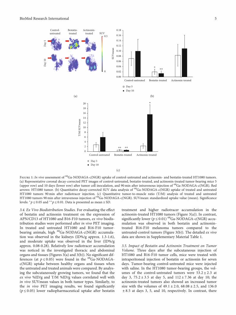
3.2. In Vivo PET Imaging of HT1080 Tumors. The effect ofactinonin and bestatin treatment on the APN/CD13 expres-sion of HT1080 tumors was monitored by in vivo PET imag-ing studies. On days 3 (five days after tumor induction) and 7(ten days after tumor induction) of the treatment, 20minstatic PET scans were acquired from the tumorous area ofcontrol-untreated and treated HT1080 tumor-bearing mice90min after intravenous injection of 68Ga-NODAGA-c(NGR). After the qualitative PET image analysis, we foundthat the untreated-control tumors and actinonin-treatedtumors were well identifiable at the investigated time points,indicating that actinonin treatment did not decrease theAPN/CD13 expression in the tumors (Figure 1(a)). Interest-ingly, in the case of actinonin-treated tumors, 68Ga-NODAGA-c(NGR) accumulation was increased comparedto the control-untreated tumors ten days after tumor cellinoculation. In contrast, in the bestatin-treated group, itwas difficult to identify the HT1080 tumors by comparingto the untreated-control tumors due to the low 68Ga-NODAGA-c(NGR) accumulation, confirming the decreasedexpression of APN/CD13 (Figure 1(a), red arrows).
After the quantitative analysis of the reconstructeddecay-corrected PET images, we found that the SUVmeanvalues of the bestatin-treated tumors (0:02 ± 0:01) were sig-nificantly (p ≤ 0:05 and p ≤ 0:01) lower than that of thecontrol-untreated tumors (0:07 ± 0:03) and the actinonin-treated tumors (0:08 ± 0:04) five days after HT1080 tumorcell implantation (Figure 1(b)). This significant differencepersisted in the 10-day study, where the SUVmean valuesof the bestatin-treated, control-untreated, and actinonin-treated tumors were 0:01 ± 0:01, 0:10 ± 0:05, and 0:11 ±0:05, respectively. By taking the SUVmean values, a moderateincrease was observed in the untreated and actinonin-treated
groups between 5 and 10 days. In contrast, there was a slightdecrease in the SUVmean data in the bestatin-treated group,but these differences were not significant at p ≤ 0:05. More-over, the accumulation of 68Ga-NODAGA-c(NGR) washigher (not significantly) in the actinonin-treated HT1080tumors than in the untreated tumors (Figure 1(b)). This phe-nomenon was also recognizable in the PET images(Figure 1(a)). The contrast of the tumors (T/M SUVmeanvalue) was also calculated in the control and treated groups,which determines the evaluability of the PET images andindicates the effectiveness of the treatment (Figure 1(c)).When the untreated and bestatin-treated HT1080 tumorswere compared, significantly (p ≤ 0:01) lower T/M SUVmeanvalues (0:57 ± 0:2 at day 5 and 0:61 ± 0:20 at day 10) wereobserved in the bestatin-treated group at each investigatedtime point. By analysing the actinonin-treated tumors, wefound a lower T/M ratio (2:25 ± 1:0) five days after tumor cellinoculation, and a significantly (p ≤ 0:05) higher T/M ratiowas observed (15:57 ± 4:0) at day 10, than that of theuntreated tumors, where the T/M ratios were 2:8 ± 1:0 and6:0 ± 2:0 at days 5 and 10, respectively (Figure 1(c)). Theseresults suggest that bestatin is a suitable and effectiveAPN/CD13 inhibitor in HT1080 experimental tumors.
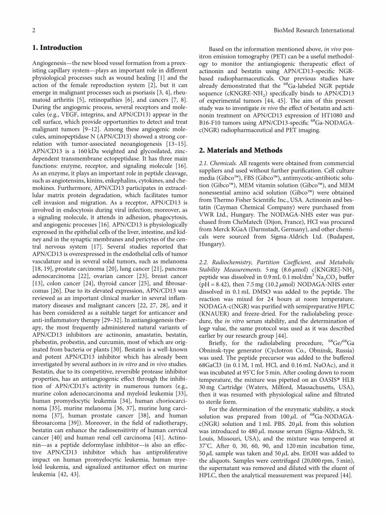
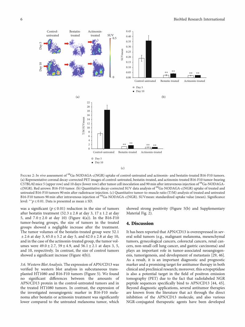
3.3. In Vivo PET Imaging of B16-F10 Melanoma Tumors. Forthe assessment of the effect of actinonin and bestatin treat-ment on the APN/CD13 expression of B16-F10 tumors,PET scans were performed 90min after the intravenousinjection of 68Ga-NODAGA-c(NGR) five and ten days aftertumor induction (Figure 2). High 68Ga-NODAGA-c(NGR)accumulation was observed in the subcutaneously growingB16-F10 melanoma tumors by qualitative PET image analy-sis. The APN/CD13 expression was clearly visualized in theuntreated group due to the high specificity of the radiotracer;furthermore, moderate radiotracer uptake was observed inthe bestatin- and actinonin-treated groups (Figure 2(a), redarrows).
By the quantitative image analysis of the decay-correctedPET images at day 5, significantly (p ≤ 0:01) lower SUVmeanvalues were observed in the actinonin-treated (0:02 ± 0:001)and bestatin-treated groups (0:03 ± 0:01) in comparison withthe control-untreated group, where elevated SUVmean(0:19 ± 0:03) values were measured (Figure 2(b)). This signif-icant difference was also observed on the 10th day, when theSUVmean values of actinonin-treated, bestatin-treated, andcontrol-untreated groups were 0:02 ± 0:001, 0:04 ± 0:001,and 0:36 ± 0:05, respectively. In the control-untreated group,a remarkable increasing of SUVmean values was seenbetween the 5th and 10th days; furthermore, negligible eleva-tion was observed in the bestatin- and actinonin-treatedgroups at the same time points, but these differences werenot significant at p ≤ 0:05. In the control-untreated group,considerably increased T/M SUVmean values were seenbetween days 5 (7:24 ± 0:80) and 10 (18:60 ± 2:32). In addi-tion, significantly (p ≤ 0:01) lower T/M ratios were observedon days 5 and 10 in the actinonin-treated group(0:75 ± 0:15 at day 5 and 0:74 ± 0:05 at day 10) and in thebestatin-treated group (1:11 ± 0:01 and 1:63 ± 0:09 at days5 and 10, respectively) (Figure 2(c)).
4 BioMed Research International
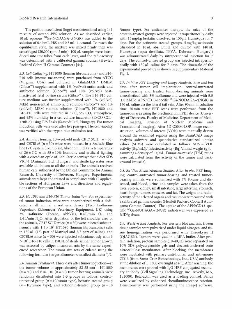
3.4. Ex Vivo Biodistribution Studies. For evaluating the effectof bestatin and actinonin treatment on the expression ofAPN/CD13 of HT1080 and B16-F10 tumors, ex vivo biodis-tribution studies were performed after in vivo PET imaging.In treated and untreated HT1080 and B16-F10 tumor-bearing animals, high 68Ga-NODAGA-c(NGR) accumula-tion was observed in the kidneys (ID%/g approx. 1.3-1.6),and moderate uptake was observed in the liver (ID%/gapprox. 0.08-0.28). Relatively low radiotracer accumulationwas noticed in the investigated thoracic and abdominalorgans and tissues (Figures 3(a) and 3(b)). No significant dif-ferences (at p ≤ 0:05) were found in the 68Ga-NODAGA-c(NGR) uptake between healthy organs and tissues whenthe untreated and treated animals were compared. By analys-ing the subcutaneously growing tumors, we found that theex vivo %ID/g and T/M %ID/g values correlated well within vivo SUVmean values in both tumor types. Similarly, tothe in vivo PET imaging results, we found significantly(p ≤ 0:05) lower radiopharmaceutical uptake after bestatin
treatment and higher radiotracer accumulation in theactinonin-treated HT1080 tumors (Figure 3(a)). In contrast,significantly lower (p ≤ 0:01) 68Ga-NODAGA-c(NGR) accu-mulation was observed in both bestatin and actinonin-treated B16-F10 melanoma tumors compared to theuntreated-control tumors (Figure 3(b)). The detailed ex vivodata are shown in Supplementary Material Table 1.
3.5. Impact of Bestatin and Actinonin Treatment on TumorVolume. Three days after the subcutaneous injection ofHT1080 and B16-F10 tumor cells, mice were treated withintraperitoneal injection of bestatin or actinonin for sevendays. Tumor-bearing control-untreated mice were injectedwith saline. In the HT1080 tumor-bearing groups, the vol-umes of the control-untreated tumors were 53:2 ± 2:3 atday 3, 75:2 ± 3:5 at day 5, and 112 ± 7:36 at day 10; theactinonin-treated tumors also showed an increased tumorsize with the volumes of 49:1 ± 2:0, 68:08 ± 2:5, and 136:0± 8:5 at days 3, 5, and 10, respectively. In contrast, there
Control-untreated
Bestatin-treated
Actinonin-treated SUV
0.5
0
Day
5D
ay 1
0
(a)
0.00
0.02
0.04
0.06
0.08
0.10
0.12
0.14
0.16
0.18
Control-untreated Bestatin-treated Actinonin-treated
SUV
mea
n
Day 5Day 10
⁎
⁎⁎
(b)
Day 5Day 10
0
2
4
6
8
10
12
14
16
18
20
Control-untreated Bestatin-treated Actinonin-treated
T/M
SU
Vm
ean
⁎
⁎⁎ ⁎⁎
(c)
Figure 1: In vivo assessment of 68Ga-NODAGA-c(NGR) uptake of control-untreated and actinonin- and bestatin-treated HT1080 tumors.(a) Representative coronal decay-corrected PET images of control-untreated, bestatin-treated, and actinonin-treated tumor-bearing mice 5(upper row) and 10 days (lower row) after tumor cell inoculation, and 90min after intravenous injection of 68Ga-NODAGA-c(NGR). Redarrows: HT1080 tumor. (b) Quantitative decay-corrected SUV data analysis of 68Ga-NODAGA-c(NGR) uptake of treated and untreatedHT1080 tumors 90min after radiotracer injection. (c) Quantitative tumor-to-muscle ratio (T/M) analysis of treated and untreatedHT1080 tumors 90min after intravenous injection of 68Ga-NODAGA-c(NGR). SUVmean: standardized uptake value (mean). Significancelevels: ∗p ≤ 0:05 and ∗∗p ≤ 0:01. Data is presented as mean ± SD.
5BioMed Research International
was a significant (p ≤ 0:01) reduction in the size of tumorsafter bestatin treatment (52:3 ± 2:8 at day 3, 17 ± 1:2 at day5, and 7:0 ± 2:0 at day 10) (Figure 4(a)). In the B16-F10tumor-bearing groups, the size of tumors in the treatedgroups showed a negligible increase after the treatment.The tumor volumes of the bestatin-treated group were 52:1± 2:6 at day 3, 65:0 ± 5:2 at day 5, and 62:0 ± 2:8 at day 10,and in the case of the actinonin-treated group, the tumor vol-umes were 49:0 ± 2:7, 59 ± 4:9, and 56:1 ± 2:1 at days 3, 5,and 10, respectively. In contrast, the size of control tumorsshowed a significant increase (Figure 4(b)).
3.6. Western Blot Analysis. The expression of APN/CD13 wasverified by western blot analysis in subcutaneous trans-planted HT1080 and B16-F10 tumors (Figure 5). We foundno significant differences between the amounts ofAPN/CD13 protein in the control-untreated tumors and inthe treated HT1080 tumors. In contrast, the expression ofthe investigated neoangiogenic marker in B16-F10 mela-noma after bestatin or actinonin treatment was significantlylower compared to the untreated melanoma tumor, which
showed strong positivity (Figure 5(b) and SupplementaryMaterial Fig. 2).
4. Discussion
It has been reported that APN/CD13 is overexpressed in sev-eral solid tumors (e.g., malignant melanoma, mesenchymaltumors, gynecological cancers, colorectal cancers, renal can-cers, non-small cell lung cancer, and gastric carcinoma) andplays an important role in tumor-associated neoangiogen-esis, tumorigenesis, and development of metastasis [29, 46].As a result, it is an important diagnostic and prognosticmarker and a promising target for antitumor therapy in bothclinical and preclinical research; moreover, this ectopeptidaseis also a potential target in the field of positron emissiontomography (PET) due to the fact that radiolabeled NGRpeptide sequences specifically bind to APN/CD13 [44, 45].Beyond diagnostic applications, several antitumor therapiesare known from the literature that act through the directinhibition of the APN/CD13 molecule, and also variousNGR-conjugated therapeutic agents have been developed
Control-untreated
Bestatin-treated
Actinonin-treated SUV
0.5
0
Day
5D
ay 1
0
(a)
0.00
0.05
0.10
0.15
0.20
0.25
0.30
0.35
0.40
0.45
Control-untreated Bestatin-treated Actinonin-treated
SUV
mea
n
⁎⁎⁎⁎
⁎⁎ ⁎⁎
Day 5Day 10
(b)
02468
10121416182022
Control-untreated Bestatin-treated Actinonin-treated
T/M
SU
Vm
ean
⁎⁎⁎⁎
⁎⁎ ⁎⁎
Day 5Day 10
(c)
Figure 2: In vivo assessment of 68Ga-NODAGA-c(NGR) uptake of control-untreated and actinonin- and bestatin-treated B16-F10 tumors.(a) Representative coronal decay-corrected PET images of control-untreated, bestatin-treated, and actinonin-treated B16-F10 tumor-bearingC57BL/6J mice 5 (upper row) and 10 days (lower row) after tumor cell inoculation and 90min after intravenous injection of 68Ga-NODAGA-c(NGR). Red arrows: B16-F10 tumor. (b) Quantitative decay-corrected SUV data analysis of 68Ga-NODAGA-c(NGR) uptake of treated anduntreated B16-F10 tumors 90min after radiotracer injection. (c) Quantitative tumor-to-muscle ratio (T/M) analysis of treated and untreatedB16-F10 tumors 90min after intravenous injection of 68Ga-NODAGA-c(NGR). SUVmean: standardized uptake value (mean). Significancelevel: ∗∗p ≤ 0:01. Data is presented as mean ± SD.
6 BioMed Research International

that deliver different cytotoxins (e.g., NGR-coated liposomes,doxorubicin-NGR conjugates, and NGR-TNF alpha conju-gates) to APN/CD13-positive solid tumors [29].
Two main types of APN/CD13 inhibitors are known, thesynthetic and the naturally occurring form. Molecules ofboth types generally have a zinc-binding group and are effec-tive in the inhibition of tumor angiogenesis and cell migra-tion. The naturally occurring APN/CD13 inhibitors are
originating from bacterial cultures; accordingly, bestatinoriginates from Streptomyces olivoreticuli and actinonin—asan antibiotic derivative of L-prolinol—was isolated fromStreptomyces cutter C/2 [30, 43]. The antitumor activity ofthese naturally occurring APN/CD13 inhibitors is mediatedby several, often independent mechanisms. Actinonin andbestatin have immunomodulatory and host-mediated antitu-mor activities, and by binding to the zinc domains of
0.0
0.2
0.4
0.6
0.8
1.0
1.2
1.4
1.6
1.8
2.0
%ID
(g)
05
10152025
T/M
%ID
(g)
Control-untreatedBestatin-treatedActinonin-treated
bloo
d
liver
kidn
ey
sple
en
smal
l int
estin
e
larg
e int
estin
e
stom
ach
mus
cle
lung
hear
t
fat
HT
1080
tum
or
Control-untreated
Bestatin-treated
Actinonin-treated
⁎
⁎
(a)
0.0
0.2
0.4
0.6
0.8
1.0
1.2
1.4
1.6
1.8
2.0
%ID
(g) 0
5
10
15
20
T/M
%ID
(g)
bloo
d
liver
kidn
ey
sple
en
smal
l int
estin
e
larg
e int
estin
e
stom
ach
mus
cle
lung
hear
t
fat
B16-
F10
tum
or
Control-untreated
Bestatin-treated
Actinonin-treated
⁎⁎
⁎⁎
Control-untreatedBestatin-treatedActinonin-treated
(b)
Figure 3: Ex vivo biodistribution studies of 68Ga-NODAGA-c(NGR) in control-untreated, bestatin-treated, and actinonin-treated HT1080(a) and B16-F10 (b) tumor-bearing mice. Inserts: T/M (%ID/g tumor/%ID/g muscle) ratios. Significance levels: ∗p ≤ 0:05 and ∗∗p ≤ 0:01.
7BioMed Research International

APN/CD13 in cancer cells, they inhibit the function of thisexopeptidase. In contrast, it was found that, e.g., actinoninalso inhibited the growth of CD13-negative tumor cells, sug-gesting that the antitumor effect is not only mediated by cellsurface APN/CD13. It was observed that the effect of theseinhibitors was mediated partly through the inhibition ofother zinc-dependent intracellular enzymes resulting in cellcycle G1 arrest and apoptosis. Among the direct inhibitors,bestatin inhibits at least 12 different aminopeptidases, asthese exopeptidases—including APN/CD13—generally havea diverse and overlapping substrate specificity. Despite thefact that these inhibitors are not specific for only one typeof APN, because of their strong antitumor activity, severalAPN inhibitors have entered clinical trials, e.g., bestatin (ube-nimex) [29, 34, 35, 43].
In this present study, the effect of bestatin and actinonintreatment on APN/CD13 expression of HT1080 and B16-F10 tumors was investigated and followed by usingAPN/CD13-specific 68Ga-NODAGA-c(NGR) radiopharma-ceutical and in vivo PET imaging.
For the in vivo assessment of the antitumor effect of bes-tatin and actinonin treatment, HT1080 and B16-F10 cell
lines were used to establish tumor-bearing animals. Previousstudies have shown that these cell lines showed APN/CD13positivity by western blot analysis, flow cytometry, immuno-fluorescence analysis, immunohistochemistry, RT-PCR, andoptical imaging [19, 26, 39, 47]. Our in vivo PET imagingresults (Figures 1(a) and 2(a)) correlated well with these in vi-tro findings where the control-untreated HT1080 and B16-F10 tumors were clearly visualized by the APN/CD13-spe-cific 68Ga-NODAGA-c(NGR) radiopharmaceutical. Thewestern blot analysis also confirmed the strong APN/CD13positivity in the investigated tumors (Figure 5).
In our further in vivo experiments, the efficacy of the acti-nonin and bestatin treatments was investigated on experi-mental tumors. The antitumor effect of actinonin wasinvestigated earlier by Xu and coworkers [43] on human(HL60, NB4) and murine (AKR) leukemia and lymphoma(RAJI, DAUDI) cell lines. In these in vitro studies, actinoninblocked the tumor growth of APN/CD13-positive leukemiaand arrested the growth of APN/CD13-negative lymphomasinterestingly. The authors supposed that the antitumor effectof actinonin is not derived only by the inhibition ofAPN/CD13. In in vivo studies, syngeneic AKR tumor-
Tum
or vo
lum
e (m
m3 )
Time (days)
0
20
40
60
80
100
120
140
160
3 5 10
HT1080 control-untreatedHT1080 bestatin-treatedHT1080 actinonin-treated
⁎⁎
⁎⁎
(a)
0
50
100
150
200
250
300
3 5 10Time (days)
B16-F10 control-untreatedB16-F10 bestatin-treatedB16-F10 actinonin-treated
⁎⁎ ⁎⁎
Tum
or vo
lum
e (m
m3 )
(b)
Figure 4: Impact of bestatin and actinonin treatment on tumor growth. Treatment began 3 days after HT1080 and B16-F10 tumor cellinoculation at the tumor volume of approx. 52-55mm3. (a) Impact of treatment on HT1080 tumors (n = 10/group). (b) Impact oftreatment on B16-F10 melanoma tumors (n = 10/group). Significance level: ∗∗p ≤ 0:01.
Control-untreated
Bestatin-treated
Actinonin-treated
HT1
080
APN/CD13(150 kDa)
Actin(45 kDa)
(a)
B16-
F10
Control-untreated
Bestatin-treated
Actinonin-treated
APN/CD13(150 kDa)
Actin(45 kDa)
(b)
Figure 5: Qualitative western blot analysis of APN/CD13 expression in subcutaneously transplanted untreated and treated HT1080 (a) andB16-F10 (b) tumors.
8 BioMed Research International
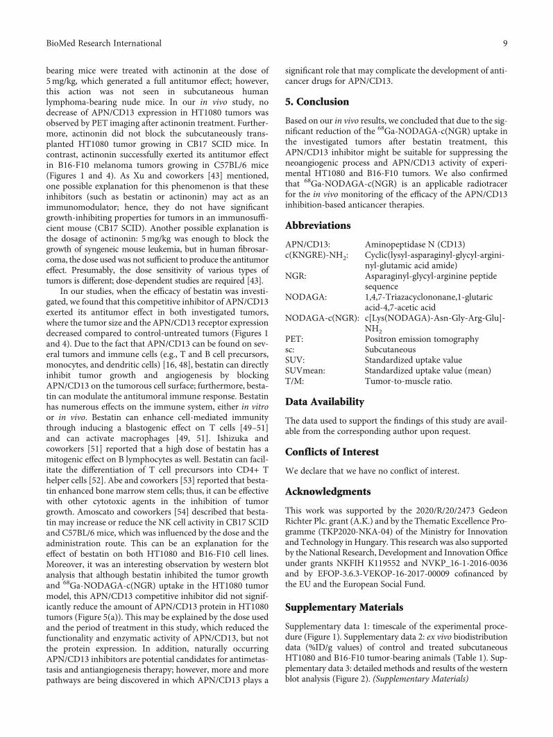
bearing mice were treated with actinonin at the dose of5mg/kg, which generated a full antitumor effect; however,this action was not seen in subcutaneous humanlymphoma-bearing nude mice. In our in vivo study, nodecrease of APN/CD13 expression in HT1080 tumors wasobserved by PET imaging after actinonin treatment. Further-more, actinonin did not block the subcutaneously trans-planted HT1080 tumor growing in CB17 SCID mice. Incontrast, actinonin successfully exerted its antitumor effectin B16-F10 melanoma tumors growing in C57BL/6 mice(Figures 1 and 4). As Xu and coworkers [43] mentioned,one possible explanation for this phenomenon is that theseinhibitors (such as bestatin or actinonin) may act as animmunomodulator; hence, they do not have significantgrowth-inhibiting properties for tumors in an immunosuffi-cient mouse (CB17 SCID). Another possible explanation isthe dosage of actinonin: 5mg/kg was enough to block thegrowth of syngeneic mouse leukemia, but in human fibrosar-coma, the dose used was not sufficient to produce the antitumoreffect. Presumably, the dose sensitivity of various types oftumors is different; dose-dependent studies are required [43].
In our studies, when the efficacy of bestatin was investi-gated, we found that this competitive inhibitor of APN/CD13exerted its antitumor effect in both investigated tumors,where the tumor size and the APN/CD13 receptor expressiondecreased compared to control-untreated tumors (Figures 1and 4). Due to the fact that APN/CD13 can be found on sev-eral tumors and immune cells (e.g., T and B cell precursors,monocytes, and dendritic cells) [16, 48], bestatin can directlyinhibit tumor growth and angiogenesis by blockingAPN/CD13 on the tumorous cell surface; furthermore, besta-tin can modulate the antitumoral immune response. Bestatinhas numerous effects on the immune system, either in vitroor in vivo. Bestatin can enhance cell-mediated immunitythrough inducing a blastogenic effect on T cells [49–51]and can activate macrophages [49, 51]. Ishizuka andcoworkers [51] reported that a high dose of bestatin has amitogenic effect on B lymphocytes as well. Bestatin can facil-itate the differentiation of T cell precursors into CD4+ Thelper cells [52]. Abe and coworkers [53] reported that besta-tin enhanced bone marrow stem cells; thus, it can be effectivewith other cytotoxic agents in the inhibition of tumorgrowth. Amoscato and coworkers [54] described that besta-tin may increase or reduce the NK cell activity in CB17 SCIDand C57BL/6 mice, which was influenced by the dose and theadministration route. This can be an explanation for theeffect of bestatin on both HT1080 and B16-F10 cell lines.Moreover, it was an interesting observation by western blotanalysis that although bestatin inhibited the tumor growthand 68Ga-NODAGA-c(NGR) uptake in the HT1080 tumormodel, this APN/CD13 competitive inhibitor did not signif-icantly reduce the amount of APN/CD13 protein in HT1080tumors (Figure 5(a)). This may be explained by the dose usedand the period of treatment in this study, which reduced thefunctionality and enzymatic activity of APN/CD13, but notthe protein expression. In addition, naturally occurringAPN/CD13 inhibitors are potential candidates for antimetas-tasis and antiangiogenesis therapy; however, more and morepathways are being discovered in which APN/CD13 plays a
significant role that may complicate the development of anti-cancer drugs for APN/CD13.
5. Conclusion
Based on our in vivo results, we concluded that due to the sig-nificant reduction of the 68Ga-NODAGA-c(NGR) uptake inthe investigated tumors after bestatin treatment, thisAPN/CD13 inhibitor might be suitable for suppressing theneoangiogenic process and APN/CD13 activity of experi-mental HT1080 and B16-F10 tumors. We also confirmedthat 68Ga-NODAGA-c(NGR) is an applicable radiotracerfor the in vivo monitoring of the efficacy of the APN/CD13inhibition-based anticancer therapies.
Abbreviations
APN/CD13: Aminopeptidase N (CD13)c(KNGRE)-NH2: Cyclic(lysyl-asparaginyl-glycyl-argini-
nyl-glutamic acid amide)NGR: Asparaginyl-glycyl-arginine peptide
sequenceNODAGA: 1,4,7-Triazacyclononane,1-glutaric
acid-4,7-acetic acidNODAGA-c(NGR): c[Lys(NODAGA)-Asn-Gly-Arg-Glu]-
NH2PET: Positron emission tomographysc: SubcutaneousSUV: Standardized uptake valueSUVmean: Standardized uptake value (mean)T/M: Tumor-to-muscle ratio.
Data Availability
The data used to support the findings of this study are avail-able from the corresponding author upon request.
Conflicts of Interest
We declare that we have no conflict of interest.
Acknowledgments
This work was supported by the 2020/R/20/2473 GedeonRichter Plc. grant (A.K.) and by the Thematic Excellence Pro-gramme (TKP2020-NKA-04) of the Ministry for Innovationand Technology in Hungary. This research was also supportedby the National Research, Development and Innovation Officeunder grants NKFIH K119552 and NVKP_16-1-2016-0036and by EFOP-3.6.3-VEKOP-16-2017-00009 cofinanced bythe EU and the European Social Fund.
Supplementary Materials
Supplementary data 1: timescale of the experimental proce-dure (Figure 1). Supplementary data 2: ex vivo biodistributiondata (%ID/g values) of control and treated subcutaneousHT1080 and B16-F10 tumor-bearing animals (Table 1). Sup-plementary data 3: detailedmethods and results of the westernblot analysis (Figure 2). (Supplementary Materials)
9BioMed Research International

References
[1] M. G. Tonnesen, X. Feng, and R. A. Clark, “Angiogenesis inwound healing,” The Journal of Investigative DermatologySymposium Proceedings, vol. 5, no. 1, pp. 40–46, 2000.
[2] H. M. Fraser and S. F. Lunn, “Angiogenesis and its control inthe female reproductive system,” British Medical Bulletin,vol. 56, no. 3, pp. 787–797, 2000.
[3] S. Guérard and R. Pouliot, “The role of angiogenesis in thepathogenesis of psoriasis: mechanisms and clinical implica-tions,” Journal of Clinical & Experimental DermatologyResearch, vol. 4, no. 3, 2014.
[4] R. Heidenreich, M. Röcken, and K. Ghoreschi, “Angiogenesisdrives psoriasis pathogenesis,” International Journal of Exper-imental Pathology, vol. 90, no. 3, pp. 232–248, 2009.
[5] D. G. Stupack, C. M. Storgard, and D. A. Cheresh, “A role forangiogenesis in rheumatoid arthritis,” Brazilian Journal ofMedical and Biological Research, vol. 32, no. 5, pp. 573–581,1999.
[6] G. Tremolada, C. del Turco, R. Lattanzio et al., “The role ofangiogenesis in the development of proliferative diabetic reti-nopathy: impact of intravitreal anti-VEGF treatment,” Experi-mental Diabetes Research, vol. 2012, Article ID 728325, 8pages, 2012.
[7] N. Nishida, H. Yano, T. Nishida, T. Kamura, and M. Kojiro,“Angiogenesis in cancer,” Vascular Health and Risk Manage-ment, vol. 2, no. 3, pp. 213–219, 2006.
[8] R. S. Kerbel, “Tumor angiogenesis,” The New England Journalof Medicine, vol. 358, no. 19, pp. 2039–2049, 2008.
[9] M. Klagsbrun and M. A. Moses, “Molecular angiogenesis,”Chemistry & Biology, vol. 6, no. 8, pp. R217–R224, 1999.
[10] D. G. Stupack and D. A. Cheresh, “Integrins and angiogene-sis,” Current Topics in Developmental Biology, vol. 64,pp. 207–238, 2004.
[11] P. Carmeliet and D. Collen, “Molecular basis of angiogenesis.Role of VEGF and VE-cadherin,” Annals of the New YorkAcademy of Sciences, vol. 902, no. 1, pp. 249–264, 2000.
[12] P. Carmeliet and R. K. Jain, “Molecular mechanisms and clin-ical applications of angiogenesis,” Nature, vol. 473, no. 7347,pp. 298–307, 2011.
[13] R. Pasqualini, E. Koivunen, R. Kain et al., “Aminopeptidase Nis a receptor for tumor-homing peptides and a target for inhi-biting angiogenesis,” Cancer Research, vol. 60, no. 3, pp. 722–727, 2000.
[14] S. V. Bhagwat, J. Lahdenranta, R. Giordano, W. Arap,R. Pasqualini, and L. H. Shapiro, “CD13/APN is activated byangiogenic signals and is essential for capillary tube forma-tion,” Blood, vol. 97, no. 3, pp. 652–659, 2001.
[15] K. Fukasawa, H. Fujii, Y. Saitoh et al., “Aminopeptidase N(APN/CD13) is selectively expressed in vascular endothelialcells and plays multiple roles in angiogenesis,” Cancer Letters,vol. 243, no. 1, pp. 135–143, 2006.
[16] P. Mina-Osorio, “The moonlighting enzyme CD13: old andnew functions to target,” Trends in Molecular Medicine,vol. 14, no. 8, pp. 361–371, 2008.
[17] J. Olsen, K. Kokholm, O. Norén, and H. Sjöström, “Structureand expression of aminopeptidase N,” Advances in Experimen-tal Medicine and Biology, vol. 421, pp. 47–57, 1997.
[18] H. Fujii, M. Nakajima, I. Saiki, J. Yoneda, I. Azuma, andT. Tsuruo, “Human melanoma invasion and metastasisenhancement by high expression of aminopeptidase
N/CD13,” Clinical & Experimental Metastasis, vol. 13, no. 5,pp. 337–344, 1995.
[19] L. Guzman-Rojas, R. Rangel, A. Salameh et al., “Cooperativeeffects of aminopeptidase N (CD13) expressed by nonmalig-nant and cancer cells within the tumor microenvironment,”Proceedings of the National Academy of Sciences of the UnitedStates of America, vol. 109, no. 5, pp. 1637–1642, 2012.
[20] K. ISHII, S. USUI, Y. SUGIMURA, H. YAMAMOTO,K. YOSHIKAWA, and K. HIRANO, “Inhibition of aminopep-tidase N (AP-N) and urokinase-type plasminogen activator(uPA) by zinc suppresses the invasion activity in human uro-logical cancer cells,” Biological & Pharmaceutical Bulletin,vol. 24, no. 3, pp. 226–230, 2001.
[21] Q. Zhang, Q. Zhang, J. Wang, H. Zhang, D. Zhao, andZ. Zhang, “Expression and clinical significance of aminopepti-dase N/CD13 in non-small cell lung cancer,” Journal of CancerResearch and Therapeutics, vol. 11, no. 1, pp. 223–228, 2015.
[22] N. Ikeda, Y. Nakajima, T. Tokuhara et al., “Clinical signifi-cance of aminopeptidase N/CD13 expression in human pan-creatic carcinoma,” Clinical Cancer Research, vol. 9, no. 4,pp. 1503–1508, 2003.
[23] P. Surowiak, M. Drag, V. Materna et al., “Expression of amino-peptidase N/CD13 in human ovarian cancers,” InternationalJournal of Gynecological Cancer, vol. 16, no. 5, pp. 1783–1788, 2006.
[24] H. Hashida, A. Takabayashi, M. Kanai et al., “AminopeptidaseN is involved in cell motility and angiogenesis: its clinical sig-nificance in human colon cancer,” Gastroenterology, vol. 122,no. 2, pp. 376–386, 2002.
[25] A. Kehlen, U. Lendeckel, H. Dralle, J. Langner, and C. Hoang-Vu, “Biological significance of aminopeptidase N/CD13 inthyroid carcinomas,” Cancer Research, vol. 63, no. 23,pp. 8500–8506, 2003.
[26] J. S. Shim, J. H. Kim, H. Y. Cho et al., “Irreversible inhibition ofCD13/aminopeptidase N by the antiangiogenic agent curcu-min,” Chemistry & Biology, vol. 10, no. 8, pp. 695–704, 2003.
[27] L. Pang, N. Zhang, Y. Xia, D. Wang, G. Wang, and X. Meng,“Serum APN/CD13 as a novel diagnostic and prognostic bio-marker of pancreatic cancer,” Oncotarget, vol. 7, no. 47,pp. 77854–77864, 2016.
[28] T. Shimizu, K. Tani, K. Hase et al., “CD13/aminopeptidase N–induced lymphocyte involvement in inflamed joints of patientswith rheumatoid arthritis,” Arthritis and Rheumatism, vol. 46,no. 9, pp. 2330–2338, 2002.
[29] M. Wickström, R. Larsson, P. Nygren, and J. Gullbo, “Amino-peptidase N (CD13) as a target for cancer chemotherapy,”Cancer Science, vol. 102, no. 3, pp. 501–508, 2011.
[30] B. Bauvois and D. Dauzonne, “Aminopeptidase-N/CD13 (EC3.4.11.2) inhibitors: chemistry, biological evaluations, andtherapeutic prospects,” Medicinal Research Reviews, vol. 26,no. 1, pp. 88–130, 2006.
[31] U. Bank, A. Heimburg, M. Helmuth et al., “Triggering endog-enous immunosuppressive mechanisms by combined target-ing of dipeptidyl peptidase IV (DPIV/CD26) andaminopeptidase N (APN/ CD13) — a novel approach for thetreatment of inflammatory bowel disease,” InternationalImmunopharmacology, vol. 6, no. 13-14, pp. 1925–1934, 2006.
[32] S. Ansorge, U. Bank, A. Heimburg et al., “Recent insights intothe role of dipeptidyl aminopeptidase IV (DPIV) and aminopep-tidase N (APN) families in immune functions,” Clinical Chemis-try and Laboratory Medicine, vol. 47, no. 3, pp. 253–261, 2009.
10 BioMed Research International

[33] F. Abe, K. Shibuya, M. Uchida et al., “Effect of bestatin on syn-geneic tumors in mice,” GANN Japanese Journal of CancerResearch, vol. 75, no. 1, pp. 89–94, 1984.
[34] N. Imamura and A. Kimura, “Effect of ubenimex (bestatin) onthe cell growth and phenotype of HL-60 and HL-60R cell lines:up-and down-regulation of CD13/aminopeptidase N,” Leuke-mia & Lymphoma, vol. 37, no. 5-6, pp. 663–667, 2000.
[35] K. Inoi, S. Goto, S. Nomura et al., “Aminopeptidase inhibitorubenimex (bestatin) inhibits the growth of human choriocarci-noma in nude mice through its direct cytostatic activity,” Anti-cancer Research, vol. 15, no. 5B, pp. 2081–2087, 1995.
[36] Y. Aozuka, K. Koizumi, Y. Saitoh, Y. Ueda, H. Sakurai, andI. Saiki, “Anti-tumor angiogenesis effect of aminopeptidaseinhibitor bestatin against B16-BL6 melanoma cells orthotopi-cally implanted into syngeneic mice,” Cancer Letters,vol. 216, no. 1, pp. 35–42, 2004.
[37] I. Saiki, J. Murata, K. Watanabe, H. Fujii, F. Abe, and I. Azuma,“Inhibition of tumor cell invasion by ubenimex (bestatin)in vitro,” Japanese Journal of Cancer Research, vol. 80, no. 9,pp. 873–878, 1989.
[38] X. Wang, Z. Niu, Y. Jia et al., “Ubenimex inhibits cell prolifer-ation, migration and invasion by inhibiting the expression ofAPN and inducing autophagic cell death in prostate cancercells,” Oncology Reports, vol. 35, no. 4, pp. 2121–2130, 2016.
[39] I. Saiki, H. Fujii, and J. Yoneda, “Role of aminopeptidase N(CD13) in tumor-cell invasion and extracellular matrix degra-dation,” International Journal of Cancer, vol. 54, no. 1,pp. 137–143, 1993.
[40] H. Tsukamoto, K. Shibata, H. Kajiyama, M. Terauchi,A. Nawa, and F. Kikkawa, “Aminopeptidase N (APN)/CD13inhibitor, ubenimex, enhances radiation sensitivity in humancervical cancer,” BMC Cancer, vol. 8, no. 1, p. 74, 2008.
[41] S. Liu, X. Wang, J. Lu et al., “Ubenimex enhances the radiosen-sitivity of renal cell carcinoma cells by inducing autophagic celldeath,” Oncology Letters, vol. 12, no. 5, pp. 3403–3410, 2016.
[42] D. Z. Chen, D. V. Patel, C. J. Hackbarth et al., “Actinonin, anaturally occurring antibacterial agent, is a potent deformylaseinhibitor,” Biochemistry, vol. 39, no. 6, pp. 1256–1262, 2000.
[43] Y. Xu, L. T. Lai, J. L. Gabrilove, and D. A. Scheinberg, “Antitu-mor activity of actinonin in vitro and in vivo,” Clinical CancerResearch, vol. 4, no. 1, pp. 171–176, 1998.
[44] A. Kis, N. Dénes, J. P. Szabó et al., “In vivo assessment of ami-nopeptidase N (APN/CD13) specificity of different 68Ga-labelled NGR derivatives using PET/MRI imaging,” Interna-tional Journal of Pharmaceutics, vol. 589, article 119881, 2020.
[45] G. Máté, I. Kertész, K. N. Enyedi et al., “In vivo imaging ofaminopeptidase N (CD13) receptors in experimental renaltumors using the novel radiotracer 68Ga-NOTA-c(NGR),”European Journal of Pharmaceutical Sciences, vol. 69, pp. 61–71, 2015.
[46] S. Joshi, L. Chen, M. B. Winter et al., “The rational design oftherapeutic peptides for aminopeptidase N using a substrate-based approach,” Scientific Reports, vol. 7, no. 1, p. 1424, 2017.
[47] A. vonWallbrunn, J. Waldeck, C. Höltke et al., “In vivo opticalimaging of CD13/APN-expression in tumor xenografts,” Jour-nal of Biomedical Optics, vol. 13, no. 1, article 011007, 2008.
[48] U. Lendeckel, T. Kähne, D. Riemann, K. Neubert, M. Arndt,and D. Reinhold, “Review: the role of membrane peptidasesin immune functions,” in Cellular Peptidases in Immune Func-tions and Diseases 2, J. Langner and S. Ansorge, Eds., vol. 477
of Advances in Experimental Medicine and Biology, pp. 1–24,Springer, Boston, MA, 2002.
[49] J. E. Talmadge, B. F. Lenz, R. Pennington et al., “Immunomod-ulatory and therapeutic properties of bestatin in mice,” CancerResearch, vol. 46, no. 9, pp. 4505–4510, 1986.
[50] M. Saito, K. Takegoshi, T. Aoyagi, H. Umezawa, and Y. Nagai,“Stimulatory effect of bestatin, a new specific inhibitor of ami-nopeptidases, on the blastogenesis of guinea pig lymphocytes,”Cellular Immunology, vol. 40, no. 2, pp. 247–262, 1978.
[51] M. Ishizuka, J. Sato, Y. Sugiyama, T. Takeuchi, andH. Umezawa, “Mitogenic effect of bestatin on lymphocytes,”The Journal of Antibiotics, vol. 33, no. 6, pp. 653–662, 1980.
[52] Y. Wakabayashi, M. Hashimoto, K. Saitoh, H. Osawa,M. Koike, and S. I. Hirose, “Effects of bestatin (ubenimex) onhuman T-cell colony formation,” Anti-Cancer Drugs, vol. 2,no. 1, pp. 39–44, 1991.
[53] F. Abe, K. Shibuya, J. Ashizawa et al., “Enhancement of antitu-mor effect of cytotoxic agents by bestatin,” The Journal of Anti-biotics, vol. 38, no. 3, pp. 411–414, 1985.
[54] A. A. Amoscato, R. R. Spiess, A. M. Brumfield, R. B. Herber-man, and W. H. Chambers, “Surface aminopeptidase activityof rat natural killer cells I. Biochemical and biological proper-ties,” Biochimica et Biophysica Acta, vol. 1221, no. 3, pp. 221–232, 1994.
11BioMed Research International