RESEARCH ARTICLE Impact Factor: 5.019 ANTI-DIABETIC ...
15
Asian Journal of Pharmaceutical Education and Research Vol -7, Issue-2, April-June 2018 ISSN: 2278 7496 AJPER April – June 2018, Vol 7, Issue 2 (45-59) ANTI-DIABETIC ACTIVITY OF SILVER NANOPARTICLES SYNTHESIZED FROM ASHWAGANDHA ROOT AQUEOUS EXTRACT Md. Imad Uddin*, Kavya G, Soundarya G, Ropa G, Sridhar CH Department of pharmacology, Pulla Reddy Institute of Pharmacy, Domadugu, SangaReddy, Telangana, India-502313. *Corresponding Author’s E mail: [email protected] Received 26 Mar. 2018; Revised 28 Mar. 2018; Accepted 30 Mar. 2018, Available online 15 Apr. 2018 ABSTRACT According to traditional medicine, Withania somnifera also known as Indian ginseng is a plant with a cure for many diseases. This study was conducted to develop an ecofriendly, cheap and effective procedure for the green synthesis of silver nanoparticles (AgNPs) form ashwagandha root aqueous extract (ARAE) and evaluating their in-vitro anti diabetic activity. Addition of ARAE to AgNO3 (1mM) solution kept on stirring at 60 o C resulted in the formation of Ashwagandha Root Aqueous Extract Nano particles (ARAENP). Synthesized nanoparticles are subjected to characterization by various procedures. UV-Visible spectroscopy showed absorption peak at 440nm which was absent in ARAE and AgNO3. FTIR spectroscopy revealed the absorption peaks of different functional groups involved in the formation of ARAENP and further emphasizes role of withanolides in formation of ARAENP. Scanning Electron Microscopy (SEM) revealed average size of ARAENP as 123.23nm. 2 ߠpeaks at 28.98 o , 39.80 o , 42.24 o , 64.42 o and 79.76 o were identified by X-ray Diffraction studies (XRD). Stability of ARAENP was determined by zeta potential which was found to be -10mV. Successfully characterized ARAENP were evaluated for their in-vitro anti diabetic potential by α-amylase and α –glucosidase inhibitory activities. Acarbose was used as a standard. Inhibitory potential of ARAE and ARAENP against α-amylase and α –glucosidase proves their therapeutic role. Inhibition of enzyme was probably due to the withanolides capped on ARAENP. Keywords: α-Amylase, α –Glucosidase, Ashwagandha, silver nanoparticles, Diabetes mellitus. INTRODUCTION Diabetes mellitus is an epidemic, endocrine disease characterized by increased blood glucose level and glycosuria affecting 6% of the worldwide population. Type 1 and type 2 are the two major types of DM. type 2 accounts for 90% of all cases and may occur due to inadequate secretion of insulin 1 and resistance of insulin receptors 2 . Two important enzymes viz., α-amylase and α –glucosidase are involved in the hydrolysis of starch 3 . Pancreatic α-amylase converts starch in to disaccharides and oligosaccharides; these are further converted to glucose by intestinal α –glucosidase. These two enzymes have a very important role in the onset of Type 2 DM 4 . Marketed anti-diabetic drugs like Acarbose, Voglibose and miglitol act by inhibiting these enzymes but are associated with serious side effects like diarrhea, bloating RESEARCH ARTICLE Impact Factor: 5.019
Transcript of RESEARCH ARTICLE Impact Factor: 5.019 ANTI-DIABETIC ...

Asian Journal of Pharmaceutical Education and Research Vol -7, Issue-2, April-June 2018
ISSN: 2278 7496
AJPER April – June 2018, Vol 7, Issue 2 (45-59)
ANTI-DIABETIC ACTIVITY OF SILVER NANOPARTICLES SYNTHESIZED FROM ASHWAGANDHA ROOT AQUEOUS EXTRACT
Md. Imad Uddin*, Kavya G, Soundarya G, Ropa G, Sridhar CH
Department of pharmacology, Pulla Reddy Institute of Pharmacy, Domadugu, SangaReddy, Telangana, India-502313.
*Corresponding Author’s E mail: [email protected]
Received 26 Mar. 2018; Revised 28 Mar. 2018; Accepted 30 Mar. 2018, Available online 15 Apr. 2018
ABSTRACT According to traditional medicine, Withania somnifera also known as Indian ginseng is a plant with a cure for many diseases. This study was conducted to develop an ecofriendly, cheap and effective procedure for the green synthesis of silver nanoparticles (AgNPs) form ashwagandha root aqueous extract (ARAE) and evaluating their in-vitro anti diabetic activity. Addition of ARAE to AgNO3 (1mM) solution kept on stirring at 60oC resulted in the formation of Ashwagandha Root Aqueous Extract Nano particles (ARAENP). Synthesized nanoparticles are subjected to characterization by various procedures. UV-Visible spectroscopy showed absorption peak at 440nm which was absent in ARAE and AgNO3. FTIR spectroscopy revealed the absorption peaks of different functional groups involved in the formation of ARAENP and further emphasizes role of withanolides in formation of ARAENP. Scanning Electron Microscopy (SEM) revealed average size of ARAENP as 123.23nm. 2 ߠ peaks at 28.98o, 39.80o, 42.24o, 64.42o and 79.76o were identified by X-ray Diffraction studies (XRD). Stability of ARAENP was determined by zeta potential which was found to be -10mV. Successfully characterized ARAENP were evaluated for their in-vitro anti diabetic potential by α-amylase and α –glucosidase inhibitory activities. Acarbose was used as a standard. Inhibitory potential of ARAE and ARAENP against α-amylase and α –glucosidase proves their therapeutic role. Inhibition of enzyme was probably due to the withanolides capped on ARAENP.
Keywords: α-Amylase, α –Glucosidase, Ashwagandha, silver nanoparticles, Diabetes mellitus.
INTRODUCTION
Diabetes mellitus is an epidemic, endocrine disease characterized by increased blood glucose level and
glycosuria affecting 6% of the worldwide population. Type 1 and type 2 are the two major types of DM.
type 2 accounts for 90% of all cases and may occur due to inadequate secretion of insulin1 and resistance
of insulin receptors2. Two important enzymes viz., α-amylase and α –glucosidase are involved in the
hydrolysis of starch3. Pancreatic α-amylase converts starch in to disaccharides and oligosaccharides;
these are further converted to glucose by intestinal α –glucosidase. These two enzymes have a very
important role in the onset of Type 2 DM4. Marketed anti-diabetic drugs like Acarbose, Voglibose and
miglitol act by inhibiting these enzymes but are associated with serious side effects like diarrhea, bloating
RESEARCH ARTICLE Impact Factor: 5.019

Imad Uddin et al. Anti-Diabetic Activity of Silver Nanoparticles Synthesized from Ashwagandha Root Aqueous Extract
AJPER April – June 2018, Vol 7, Issue 2 (45-59)
meteorism and distention5, 6. To surmount the drawbacks of available drugs, use of medicinal plants is
increasing all over the world because of their low cost, safe and effectiveness.
Withania somnifera commonly known as ashwagandha belonging to family Solanaceae contains
withanolides, a group of steroidal lactones in roots7. Apart from withanolides other constituents which
are present in roots are withaferins8 and withanosides9. Pharmacological activity of this plant include
anxiolytic-anti-depressive10, antifungal11, antimalarial12 apoptotic13, Hypoglycemic effects14, anticancer
activity15, 16, anti-inflammatory, antitumor, anti-stress, antioxidant, immunomodulatory17, 18,
antidiabetic19. Leaves also exhibit antibacterial, anti-fungal and antitumor properties20.
Distinctive property of nanoparticles is their large surface area to volume ratio. Silver, gold, palladium
and platinum are the metals used in the synthesis but silver is superior among all the metals21. Physical
and chemical methods used for the synthesis are harmful and expensive so there is a need of ecofriendly
method for synthesis of nanoparticles22. Green synthesis is another approach for synthesis of
nanoparticles increased enormously in current research because of safety, effectiveness and low cost.
This innovative technique involves the reduction of Ag+ to Ago and stabilization of AgNPs by
biomolecules which are present in plant extract23. Based on these benefits, present study was designed to
use ARAE for the synthesis of silver nanoparticles and further evaluating their in-vitro anti-diabetic
activity.
MATERIALS AND METHODS
Plant Material Collection and Extraction:
Roots of Ashwagandha plant were collected from in and around areas of Sangareddy Dist. in Dec 2017.
Plant material was authenticated at Botanical Survey of India, Hyderabad, and Telangana, India.
PRIP/PCOG/17-18/001 is the reference number given to herbarium and it is stored in dept. of
Pharmacognosy, Pulla Reddy Institute of Pharmacy, Hyderabad, India. Freshly collected roots were
washed under running tap water, dried in shade for 15 days, sliced in to small pieces and grinded to
coarse powder. 20gm of powder was boiled with 200ml of double distilled water (DDW) for 1hr. After
cooling to room temperature, mixture was filtered with whatman filter paper no.1 and filtrate is
refrigerated (4oC) for future use.
Synthesis of ARAENP:
190ml of 1mM AgNO3 solution was prepared by using DDW, 10ml of aqueous extract was added slowly
to it, reaction was carried out in a conical flask kept on stirring at a temp. of 60oC for 8hrs. Change in the

Imad Uddin et al. Anti-Diabetic Activity of Silver Nanoparticles Synthesized from Ashwagandha Root Aqueous Extract
AJPER April – June 2018, Vol 7, Issue 2 (45-59)
color of solution specifies the formation of ARAENP. Solution was centrifuged at 4000rpm to obtain the
pellet of nanoparticles which was stored for future use24.
Characterization of ARAENP:
UV-Visible spectroscopy and Visual identification of ARAENP:
Color of ARAE and AgNO3 solution was taken as control and change in the color of reaction mixture
after addition of ARAE to AgNO3 solution is the first indication for the formation of ARAENP. UV-
visible spectroscopy was conducted in a range of 200-800nm by using UV-VIS Spectrophotometer
(UV3000, LBINDIA) model No. 18-1885-01-0115.
FTIR spectroscopy of ARAE and ARAENP:
In order to determine the involvement of functional groups in the formation of ARAENP FTIR
spectroscopy (BRUKER, ALPHA) was conducted in the range of 4000 to 500 cm-1. ARAENP
synthesized also contains biomolecules which are not capped on nanoparticles; they are removed by
dissolving in DDW and centrifuged at 5000rpm for 10 min, procedure is repeated for 3 times and final
pellet obtained is dried in at 600C in hot air oven and used for characterization.
SEM studies of ARAENP:
Size, shape and morphology of ARAENP was determined by using SEM (ZEISS) operated at
accelerating voltage 10.00kV, magnification 50.00 KX, working distance 8.5mm. Minute quantity of
ARAENP was dropped on copper grid coated with carbon, extra solution was removed by using blotting
paper and gird was used for study.
XRD studies of ARAENP:
Crystallographic structure of ARAENP was determined by using XRD (SHIMADZU, XRD7000)
operated at scan speed – 5.0 deg/min, sampling pitch – 0.02 deg, preset time is 0.24 sec, 2 ߠ range from
100 to 800, 30mA current, 40kV voltage with Kα1 Cu radiation, 1.54 = ߣ A0. Crystallographic structure
of ARAENP was calculated by using peaks of XRD. Average size of ARAENP was assessed by using
the Debye-Scherrer equation25.
Where D = average size of ARAENP, ݇ = constant (0.94), ߣ = wavelength of X-rays (1.546Å), ߚ = full
width at half maximum (FWHM), ߠ = diffraction angle in degrees.

Imad Uddin et al. Anti-Diabetic Activity of Silver Nanoparticles Synthesized from Ashwagandha Root Aqueous Extract
AJPER April – June 2018, Vol 7, Issue 2 (45-59)
Zeta potential and Dynamic Light Scattering (DLS) studies of ARAENP:
Stability of ARAENP was determined by using light scattering method for measuring zeta potential
(HORIBA SCIENTIFIC, SZ100) operated at electrode Voltage - 3.4V, Conductivity - 0.168mS/cm,
temperature (250 C) and Viscosity of dispersion medium (0.894mPa.s). Particle size distribution was
estimated by DLS measurements.
In-vitro anti diabetic activity:
Effect of Acarbose, ARAE and ARAENP on α-amylase activity:
Various concentrations (2 to 10mg/L) of Acarbose, ARAE and ARAENP were prepared by using DDW.
100μL of each of the concentrations was taken in a test tube; to this add 500 μL of 20mM sodium
phosphate buffer with 6mM NaCl pH 6.9 containing porcine pancreatic α-amylase (0.5 mg/mL). Mixture
was incubated for 10min at 25 °C. 500 μL of 1% starch solution prepared in 20mM sodium phosphate
buffer with 6mM NaCl pH 6.9 was added to each test tube. Reaction mixture was again incubated at 25
°C for 10 minutes. 1ml of 3, 5 dinitro salicylic acid was added to stop the reaction; mixture was further
incubated for 5 min in boiling water bath. Cooled to room temperature and 10ml of DDW was added and
absorbance was measured by using UV-Visible spectrophotometer at 540 nm. Without Acarbose, ARAE
and ARAENP, a complete reaction mixture was taken as control. All the tests are carried out in triplicates.
Inhibitory activity of α-amylase was calculated by using the formula26:
% inhibition = (Acontrol – Asample / Acontrol) × 100.
Effect of Acarbose, ARAE and ARAENP on α-glucosidase activity:
Various concentrations (2 to 10mg/L) of Acarbose, ARAE and ARAENP were prepared by using DDW.
20 μL of each of the concentration was taken in a test tube. To this add 100 μL of α-glucosidase (16.9
U/ml) solution in 0.1M phosphate buffer pH 6.9. Incubate at 250 C for 10 minutes and then add 50 μL of
5mM p-nitrophenyl-α-D-glucopyranoside solution prepared in 0.1M phosphate buffer pH 6.9. Reaction
mixture was incubated at 250 C for 5 minutes and absorbance was measured by using UV-Visible
spectrophotometer at 405 nm. Control contains complete reaction mixture without Acarbose, ARAE or
ARAENP27. All the tests are carried out in triplicates. Inhibitory activity was expressed as percentage
inhibition and determined by using the formula:
% inhibition = (Acontrol – Asample / Acontrol) × 100

Imad Uddin et al. Anti-Diabetic Activity of Silver Nanoparticles Synthesized from Ashwagandha Root Aqueous Extract
AJPER April – June 2018, Vol 7, Issue 2 (45-59)
RESULTS AND DISCUSSION
UV-Visible spectroscopy and Visual identification of ARAENP:
Visual proof for the formation of ARAENP is change of color of AgNO3 solution from colorless to dark
brown. Excitation of surface Plasmon resonance and reduction of Ag+ to Ago are the two major causes
for the change of color of solution (Figure 1). Temperature, time and stirring accelerated the reaction.
Our results are in accordance with the results of Rashid et al28 where they also showed change in color
solution. UV spectra of ARAE, silver nitrate and ARAENP are presented in figure 2. Many studies
reported the characteristic peak of SNPs in the range of 400-450nm which was absent in ARAE and
AgNO3, whereas a peak at 440nm was successfully appeared in ARAENP spectrum. This is very close
to the peak at 443nm obtained by Haytham M.M.Ibrahim29 with the AgNPs synthesized using banana
peel extract.
Figure 1: ARAE which is light brown in color when added to AgNO3 solution 1mM it forms dark
brown coloured ARAENP. change of color indicates formation of nanoparticles.

Imad Uddin et al. Anti-Diabetic Activity of Silver Nanoparticles Synthesized from Ashwagandha Root Aqueous Extract
AJPER April – June 2018, Vol 7, Issue 2 (45-59)
Figure 2: UV spectra of AgNO3, ARAE and ARAENP.
The melting points of all synthesized compounds were found in open capillary tubes and readings were
uncorrected. The structures of the synthesized compounds were supported by physical data (Table-1) and
following spectral analysis.
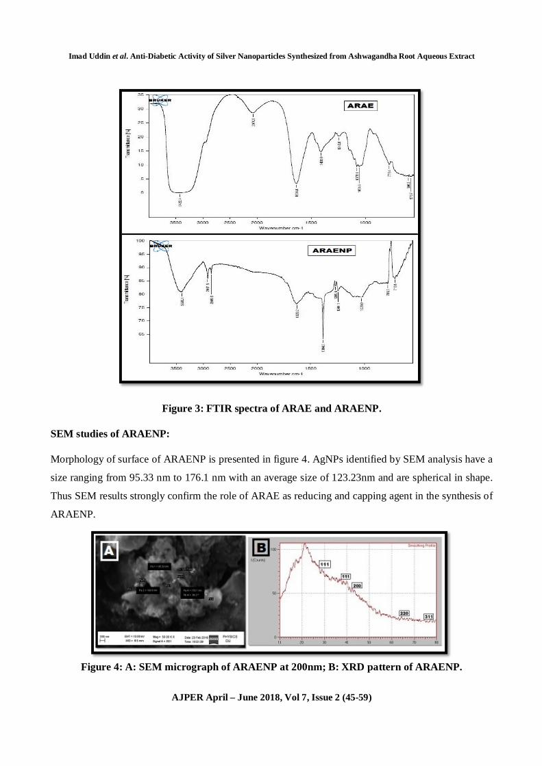
FTIR spectroscopy of ARAE and ARAENP:
FTIR spectroscopy revealed the role of different functional groups involved in the formation of AgNPs
depicted in figure 3. Different absorption peaks of ARAE and their shift in ARAENP and their
corresponding functional groups are discussed in table 1. Proteins, polysaccharides and enzymes present
in extract contains –OH group which undergoes stretching vibrations and produces peak at 3422.4 in
ARAE, which is shifted to 3399.1 in ARAENP indicating their role in the formation of SNPs. FTIR
spectroscopy proved the capping of functional groups of different chemical constituents present in ARAE
on AgNPs. Our results are in accordance with the study of Gole et al where they presented the role of
free amino acids and carboxyl groups of proteins present in extract for the formation of AgNPs30. ARAE
peaks (1242, 1408 and 1634) and ARAENP peaks (1269.6, 1244.1 and 1629) corresponds to carbon
skeleton of withanolides. ARAE peaks (1078.1, 1054.4) and ARAENP peaks (1029) corresponds to with
anolides ring band and flavonoids like structure. Thus from these results we can report that withanolides
which are main constituents of ARAE are involved in formation of ARAENP. Our results are in

Imad Uddin et al. Anti-Diabetic Activity of Silver Nanoparticles Synthesized from Ashwagandha Root Aqueous Extract
AJPER April – June 2018, Vol 7, Issue 2 (45-59)
accordance with the results of Anil Kumar et al., where they reported the peaks in the range of 1001-
1076 cm-1 corresponding to withanolides ring band and flavonoids and peaks at 1236 cm-1, 1400 cm-1 and
1614 cm-1 representing the carbon skeleton of withanolides31.
Table 1: FTIR peak values and corresponding functional groups of ARAE and ARAENP
S No.
Peak in ARAE (cm-1)
Peak in ARAENP (cm-1) Corresponding functional group
1. 3422.4 3399.1 Stretching vibrations of –OH group of phenolic acid compounds and carbohydrates with –H bonding.
2. 1634.6 1629.2 Stretching vibrations of C=C of alkenes
3. 1242.8 1269.6 &1244.1 Stretching vibrations of C-O group of Carboxylic acids
4. 1078.1 & 1054.4
1029.0 Stretching vibrations of C-N group of amines
5. 778.4 781.3 & 715.8 Stretching vibrations of C-H group of alkenes
6. 2070.5 --- Stretching vibrations of N=C=S of isothiocyanate
7. 1408.0 --- Bending vibration of O-H group of polyphenol
8. 600.2 & 576.7 --- Stretching vibrations of C-Br
9. --- 2917.5 & 2849.6 Stretching vibrations of C-H group of aldehydes
10. --- 1384.2 Bending vibrations of C-H group of alkanes
Imad Uddin et al. Anti-Diabetic Activity of Silver Nanoparticles Synthesized from Ashwagandha Root Aqueous Extract
AJPER April – June 2018, Vol 7, Issue 2 (45-59)
Figure 3: FTIR spectra of ARAE and ARAENP.
SEM studies of ARAENP:
Morphology of surface of ARAENP is presented in figure 4. AgNPs identified by SEM analysis have a
size ranging from 95.33 nm to 176.1 nm with an average size of 123.23nm and are spherical in shape.
Thus SEM results strongly confirm the role of ARAE as reducing and capping agent in the synthesis of
ARAENP.
Figure 4: A: SEM micrograph of ARAENP at 200nm; B: XRD pattern of ARAENP.

Imad Uddin et al. Anti-Diabetic Activity of Silver Nanoparticles Synthesized from Ashwagandha Root Aqueous Extract
AJPER April – June 2018, Vol 7, Issue 2 (45-59)
XRD studies of ARAENP:
peaks at 28.98o, 39.80 o, 42.24 o, 64.42 o and 79.76 o corresponds to 111, 200, 220 and 311 planes of ߠ 2
Bragg’s reflection of silver. Figure 4 and Table 2 represent the data of XRD analysis. These planes clearly
indicate the face centered cubic structure of silver. 12.02nm is the average size of ARAENPs. Our results
are in accordance with the study of Selvaraj Raja et al., where they reported the average size of
nanoparticles by XRD as 13.07nm32.
Table 2: Size of ARAENP by using Debye-Scherrer equation:
S No. 2ߠ (deg) D (angle) FWHM
(deg) Int. I
(cps deg) Size (nm)
1. 28.98 3.07860 0.0800 51 18.91 2. 39.80 2.26306 0.1200 46 12.88 3. 42.24 2.13781 0.1600 41 9.81 4. 64.42 1.44515 0.1200 48 14.53 5. 79.76 1.20138 0.4800 60 3.99 Average size of ARAENP 12.02
Zeta Potential and DLS studies of ARAENP:
Zeta potential was conducted to determine the stability of ARAENP. Mean Zeta potential was found to
be -10mV. Negative value indicates the capping of constituents present in ARAE on surface of ARAENP.
Moreover, negative charge also proves the stability and thus preventing them from agglomeration33.
Average particle size measured by DLS was found to be 418.7nm (Figure 5). Size detected by DLS was
higher than SEM analysis because SEM measures physical size of particle without capping agent where
as DLS measures size of particle along with capped biomolecules34.
Figure 5: A: zeta potential of ARAENP; B: Size distribution of ARAENP with maximum
intensity at 418.7 nm.

Imad Uddin et al. Anti-Diabetic Activity of Silver Nanoparticles Synthesized from Ashwagandha Root Aqueous Extract
AJPER April – June 2018, Vol 7, Issue 2 (45-59)
In-vitro anti diabetic activity:
Effect of Acarbose, ARAE and ARAENP on α-amylase activity:
Inhibitory effect of Acarbose, ARAE and ARAENP on pancreatic alpha amylase is represented in figure
6. % inhibition increased dose dependently in all three treatments. At 10mg/L concentration of each of
Acarbose, ARAE and ARAENP % inhibition was found as 58.16%, 29.08% and 81.67% respectively.
% inhibition produced by Acarbose is more than ARAE at all concentrations whereas % inhibition
produced by ARAENP is more than both Acarbose and ARAE at all concentrations indicating its
effectiveness. α-amylase is an important enzyme in the human body responsible for metabolism of starch
i.e., it converts complex polysaccharides in to disaccharides and oligosaccharides. Acarbose is a well
known inhibitor of alpha amylase enzyme controlling postprandial blood glucose level in types 2 diabetes
mellitus35, 36, 37. Inhibition of these enzymes provided prophylaxis against incidence of serious vascular
complications in type-2 diabetes mellitus patients38. Thus according to results of our study ARAE and
ARAENP inhibited the enzyme at the same pace as that of Acarbose and inhibited the hydrolysis of
complex polysaccharides.
Figure 6: in-vitro inhibitory effect of Acarbose, ARAE and ARAENP on α-amylase activity.
Effect of Acarbose, ARAE and ARAENP on α-glucosidase activity:
At 10mg/L % inhibition produced by Acarbose, ARAE and ARAENP was found to be 94.90%, 85.83%
and 94.11% respectively. There was a dose dependent increase in % inhibition of all three treatments
(Figure 7). Moreover, Acarbose and ARAENP produced more inhibition than ARAE. α-glucosidase is
responsible for conversion of oligosaccharides and disaccharides in to monosaccharides which are than
absorbed in to blood circulation causing elevated postprandial blood glucose level which is one of the
0
20
40
60
80
100
2 mg 4mg 6mg 8mg 10mg
% In
hibi
tion
Concenteartion mg/L
alpha- Amylase Inhibtion Assay
Acarbose
ARAENP
ARAE

Imad Uddin et al. Anti-Diabetic Activity of Silver Nanoparticles Synthesized from Ashwagandha Root Aqueous Extract
AJPER April – June 2018, Vol 7, Issue 2 (45-59)
most important cause of Type-2 Diabetes Mellitus39, 40. Based on the results, ARAE, ARAENP and
Acarbose inhibited the enzyme at the same pace and thus prevent the further hydrolysis of
oligosaccharides and disaccharides, and control elevation of postprandial blood glucose level in type -2
diabetes patients.
Figure 7: in-vitro inhibitory effect of Acarbose, ARAE and ARAENP on α-Glucosidase activity.
Acarbose was reported with many side effects and also increase in the levels of liver enzymes41. Because
of these adverse effects there is a need of discovering an alternative treatment with high therapeutic
efficacy and fewer side effects. Medicinal plants are marvelous source for treatment of diabetes with
minimum side effects. In this context our ARAENP synthesized form ARAE proved to be an excellent
candidate for the treatment of type-2 diabetes mellitus with fewer side effects. Effect produced by
ARAENP is due to the withanolides which are capped on them.
CONCLUSION
This study presents, green synthesis as safe, cheaper and eminent tool for the production of nanoparticles.
Different characterization methods prove the formation of ARAENP. UV- Visible spectroscopy shows
peak at 440nm, functional groups and withanolides as capping material on ARAENP are identified by
FTIR, particle size confirmed by SEM, XRD presents the crystalline structure and stability was proved
by zeta potential. In addition, data obtained in this study also proves the inhibitory potential against α-
Amylase, α –Glucosidase enzymes of ARAENP which is probably due to with anolides which are capped
on it and thus establishes their effectiveness in treatment of diabetes mellitus. However further in depth
studies are required to be conducted in terms of in-vitro and in-vivo procedures to develop ARAENP as
0
20
40
60
80
100
120
2 mg 4mg 6mg 8mg 10mg
% In
hibi
tion
Concenteartion mg/L
alpha- Glucosidase Inhibtion Assay
Acarbose
ARAENP
ARAE

Imad Uddin et al. Anti-Diabetic Activity of Silver Nanoparticles Synthesized from Ashwagandha Root Aqueous Extract
AJPER April – June 2018, Vol 7, Issue 2 (45-59)
potent contender with high therapeutic efficacy and low side effects for the treatment of type 2 diabetes
mellitus.
ACKNOWLEDGEMENT
This research did not receive any specific grants from funding agencies in the public,
commercial, or not-for-profit sectors.
REFERENCES
1. Rorsman P. Review: insulin secretion: function and therapy of pancreatic betacells in diabetes. Br J
Diabetes Vasc Dis. 2005; 5: 187-91.
2. Pfeifer MA, Halter JB and Porte Jr D. Insulin secretion in diabetes mellitus. Am J Med. 1981; 70:
579-88.
3. Oboh G, Akinyemi AJ, Ademiluyi AO and Adefegha SA. Inhibitory effect of aqueous extract of two
varieties of ginger on key enzymes linked with type-2 diabetes. J Food Nutr Res. 2010; 49: 14-20.
4. Kwon YI, Apostolidis E, Kim Y and Shetty K. Health benefits of traditional corn, beans and pumpkin:
in vitro studies for hyperglycemia and hypertension management. J Med Food. 2007; 10(2): 266-75.
5. Cheng AY and Fantus IG. Oral anti-hyperglycemic therapy for type 2 diabetes mellitus. Can Med
Assoc J. 2005; 172: 213-26.
6. Kimmel B and Inzucchi S. Oral agents for type 2 diabetes: an update. Clin Diabetes. 2005; 23: 64–
76.
7. Budhiraja RD and Sudhir S. Review of biological activity of withanolides. J. Sci. Ind. Res. 1987; 42:
488-91.
8. Boparai A, Niazi J, Bajwa N and Singh AP. A Review Update on Dillenia Indica F. Elongata
(MIQ.)MIQ. Journal of Drug Delivery & Therapeutics. 2016; 6(2): 62-70. DOI:
http://dx.doi.org/10.22270/jddt.v6i5.1226
9. Senthil K, Sumithradevi S and Pradeepa D. A simple method to purify with anolide A from the roots
of Withania somnifera dunal. International Journal of Pharma and Bio Sciences. 2011; 2(2): 231-36.
10. Bhattacharya SK, Bhattacharya A, Sairam K and Ghosal S. Anxiolytic-antidepressant activity of
Withania somnifera glycowithanolides: An experimental study. Phytomedicine. 2000; 7(6): 463-9.
11. Girish KS, Machiah KD, Ushanandini S, Harish Kumar K, Nagaraju S and Govindappa M.
Antimicrobial properties of a non-toxic glycoprotein (WSG) from Withania somnifera
(Ashwagandha). J Basic Microbiol. 2006; 46(5): 365-74.

Imad Uddin et al. Anti-Diabetic Activity of Silver Nanoparticles Synthesized from Ashwagandha Root Aqueous Extract
AJPER April – June 2018, Vol 7, Issue 2 (45-59)
12. Dikasso D, Makonnen E, Debella A, Abebe D, Urga K and Makonnen W. Anti-malarial activity of
Withania somnifera L. Dunal extracts in mice. Ethiop Med J. 2006; 44(3): 279-85.
13. Senthil V, Ramadevi S, Venkatakrishnan V, Giridharan P, Lakshmi BS and Vishwakarma RA.
Withanolide induces apoptosis in HL-60 leukemia cells via mitochondria mediated cytochrome c
release and caspase activation. Chem Biol Interact. 2007; 167(1): 19-30.
14. Andallu B, Radhika B and Dawar R. Hypoglycaemic, diuretic and hypocholesterolemic effects of
winter cherry Withania somnifera (L.) Dunal root. Indian J. Exp. Biol. 2000; 6: 607-09.
15. Singh G and Padma K. Antimicrobial efficacy of alkaloids of Withania somnifera (Ashwagandha)
an indigenous medicinal plant against some pathogens. Journal of Pharmacy Research. 2011; 4(3):
807-09.
16. Mehrotra V, Mehrotra S, Kirar V, Shyam R, Misra K, Ashwani KS and Nandi SP. Antioxidant and
antimicrobial activities of aqueous extract of Withania somnifera against methicillin-resistant
Staphylococcus aureus. Journal of Microbiology and Biotechnology Research. 2011; 1 (1): 40-45.
17. Ghosal S, Srivastava RS, Bhattacharya SK, Upadhyay SN, Jaiswal AK and Chattopadhyay U.
Immunomodulatory and CNS effects of sitoindosides IX and X, two new glycowithanolides from
Withania somnifera. Phytother Res. 1989; 2: 201–06.
18. Mishra L, Singh BB and Dagenais S. Scientific basis for the therapeutic use of Withania somnifera
(Ashwagandha): a review. Altern Med Rev. 2000; 5: 334–46.
19. Udayakumar R, Kasthurirengan S, Mariashibu TS, Rajesh M, Anbazhagan VR, Kim SC, Ganapathi
A and Choi CW. Hypoglycaemic and Hypolipidaemic Effects of Withania somnifera Root and Leaf
Extracts on Alloxan-Induced Diabetic Rats. Int. J. Mol. Sci, 2009; 10: 2367-82; doi:
10.3390/ijms10052367.
20. Devi PU, Sharada AC and Solomon FE. Anti-tumor and radiosensitizing effects of Withania
somnifera (Ashwagandha) on a transplantable mouse tumor sarcoma 180. Indian J. Exp. Biol. 1993;
31: 607-11.
21. Sintubin L, De Windt W, Dick J, Mast J, Ha DV, Verstraete W and Boon N. Lactic acid bacteria as
reducing and capping agent for the fast and efficient production of silver nanoparticles. Appl
Microbiol Biotechnol. 2009; 84(4): 741–49.
22. Yang WT, Li H, Gong Y, Chen WY and Gaidau C. Preparation of silver nanoparticles of enhanced
antibacterial effect with benzalkonium bromide. Journal of optoelectronics and advanced materials.
2011; 13(6): 661-65.
23. Mittal AK, Chisti Y and Banerjee UC. Synthesis of metallic nanoparticles using plant extracts.
Biotechnol Adv. 2013; 31: 346-56.

Imad Uddin et al. Anti-Diabetic Activity of Silver Nanoparticles Synthesized from Ashwagandha Root Aqueous Extract
AJPER April – June 2018, Vol 7, Issue 2 (45-59)
24. Sudha A, Jeyakanthan J and Srinivasan P. Green synthesis of silver nanoparticles using Lippia
nodiflora aerial extract and evaluation of their antioxidant, antibacterial and cytotoxic effects.
Resource-Efficient Technologies. 2017; 3:506–15.
25. Wang ZL: A Text book of Characterization of Nanophase Materials. Wiley-VCH, Germany, First
Edition 2000.
26. Bernfield H. Enzymes of starch degradation and synthesis. Adv Enzy mol Relat Subj Biochem. 1951;
12: 379–28.
27. Apostolidis E, Kwon YI and Shetty K. Inhibitory potential of herb, fruit, and fungal-enriched cheese
against key enzymes linked to type 2 diabetes and hypertension. Innov Food Sci Emerg Technol.
2007; 8(1): 46–54.
28. Rashid MMO, Ferdous J, Banik S, Islam MDR, Mazbahuddin AHM and Robel FN. Anthelmintic
activity of silver-extract nanoparticles synthesized from the combination of silver nanoparticles and
M.charantia fruit extract. BMC Complem Altern M. 2013; 16: 242: 1-6.
29. Haytham MMI. Green synthesis and characterization of silver nanoparticles using banana peel extract
and their antimicrobial activity against representative microorganisms. Journal of Radiation Research
and Applied Sciences. 2015; 8:265–75.
30. Gole A, Dash C and Ramakrishnan V. Pepsin-gold colloid conjugates: preparation, characterization,
and enzymatic activity. Langmuir. 2001; 17: 1674-79.
31. Anil Kumar VS, Dinesh Babu KV, Salini R, Eapen J and Deepa MS. FTIR Spectroscopy data as A
Fingerprint of Withania somnifera Root Tissues: A Case Study With Accessions of The Species
From Kerala, South India. Indo American Journal of Pharmaceutical Research. 2016; 6(06): 5748-
56.
32. Raja S, Ramesh V and Thivaharan V. Green biosynthesis of silver nanoparticles using calliandra
haematocephala leaf extract, their anti-bacterial activity and hydrogen peroxide sensing capability.
Arabian journal of chemistry. 2017; 10:253-61.
33. Suriyakalaa U, Antony JJ, Suganya S, Siva D, Sukirtha R, Kamalakkannan S, Pichiah PB and
Achiraman S. Hepatocurative activity of biosynthesized silver nanoparticles fabricated using
Andrographis paniculata.Colloids Surf. B Biointerface. 2013; 102: 189–94.
34. Erjaee H, Rajaian H and Nazifi S. Synthesis and characterization of novel silver nanoparticles using
Chamaemelum nobile extract for antibacterial application. Adv. Nat. Sci. Nanosci. Nanotechnol.
2017; 8: 1-9.
35. Narkhede MB. Investigation of in vitro α-amylase and α-glucosidase inhibitory activities of
polyherbal extract. Int J Pharm Res Dev. 2011; 3(8): 97–103.

Imad Uddin et al. Anti-Diabetic Activity of Silver Nanoparticles Synthesized from Ashwagandha Root Aqueous Extract
AJPER April – June 2018, Vol 7, Issue 2 (45-59)
36. Shimabukuro M, Higa N, Chinen I, Yamakawa K and Takasu N. Effects of a single administration
of acarbose on postprandial glucose excursion and endothelial dysfunction in type 2 diabetic patients:
a randomized crossover study. J Clin Endocrinol Metab. 2006; 91: 837–42.
37. Zhang L, Li J, Hogan S, Chung H, Welbaum GE and Zhou K. Inhibitory effect of raspberries on
starch digestive enzyme and their antioxidant properties and phenolic composition. Food Chem.
2010; 119: 592–99.
38. Scorpiglione N, Belfiglio M, Carinci F, Cavaliere D, De Curtis A and Franciosi M. The effectiveness,
safety and epidemiology of the use of acarbose in the treatment of patients with type II diabetes
mellitus. A model of medicine based evidence. Eur J Clin Pharmacol. 1999; 55(4): 239–49.
39. Ranilla LG, Kwon YI, Apostolidis E and Shetty K. Phenolic compounds, antioxidant activity and in
vitro inhibitory potential against key enzymes relevant for hyperglycemia and hypertension of
commonly used medicinal plants, herbs and spices in Latin America. Bioresour Technol. 2010;
101(12): 4676-89.
40. El-Kaissi S and Sherbeeni S. Pharmacological management of type 2 diabetes mellitus: an update.
Curr Diabetes Rev. 2011; 7(6): 392-405.
41. Gentile S, Turco S, Guarino G, Sasso FC and Torella R. Aminotransferase activity and acarbose
treatment in patients with type 2 diabetes. Diabetes Care. 1998; 22: 1217-18.


![The Guide - Diabetic Retinopathy - Vision Lossvisionloss.org.au/wp-content/uploads/2016/05/The... · the guide [diabetic retinopathy] What is Diabetic Retinopathy? Diabetic Retinopathy](https://static.fdocuments.in/doc/165x107/5e3ed00bf9c32e41ea6578a8/the-guide-diabetic-retinopathy-vision-the-guide-diabetic-retinopathy-what.jpg)