Research Article Fabrication of Au Nanoparticle- Decorated ...
3
Central JSM Nanotechnology & Nanomedicine Cite this article: Kato T, Xu B, Suzuki H, Kaneko T (2014) Fabrication of Au Nanoparticle-Decorated Standing Graphene/Carbon Paper Composite. JSM Nanotechnol Nanomed 2(1): 1018. Corresponding author Toshiaki Kato, Department of Electronic Engineering, Tohoku University, Aoba 6-6-05, Aramaki, Aobak-Ku 980-8579, Sendai, Japan, Tel: +81-22-795-7046; Fax: +81-22-263-9225; E-mail: Submitted: 25 November 2013 Accepted: 06 December 2013 Published: 06 December 2013 Copyright © 2014 da Kato et al. OPEN ACCESS Keywords • Standing graphene • Gold nanoparticle • Rapid thermal annealing Research Article Fabrication of Au Nanoparticle- Decorated Standing Graphene/ Carbon Paper Composite Toshiaki Kato*, Bin Xu, Hiroo Suzuki, and Toshiro Kaneko Department of Electronic Engineering, Tohoku University, Japan Abstract A gold nanoparticle (Au NP)-functionalized standing graphene/carbon paper composite was fabricated using an all-dry process. After the growth of standing graphene on the surface of the carbon paper, the surface area of the paper increased by up to 366%. The size of the Au NPs was controlled by adjusting the thickness of the initial Au thin film. Since the all-dry procedure was used, it should prove to be easy to scale up. The synthesized composite material should find use in a number of devices such as fuel cells and biomedical sensors. ABBREVIATIONS RTA: Rapid Thermal Annealing; CVD: Chemical Vapor Deposition; NP: Nanoparticles; CP: Carbon Paper; SG: Standing Graphene; 2D: Two-Dimensional; SEM: Scanning Electron Microscopy. INTRODUCTION Carbon paper (CP), which consists of micrometer-sized carbon fibers, has found application in a variety of fields, including as the gas diffusion layer in fuel cells and pseudocapacitors and as an efficient heat-exchange material in electrical devices [1-3]. Increasing the surface area of CP can significantly improve device performance because the area of the reaction site is proportional to the CP surface area. Standing graphene (SG), also known as carbon nanowalls [4-6], has a large surface area, in addition to exhibiting all the outstanding characteristics of graphene [7]. Graphene is a one-atom-thick, two-dimensional (2D) sheet and exhibits high carrier mobility, mechanical flexibility, and chemical stability. Therefore, a composite of CP and SG should be a promising material for numerous applications, owing to its very large surface area and high chemical stability. The growth of SG itself has been investigated widely, and it is possible to control the number of layers and size of the grown graphene sheet [4-6]. However, there have been few reports on the fabrication of a composite material based on CP and SG [8,9]. Adding greater functionality to such a composite would make it even more attractive as a material. Nanoparticles (NPs) should be ideal for affording a CP/SG composite greater functionality, because they exhibit unique physical and chemical features such as quantum confinement, surface plasmon resonance, and superparamagnetism [10-12]. Platinum (Pt) NPs exhibit excellent performance as electrochemical catalysts in low-temperature proton exchange fuel cells [13]. Gold (Au) NPs are used as sensitive detectors in biomedical sensing applications [14]. Although the functionalization of NPs on SG has been reported by many groups, almost all of the fabrication methods used have been based on wet chemical processes [15-17]. Wet chemical processes are relatively easy to implement and are cost effective. However, the aggregation of thus-functionalized NPs and their SG Au coang RTA Au NP (a) CP SG growth SG/CP SG Au NP / SG /CP (b) (c) Figure 1 Fabrication process of Au-NP/SG/CP composite. (a-c) Detailed structures of (a) CP, (b) SG/CP, and (c) Au NPs/SG/CP.
Transcript of Research Article Fabrication of Au Nanoparticle- Decorated ...
Central JSM Nanotechnology & Nanomedicine
Cite this article: Kato T, Xu B, Suzuki H, Kaneko T (2014) Fabrication of Au Nanoparticle-Decorated Standing Graphene/Carbon Paper Composite. JSM Nanotechnol Nanomed 2(1): 1018.
Corresponding authorToshiaki Kato, Department of Electronic Engineering, Tohoku University, Aoba 6-6-05, Aramaki, Aobak-Ku 980-8579, Sendai, Japan, Tel: +81-22-795-7046; Fax: +81-22-263-9225; E-mail:
Submitted: 25 November 2013
Accepted: 06 December 2013
Published: 06 December 2013
Copyright© 2014 da Kato et al.
OPEN ACCESS
Keywords•Standing graphene•Gold nanoparticle•Rapid thermal annealing
Research Article
Fabrication of Au Nanoparticle-Decorated Standing Graphene/Carbon Paper CompositeToshiaki Kato*, Bin Xu, Hiroo Suzuki, and Toshiro KanekoDepartment of Electronic Engineering, Tohoku University, Japan
Abstract
A gold nanoparticle (Au NP)-functionalized standing graphene/carbon paper composite was fabricated using an all-dry process. After the growth of standing graphene on the surface of the carbon paper, the surface area of the paper increased by up to 366%. The size of the Au NPs was controlled by adjusting the thickness of the initial Au thin film. Since the all-dry procedure was used, it should prove to be easy to scale up. The synthesized composite material should find use in a number of devices such as fuel cells and biomedical sensors.
ABBREVIATIONSRTA: Rapid Thermal Annealing; CVD: Chemical Vapor
Deposition; NP: Nanoparticles; CP: Carbon Paper; SG: Standing Graphene; 2D: Two-Dimensional; SEM: Scanning Electron Microscopy.
INTRODUCTIONCarbon paper (CP), which consists of micrometer-sized
carbon fibers, has found application in a variety of fields, including as the gas diffusion layer in fuel cells and pseudocapacitors and as an efficient heat-exchange material in electrical devices [1-3]. Increasing the surface area of CP can significantly improve device performance because the area of the reaction site is proportional to the CP surface area. Standing graphene (SG), also known as carbon nanowalls [4-6], has a large surface area, in addition to exhibiting all the outstanding characteristics of graphene [7]. Graphene is a one-atom-thick, two-dimensional (2D) sheet and exhibits high carrier mobility, mechanical flexibility, and chemical stability. Therefore, a composite of CP and SG should be a promising material for numerous applications, owing to its very large surface area and high chemical stability. The growth of SG itself has been investigated widely, and it is possible to control the number of layers and size of the grown graphene sheet [4-6]. However, there have been few reports on the fabrication of a composite material based on CP and SG [8,9]. Adding greater functionality to such a composite would make it even more attractive as a material. Nanoparticles (NPs) should be ideal for affording a CP/SG composite greater functionality, because they exhibit unique physical and chemical features such as quantum confinement, surface plasmon resonance, and superparamagnetism [10-12]. Platinum (Pt) NPs exhibit excellent performance as electrochemical catalysts in low-temperature
proton exchange fuel cells [13]. Gold (Au) NPs are used as sensitive detectors in biomedical sensing applications [14]. Although the functionalization of NPs on SG has been reported by many groups, almost all of the fabrication methods used have been based on wet chemical processes [15-17]. Wet chemical processes are relatively easy to implement and are cost effective. However, the aggregation of thus-functionalized NPs and their
SG
Au coating
RTA
Au NP
(a) CP
SG growth
SG/CP
SG
Au NP / SG /CP
(b)
(c)
Figure 1 Fabrication process of Au-NP/SG/CP composite. (a-c) Detailed structures of (a) CP, (b) SG/CP, and (c) Au NPs/SG/CP.
Central
Kato et al. (2014)Email:
JSM Nanotechnol Nanomed 2(1): 1018 (2014) 2/3
contamination with impurities owing to the solution-base reactions involved remain problems. The difficulty encountered in large-scale fabrication and the nonuniformity of the thus-synthesized NPs are also critical issues from an industrial point of view. Thus, the development of an all-dry process for NP functionalization is very important. There have been a few reports on NP synthesis through dry processes [15,18]. These processes include atomic layer deposition and supercritical fluid chemical vapor deposition; however, they require relatively expensive equipment. The NP functionalization of a SG/CP composite is yet to be realized using either wet chemical processes or dry ones.
Here we describe a simple, all-dry process for the fabrication of an Au NP-functionalized SG/CP composite. Since Au NPs could be functionalized in high density on a large-surface-area SG/CP composite, significant improvements in device performance can be expected through the use of this unique composite material.
MATERIALS AND METHODS SG was grown using a laboratory-made helicon plasma
chemical vapor deposition (CVD) system. CH4 gas was used as the carbon source for the growth of SG. A weak magnetic field (40 G) was applied to the plasma generation and diffusion region. Since a helicon wave could be generated by combining a radiofrequency (13.56 MHz) electric field, supplied to the solenoid coils, and externally applied magnetic field, uniform, large-area plasma could be generated. The Au NPs were decorated on the SG/CP composite through thin-film deposition, followed by rapid thermal annealing (RTA) (∼900°C) under high vacuum (< 10-3 Pa). The structures of SG and the Au NPs were analyzed using scanning electron microscopy (SEM) (Hitachi SU1510).
RESULTS AND DISCUSSION Figure 1 shows a schematic illustration of the fabrication
process for the Au NP-decorated SG/CP composite. SG was grown on the surface of the CP by helicon plasma CVD (Figure 1a). Then, an Au thin film (5–14 nm) was deposited on the surface of the SG by vacuum evaporation. Finally, RTA was performed under high vacuum. Dense Au nanoparticles were formed on the surface of the SG without there being any damage to the SG and CP (Figure 1b). Thus, the process enabled us to obtain Au NP-decorated SG on CP (Figure 1c).
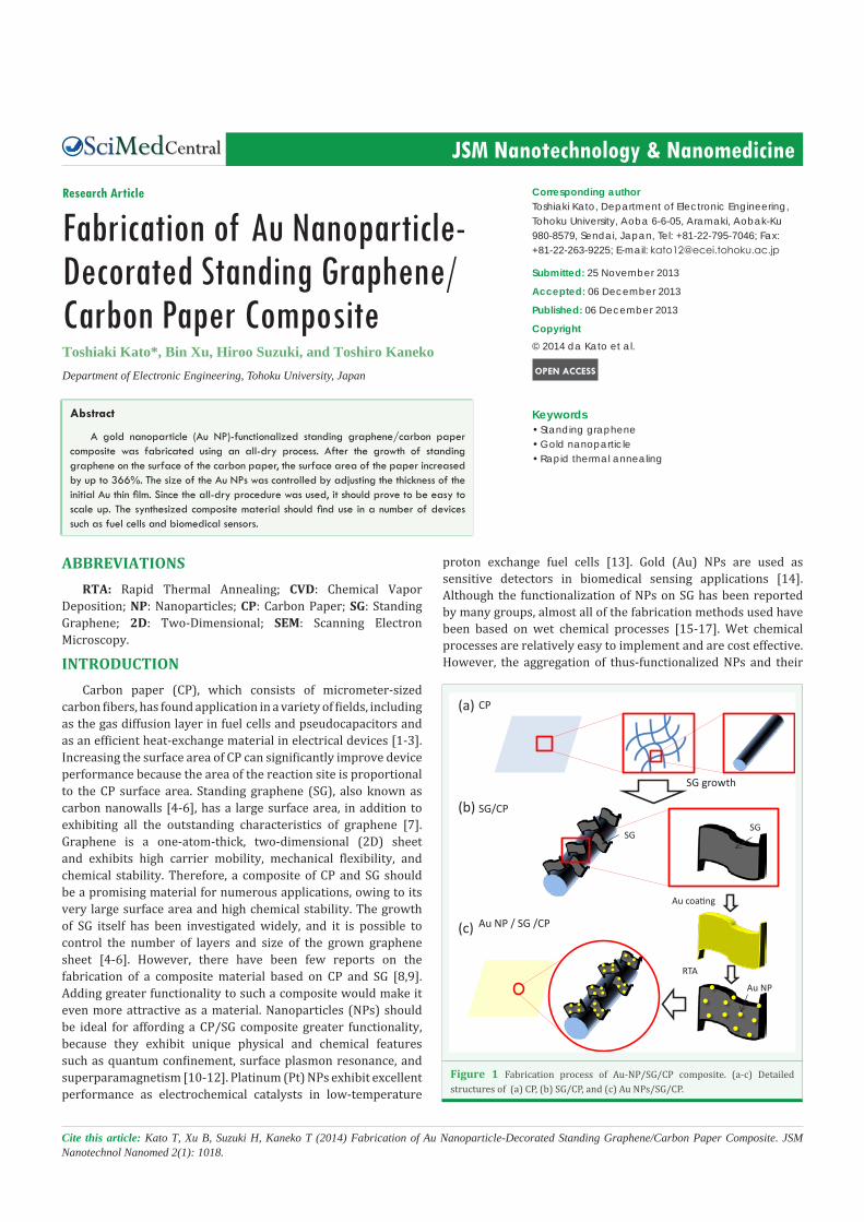
The results of SG growth on the surface of the CP are shown in (Figure 2). The CP consisted of micron-scaled carbon fibers. Every carbon fiber had a straight structure, and its surface was relatively smooth (Figure 2a). After the helicon plasma CVD, the surfaces of the carbon fibers were covered with dense SG without there being any changes in the overall structure of the carbon fibers (Figures 2b-d). This shows that the overall structure of CP can be maintained even after plasma CVD. Further, the surface area of the CP increased significantly after the SG had covered the carbon fibers. A simple calculation revealed that the surface area of the CP after SG growth was 366% greater than that of the pristine CP. Such a large surface area could result in significant improvements in device performance with respect to various applications.
One possible application for this large-surface-area material
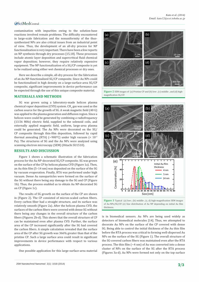
is in biomedical sensors. Au NPs are being used widely as detectors of biomedical molecules [14]. Thus, we attempted to decorate Au NPs on the surface of the CP covered with dense SG. Being able to control the initial thickness of the Au thin film before the RTA process was critical to forming well-dispersed Au NPs on the surface of the SG (Figure 1). The overall structure of the SG-covered carbon fibers was maintained even after the RTA process. The thin film (∼4 nm) of Au was converted into a dense cluster of NPs on the surface of the SG after the RTA process (Figures 3a-d). Au NPs were formed not only on the top surface
Figure 2 SEM images of (a) Pristine CP and (b) low-, (c) middle-, and (d) high-magnification SG/CP.
0 5 10 15 20 25 30 35 40 45 50 55 60 65 70 75 800
20
40
60
Size [nm]
Conc
entr
ation
[%]
(e)
14 nm5 nm
Initial Au film thickness
4 nm
(b)
(c) (d)
(a)
10 µm 1 µm
500 nm 500 nm
Figure 3 Typical (a) low-, (b) middle-, (c, d) high-magnification SEM images of Au-NPs/SG/CP. (e) Size distribution of Au NP depending on initial Au film thickness.

Central
Kato et al. (2014)Email:
JSM Nanotechnol Nanomed 2(1): 1018 (2014) 3/3
Kato T, Xu B, Suzuki H, Kaneko T (2014) Fabrication of Au Nanoparticle-Decorated Standing Graphene/Carbon Paper Composite. JSM Nanotechnol Nanomed 2(1): 1018.
Cite this article
of the SG but also on the bottom of the SG (Figures 3c,d). The size distribution of the Au NPs, shown in (Figure 3(e)), reveals that the mean diameter of the Au NPs was approximately 20 nm. The size of the Au NPs could be controlled by changing the thickness of the initial Au film. With an increase in the Au film thickness, the average Au NP diameter increased from 20 nm to 30 nm. This ability to tune the size of the Au NPs is important for controlling the sensitivity of biomedical sensors. Very recently, similar results have been reported in the case of Pt NP functionalization on a carbon nanowalls/CP composite for fuel cell applications [19]. However, the process for NP synthesis used in the current study is completely different from the one used to fabricate the Pt NP.
CONCLUSIONWe were able to fabricate Au NPs on a SG/CP composite.
After the growth of SG, the surface area of the CP increased by 366%. Dense Au NPs could be decorated on the surface of the SG by a simple, all-dry process. Thin Au film deposition followed by RTA allowed well-dispersed Au NPs to be formed on the SG/CP composite in high density. Furthermore, the size distribution of the Au NPs could be controlled by changing the thickness of the initial Au film. This Au NP-decorated SG/CP composite should be useful in various devices such as fuel cells and biomedical sensors.
ACKNOWLEDGEMENTSThis work was supported in part by Grants-in-Aid for
Scientific Research (KAKENHI) grant (25706028) from the Japan Society for the Promotion of Science (JSPS). Support was also provided by the Ozawa-Yoshikawa Memorial Electronics Research Foundation and the Iketani Science and Technology Foundation. We are grateful to Ms. N. Sato for their help with the SEM measurements.
REFERENCES1. Bevers D, Rogers R, Bradke MV. Examination of the influence of PTFE
coating on the properties of carbon paper in polymer electrolyte fuel cells. J Power Sources. 1996; 63: 193-201.
2. Cheng S, Yang L, Liu Y, Lin W, Huang L, Chen D, et al. Carbon fiber paper supported hybrid nanonet/nanoflower nickel oxide electrodes for high-performance pseudo-capacitors. J Mater Chem A; 2013; 1: 7709-7716.
3. Wang Y, Gundevia M. Measurement of thermal conductivity and heat pipe effect in hydrophilic and hydrophobic carbon papers. Int J Heat Mass Transf. 2013; 60: 134-142.
4. Wu YH, Qiao PW, Chong TC, Shen ZX, Carbon nanowalls grown by microwave plasma enhanced chemical vapor deposition. Adv Mater. 2002; 14: 64-67.
5. Sato G, Morio T, Kato T, and Hatakeyama R. Fast growth of carbon nanowalls from pure methane using helicon plasma-enhanced chemical vapor deposition. Jpn J Appl Phys. 2006; 45: 5210-5212.
6. Sato G, Kato T, Oohara W, and Hatakeyama R. Production and application of reactive plasmas using helicon-wave discharge in very low magnetic fields. Thin Solid Films. 2006; 506-507: 550-554.
7. Novoselov KS, Geim AK, Morozov SV, Jiang D, Zhang Y, Dubonos SV, et al. Electric field effect in atomically thin carbon films. Science. 2004; 306: 666-669.
8. Li J, Su S, Zhou L, Kundrat V, Abbot AM, Mushtaq F, et al. Carbon nanowalls grown by microwave plasma enhanced chemical vapor deposition during the carbonization of polyacrylonitrile fibers. J Appl Phys. 2013; 113: 024313.
9. Lisi N, Giorgi R, Re M, Dikonimos T, Giorgi L, Salernitano E, et al. Carbon nanowall growth on carbon paper by hot filament chemical vapor deposition and its microstructure. Carbon. 2011; 49: 2134-2140.
10. Alivisatos AP. Semiconductor clusters, nanocrystals, and quantum dots. Science. 1996; 271: 933-937.
11. Fang N, Lee H, Sun C, Zhang X. Sub-diffraction-limited optical imaging with a silver superlens. Science. 2005; 308: 534-537.
12. Suh SK, Yuet K, Hwang DK, Bong KW, Doyle PS, Hatton TA. Synthesis of nonspherical superparamagnetic particles: in situ coprecipitation of magnetic nanoparticles in microgels prepared by stop-flow lithography. J Am Chem Soc. 2012; 134: 7337-7343.
13. Aricò AS, Bruce P, Scrosati B, Tarascon JM, van Schalkwijk W. Nanostructured materials for advanced energy conversion and storage devices. Nat Mater. 2005; 4: 366-377.
14. Xiao Y, Patolsky F, Katz E, Hainfeld JF, Willner I. “Plugging into Enzymes”: nanowiring of redox enzymes by a gold nanoparticle. Science. 2003; 299: 1877-1881.
15. Wu YH, Yang BJ, Han GC, Zong BY, Ni HQ, Luo P, Chong TC, Low TS, Shen ZX, Fabrication of a class of nanostructured materials using carbon nanowalls as the templates. Adv Fun Mater. 2002; 12: 489-494.
16. Giorgi L, Makris TD, Giorgi R, Lisi N, Salernitano E. Electrochemical properties of carbon nanowalls synthesized by HF-CVD. Sensors and Actuators B. 2007; 126: 144-152.
17. Rout CS, Kumar A, Fisher TS. Carbon nanowalls amplify the surface-enhanced Raman scattering from Ag nanoparticles. Nanotechnology. 2011; 22: 395704.
18. Machino T, Takeuchi W, Kano H, Hiramatsu M, Hori M. Synthesis of platinum nanoparticles on two-dimensional carbon nanostructures with an ultrahigh aspect ratio employing supercritical fluid chemical vapor deposition process. Appl Phys Exp. 2009; 2: 025001-1-3.
19. Hiramatsu M, Mitsuguchi S, Horibe T, Kondo H, Hori M, Kano H. Fabrication of carbon nanowalls on carbon fiber paper for fuel cell application. Jpn J Appl Phys. 2013; 52: 01AK03-1-5.