Research Article Antibacterial Activity and...
11
Research Article Antibacterial Activity and Cytocompatibility of PLGA/CuO Hybrid Nanofiber Scaffolds Prepared by Electrospinning Adnan Haider, 1 Sanghwa Kwak, 1 Kailash Chandra Gupta, 1,2 and Inn-Kyu Kang 1 1 Department of Polymer Science and Engineering, School of Applied Chemical Engineering, Kyungpook National University, Daegu 702-701, Republic of Korea 2 Polymer Research Laboratory, Department of Chemistry, IIT, Roorkee 247667, India Correspondence should be addressed to Inn-Kyu Kang; [email protected] Received 8 December 2014; Revised 21 February 2015; Accepted 5 March 2015 Academic Editor: Tamer Uyar Copyright © 2015 Adnan Haider et al. is is an open access article distributed under the Creative Commons Attribution License, which permits unrestricted use, distribution, and reproduction in any medium, provided the original work is properly cited. e PLGA/CuO hybrid nanofibers scaffolds were prepared via electrospinning technique. e presence of CuO in the PLGA scaffolds was confirmed by transmission electron microscope (TEM) and X-ray photoelectron spectroscopy (XPS). e scaffolds were subjected to various antibacterial and cytobiocompatibility tests. e results not only showed excellent adhesion, proliferation, and viability (live/dead staining) for fibroblastic cells but also revealed that PLGA/CuO hybrid nanofiber scaffolds inhibited both Gram-positive and Gram-negative bacterial growth. e mechanism of the antibacterial activity was concluded to be based on the CuO nanoparticles and Cu ++ ions release. It is, therefore, evaluated that PLGA/CuO hybrid nanofiber scaffolds can be a useful candidate for wound dressing. 1. Introduction e emergence of infectious diseases due to microbes in general and the antibiotic-resistant bacterial strains (Gram- positive and Gram-negative) in particular poses a serious threat to public health worldwide [1]. Both Gram-positive and Gram-negative bacterial strains are considered a major threat to animal’s life [1, 2]. In the last century, antibiotics were used as the only source for controlling and/or curing the infections caused by microbes. e current advances in the field of nanotechnology, especially, having the facility and ability to prepare metal or metal oxide nanoparticles of specific size and shape, have enabled human beings to invent new techniques for the preparation of antimicrobial agents against the microbes [2]. Microbial activity of the nanoparticles in part depends on their size, stability, and concentration of the nanoparticles in the growth medium. ese properties of nanoparticles can easily be altered. Nanoparticles have therefore received great attention in the medicine field [3]. Considering the unique properties, nano- sized inorganic particles such as metal oxides of zinc (Zn), magnesium (Mg), silver (Ag), and copper (Cu) are being generated for the ultimate use in biomedical nanotechnology [4, 5]. Antimicrobial activity of nanoparticles has largely been studied with human pathogenic bacteria such as Escherichia coli and Staphylococcus aureus [4]. e microbial cells and their colonies showed vulnerability to metals nanoparticles such as Zn, Ag, Cu, and Mg [3, 5, 6]. e bacterial population growth is inhibited by specific interaction of these metal nanoparticles with bacterial strains [1, 6]. Among these metal nanoparticles, Cu and copper oxide (CuO) nanoparticles have attracted considerable attention because Cu is easily available and one of the most widely used metals in the modern research world [7]. Since researchers have well documented the synthesis and optical, catalytic, and electrical properties and medicinal properties of CuO nanoparticles [8, 9], this study has chosen compositing CuO with the polymer for the ease of processability and developing a hybrid antibacterial scaffold [10]. Polymeric scaffolds play a vital role in tissue regeneration [11]. ey are intended to provide favorable environment to the cells that ultimately help in stimulating their growth and normal cellular functions. Polymeric scaffolds help in the movement of various growth factors, metabolites, and soluble drugs that help in stimulating cell growth and help in tissue regeneration. Poly(lactide-co-glycolide) (PLGA) has Hindawi Publishing Corporation Journal of Nanomaterials Volume 2015, Article ID 832762, 10 pages http://dx.doi.org/10.1155/2015/832762
Transcript of Research Article Antibacterial Activity and...

Research ArticleAntibacterial Activity and Cytocompatibility of PLGA/CuOHybrid Nanofiber Scaffolds Prepared by Electrospinning
Adnan Haider,1 Sanghwa Kwak,1 Kailash Chandra Gupta,1,2 and Inn-Kyu Kang1
1Department of Polymer Science and Engineering, School of Applied Chemical Engineering, Kyungpook National University,Daegu 702-701, Republic of Korea2Polymer Research Laboratory, Department of Chemistry, IIT, Roorkee 247667, India
Correspondence should be addressed to Inn-Kyu Kang; [email protected]
Received 8 December 2014; Revised 21 February 2015; Accepted 5 March 2015
Academic Editor: Tamer Uyar
Copyright © 2015 Adnan Haider et al. This is an open access article distributed under the Creative Commons Attribution License,which permits unrestricted use, distribution, and reproduction in any medium, provided the original work is properly cited.
The PLGA/CuO hybrid nanofibers scaffolds were prepared via electrospinning technique. The presence of CuO in the PLGAscaffolds was confirmed by transmission electron microscope (TEM) and X-ray photoelectron spectroscopy (XPS). The scaffoldswere subjected to various antibacterial and cytobiocompatibility tests.The results not only showed excellent adhesion, proliferation,and viability (live/dead staining) for fibroblastic cells but also revealed that PLGA/CuO hybrid nanofiber scaffolds inhibited bothGram-positive and Gram-negative bacterial growth. The mechanism of the antibacterial activity was concluded to be based on theCuO nanoparticles and Cu++ ions release. It is, therefore, evaluated that PLGA/CuO hybrid nanofiber scaffolds can be a usefulcandidate for wound dressing.
1. Introduction
The emergence of infectious diseases due to microbes ingeneral and the antibiotic-resistant bacterial strains (Gram-positive and Gram-negative) in particular poses a seriousthreat to public health worldwide [1]. Both Gram-positiveand Gram-negative bacterial strains are considered a majorthreat to animal’s life [1, 2]. In the last century, antibioticswere used as the only source for controlling and/or curingthe infections caused by microbes. The current advances inthe field of nanotechnology, especially, having the facilityand ability to prepare metal or metal oxide nanoparticlesof specific size and shape, have enabled human beings toinvent new techniques for the preparation of antimicrobialagents against the microbes [2]. Microbial activity of thenanoparticles in part depends on their size, stability, andconcentration of the nanoparticles in the growth medium.These properties of nanoparticles can easily be altered.Nanoparticles have therefore received great attention in themedicine field [3]. Considering the unique properties, nano-sized inorganic particles such as metal oxides of zinc (Zn),magnesium (Mg), silver (Ag), and copper (Cu) are beinggenerated for the ultimate use in biomedical nanotechnology
[4, 5]. Antimicrobial activity of nanoparticles has largely beenstudied with human pathogenic bacteria such as Escherichiacoli and Staphylococcus aureus [4]. The microbial cells andtheir colonies showed vulnerability to metals nanoparticlessuch as Zn, Ag, Cu, andMg [3, 5, 6].The bacterial populationgrowth is inhibited by specific interaction of these metalnanoparticles with bacterial strains [1, 6]. Among these metalnanoparticles, Cu and copper oxide (CuO) nanoparticleshave attracted considerable attention because Cu is easilyavailable and one of the most widely used metals in themodern research world [7]. Since researchers have welldocumented the synthesis and optical, catalytic, and electricalproperties and medicinal properties of CuO nanoparticles[8, 9], this study has chosen compositing CuO with thepolymer for the ease of processability and developing a hybridantibacterial scaffold [10].
Polymeric scaffolds play a vital role in tissue regeneration[11]. They are intended to provide favorable environmentto the cells that ultimately help in stimulating their growthand normal cellular functions. Polymeric scaffolds help inthe movement of various growth factors, metabolites, andsoluble drugs that help in stimulating cell growth and helpin tissue regeneration. Poly(lactide-co-glycolide) (PLGA) has
Hindawi Publishing CorporationJournal of NanomaterialsVolume 2015, Article ID 832762, 10 pageshttp://dx.doi.org/10.1155/2015/832762
2 Journal of Nanomaterials
been the most frequently used polymeric materials amongthe various polymeric materials used for the preparation ofelectrospunnanofiber scaffolds [11]. PLGAhas been approvedby the Food and Drug Administration (FDA) authoritydue to its biocompatible and biodegradable nature [12, 13].Furthermore PLGA has been extensively studied in thefield of drug delivery and tissue regeneration. Apart fromthe biocompatible nature, PLGA offers different degradationtime by tailoring the monomer ratio of lactide and glycolide[12].
Till now various techniques have been introduced forthe preparation of various polymeric scaffolds includinggas forming, freeze-drying, phase separation, and solventcasting. Among the so far used techniques, electrospinningis a simple, versatile, and cost effective technique used forgenerating fibers with size ranging from submicron (𝜇m) tonanometer (nm) scale diameter [13, 14]. Electrospinning isused for producingmultifunctional nanofiber from both nat-ural and artificial polymers, polymer blends, and composite[15]. Along with high surface area, inter- and intrafibrouspores, electrospun nanofiber scaffolds have a strong adhesiveforce, good air filtration, high adhesion barrier activity,and heat resistance [15–17]. These properties make thesenanofiber scaffolds quite similar in shape to human skin.Thescope of electrospun nanofiber has been explored specificallyin biomedical area for various reasons such as biofilms,wound dressing materials, hemostatic materials, artificialblood vessels, drug and gene delivery, antimicrobial agent,and tissue regeneration scaffolds [3, 18].
Keeping these antecedents in mind, the purpose of thisstudy was to test and determine the antibacterial activity ofPLGA/CuO hybrid nanofiber scaffolds on various bacterialstrains. Furthermore, we aimed to study the interactionof fibroblast (skin cells) with PLGA/CuO hybrid nanofiberscaffolds so that it can be used as an internal and externalwound dressing agent. This kind of study will pave new waysin designing the future wound dressing materials, which canbehave as an antibacterial agent without compromising itscytocompatibility.
2. Experimental
2.1. Materials and Methods. Poly(lactide-co-glycolide)(PLGA), copper (Cu), dimethylformamide (DMF), and tetra-hydrofuran (THF) were purchased from Sigma Aldrich.Fibroblastic cell line (NIH3T3) was obtained fromKorea CellBank. MTT assay kit was purchased from Gibco, Invitrogen,USA, whereas Dulbecco’smodified eagle’smedium (DMEM),fetal bovine serum (FBS), and penicillin G-streptomycinwere acquired from Gibco, Japan. Escherichia coli (KCCM12119) and staphylococcus aureus (KCCM 12256) wereobtained from Korean Culture Center of Microorganisms(KCCM). All the reagents and chemicals in this study wereused as received.
2.2. Synthesis of Copper Oxide (CuO) Nanoparticles. For thesynthesis of CuO nanoparticles, 3mg of Cu powder wasadded to 30mL of distilled water in a glass vial and was
sonicated for about 25 minutes in a sonicator. The reactionmixture was transferred into a glass bottle and sealed underordinary conditions by wrapping the bottle cap with Teflonfoil. The sealed glass bottle containing reaction solutionwas autoclaved at 140∘C for 24 hours. After 24 hours thesolution was allowed to cool at room temperature and thencentrifuged to retrieve CuO nanoparticles.The nanoparticleswere washed repeatedly with double distilled water, freeze-dried, and stored [19].
2.3. Solution Preparation and Fabrication of Nanofiber Scaf-folds. Polymer solutions at the concentration of 5 to 20wt%were prepared by dissolving PLGA in a binary solvent of THFand DMF in 3 : 1 ratio. The solution was stirred overnight atroom temperature until complete dissolution. The preparedsolution was then subjected to electrospinning process. Thetypical electrospinning apparatus and the basic concept ofthe fiber formation are summarized in Figure 1. Briefly, thesolution was transferred to a 10mL glass syringe fitted with aneedlewith an inner diameter of 0.9mm.The electrospinningexperiment was based on our previously reported study [13].The optimized electrospinning conditions used in the presentstudy were tip to collector distance 20 cm, applied voltage20 kV, and a solution flow rate 1mL/h which was maintainedthroughout the electrospinning process. The electrospunnanofiber scaffolds were collected onto the aluminium foilwrapped over the metallic collector. After the completionof the process, the electrospun nanofiber scaffolds wereremoved from the metallic collector along with the alu-minium foil. The electrospun nanofiber scaffold was vacuumdried overnight at 40∘C to remove the solvent. The sameprocedure was adapted for the preparation of the electrospunPLGA/CuO hybrid nanofiber scaffolds containing 0.5 wt% ofCuO nanoparticles.
2.4. Characterization of the Scaffolds. The viscosity of thePLGA polymer solutions in the binary solvent (THF :DMF =3 : 1 ratio) was measured at room temperature using a vis-cometer (Brookfield Viscometer DV-II Pro) with spindlenumber 6 at 100 revolutions per minute (rpm).Themorphol-ogy of the PLGA/CuO hybrid nanofiber scaffolds was eval-uated by field emission scanning electron microscopy (FE-SEM, 400 Hitachi, Tokyo, Japan) and transmission electronmicroscope (BioTEM, Hitachi, Tokyo, Japan). The particlesize was analyzed via dynamic light scattering (DLS). Thequalitative and quantitative analysis of CuO, PLGA/CuO, andPLGA nanofiber scaffolds were carried out by X-ray pho-toelectron spectroscopy (XPS, ESCA LAB VIG microtech,Mt 500/1, etc., East Grinstead, UK) equipped with Mg K𝛼radiation at 1,253.6 eV and a 150-W powermode at the anode.A survey scan spectrum was taken and the surface elementalcompositions relative to the carbon were calculated from thepeak heights taking into account the atomic sensitivity.
2.5. Antibacterial Activity. The antibacterial activities ofpristine PLGA and PLGA/CuO nanofiber scaffolds wereinvestigated against model microbial species includingEscherichia coli (Gram-negative) and Staphylococcus aureus,

Journal of Nanomaterials 3
Ground
Drum collector
Fibers
Syringe
Power sourcepump
(a)
Char
ges e
nds u
p on
th
e out
side
Liqu
idSo
lid
Dire
ctio
n of
flow
(b)
Taylor cone
Spinning tip
Ohm
ic
flow
Con
vect
ion
flow
Geometry of cone isgoverned by the ratioof surface tension to
electrostatic repulsion
Zone of transitionbetween liquid and
solid
Drum collector
(c)
Figure 1: The schematic of the basic concept of electrospinning: electrospinning apparatus (a), accumulation of charges on the needle andconsequently on the liquid (b), and Taylor cone and basic schematic for nanofiber preparation (c).
S. aureus (Gram-positive), through two well-knownmethodsdescribed below.
2.5.1. Agar Disc Diffusion Method. In this method, theantibacterial activities of the pristine PLGA and PLGA-CuOhybrid nanofiber scaffolds were measured by disc diffusionmethod [20]. Microbial species, E. coli (KCCM 12119), wasgrown on nutrient agar (DIFCO 0001) containing 3 g/L beefextract, 5 g/L peptones, and 15 g/L agar in distilled water,whereas S. aureus (KCCM 12256) was grown on trypticasesoy agar (BBL 211768) containing 17 g/L pancreatic digest ofcasein, 3 g/L pancreatic digest of soybean meal, and 15 g/Lagar in distilled water. The pH of the agar media for bothmicrobial species was maintained at 7.0 [20]. Disc shapesamples of PLGA and PLGA/CuO hybrid nanofiber scaffoldswith 1×1 cm2 dimension were prepared and subjected to theinhibition zone test.The sampleswere sterilizedwithUV for 2hours and subsequently placed on E. coli and S. aureus cultureplates. The plates were incubated for 24 hours at 37∘C. Therelative antibacterial effect was found by measuring the clearzones of inhibition formed around the discs [21].
2.5.2. Optical DensityMethod. In thismethod, pristine PLGAand PLGA-CuO hybrid nanofiber scaffolds were sliced intosmall pieces and sterilized at 121∘C for 15 minutes. Next,50mL of growth medium for both bacterial species wastaken in 100mL flasks, followed by addition of 0.005 g/mLfinely sliced solid PLGA and PLGA-CuO hybrid nanofiberscaffolds. The tubes were then seeded with 1mL fresh cultureof bacterial strains and incubated in a shaking incubatorat 37∘C and for 24 hours. The turbidity of the media wasobserved at various time intervals at 610 nm using a UVspectrophotometer (T60U, China) [20, 22].
2.6. Cell Adhesion Study. To examine the response of thefibroblastic cells to the pristine PLGA and PLGA/CuOhybridnanofiber scaffolds, small circular samples of 1 × 1 cm2dimension of both scaffolds were prepared and used. Briefly,the circular samples of the PLGA and PLGA/CuO hybridnanofiber scaffolds were fitted in a 24-well culture dish andsubsequently immersed in a DMEM medium containing10% FBS and 1% penicillin G-streptomycin. One milliliterof a NIH3T3 cell solution (5 × 10⁵ cells/mL) was seeded onthe electrospun pristine and PLGA/CuO hybrid nanofiberscaffolds and incubated in a humidified atmosphere (5%CO
2
and at 37∘C) for 1 and 3 days in order to determine thecell adhesion on the nanofiber scaffolds. After incubation,the supernatant was removed, washed twice with PBS, andfixed with an aqueous 2.5% glutardialdehyde solution for 20minutes. The sample sheet was then dehydrated, dried in acritical point drier, and stored for characterization [3].
2.7. Cell Proliferation and Viability Study. 3-(4, 5-Dimeth-ylazol-2-yl)-2,5-diphenyl-2H-tetrazolium bromide (MTT)assay was used to determine the proliferation of fibroblasticcells on the PLGA and PLGA/CuO hybrid nanofiber scaf-folds. Briefly, NIH3T3 cells were seeded at a concentrationof 3 × 10⁴ cells/mL onto pristine PLGA and PLGA/CuOhybrid nanofiber scaffolds, which were fitted in a 24-wellplate, and cell proliferation was monitored after 1 and 3days of incubation. A MTT solution (50 𝜇L, 5mg/mL inPBS) was added to each well and incubated in a humidifiedatmosphere containing 5% CO
2at 37∘C for 4 hours. After
removing the medium, the converted dye was dissolved inacidic isopropanol (0.04N HCl-isopropanol) and kept in thedark at room temperature for 30 minutes. From each sample,100 𝜇L medium was taken, transferred to a 96-well plate, and

4 Journal of Nanomaterials
Fiber diameter (nm)300 400 500 600 700 800
Freq
uenc
y
02468
10121416
200𝜇m
(a)
Fiber diameter (nm)300 400 500 600 700
Freq
uenc
y
02468
10121416
200𝜇m
(b)
Figure 2: FE-SEM images of PLGA (a) and PLGA/CuO hybrid nanofiber scaffolds (b).
subjected to ultraviolet measurements for the converted dyeat a wavelength of 570 nm on a kinetic microplate reader(EL × 800, Bio-T Instruments, Inc., Highland Park, USA).
A standard live/dead assay was conducted for the eval-uation of NIH3T3 cell viability after culturing NIH3T3 cellson pristine PLGAandPLGA/CuOhybrid nanofiber scaffolds.The cell viability experiment was performed in accordancewith the previous articles [3, 23].
2.8. Cu++ Ion Release. The Cu++ ions release experiment wascarried out to find the ionization potential of CuO from thePLGA/CuO hybrid nanofiber scaffolds. The amount of Cu++ions released from the sample was determined by immersingthe PLGA/CuO hybrid nanofiber scaffolds (4 × 4 cm2, 0.2 g)into 10mL distilled water for different time periods. Theamount of Cu++ ions in the double distilled water was deter-mined by inductively coupled plasma spectrophotometer(ICP, Thermo Jarrell Ash IRIS-AP) [3].
3. Results and Discussion
3.1. Morphology of Nanofiber and CuO. Figure 2 depicts theFE-SEM images of the electrospun PLGA and PLGA/CuOhybrid nanofiber scaffolds. It is evident from the FE-SEMimages of PLGA nanofiber (Figure 2(a)) and PLGA/CuOhybrid nanofiber scaffolds (Figure 2(b)) that after implement-ing optimized electrospinning parameters the electrospunnanofiber was smooth and uniform. The average diameterscalculated from the histograms of the electrospun pris-tine PLGA (inset of (Figure 2(a)) and PLGA/CuO (insetof (Figure 2(b)) hybrid nanofiber scaffolds were 548 and553 nm, respectively.This small change in the average diame-ter of the fibers shows that the addition of CuO to PLGA didnot affect the average diameter of the fibers to a great extent.
TEM images of the CuO nanoparticles and PLGA/CuOhybrid nanofiber are depicted in Figures 3(a) and 3(b).
Images show that CuO nanoparticles (Figure 3(a)) were suc-cessfully incorporated into the PLGAnanofiber (Figure 3(b)).The size distribution of CuO nanoparticles calculated fromTEM (Figure 3(a)) was ranging from 40 to 100 nm, whereasthe relative size distribution obtained from DLS (inset ofFigure 3(a)) was in the range of 60–128 nm with the max-imum distribution peak observed at 95 nm. Hence it wasconcluded that the size of the CuO ranged from 40 to 128 nm.
3.2. XPS Study. ESCA survey scan spectra were used toconfirm the presence of CuO nanoparticles in the matrixof PLGA electrospun nanofiber. Cu peak is the marker ofchoice for confirming the presence of CuO nanoparticles inthe PLGA/CuO hybrid nanofiber scaffolds. The survey scanspectra of pristine PLGA nanofiber scaffolds show typicalpeaks of carbon C1s at 284.6 eV and oxygen O1s at 536.1 eV(Figure 4(a)). Similarly, two characteristic signals of Cu at932.4 eV and oxygen O1s at 536.1 eV were detected in thesurvey scan spectra of pure CuO nanoparticles (Figure 4(c)).The appearance of the characteristic peaks of Cu, carbon, andoxygen at 932.4 eV, 284.6 eV, and 536.1 eV, respectively, in thesurvey scan spectra of PLGA/CuO hybrid nanofiber scaffoldsconfirmed the presence of CuO nanoparticles in the PLGApolymermatrix (Figure 4(b)). Besides the qualitative analysisof the survey scans from the peak positions, quantitativedetermination of the major elements was performed byanalyzing the peak intensity and summarized in Table 1.The analysis of the data showed that after the successfulincorporation of CuO nanoparticles into the PLGA/CuOhybrid nanofiber scaffold, a change in the%weights of carbon(62.10), oxygen (47.53), and CuO (0.40) for PLGA/CuOhybrid nanofiber scaffolds was observed as compared to thepristine CuO nanoparticles and PLGA nanofiber. Therefore,it was confirmed from the ESCA that CuOnanoparticles weresuccessfully incorporated into PLGA during electrospinningto prepare PLGA/CuO hybrid nanofiber scaffold.

Journal of Nanomaterials 5
Size (r.nm)50 70 90 110 130
Inte
nsity
(%)
02468
1012141618202224
100nm
(a)
100nm
(b)
Figure 3: TEM images of the CuO nanoparticles (a) and PLGA/CuO electrospun hybrid nanofiber (b).
800 700 600 500 400 300 200 100 0
O1s
C1s
Cou
nts/
s
Binding energy (e.V)
O2s
5
4
3
2
1
0
×104
(a)
Cou
nts/
s
1000 800900 700 600 500 400 300 200 100 0Binding energy (e.V)
Si2p
Si2s
Cu3p
CuLM
M3
CuLM
M2-
CuLM
M1
O1s
C1s
O2s
Cu3s
Cu2p
3-Cu
2pCu
2p1
6
4
5
3
2
1
0
×104
(b)
Cou
nts/
s
1000 800900 700 600 500 400 300 200 100 0Binding energy (e.V)
Cu2p
1 Cu2p
3-Cu
2p
CuLM
M3
O2s
Cu3p
Cu3s
CuLM
M-C
uLM
M1
C1s
O1s
CuLM
M2
8
6
7
4
5
3
2
1
0
×104
(c)
Figure 4: ESCA survey scan spectra of pristine PLGA (a), CuO nanoparticles (b), and (c) PLGA/CuO hybrid nanofiber scaffold.

6 Journal of Nanomaterials
S. aureus
E. coli
(a)
Time (h)0 2 4 6 8 10 12 14
Abso
rban
ce (a
.u.)
0.0
0.1
0.2
0.3
0.4
0.5
0.6
0.7
Inhi
bitio
n
PLGA (S. aureus)PLGA (E. coli)
PLGA-CuO (S. aureus)PLGA-CuO (E. coli)
(b)
Figure 5: Inhibition zones of the PLGA and PLGA/CuO nanofiber scaffolds against E. coli and S. aureus determined by disc diffusionmethod(a) and antibacterial activities of PLGA and PLGA/CuOnanofiber scaffolds against E. coli and S. aureus determined by optical densitymethod(b).
Table 1: Chemical composition of the CuO, PLGA, and PLGA/CuOcomposite nanofiber scaffolds calculated from ESCA survey scanspectra.
Substrates Atomic percent (%)C O Cu
PLGA 64.61 35.39CuO 6.23 43.40 49.70PLGA/CuO 62.10 35.53 0.40
3.3. Antibacterial Properties of PLGA and PLGA-CuO HybridNanofiber Scaffolds. Lack of antibacterial properties is oneof the serious obstacles limiting biomedical applicationsof biopolymers including PLGA. As being used in woundhealing and tissue regeneration, the PLGA scaffolds shouldkeep the ability of protection against pathogenic species [12,13]. The current PLGA/CuO hybrid nanofiber scaffolds wereprepared with the aim of introducing antibacterial activitiesinto the PLGA electrospun nanofiber so as to enhance theirapplicability in medical field. Figures 5(a) and 5(b) illustratethe comparative results of the antibacterial activities of pris-tine PLGA and PLGA/CuO nanofiber scaffolds. The resultsobtained from disc diffusion method against E. coli and S.aureus indicated that the pristine PLGA nanofiber scaffolddid not produce any zone of inhibition against both species(E. coli and S. aureus (Figure 5(a)). Thus the results clearlydemonstrate that the pristine PLGA has nonbactericidalnature. On the other hand, the PLGA/CuO hybrid nanofiberscaffold produced zones of inhibition against both E. coli
and S. aureus (Figure 5(a)) indicating antibacterial activitybased on the incorporation of CuO into PLGA nanofiber.Thus PLGA/CuO hybrid nanofiber scaffold has obtainedthe bactericidal ability. A similar phenomenon was alsoindicated by the optical density method (Figure 5(b)). Anincreasing trend in the cell density is observed in the samplescontaining PLGA; however, the growth of both the E. coliand S. aureus is greatly inhibited by PLGA/CuO nanofiberscaffold. The inhibition trend remained the same as wasshown by disc diffusion method; that is, E. coli is inhibitedmore as compared to S. aureus. Thus it is concluded thatthe disc diffusion as well as optical density method showedsignificant (Figures 5(a) and 5(b)) antibacterial activity ofPLGA/CuO nanofiber scaffold. The antibacterial activitiesof Cu and CuO are well established [24]. CuO, not onlyin colloidal form but also in composite form, have alsoshown tremendous antibacterial properties [20, 25]. Hereinthe produced inhibition zones and the higher inhibition inthe growth of both E. coli and S. aureus could be attributed tothe bactericidal nature of CuO [26–28].
Schematics of the potential mechanisms which can beeffective in retarding the bacterial growth are depicted inFigure 6. Exact mechanism of the antibacterial activities ofnanomaterials is still not known [2, 29].The bacterial growthcan be inhibited by the penetration of CuO nanoparticlesor Cu++ ions into the cell via cell membrane or may inhibitbacterial growth by the reactive oxygen species (ROS) gen-eration. However, the most established mechanism is theadhesion of CuO nanoparticles/Cu++ ions to the proteincontaining sulphur present in the cell wall of the bacterial

Journal of Nanomaterials 7
SH S S
SH
Nanoparticles
ROS
1 2
3
Protein
(1) Direct membrane damage(2) ROS generation(3) Protein damage
Figure 6: Schematic representingmechanism related to the antibac-terial activity of CuO nanoparticles/Cu++ ions.
Time (day)0 2 4 6 8 10 12
0.0
0.2
0.4
0.6
0.8
1.0
1.2
1.4
1.6
1.8
2.0
Cu2+
ions
rele
ase i
n w
ater
(ppm
)
Figure 7: Cu++ ions release in water from the PLGA/CuO hybridnanofiber scaffolds with respect to incubation time.
cells, which leads to the malfunctioning of the bacterial cellwall and ultimately causes the bacterial cell death [27, 28, 30].
3.4. Cu++ Ions Release Study. Figure 7 depicts the resultsobtained from the Cu++ ions release experiment. It wasobserved that Cu++ ions from the PLGA/CuO hybridnanofiber scaffold were released in a sustained manner,therefore enabling the PLGA/CuO hybrid nanofiber scaffoldsto exhibit antibacterial effect for longer duration.The amountof Cu++ ions released from the PLGA/CuO hybrid nanofiberscaffold reached 1.6 ppm after 10 days of incubation in water.The Cu++ ions release and the inhibition of E. coli and S.aureus by the PLGA/CuO nanofiber scaffold (Figure 5(b))
follow the same pattern; this is an indication that theoperating mechanism of inhibition is governed by Cu++ions. However, for Cu++ ions release, the choice of mediais important, as the body fluid has a complex compositionand the components comprising body fluid exhibit differentbinding abilities to Cu++ ions [28, 31, 32]. The amount ofCu++ ions in the double distilled water was determined byinductively coupled plasma spectrophotometer.
3.5. Cytocompatibility Study
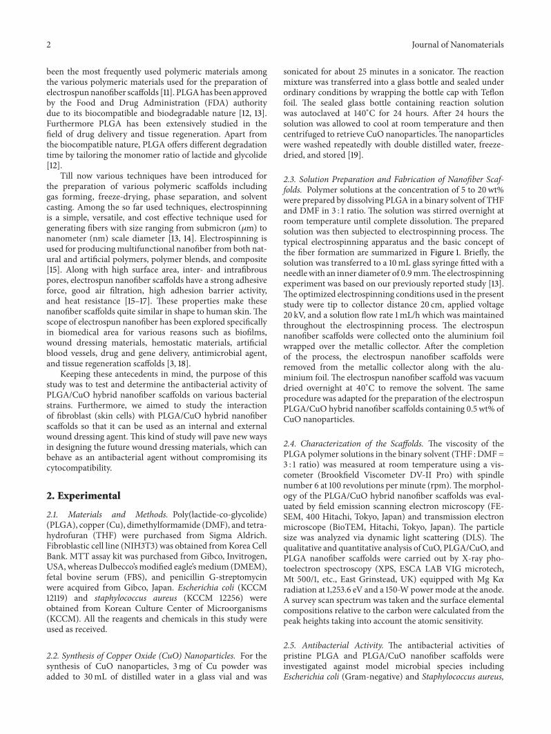
3.5.1. Cell Adhesion. NIH3T3 cells tend to get adhered tothe surface of the biocompatible materials; therefore thevarious cell fate processes including proliferation, migration,apoptosis, and differentiation are highly affected by cellsadhesion to cell-binding epitopes in the extracellular matrix(ECM) [33]. Figure 8 depicts the FE-SEM images of theNIH3T3 cells adhered to the pristine PLGA and PLGA/CuOhybrid nanofiber scaffolds with different incubation time. Itis clearly visible that NIH3T3 cells were adhered to bothpristine PLGA (Figures 8(a) and 8(c)) and PLGA/CuOhybridnanofiber scaffolds (Figures 8(b) and 8(d)), thus provingthat both pristine PLGA and PLGA/CuO hybrid nanofiberscaffolds provided good cytocompatibility environment forNIH3T3 cells. The spreading pattern of the NIH3T3 cellson the PLGA and PLGA/CuO hybrid nanofiber scaffoldsfurther confirmed the noncytotoxic nature of the scaffolds.Furthermore, the observed increase in the number of adheredcells to the scaffolds was directly proportional to the increasein incubation time (Figures 8(c) and 8(d)) [3, 12].
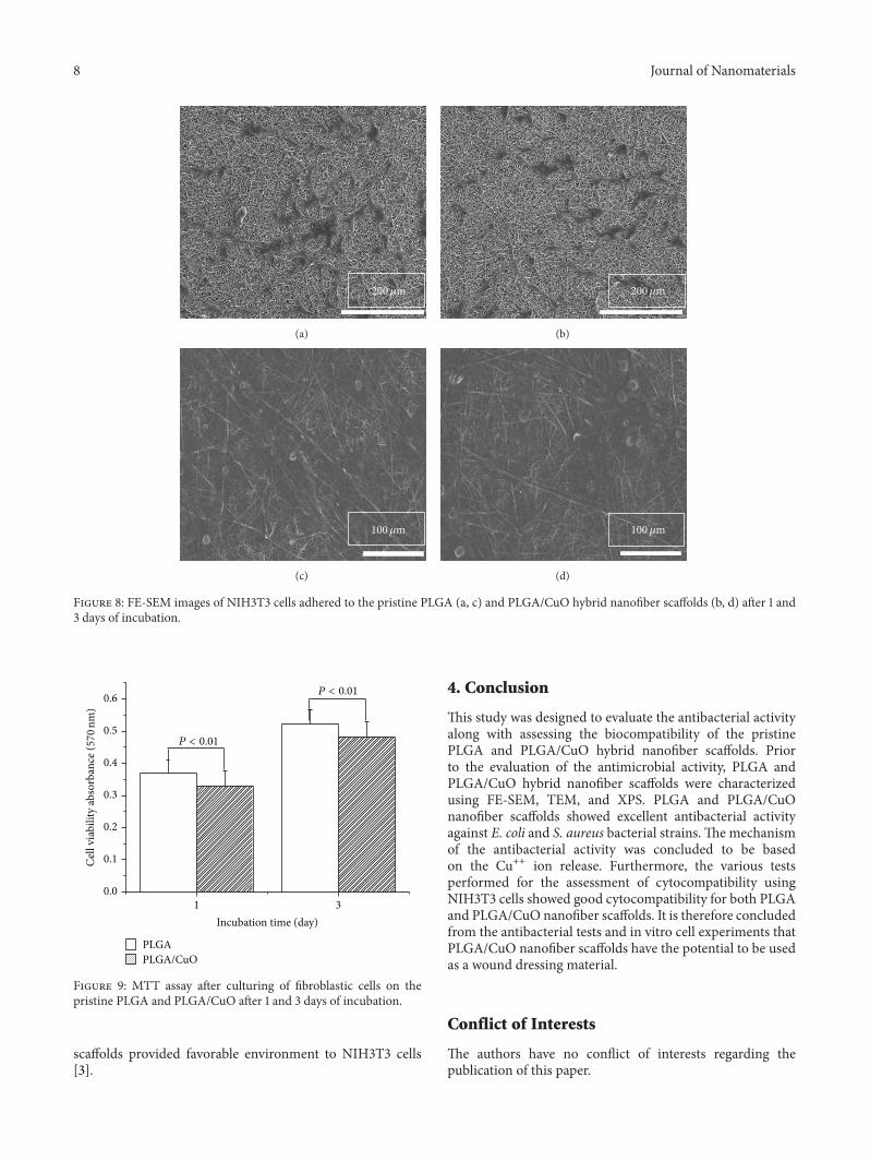
3.5.2. Cell Proliferation and Viability. Many colorimetricmethods have been adapted in order to estimate the exact cellnumber, among which the methods based on cells metabolicviability test are widely employed [34]. MTT reagents arewidely used for this purpose. Figure 9 illustrates the dataobtained from MTT assay of the proliferation of fibroblasticcells seeded on the PLGA and PLGA/CuO hybrid nanofiberscaffolds. From the MTT assay it was observed that NIH3T3cells proliferated on the PLGA and PLGA/CuO hybridnanofiber scaffolds. The NIH3T3 cells proliferation on thePLGA/CuO hybrid nanofiber scaffolds was in the range ofstandard deviation compared with pristine PLGA nanofiber;thus both scaffolds exhibit cytocompatibility nature. Fur-thermore, MTT assay was conclusive in elucidating theproliferation of NIH3T3 cells on the pristine PLGA andPLGA/CuO hybrid nanofiber scaffolds.
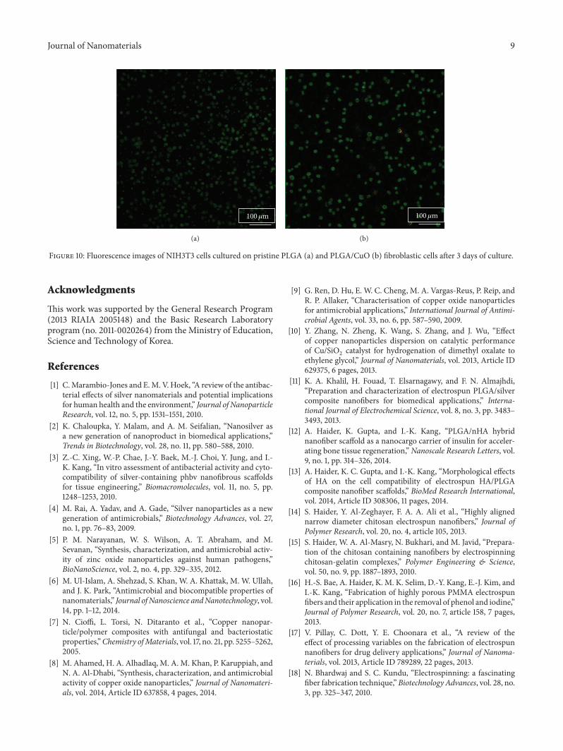
Figures 10(a) and 10(b) show the fluorescence imagesof the NIH3T3 cells cultured on the PLGA/CuO hybridnanofiber scaffold for 8 days using the live/dead assay [23].The fluorescence color of cells cultured on the PLGA andPLGA/CuO hybrid nanofiber scaffolds was totally green,indicating a good viability of the fibroblastic cells. Rarely,few dead cells showed red fluorescence of propidium iodidestaining (Figure 10(b)) when cultured on the PLGA/CuOhybrid nanofiber scaffolds [3]. These observations of thelive/dead assay clearly suggest that the PLGA/CuO nanofiber
8 Journal of Nanomaterials
200𝜇m
(a)
200𝜇m
(b)
100𝜇m
(c)
100𝜇m
(d)
Figure 8: FE-SEM images of NIH3T3 cells adhered to the pristine PLGA (a, c) and PLGA/CuO hybrid nanofiber scaffolds (b, d) after 1 and3 days of incubation.
1 30.0
0.1
0.2
0.3
0.4
0.5
0.6
Incubation time (day)
Cel
l via
bilit
y ab
sorb
ance
( 570
nm)
P < 0.01
P < 0.01
PLGAPLGA/CuO
Figure 9: MTT assay after culturing of fibroblastic cells on thepristine PLGA and PLGA/CuO after 1 and 3 days of incubation.
scaffolds provided favorable environment to NIH3T3 cells[3].
4. Conclusion
This study was designed to evaluate the antibacterial activityalong with assessing the biocompatibility of the pristinePLGA and PLGA/CuO hybrid nanofiber scaffolds. Priorto the evaluation of the antimicrobial activity, PLGA andPLGA/CuO hybrid nanofiber scaffolds were characterizedusing FE-SEM, TEM, and XPS. PLGA and PLGA/CuOnanofiber scaffolds showed excellent antibacterial activityagainst E. coli and S. aureus bacterial strains. The mechanismof the antibacterial activity was concluded to be basedon the Cu++ ion release. Furthermore, the various testsperformed for the assessment of cytocompatibility usingNIH3T3 cells showed good cytocompatibility for both PLGAand PLGA/CuO nanofiber scaffolds. It is therefore concludedfrom the antibacterial tests and in vitro cell experiments thatPLGA/CuO nanofiber scaffolds have the potential to be usedas a wound dressing material.
Conflict of Interests
The authors have no conflict of interests regarding thepublication of this paper.
Journal of Nanomaterials 9
100𝜇m
(a)
100𝜇m
(b)
Figure 10: Fluorescence images of NIH3T3 cells cultured on pristine PLGA (a) and PLGA/CuO (b) fibroblastic cells after 3 days of culture.
Acknowledgments
This work was supported by the General Research Program(2013 RIAIA 2005148) and the Basic Research Laboratoryprogram (no. 2011-0020264) from the Ministry of Education,Science and Technology of Korea.
References
[1] C.Marambio-Jones and E.M. V.Hoek, “A review of the antibac-terial effects of silver nanomaterials and potential implicationsfor human health and the environment,” Journal of NanoparticleResearch, vol. 12, no. 5, pp. 1531–1551, 2010.
[2] K. Chaloupka, Y. Malam, and A. M. Seifalian, “Nanosilver asa new generation of nanoproduct in biomedical applications,”Trends in Biotechnology, vol. 28, no. 11, pp. 580–588, 2010.
[3] Z.-C. Xing, W.-P. Chae, J.-Y. Baek, M.-J. Choi, Y. Jung, and I.-K. Kang, “In vitro assessment of antibacterial activity and cyto-compatibility of silver-containing phbv nanofibrous scaffoldsfor tissue engineering,” Biomacromolecules, vol. 11, no. 5, pp.1248–1253, 2010.
[4] M. Rai, A. Yadav, and A. Gade, “Silver nanoparticles as a newgeneration of antimicrobials,” Biotechnology Advances, vol. 27,no. 1, pp. 76–83, 2009.
[5] P. M. Narayanan, W. S. Wilson, A. T. Abraham, and M.Sevanan, “Synthesis, characterization, and antimicrobial activ-ity of zinc oxide nanoparticles against human pathogens,”BioNanoScience, vol. 2, no. 4, pp. 329–335, 2012.
[6] M. Ul-Islam, A. Shehzad, S. Khan, W. A. Khattak, M. W. Ullah,and J. K. Park, “Antimicrobial and biocompatible properties ofnanomaterials,” Journal ofNanoscience andNanotechnology, vol.14, pp. 1–12, 2014.
[7] N. Cioffi, L. Torsi, N. Ditaranto et al., “Copper nanopar-ticle/polymer composites with antifungal and bacteriostaticproperties,”Chemistry ofMaterials, vol. 17, no. 21, pp. 5255–5262,2005.
[8] M. Ahamed, H. A. Alhadlaq, M. A. M. Khan, P. Karuppiah, andN. A. Al-Dhabi, “Synthesis, characterization, and antimicrobialactivity of copper oxide nanoparticles,” Journal of Nanomateri-als, vol. 2014, Article ID 637858, 4 pages, 2014.
[9] G. Ren, D. Hu, E. W. C. Cheng, M. A. Vargas-Reus, P. Reip, andR. P. Allaker, “Characterisation of copper oxide nanoparticlesfor antimicrobial applications,” International Journal of Antimi-crobial Agents, vol. 33, no. 6, pp. 587–590, 2009.
[10] Y. Zhang, N. Zheng, K. Wang, S. Zhang, and J. Wu, “Effectof copper nanoparticles dispersion on catalytic performanceof Cu/SiO
2catalyst for hydrogenation of dimethyl oxalate to
ethylene glycol,” Journal of Nanomaterials, vol. 2013, Article ID629375, 6 pages, 2013.
[11] K. A. Khalil, H. Fouad, T. Elsarnagawy, and F. N. Almajhdi,“Preparation and characterization of electrospun PLGA/silvercomposite nanofibers for biomedical applications,” Interna-tional Journal of Electrochemical Science, vol. 8, no. 3, pp. 3483–3493, 2013.
[12] A. Haider, K. Gupta, and I.-K. Kang, “PLGA/nHA hybridnanofiber scaffold as a nanocargo carrier of insulin for acceler-ating bone tissue regeneration,” Nanoscale Research Letters, vol.9, no. 1, pp. 314–326, 2014.
[13] A. Haider, K. C. Gupta, and I.-K. Kang, “Morphological effectsof HA on the cell compatibility of electrospun HA/PLGAcomposite nanofiber scaffolds,” BioMed Research International,vol. 2014, Article ID 308306, 11 pages, 2014.
[14] S. Haider, Y. Al-Zeghayer, F. A. A. Ali et al., “Highly alignednarrow diameter chitosan electrospun nanofibers,” Journal ofPolymer Research, vol. 20, no. 4, article 105, 2013.
[15] S. Haider, W. A. Al-Masry, N. Bukhari, and M. Javid, “Prepara-tion of the chitosan containing nanofibers by electrospinningchitosan-gelatin complexes,” Polymer Engineering & Science,vol. 50, no. 9, pp. 1887–1893, 2010.
[16] H.-S. Bae, A. Haider, K. M. K. Selim, D.-Y. Kang, E.-J. Kim, andI.-K. Kang, “Fabrication of highly porous PMMA electrospunfibers and their application in the removal of phenol and iodine,”Journal of Polymer Research, vol. 20, no. 7, article 158, 7 pages,2013.
[17] V. Pillay, C. Dott, Y. E. Choonara et al., “A review of theeffect of processing variables on the fabrication of electrospunnanofibers for drug delivery applications,” Journal of Nanoma-terials, vol. 2013, Article ID 789289, 22 pages, 2013.
[18] N. Bhardwaj and S. C. Kundu, “Electrospinning: a fascinatingfiber fabrication technique,”Biotechnology Advances, vol. 28, no.3, pp. 325–347, 2010.

10 Journal of Nanomaterials
[19] M. A. Shah and M. S. Al-Ghamdi, “Preparation of copper (Cu)and copper oxide (Cu
2O) nanoparticles under supercritical
conditions,”Materials Sciences and Application, vol. 2, no. 8, pp.977–980, 2011.
[20] M. Ul-Islam, T. Khan, W. A. Khattak, and J. K. Park, “Bacterialcellulose-MMTs nanoreinforced composite films: novel wounddressing material with antibacterial properties,” Cellulose, vol.20, no. 2, pp. 589–596, 2013.
[21] T. Maneerung, S. Tokura, and R. Rujiravanit, “Impregnationof silver nanoparticles into bacterial cellulose for antimicrobialwound dressing,” Carbohydrate Polymers, vol. 72, no. 1, pp. 43–51, 2008.
[22] S. A. Holowachuk, M. F. Bal’a, and R. K. Buddington, “A kineticmicroplate method for quantifying the antibacterial propertiesof biological fluids,” Journal of Microbiological Methods, vol. 55,no. 2, pp. 441–446, 2003.
[23] K. Trescher, N. Scharnagl, K. Kratz, T. Roch, A. Lendlein, and F.Jung, “Adherence and viability of primary human keratinocytesand primary human dermal fibroblasts on acrylonitrile-basedcopolymers with different concentrations of positively chargedfunctional groups,” Clinical Hemorheology and Microcircula-tion, vol. 52, no. 2–4, pp. 391–401, 2012.
[24] V. K. Champagne and D. J. Helfritch, “A demonstration of theantimicrobial effectiveness of various copper surfaces,” Journalof Biological Engineering, vol. 7, article 8, 2013.
[25] Y. Haldorai and J.-J. Shim, “Multifunctional chitosan-copperoxide hybrid material: photocatalytic and antibacterial activi-ties,” International Journal of Photoenergy, vol. 2013, Article ID245646, 8 pages, 2013.
[26] A. A. Tayel, W. F. El-Tras, S. Moussa et al., “Antibacterial actionof zinc oxide nanoparticles against foodborne pathogens,”Journal of Food Safety, vol. 31, no. 2, pp. 211–218, 2011.
[27] A. Azam, A. S. Ahmed, M. Oves, M. S. Khan, S. S. Habib, andA. Memic, “Antimicrobial activity of metal oxide nanoparticlesagainst Gram-positive and Gram-negative bacteria: a compar-ative study,” International Journal of Nanomedicine, vol. 7, pp.6003–6009, 2012.
[28] X. Hu, S. Cook, P.Wang, andH.-M. Hwang, “In vitro evaluationof cytotoxicity of engineeredmetal oxide nanoparticles,” Scienceof The Total Environment, vol. 407, no. 8, pp. 3070–3072, 2009.
[29] B. Reidy, A. Haase, A. Luch, K. A. Dawson, and I. Lynch,“Mechanisms of silver nanoparticle release, transformation andtoxicity: a critical review of current knowledge and recommen-dations for future studies and applications,”Materials, vol. 6, no.6, pp. 2295–2350, 2013.
[30] A. Haider and I.-K. Kang, “Preparation of silver nanoparticlesand their industrial and biomedical applications: a comprehen-sive review,” Advances in Materials Science and Engineering, vol.2014, Article ID 165257, 16 pages, 2014.
[31] A. Semisch, J. Ohle, B. Witt, and A. Hartwig, “Cytotoxicity andgenotoxicity of nano- and microparticulate copper oxide: roleof solubility and intracellular bioavailability,” Particle and FibreToxicology, vol. 11, article 10, 2014.
[32] M. S. Hassan, T. Amna, H. Y. Kim, and M.-S. Khil, “Enhancedbactericidal effect of novel CuO/TiO
2composite nanorods and
a mechanism thereof,” Composites Part B: Engineering, vol. 45,no. 1, pp. 904–910, 2013.
[33] X. Cao, K. Kwek, J. K. Y. Chan, C. K. H. Chan, and M. Lim,“Electrospun nanofibers as a bioadhesive platform for captur-ing adherent leukemia cells,” Journal of Biomedical MaterialsResearch Part A, vol. 102, no. 2, pp. 523–531, 2014.
[34] A. G. Karakecili, T. T. Demirtas, C. Satriano, M. Gumusder-elioglu, and G. Marletta, “Evaluation of L929 fibroblastattachment and proliferation on Arg-Gly-Asp-Ser (RGDS)-immobilized chitosan in serum-containing/serum-free cul-tures,” Journal of Bioscience and Bioengineering, vol. 104, no. 1,pp. 69–77, 2007.

Submit your manuscripts athttp://www.hindawi.com
ScientificaHindawi Publishing Corporationhttp://www.hindawi.com Volume 2014
CorrosionInternational Journal of
Hindawi Publishing Corporationhttp://www.hindawi.com Volume 2014
Polymer ScienceInternational Journal of
Hindawi Publishing Corporationhttp://www.hindawi.com Volume 2014
Hindawi Publishing Corporationhttp://www.hindawi.com Volume 2014
CeramicsJournal of
Hindawi Publishing Corporationhttp://www.hindawi.com Volume 2014
CompositesJournal of
NanoparticlesJournal of
Hindawi Publishing Corporationhttp://www.hindawi.com Volume 2014
Hindawi Publishing Corporationhttp://www.hindawi.com Volume 2014
International Journal of
Biomaterials
Hindawi Publishing Corporationhttp://www.hindawi.com Volume 2014
NanoscienceJournal of
TextilesHindawi Publishing Corporation http://www.hindawi.com Volume 2014
Journal of
NanotechnologyHindawi Publishing Corporationhttp://www.hindawi.com Volume 2014
Journal of
CrystallographyJournal of
Hindawi Publishing Corporationhttp://www.hindawi.com Volume 2014
The Scientific World JournalHindawi Publishing Corporation http://www.hindawi.com Volume 2014
Hindawi Publishing Corporationhttp://www.hindawi.com Volume 2014
CoatingsJournal of
Advances in
Materials Science and EngineeringHindawi Publishing Corporationhttp://www.hindawi.com Volume 2014
Smart Materials Research
Hindawi Publishing Corporationhttp://www.hindawi.com Volume 2014
Hindawi Publishing Corporationhttp://www.hindawi.com Volume 2014
MetallurgyJournal of
Hindawi Publishing Corporationhttp://www.hindawi.com Volume 2014
BioMed Research International
MaterialsJournal of
Hindawi Publishing Corporationhttp://www.hindawi.com Volume 2014
Nano
materials
Hindawi Publishing Corporationhttp://www.hindawi.com Volume 2014
Journal ofNanomaterials