Research Article 951 - Home | Journal of Cell...
7
Research Article 951 Introduction Quadriceps myopathy (QM) is a rare muscle disease predominately affecting the quadriceps muscles with clinical symptoms arising in adulthood (Sunohara et al., 1990). There is considerable heterogeneity in QM because etiologies can range from a form of limb-girdle muscular dystrophy (Swash and Heathfield, 1983) to polymyositis (Mohr and Knowlson, 1977; Turner and Heathfield, 1961). Genetic variants of QM are suspected to arise from autosomal dominant (Charniot et al., 2006; Espir and Matthews, 1973), autosomal recessive (Jarry et al., 2007; Mahjneh et al., 2003), and X-linked recessive (Kumari et al., 2000; Wada et al., 1990) inheritance patterns, indicating that several gene products probably play a role in QM pathology. However, multiple reports of alterations in the dystrophin gene, the gene mutated in males afflicted with Duchenne muscular dystrophy (Hoffman et al., 1987), resulted in a QM diagnosis (Beggs et al., 1991; Kumari et al., 2000; Sunohara et al., 1990; Wada et al., 1990). Molecular characterization of the dystrophin gene in a subset of QM patients predicted deletion of exons 45–48 in one patient (Wada et al., 1990) and deletion of exons 45–47 in three additional patients (Beggs et al., 1991; Kumari et al., 2000). These deletions result in a truncated dystrophin that lacks fully functional spectrin- like repeats 17 and 18. Previous studies indicated that spectrin- like repeat 17 is localized to the second actin binding domain of dystrophin (Amann et al., 1999) and is also important for anchoring neuronal nitric oxide synthase (nNOS) to the sarcolemma (Lai et al., 2009). Moreover, both spectrin-like repeat 17 and 18 can bind anionic phospholipids (Legardinier et al., 2009). Thus, loss of spectrin-like repeats 17 and 18 might compromise interactions between dystrophin and actin filaments, nNOS, anionic phospholipids, or some combination of all three, and possibly explain the underlying defect in this particular subset of QM patients. Previous studies in mouse models have investigated the function of known binding partners for spectrin-like repeat 17 of dystrophin, but did not observe a QM phenotype. First, nNOS-deficient mice exhibited decreased muscle mass in males (Percival et al., 2008) and increased fatigue following exercise (Kobayashi et al., 2008), but no evidence of muscle cell death, and regeneration was observed (Kobayashi et al., 2008). In regard to the importance of a dystrophin–actin interaction, we showed that dystrophin mechanically linked costameric cyto -actin to the sarcolemma (Rybakova et al., 2000). However, skeletal muscle-specific ablation of cyto -actin did not result in a QM; instead a myopathy unrelated to dystrophin misregulation was observed (Sonnemann et al., 2006). Collectively, these results indicate that gene deletion of known binding partners of spectrin-like repeat 17 does not result in a QM, which suggests other proteins might also interact with spectrin-like repeat 17 and could potentially be perturbed in this particular subset of QM. The actin-filament binding activity of spectrin-like repeat 17 (Amann et al., 1999) combined with the fact that dystrophin binds filamentous actin composed of different actin isoforms with similar affinities (Renley et al., 1998) led us to hypothesize that spectrin- like repeat 17 might also interact with cyto -actin. Although we did not detect cyto -actin on peeled sarcolemma (Rybakova et al., 2000), another study showed sarcolemmal localization of cyto - actin on an in situ skeletal muscle preparation (Lubit and Schwartz, 1980). Differences in antibody sources, tissue preparation, and the documented difficulty imaging cytoplasmic actins (Dugina et al., 2009) might explain these seemingly contradictory results. Nonetheless, the results from Lubit et al. indicate dystrophin and cyto -actin might localize to the same subcellular space in skeletal muscle. Therefore, it is possible that a link between cyto -actin and dystrophin could be perturbed by the loss of spectrin-like repeat 17. Summary Quadriceps myopathy (QM) is a rare form of muscle disease characterized by pathological changes predominately localized to the quadriceps. Although numerous inheritance patterns have been implicated in QM, several QM patients harbor deletions in dystrophin. Two defined deletions predicted loss of functional spectrin-like repeats 17 and 18. Spectrin-like repeat 17 participates in actin-filament binding, and thus we hypothesized that disruption of a dystrophin–cytoplasmic actin interaction might be one of the mechanisms underlying QM. To test this hypothesis, we generated mice deficient for cyto -actin in skeletal muscles (Actb-msKO). Actb-msKO mice presented with a progressive increase in the proportion of centrally nucleated fibers in the quadriceps, an approximately 50% decrease in dystrophin protein expression without alteration in transcript levels, deficits in repeated maximal treadmill tests, and heightened sensitivity to eccentric contractions. Collectively, these results suggest that perturbing a dystrophin– cyto -actin linkage decreases dystrophin stability, which results in a QM, and implicates cyto -actin as a possible candidate gene in QM pathology. Key words: Actin, Dystrophin, Myopathy, Skeletal muscle Accepted 8 November 2010 Journal of Cell Science 124, 951-957 © 2011. Published by The Company of Biologists Ltd doi:10.1242/jcs.079848 Quadriceps myopathy caused by skeletal muscle- specific ablation of cyto -actin Kurt W. Prins 1 , Jarrod A. Call 2 , Dawn A. Lowe 2 and James M. Ervasti 1, * 1 Department of Biochemistry, Molecular Biology, and Biophysics, University of Minnesota, Minneapolis, MN 55455, USA 2 Department of Physical Medicine and Rehabilitation, University of Minnesota, Minneapolis, MN 55455, USA *Author for correspondence ([email protected]) Journal of Cell Science
Transcript of Research Article 951 - Home | Journal of Cell...
Research Article 951
IntroductionQuadriceps myopathy (QM) is a rare muscle disease predominatelyaffecting the quadriceps muscles with clinical symptoms arising inadulthood (Sunohara et al., 1990). There is considerableheterogeneity in QM because etiologies can range from a form oflimb-girdle muscular dystrophy (Swash and Heathfield, 1983) topolymyositis (Mohr and Knowlson, 1977; Turner and Heathfield,1961). Genetic variants of QM are suspected to arise fromautosomal dominant (Charniot et al., 2006; Espir and Matthews,1973), autosomal recessive (Jarry et al., 2007; Mahjneh et al.,2003), and X-linked recessive (Kumari et al., 2000; Wada et al.,1990) inheritance patterns, indicating that several gene productsprobably play a role in QM pathology. However, multiple reportsof alterations in the dystrophin gene, the gene mutated in malesafflicted with Duchenne muscular dystrophy (Hoffman et al., 1987),resulted in a QM diagnosis (Beggs et al., 1991; Kumari et al.,2000; Sunohara et al., 1990; Wada et al., 1990).
Molecular characterization of the dystrophin gene in a subsetof QM patients predicted deletion of exons 45–48 in one patient(Wada et al., 1990) and deletion of exons 45–47 in three additionalpatients (Beggs et al., 1991; Kumari et al., 2000). These deletionsresult in a truncated dystrophin that lacks fully functional spectrin-like repeats 17 and 18. Previous studies indicated that spectrin-like repeat 17 is localized to the second actin binding domain ofdystrophin (Amann et al., 1999) and is also important foranchoring neuronal nitric oxide synthase (nNOS) to thesarcolemma (Lai et al., 2009). Moreover, both spectrin-like repeat17 and 18 can bind anionic phospholipids (Legardinier et al.,2009). Thus, loss of spectrin-like repeats 17 and 18 mightcompromise interactions between dystrophin and actin filaments,nNOS, anionic phospholipids, or some combination of all three,and possibly explain the underlying defect in this particular subsetof QM patients.
Previous studies in mouse models have investigated the functionof known binding partners for spectrin-like repeat 17 of dystrophin,but did not observe a QM phenotype. First, nNOS-deficient miceexhibited decreased muscle mass in males (Percival et al., 2008)and increased fatigue following exercise (Kobayashi et al., 2008),but no evidence of muscle cell death, and regeneration was observed(Kobayashi et al., 2008). In regard to the importance of adystrophin–actin interaction, we showed that dystrophinmechanically linked costameric cyto-actin to the sarcolemma(Rybakova et al., 2000). However, skeletal muscle-specific ablationof cyto-actin did not result in a QM; instead a myopathy unrelatedto dystrophin misregulation was observed (Sonnemann et al., 2006).Collectively, these results indicate that gene deletion of knownbinding partners of spectrin-like repeat 17 does not result in a QM,which suggests other proteins might also interact with spectrin-likerepeat 17 and could potentially be perturbed in this particularsubset of QM.
The actin-filament binding activity of spectrin-like repeat 17(Amann et al., 1999) combined with the fact that dystrophin bindsfilamentous actin composed of different actin isoforms with similaraffinities (Renley et al., 1998) led us to hypothesize that spectrin-like repeat 17 might also interact with cyto-actin. Although we didnot detect cyto-actin on peeled sarcolemma (Rybakova et al.,2000), another study showed sarcolemmal localization of cyto-actin on an in situ skeletal muscle preparation (Lubit and Schwartz,1980). Differences in antibody sources, tissue preparation, and thedocumented difficulty imaging cytoplasmic actins (Dugina et al.,2009) might explain these seemingly contradictory results.Nonetheless, the results from Lubit et al. indicate dystrophin andcyto-actin might localize to the same subcellular space in skeletalmuscle. Therefore, it is possible that a link between cyto-actin anddystrophin could be perturbed by the loss of spectrin-like repeat17.
SummaryQuadriceps myopathy (QM) is a rare form of muscle disease characterized by pathological changes predominately localized to thequadriceps. Although numerous inheritance patterns have been implicated in QM, several QM patients harbor deletions in dystrophin.Two defined deletions predicted loss of functional spectrin-like repeats 17 and 18. Spectrin-like repeat 17 participates in actin-filamentbinding, and thus we hypothesized that disruption of a dystrophin–cytoplasmic actin interaction might be one of the mechanismsunderlying QM. To test this hypothesis, we generated mice deficient for cyto-actin in skeletal muscles (Actb-msKO). Actb-msKO micepresented with a progressive increase in the proportion of centrally nucleated fibers in the quadriceps, an approximately 50% decreasein dystrophin protein expression without alteration in transcript levels, deficits in repeated maximal treadmill tests, and heightenedsensitivity to eccentric contractions. Collectively, these results suggest that perturbing a dystrophin–cyto-actin linkage decreasesdystrophin stability, which results in a QM, and implicates cyto-actin as a possible candidate gene in QM pathology.
Key words: Actin, Dystrophin, Myopathy, Skeletal muscle
Accepted 8 November 2010Journal of Cell Science 124, 951-957 © 2011. Published by The Company of Biologists Ltddoi:10.1242/jcs.079848
Quadriceps myopathy caused by skeletal muscle-specific ablation of cyto-actinKurt W. Prins1, Jarrod A. Call2, Dawn A. Lowe2 and James M. Ervasti1,*1Department of Biochemistry, Molecular Biology, and Biophysics, University of Minnesota, Minneapolis, MN 55455, USA2Department of Physical Medicine and Rehabilitation, University of Minnesota, Minneapolis, MN 55455, USA*Author for correspondence ([email protected])
Jour
nal o
f Cel
l Sci
ence
To investigate the importance of a dystrophin–cyto-actininteraction in QM pathogenesis, we generated mice that lackedcyto-actin in skeletal muscle (Actb-msKO) by breeding mice thatharbored the conditional Actb allele to mice expressing Crerecombinase under control of the human -skeletal actin promoter(Miniou et al., 1999). First, we confirmed cyto-actinimmunostaining at the sarcolemma in control skeletal muscle,which was absent in Actb-msKO skeletal muscle. cyto-actin-deficient skeletal muscle showed a progressive increase in theamount of centrally nucleated fibers, primarily in the quadriceps,which was consistent with a QM phenotype. Moreover, ablation ofcyto-actin resulted in a 50% reduction in dystrophin protein levelswith no difference in dystrophin transcript levels. Although Actb-msKO mice presented with a 50% decrease in dystrophin protein,there was no alteration in cage behavior or reduction in muscleforce production. However, significant reductions in performancewere recorded when Actb-msKO mice were challenged withrepeated treadmill tests or eccentric contraction protocols.Collectively, these results suggest loss of a cyto-actin-dystrophinlink results in QM due to decreased dystrophin stability. Finally,our results might provide an explanation for disease associatedwith disruption of spectrin-like repeats 17 and 18 of dystrophin.
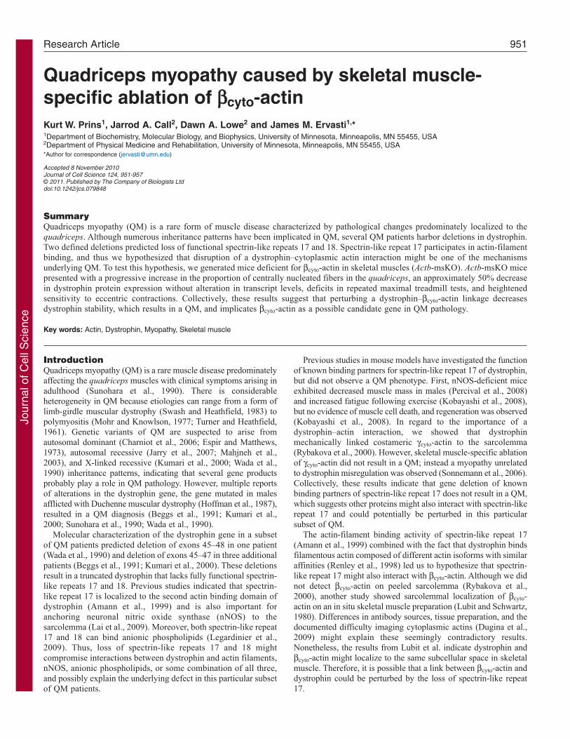
ResultsTo determine whether disruption of a dystrophin–cyto-actininteraction results in a QM phenotype, we first generated mice thatconditionally lacked cyto-actin in skeletal muscle. Subsequently,protein levels of actin isoforms were assessed by quantitativewestern blot analysis of actin-rich eluates from skeletal muscle.Actb-msKOs actin extracts showed a 59% reduction in the levelsof cyto-actin, with no detectable compensatory upregulation ofcyto-, sm-, or sm-actin (Fig. 1,A,B). To determine what cell typewas responsible for the remaining cyto-actin signal observed inActb-msKO skeletal muscle, we subjected control and Actb-msKOskeletal muscle sections to immunofluorescence microscopy. Bothcontrol mice and Actb-msKO sections showed strong cyto-actinimmunoreactivity localizing to endomyosial capillaries (Fig. 1Carrowheads), while a weaker subsarcolemmal staining was observedin control but not Actb-msKO (Fig. 1C). Thus, the cyto-actinpresent in capillary endothelial cells likely represents the observedcyto-actin protein detected in Actb-msKO skeletal muscle extracts.
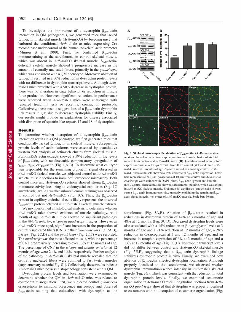
We next performed a histological analysis to determine whetherActb-msKO mice showed evidence of muscle pathology. At 1month of age, Actb-msKO mice showed no significant pathologyin the tibialis anterior, triceps or quadriceps muscles. However asActb-msKO mice aged, significant increases in the proportion ofcentrally nucleated fibers (CNF) in the tibialis anterior (Fig. 2A,B),triceps (Fig. 2C,D) and the quadriceps (Fig. 2E,F) were recorded.The quadriceps was the most affected muscle, with the percentageof CNF progressively increasing to over 13% at 12 months of age.The percentage of CNF in the triceps and tibialis anterior at 12months of age were 2.4% and 1.6%, respectively. Further analysisof the pathology in Actb-msKO skeletal muscle revealed that thecentrally nucleated fibers were confined to fast twitch muscles(supplementary material Fig. S1). Collectively, these results indicateActb-msKO mice possess histopathology consistent with a QM.
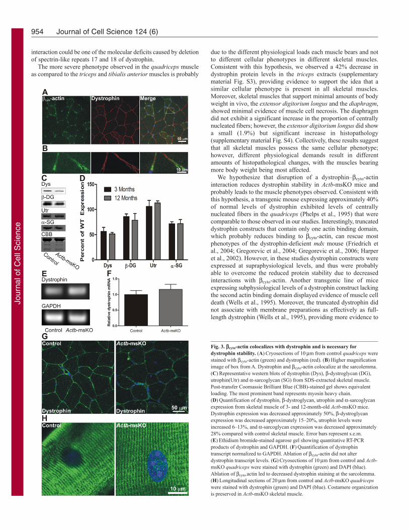
Dystrophin protein levels and localization were examined todetermine whether the QM in Actb-msKO mice was related todystrophin misregulation. First, we subjected control quadricepscryosections to immunofluorescence microscopy and observedcyto-actin staining that colocalized with dystrophin at the
sarcolemma (Fig. 3A,B). Ablation of cyto-actin resulted inreductions in dystrophin protein of 44% at 3 months of age and48% at 12 months (Fig. 3C,D). Decreased dystrophin levels werealso associated with a 15% reduction in -dytroglycan levels at 3months of age and a 21% reduction at 12 months of age, a 28%reduction in -sarcoglycan at 3 and 12 months of age, and anincrease in utrophin expression of 6% at 3 months of age and a13% at 12 months of age (Fig. 3C,D). Dystrophin transcript levelsdid not differ between control and Actb-msKO skeletal muscle(Fig. 3E,F), suggesting that a cyto-actin dystrophin linkagestabilizes dystrophin protein in vivo. Finally, we examined howablation of cyto-actin affected dystrophin localization. Althoughproperly localized to the sarcolemma, we observed weakerdystrophin immunofluorescence intensity in Actb-msKO skeletalmuscle (Fig. 3G), which was consistent with the reduction in totaldystrophin protein levels. Finally, we examined costamericorganization in Actb-msKO mice. Longitudinal sections from Actb-msKO quadriceps showed that dystrophin was properly localizedto costameres with no disruption of costameric organization (Fig.
952 Journal of Cell Science 124 (6)
Fig. 1. Skeletal muscle-specific ablation of cyto-actin. (A)Representativewestern blots of actin isoform expression from actin-rich eluates of skeletalmuscle from control and Actb-msKO mice. (B)Quantification of actin isoformexpression from quadriceps extracts from three control (WT) and three Actb-msKO mice at 3 months of age. st-actin served as a loading control. Actb-msKO skeletal muscle showed a 59% decrease in cyto-actin expression. Errorbars represent s.e.m. (C)Cryosections of 10m from control and Actb-msKOquadriceps were stained with DAPI (blue), cyto-actin (green) and laminin(red). Control skeletal muscle showed sarcolemmal staining, which was absentin Actb-msKO skeletal muscle. Endomyosial capillaries (arrowheads) showedstrong cyto-actin immunoreactivity, probably explaining the remaining cyto-actin signal in actin-rich elutes of Actb-msKO muscle. Scale bar: 50m.
Jour
nal o
f Cel
l Sci
ence
3H). In summary, these data suggest that cyto-actin localizes to thesarcolemma, where it functions to stabilize dystrophin.
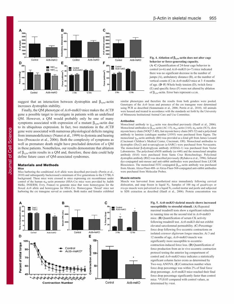
Next, we analyzed cage activity and skeletal muscle function inActb-msKO mice. To our surprise, deletion of cyto-actin did notaffect the total distance traveled (Fig. 4A), number of jumps (Fig.4B), or vertical counts (Fig. 4C) in a 24-hour period. Moreover,Actb-msKO showed equivalent in vivo pulling forces (Fig. 4D)and no differences in either twitch or specific force at 3 and 12months of age (Fig. 4E,F). Collectively, these data indicate thatablation of cyto-actin mice did not alter cage behavior or result inreduced contractile function.
Finally, we analyzed how ablation of cyto-actin affected morestrenuous tests of muscle performance by subjecting Actb-msKOmice to repeated treadmill tests and eccentric contraction protocols.During the first maximal treadmill test, control and Actb-msKOmice showed equivalent times to exhaustion (Fig. 5A). However,when the treadmill test was repeated 48 hours later, Actb-msKOmice performed significantly worse with an 18% reduction inrunning time (Fig. 5A). The decreased running time in Actb-msKOmice was not associated with altered nNOS localization(supplementary material Fig. S2) or an increase in musclemembrane permeability (Fig. 5B) probably due to preservedinteractions between dystrophin and cyto-actin (Rybakova et al.,2000), microtubules (Prins et al., 2009), and intermediate filaments(Bhosle et al., 2006; Stone et al., 2005). Subsequently, we assayeddamage contractile deficits caused by eccentric contractions bothboth ex vivo and in vivo. At both 3 and 12 months of age, isolatedextensor digitorum longus muscles from Actb-msKO mice showeda significant increase in force drop following five consecutive
eccentric contractions (Fig. 5C). The in vivo eccentric contractionprotocol resulted in reduced contractile performance in both controlsand Actb-msKOs (Fig. 5D); however, two-way ANOVA indicatedthat a significant column factor exists when comparing control andActb-msKO mice (Fig. 5D). By calculating the contraction numberat which torque loss was within 2% of the total loss, we observedthat Actb-msKOs reached their final torque significantly fasterthan controls (Fig. 5E). These results indicate that eccentric-contraction-induced damage in vivo occurs more readily whencyto-actin is deleted. In summary, these data demonstrate that lossof cyto-actin renders skeletal muscle more susceptible to damageduring maximal treadmill tests and eccentric contractions.
DiscussionAlthough skeletal muscle-specific ablation of cyto-actin led to aQM phenotype, Actb-msKO mice lacked the muscle weakness andmuscle atrophy phenotypes observed in QM patients expressingdystrophin that lacked spectrin-like repeats 17 and 18. Becausespectrin-like repeats 17 and 18 interact with nNOS and lipids, it islikely that patients expressing a dystrophin without spectrin-likerepeats 17 and 18 have a compound molecular phenotype. In fact,reduced muscle size in male mice lacking nNOS (Percival et al.,2008) combined with our results more accurately recapitulates thisparticular QM. However, it is important to note that mouse modelsdo not always phenocopy all aspects of human disease. For instance,the dystrophin-deficient mdx mouse experiences a disease muchless severe than that experienced by Duchenne patients (Banks andChamberlain, 2008). Nonetheless, the QM phenotype of Actb-msKO mice suggests that disruption of a dystrophin–cyto-actin
953-Actin in skeletal muscle
Fig. 2. Quadriceps myopathy inActb-msKO mice.(A–F)Representative 10m sectionsstained with hematoxylin and eosin,and respective quantification of theproportion of centrally nucleatedfibers from control and Actb-msKOtibialis anterior (A,B), triceps (C,D)and quadriceps (E,F) muscles at 1, 3,6 and 12 months of age (n3 miceper genotype per timepoint).*P≤0.05. Error bars represent s.e.m.Scale bar: 100m.
Jour
nal o
f Cel
l Sci
ence
interaction could be one of the molecular deficits caused by deletionof spectrin-like repeats 17 and 18 of dystrophin.
The more severe phenotype observed in the quadriceps muscleas compared to the triceps and tibialis anterior muscles is probably
due to the different physiological loads each muscle bears and notto different cellular phenotypes in different skeletal muscles.Consistent with this hypothesis, we observed a 42% decrease indystrophin protein levels in the triceps extracts (supplementarymaterial Fig. S3), providing evidence to support the idea that asimilar cellular phenotype is present in all skeletal muscles.Moreover, skeletal muscles that support minimal amounts of bodyweight in vivo, the extensor digitorium longus and the diaphragm,showed minimal evidence of muscle cell necrosis. The diaphragmdid not exhibit a significant increase in the proportion of centrallynucleated fibers; however, the extensor digitorium longus did showa small (1.9%) but significant increase in histopathology(supplementary material Fig. S4). Collectively, these results suggestthat all skeletal muscles possess the same cellular phenotype;however, different physiological demands result in differentamounts of histopathological changes, with the muscles bearingmore body weight being most affected.
We hypothesize that disruption of a dystrophin–cyto-actininteraction reduces dystrophin stability in Actb-msKO mice andprobably leads to the muscle phenotypes observed. Consistent withthis hypothesis, a transgenic mouse expressing approximately 40%of normal levels of dystrophin exhibited levels of centrallynucleated fibers in the quadriceps (Phelps et al., 1995) that werecomparable to those observed in our studies. Interestingly, truncateddystrophin constructs that contain only one actin binding domain,which probably reduces binding to cyto-actin, can rescue mostphenotypes of the dystrophin-deficient mdx mouse (Friedrich etal., 2004; Gregorevic et al., 2004; Gregorevic et al., 2006; Harperet al., 2002). However, in these studies dystrophin constructs wereexpressed at supraphysiological levels, and thus were probablyable to overcome the reduced protein stability due to decreasedinteractions with cyto-actin. Another transgenic line of miceexpressing subphysiological levels of a dystrophin construct lackingthe second actin binding domain displayed evidence of muscle celldeath (Wells et al., 1995). Moreover, the truncated dystrophin didnot associate with membrane preparations as effectively as full-length dystrophin (Wells et al., 1995), providing more evidence to
954 Journal of Cell Science 124 (6)
Fig. 3. cyto-actin colocalizes with dystrophin and is necessary fordystrophin stability. (A)Cryosections of 10m from control quadriceps werestained with cyto-actin (green) and dystrophin (red). (B)Higher magnificationimage of box from A. Dystrophin and cyto-actin colocalize at the sarcolemma.(C)Representative western blots of dystrophin (Dys), -dystroglycan (DG),utrophin(Utr) and -sarcoglycan (SG) from SDS-extracted skeletal muscle.Post-transfer Coomassie Brilliant Blue (CBB)-stained gel shows equivalentloading. The most prominent band represents myosin heavy chain.(D)Quantification of dystrophin, -dystroglycan, utrophin and -sarcoglycanexpression from skeletal muscle of 3- and 12-month-old Actb-msKO mice.Dystrophin expression was decreased approximately 50%, -dystroglycanexpression was decreased approximately 15–20%, utrophin levels wereincreased 6–13%, and -sarcoglycan expression was decreased approximately28% compared with control skeletal muscle. Error bars represent s.e.m.(E)Ethidium bromide-stained agarose gel showing quantitative RT-PCRproducts of dystrophin and GAPDH. (F)Quantification of dystrophintranscript normalized to GAPDH. Ablation of cyto-actin did not alterdystrophin transcript levels. (G)Cryosections of 10m from control and Actb-msKO quadriceps were stained with dystrophin (green) and DAPI (blue).Ablation of cyto-actin led to decreased dystrophin staining at the sarcolemma.(H)Longitudinal sections of 20m from control and Actb-msKO quadricepswere stained with dystrophin (green) and DAPI (blue). Costamere organizationis preserved in Actb-msKO skeletal muscle.
Jour
nal o
f Cel
l Sci
ence
suggest that an interaction between dystrophin and cyto-actinincreases dystrophin stability.
Finally, the QM phenotype of Actb-msKO mice makes the ACTBgene a possible target to investigate in patients with an undefinedQM. However, a QM would probably only be one of manysymptoms associated with expression of a mutant cyto-actin dueto its ubiquitous expression. In fact, two mutations in the ACTBgene were associated with numerous physiological deficits rangingfrom immunodeficiency (Nunoi et al., 1999) to dystonia and hearingloss (Procaccio et al., 2006). Both the complexity of symptoms aswell as premature death might have precluded detection of a QMin these patients. Nonetheless, our results demonstrate that ablationof cyto-actin results in a QM and, therefore, these data could helpdefine future cases of QM-associated syndromes.
Materials and MethodsMiceMice harboring the conditional Actb allele were described previously (Perrin et al.,2010) and subsequently backcrossed a minimum of five generations to the C57BL/6background. These mice were crossed to mice expressing cre recombinase undercontrol of the human sk-actin promoter (HSA-Cre mice were provided by JudithMelki, INSERM, Evry, France) to generate mice that were homozygous for thefloxed Actb allele and hemizygous for HSA-Cre. Homozygous ‘floxed’ mice notharboring the cre transgene served as controls. Both males and females exhibited
similar phenotypes and therefore the results from both genders were pooled.Genotypes of the Actb locus and presence of the cre transgene were determinedusing PCR as described (Sonnemann et al., 2006; Perrin et al., 2010). All animalswere housed and treated in accordance with the standards set forth by the Universityof Minnesota Institutional Animal Care and Use Committee.
AntibodiesMonoclonal antibody to cyto-actin was described previously (Hanft et al., 2006).Monoclonal antibodies to cyto-actin (AC-15), sm-actin (1A4), st-actin (5C5), slowmyosin heavy chain (NOQ7.5.4D), fast myosin heavy chain (MY-32) and a polyclonalantibody to laminin (catalogue number L9393) were purchased from Sigma. Themonoclonal sm-actin antibody (B4) was provided as a kind gift from James Lessard(Cincinnati Children’s Medical Center, Cincinnati, OH). Monoclonal antibodies todystrophin (Dys2) and -sarcoglycan (a-SARC) were purchased from Novacastra.The monoclonal -dystroglycan antibody (43DAG-1) was purchased from VectorLaboratories. The polyclonal nNOS antibody (sc-648) and the monoclonal utrophinantibody (8A4) were purchased from Santa Cruz Biotechnology. Polyclonaldystrophin antibody (Rb2) was described previously (Rybakova et al., 1996). Infrareddye-conjugated anti-mouse and anti-rabbit antibodies were purchased from LICORBiosciences. The monoclonal FITC-conjugated cyto-actin antibody was purchasedfrom Abcam. Alexa-Fluor-488- or Alexa-Fluor-568-conjugated anti-rabbit antibodieswere purchased from Molecular Probes.
Muscle extractsMuscle was harvested from anesthetized mice immediately following cervicaldislocation, and snap frozen in liquid N2. Samples of 100 mg of quadriceps ortriceps muscle were pulverized in a liquid N2 cooled mortar and pestle and subjectedto SDS extraction as described (Hanft et al., 2006). Protein concentration was
955-Actin in skeletal muscle
Fig. 4. Ablation of cyto-actin does not alter cagebehavior or force-generating capacity.(A–C) Quantification of 24-hour cage behavior incontrol (n6) and Actb-msKO (n7) mice indicatedthere was no significant decrease in the number ofjumps (A), ambulatory distance (B), or the number ofvertical counts (C) in Actb-msKO mice at 3–4 monthsof age. (D–F) Whole body tension (D), twitch force(E) and specific force (F) were not altered by ablationof cyto-actin. Error bars represent s.e.m.
Fig. 5. Actb-msKO skeletal muscle shows increasedsusceptibility to stressful stimuli. (A)Repeatedmaximal treadmill tests show a significant reductionin running time on the second trial in Actb-msKOmice. (B)Quantification of serum CK activityfollowing treadmill test. Actb-msKO did not exhibitelevated sarcolemmal permeability. (C)Percentageforce drop following five eccentric contractions onisolated extensor digitorum longus muscles. At 3 and12 months of age, Actb-msKO muscle wassignificantly more susceptible to eccentric-contraction-induced force loss. (D)Quantification offorce production from an in vivo eccentric contractionprotocol testing the anterior leg compartment ofcontrol and Actb-msKO mice indicates a statisticallysignificant column factor exists as determined byTwo-way ANOVA. (E)Contraction number whenforce drop percentage was within 2% of final forcedrop percentage. Actb-msKO mice reached their finalforce drop percentage significantly faster than controlmice. *P≤0.05 compared with control values, asdetermined by t-test.
Jour
nal o
f Cel
l Sci
ence
determined using the Bio-Rad DC Protein Assay. Actin preparations from skeletalmuscle were collected by subjecting muscle to a low-salt extraction and DNase-1amplification protocol as described (Hanft et al., 2006).
Immunofluorescence analysisQuadriceps and triceps muscles were dissected, coated in optimum cuttingtemperature (OCT) medium from TissueTek, and then frozen in melting isopentane.Transverse sections of 10 m were cut on a Leica CM3050 cryostat, air dried, andthen fixed in 4% paraformaldehyde for 10 minutes. Sections were washed with PBS(150 mM NaCl, 8 mM NaH2PO4, 42 mM Na2HPO4), blocked in 5% goat serum for30 minutes, and incubated with primary antibodies overnight at 4°C. Primaryantibody dilutions were: pAb anti-laminin (1:1000), pAb Rb2 (1:1000), pAb anti-nNOS (1:50), mAb anti-FMHC (1:100) and mAb anti SMHC (1:100). Sections werethen washed and blocked with 5% goat serum for 10 minutes three times and thenincubated with Alexa-Fluor-488- or Alexa-Fluor-568-conjugated secondary antibodies(1:1000 dilution) for 30 minutes at 37°C. Then, sections were washed with PBS andcoverslips were applied with a drop of Anti-Fade Reagent (Molecular Probes).Confocal images were collected on a Bio-Rad MRC 1000 scan head mounted on anupright Nikon Optishot microscope at the University of Minnesota BiomedicalImage Processing Laboratory. Images were equivalently processed using AdobePhotoshop 6.0.
When the monoclonal FITC-conjugated cyto-actin was used, sections were fixedin 4% paraformaldehyde for 10 minutes, post-fixed in 100% methanol at –20°C for10 minutes and subjected to immunofluorescence staining as described in previousparagraph. The dilution of the monoclonal FITC-conjugated cyto-actin was 1:50 andthe antibody was added during the secondary antibody incubation step. When usingthe nNOS antibody, sections were fixed in 100% methanol at –20°C, and subjectedto immunofluorescence as described in previous paragraph.
For costameric imaging, quadriceps were frozen in melting isopentane and 20 msections were collected and subjected to immunofluorescence staining as describedabove with one exception: the Rb2 antibody was used at a 1:500 dilution.
Histological assessmentQuadriceps, tibialis anterior, extensor digitorium longus, diaphragm and tricepsmuscles were dissected, coated in OCT, and then frozen in melting isopentane.Transverse sections of 10 m were cut on a Leica CM3050 cryostat and stained withhematoxylin and eosin-phloxine as described. Montage images of the entire sectionswere collected using ImagePro Software. The montages were then analyzed inImagePro and every fiber in the section was scored to determine the proportion ofcentrally nucleated fibers in 1-, 3- and 12-month-old mice (n3 for each genotypeat each timepoint). Pathology in the extensor digitorium longus and the diaphragmwas examined in 10- to 12-month-old mice.
Measurement of sarcolemma permeabilityMembrane permeability was determined by quantifying serum creatine kinase levelson Vitros CK DT slides (Ortho-Clinical Diagnostics) using a Kodak Ektachem DT60analyzer.
Western blot analysis and quantificationWestern blot analysis and quantification of skeletal muscle extracts (n3 for controland Actb-msKO at both 3 and 12 months of age) and actin-rich elutes (n3 forcontrol and Actb-msKO mice at 3 months of age) was performed as described (Prinset al., 2008). Briefly, 25 g of skeletal muscle extract or 20 l of actin-rich eluteswere subjected to SDS-PAGE and transferred to nitrocellulose membranes, whichwere washed and blocked in a 5% milk solution in PBS for 1 hour. Then, themembranes were incubated overnight with primary antibody at room temperature.Primary antibodies and dilutions employed were: mAb Dys2 (1:50), mAb 43DAG-1 (1:50), mAb a-SARC (1:50), mAb 8A4 (1:50), mAb 2-4 (1:500), mAb AC-15(1:100), mAb B4 (1:100), mAb 1A4 (1:100) and mAb 5C5 (1:1000). Membraneswere washed twice for 10 minutes in 5% milk solution at room temperature,incubated with IR-dye-conjugated secondary antibody (1:10,000 dilution) for 30minutes at room temperature, and then the membranes were washed in a 0.5%Tween solution in PBS twice for 10 minutes. Western blots were imaged andquantified with an Odyssey Infrared Imaging System. The Coomassie Blue-stainedpost-transfer gel was analyzed densitometrically using UVP software and served asthe loading control.
Quantitative real-time PCRTotal RNA was isolated from skeletal muscle from four control and four Actb-msKOmice using TRIzol reagent (Invitrogen) according to the manufacturer’s instructions.RNA was treated with RNase-free DNase (Ambion) at room temperature for 15minutes. cDNA was generated using the iScript cDNA synthesis kit (Bio-Rad). ForRT-PCR, cDNA was amplified using the SYBR GreenER qPCR ReadyMix(Invitrogen) on the Bio-Rad iQ5. Results of quantitative RT-PCR assays wereanalyzed using analysis software supplied with the Bio-Rad iQ5 system to determinedystrophin transcript levels (dystrophin forward 5�-AATGGCCTGCCCTGGGGGAT-3�, dystrophin reverse 5�-TCTGGTGTAGACCTGGCGGC-3�) normalized toGAPDH transcript levels (GAPDH forward 5�-GACCCCTCCAT TGACCTCA -ACTACATG-3�, GAPDH reverse 5�-CTCCTGGAAGATGGTG ATGGG-3�).
Whole body tensionWhole body tension (WBT) was performed as described (Sonnemann et al., 2006).Briefly, 3-month-old control and Actb-msKO mice (n4 for each genotype) weretied with silk suture to a fixed range force transducer (BioPac Systems) to record theforce evoked by a light tail pinch. Data were analyzed by comparing WBT1 (themaximal force produced by each mouse) and WBT1–5 (the average of the top fiveforce-producing pulls), with both being normalized to body weight.
Activity measurementsMice of 3–4 months of age (n6 control, n7 Actb-msKO) were monitored for 24-hourcage activities using activity chambers (Med Associates) as described (Landisch et al.,2008). In brief, mice were exposed to a 24-hour acclimation period in a mock chamber,which was identical to the activity-monitoring cage except that it lacked the infraredarrays. The activity chambers contain three infrared arrays in the x-, y-, and z-axes,with two sets of beams in the x-direction, one being elevated above the other.Ambulation was measured by arrays located in the x- and y-axes, and vertical movement(i.e., jumping and hindlimb rearing) was measured by the second elevated x-array.Infrared sensors in the chamber registered an activity count each time one of the beamswas disrupted, such that movement was simultaneously measured in all three axes. Alldata were acquired using Activity Monitor software, version 5 (Med Associates).
Ex vivo extensor digitorium longus contractile propertiesMice of 3 months and 11–12 months were anesthetized and the extensor digitorumlongus muscle was removed (n4 mice per genotype per timepoint). The proximaltendon was attached to 4–0 suture silk to a dualmode muscle lever system (model300B-LR Aurora Scientific, Aurora, ON, Canada). Muscles were equilibrated for 10minutes in a bath assembly containing Krebs–Ringer–bicarbonate buffer (119 mMNaCl, 5.0 KCl mM, 1.0 MgSO4 mM, 12.25 NaHCO3 mM, 1.0 CaCl2 mM, 1.0 mMKH2PO4, 10.0 mM glucose, 0.17 mM leucine, 0.10 mM isoleucine, 0.20 mM valineplus 10 mg/ml gentamicin sulfate and 0.10 U/ml insulin) at 25°C while beingconstantly oxygenated with 95% O2 and 5% CO2 gas. Then, the resting tension wasset to 0.4 g and twitch force was determined by stimulating the muscle with a 0.5millisecond pulse at 150 V (Grass stimulator, Grass Telefactor). Thirty seconds later,the muscle was stimulated to twitch again and the greater of two contractions wasrecorded. Tetanic contractions were elicited by stimulating the muscle with 150 Vfor 200 milliseconds at 180 Hz. The greater of two contractions separated by 2minutes of recovery time was recorded. To determine damage caused by eccentriccontractions muscles were subjected to five lengthening contractions at 3-minuteintervals. Each eccentric contraction consisted of a maximal tetanic stimulation for200 milliseconds accompanied by a stretch of 1.0 L0/second (where L0 is the optimalmuscle length for maximal force) to give a total stretch of 0.2 L0. Force drop wascalculated as (ECC1–ECC5)/ECC1, where ECC1 is the peak force generated on thefirst eccentric contraction and ECC5 is the force generated on the fifth eccentriccontraction.
In vivo eccentric contractionIn vivo eccentric contraction was carried out as previously described (Baltgalvis etal., 2009). First, mice were given a mixture of fentanyl citrate (10 mg/kg bodyweight), droperidol (0.2 mg/kg weight) and diazepam (5 mg/kg weight), and thenthe hair on the left hindlimb was shaved and removed with depilatory cream. Theleft foot of the mouse was taped to a metal foot plate, which was connected to theshaft of the servomotor (model 300B-LR, Aurora Scientific) with the left kneestabilized, ensuring the tibia was perpendicular to the foot. Two sterilized platinumsubdermal needle electrodes (model E2-12, Grass Technologies) were insertedthrough the skin 1–2 mm on either side of the left common peroneal nerve. Theservomotor passively dorsiflexed the foot 20° over a duration of 3 seconds, and thenerve was stimulated while the foot was plantar flexed 40° at a velocity of2000°/second to generate eccentric contractions. After the stimulation, the foot waspassively dorsiflexed 20° to its original resting position over a 3-second period. Onecontraction was performed every 12 seconds for a total of 150 eccentric contractions.
Treadmill exhaustion testMaximal exercise performance studies were performed on a Columbus Instrumentstreadmill with an uphill grade of 15°. An acclimation period of running at a speedof 10 m/minute for 5 minutes twice a week for 2 weeks was implemented prior tomaximal testing. To determine maximal exercise performance, mice were run on thetreadmill for 5 minutes at a rate of 10 m/minute followed by a 1 m/minute increasein speed every minute until exhaustion. Exhaustion was defined as an inability tostay off a shock bar for at least 5 seconds. Maximal exercise capacity was determinedtwice, with the trials being separated by 48 hours.
Statistical analysisAll data are presented as mean ± s.e.m. Comparison between groups was performedusing a t-test with significance defined as P≤0.05. When comparing the response oftwo groups over time, two-way ANOVA was implemented and significance wasdefined as P≤0.05.
956 Journal of Cell Science 124 (6)
Jour
nal o
f Cel
l Sci
ence
The authors thank Sarah Greising for assistance in the cage behaviormonitoring, Kevin Sonneman for generating mice with a conditionalActb allele, and Thomas Cheever and Emily Olson for their helpfuldiscussion. This work was funded by an NIH grant AR049899. K.W.P.is a member of the Medical Scientist Training Program at the Universityof Minnesota. Deposited in PMC for release after 12 months.
Supplementary material available online athttp://jcs.biologists.org/cgi/content/full/124/6/951/DC1
ReferencesAmann, K. J., Guo, A. W. and Ervasti, J. M. (1999). Utrophin lacks the rod domain
actin binding activity of dystrophin. J. Biol. Chem. 274, 35375-35380.Baltgalvis, K. A., Call, J. A., Nikas, J. B. and Lowe, D. A. (2009). Effects of prednisolone
on skeletal muscle contractility in mdx mice. Muscle Nerve 40, 443-454.Banks, G. B. and Chamberlain, J. S. (2008). The value of mammalian models for
duchenne muscular dystrophy in developing therapeutic strategies. Curr. Top. Dev. Biol.84, 431-453.
Beggs, A. H., Hoffman, E. P., Snyder, J. R., Arahata, K., Specht, L., Shapiro, F.,Angelini, C., Sugita, H. and Kunkel, L. M. (1991). Exploring the molecular basis forvariability among patients with becker muscular dystrophy: Dystrophin gene and proteinstudies. Am. J. Hum. Genet. 49, 54-67.
Bhosle, R. C., Michele, D. E., Campbell, K. P., Li, Z. and Robson, R. M. (2006).Interactions of intermediate filament protein synemin with dystrophin and utrophin.Biochem. Biophys. Res. Commun. 346, 768-777.
Charniot, J. C., Desnos, M., Zerhouni, K., Bonnefont-Rousselot, D., Albertini, J. P.,Salama, J. Z., Bassez, G., Komajda, M. and Artigou, J. Y. (2006). Severe dilatedcardiomyopathy and quadriceps myopathy due to lamin A/C gene mutation: A phenotypicstudy. Eur. J. Heart Fail. 8, 249-256.
Dugina, V., Zwaenepoel, I., Gabbiani, G., Clement, S. and Chaponnier, C. (2009). Betaand gamma-cytoplasmic actins display distinct distribution and functional diversity. J.Cell Sci. 122, 2980-2988.
Espir, M. L. and Matthews, W. B. (1973). Hereditary quadriceps myopathy. J. Neurol.Neurosurg. Psychiatr. 36, 1041-1045.
Friedrich, O., Both, M., Gillis, J. M., Chamberlain, J. S. and Fink, R. H. (2004). Mini-dystrophin restores L-type calcium currents in skeletal muscle of transgenic mdx mice.J. Physiol. 555, 251-265.
Gregorevic, P., Blankinship, M. J., Allen, J. M., Crawford, R. W., Meuse, L., Miller,D. G., Russell, D. W. and Chamberlain, J. S. (2004). Systemic delivery of genes tostriated muscles using adeno-associated viral vectors. Nat. Med. 10, 828-834.
Gregorevic, P., Allen, J. M., Minami, E., Blankinship, M. J., Haraguchi, M., Meuse,L., Finn, E., Adams, M. E., Froehner, S. C., Murry, C. E. et al. (2006). rAAV6-microdystrophin preserves muscle function and extends lifespan in severely dystrophicmice. Nat. Med. 12, 787-789.
Hanft, L. M., Rybakova, I. N., Patel, J. R., Rafael-Fortney, J. A. and Ervasti, J. M.(2006). Cytoplasmic gamma-actin contributes to a compensatory remodeling responsein dystrophin-deficient muscle. Proc. Natl. Acad. Sci. USA 103, 5385-5390.
Harper, S. Q., Hauser, M. A., DelloRusso, C., Duan, D., Crawford, R. W., Phelps, S.F., Harper, H. A., Robinson, A. S., Engelhardt, J. F., Brooks, S. V. et al. (2002).Modular flexibility of dystrophin: Implications for gene therapy of duchenne musculardystrophy. Nat. Med. 8, 253-261.
Hoffman, E. P., Brown, R. H., Jr and Kunkel, L. M. (1987). Dystrophin: The proteinproduct of the duchenne muscular dystrophy locus. Cell 51, 919-928.
Jarry, J., Rioux, M. F., Bolduc, V., Robitaille, Y., Khoury, V., Thiffault, I., Tetreault,M., Loisel, L., Bouchard, J. P. and Brais, B. (2007). A novel autosomal recessivelimb-girdle muscular dystrophy with quadriceps atrophy maps to 11p13-p12. Brain130, 368-380.
Kobayashi, Y. M., Rader, E. P., Crawford, R. W., Iyengar, N. K., Thedens, D. R.,Faulkner, J. A., Parikh, S. V., Weiss, R. M., Chamberlain, J. S., Moore, S. A. et al.(2008). Sarcolemma-localized nNOS is required to maintain activity after mild exercise.Nature 456, 511-515.
Kumari, D., Gupta, M. and Goyle, S. (2000). Detection of deletion in the dystrophingene of a patient with quadriceps myopathy. Neurol. India 48, 68-71.
Lai, Y., Thomas, G. D., Yue, Y., Yang, H. T., Li, D., Long, C., Judge, L., Bostick, B.,Chamberlain, J. S., Terjung, R. L. et al. (2009). Dystrophins carrying spectrin-likerepeats 16 and 17 anchor nNOS to the sarcolemma and enhance exercise performancein a mouse model of muscular dystrophy. J. Clin. Invest. 119, 624-635.
Landisch, R. M., Kosir, A. M., Nelson, S. A., Baltgalvis, K. A. and Lowe, D. A. (2008).Adaptive and nonadaptive responses to voluntary wheel running by mdx mice. MuscleNerve 38, 1290-1303.
Legardinier, S., Raguenes-Nicol, C., Tascon, C., Rocher, C., Hardy, S., Hubert, J. F.and Le Rumeur, E. (2009). Mapping of the lipid-binding and stability properties of thecentral rod domain of human dystrophin. J. Mol. Biol. 389, 546-558.
Lubit, B. W. and Schwartz, J. H. (1980). An antiactin antibody that distinguishesbetween cytoplasmic and skeletal muscle actins. J. Cell Biol. 86, 891-897.
Mahjneh, I., Somer, H., Paetau, A. and Marconi, G. (2003). Pure quadriceps myopathyin two sisters. Eur. J. Neurol. 10, 453-456.
Miniou, P., Tiziano, D., Frugier, T., Roblot, N., Le Meur, M. and Melki, J. (1999).Gene targeting restricted to mouse striated muscle lineage. Nucleic Acids Res. 27, e27.
Mohr, P. D. and Knowlson, T. G. (1977). Quadriceps myositis: An appraisal of thediagnostic criteria of quadriceps myopathy. Postgrad. Med. J. 53, 757-760.
Nunoi, H., Yamazaki, T., Tsuchiya, H., Kato, S., Malech, H. L., Matsuda, I. andKanegasaki, S. (1999). A heterozygous mutation of beta-actin associated with neutrophildysfunction and recurrent infection. Proc. Natl. Acad. Sci. USA 96, 8693-8698.
Percival, J. M., Anderson, K. N., Gregorevic, P., Chamberlain, J. S. and Froehner, S.C. (2008). Functional deficits in nNOSmu-deficient skeletal muscle: Myopathy innNOS knockout mice. PLoS One 3, e3387.
Perrin, B. J., Sonnemann, K. J. and Ervasti, J. M. (2010). -actin and -actin are eachdispensible for auditory hair cell development but required for stereocilia maintenance.PLoS Genet. 6, e1001158.
Phelps, S. F., Hauser, M. A., Cole, N. M., Rafael, J. A., Hinkle, R. T., Faulkner, J. A.and Chamberlain, J. S. (1995). Expression of full-length and truncated dystrophinmini-genes in transgenic mdx mice. Hum. Mol. Genet. 4, 1251-1258.
Prins, K. W., Lowe, D. A. and Ervasti, J. M. (2008). Skeletal muscle-specific ablationof gamma(cyto)-actin does not exacerbate the mdx phenotype. PLoS One 3, e2419.
Prins, K. W., Humston, J. L., Mehta, A., Tate, V., Ralston, E. and Ervasti, J. M.(2009). Dystrophin is a microtubule-associated protein. J. Cell Biol. 186, 363-369.
Procaccio, V., Salazar, G., Ono, S., Styers, M. L., Gearing, M., Davila, A., Jimenez,R., Juncos, J., Gutekunst, C. A., Meroni, G. et al. (2006). A mutation of beta -actinthat alters depolymerization dynamics is associated with autosomal dominantdevelopmental malformations, deafness, and dystonia. Am. J. Hum. Genet. 78, 947-960.
Renley, B. A., Rybakova, I. N., Amann, K. J. and Ervasti, J. M. (1998). Dystrophinbinding to nonmuscle actin. Cell Motil. Cytoskeleton 41, 264-270.
Rybakova, I. N., Amann, K. J. and Ervasti, J. M. (1996). A new model for theinteraction of dystrophin with F-actin. J. Cell Biol. 135, 661-672.
Rybakova, I. N., Patel, J. R. and Ervasti, J. M. (2000). The dystrophin complex formsa mechanically strong link between the sarcolemma and costameric actin. J. Cell Biol.150, 1209-1214.
Sonnemann, K. J., Fitzsimons, D. P., Patel, J. R., Liu, Y., Schneider, M. F., Moss, R. L.and Ervasti, J. M. (2006). Cytoplasmic gamma-actin is not required for skeletal muscledevelopment but its absence leads to a progressive myopathy. Dev. Cell 11, 387-397.
Stone, M. R., O’Neill, A., Catino, D. and Bloch, R. J. (2005). Specific interaction of theactin-binding domain of dystrophin with intermediate filaments containing keratin 19.Mol. Biol. Cell 16, 4280-4293.
Sunohara, N., Arahata, K., Hoffman, E. P., Yamada, H., Nishimiya, J., Arikawa, E.,Kaido, M., Nonaka, I. and Sugita, H. (1990). Quadriceps myopathy: Forme fruste ofbecker muscular dystrophy. Ann. Neurol. 28, 634-639.
Swash, M. and Heathfield, K. W. (1983). Quadriceps myopathy: A variant of the limb-girdle dystrophy syndrome. J. Neurol. Neurosurg. Psychiatr. 46, 355-357.
Turner, J. W. and Heathfield, K. W. (1961). Quadriceps myopathy occurring in middleage. J. Neurol. Neurosurg. Psychiatr. 24, 18-21.
Wada, Y., Itoh, Y., Furukawa, T., Tsukagoshi, H. and Arahata, K. (1990). “Quadricepsmyopathy”: A clinical variant form of becker muscular dystrophy. J. Neurol. 237, 310-312.
Wells, D. J., Wells, K. E., Asante, E. A., Turner, G., Sunada, Y., Campbell, K. P.,Walsh, F. S. and Dickson, G. (1995). Expression of human full-length andminidystrophin in transgenic mdx mice: Implications for gene therapy of duchennemuscular dystrophy. Hum. Mol. Genet. 4, 1245-1250.
957-Actin in skeletal muscle
Jour
nal o
f Cel
l Sci
ence